Publisher’s version / Version de l'éditeur:
Vous avez des questions? Nous pouvons vous aider. Pour communiquer directement avec un auteur, consultez la première page de la revue dans laquelle son article a été publié afin de trouver ses coordonnées. Si vous n’arrivez pas à les repérer, communiquez avec nous à PublicationsArchive-ArchivesPublications@nrc-cnrc.gc.ca.
Questions? Contact the NRC Publications Archive team at
PublicationsArchive-ArchivesPublications@nrc-cnrc.gc.ca. If you wish to email the authors directly, please see the first page of the publication for their contact information.
https://publications-cnrc.canada.ca/fra/droits
L’accès à ce site Web et l’utilisation de son contenu sont assujettis aux conditions présentées dans le site LISEZ CES CONDITIONS ATTENTIVEMENT AVANT D’UTILISER CE SITE WEB.
Pure and Applied Chemistry, 90, 8, pp. 1325-1356, 2018-07-12
READ THESE TERMS AND CONDITIONS CAREFULLY BEFORE USING THIS WEBSITE. https://nrc-publications.canada.ca/eng/copyright
NRC Publications Archive Record / Notice des Archives des publications du CNRC :
https://nrc-publications.canada.ca/eng/view/object/?id=074c986d-4ca0-42b4-a4ce-c49d44e40287 https://publications-cnrc.canada.ca/fra/voir/objet/?id=074c986d-4ca0-42b4-a4ce-c49d44e40287
Archives des publications du CNRC
This publication could be one of several versions: author’s original, accepted manuscript or the publisher’s version. / La version de cette publication peut être l’une des suivantes : la version prépublication de l’auteur, la version acceptée du manuscrit ou la version de l’éditeur.
For the publisher’s version, please access the DOI link below./ Pour consulter la version de l’éditeur, utilisez le lien DOI ci-dessous.
https://doi.org/10.1515/pac-2017-0102
Access and use of this website and the material on it are subject to the Terms and Conditions set forth at
Engineered nanomaterials and human health: Part 2. Applications and
nanotoxicology (IUPAC Technical Report)
Gubala, Vladimir; Johnston, Linda J.; Krug, Harald F.; Moore, Colin J.; Ober,
Christopher K.; Schwenk, Michael; Vert, Michel
IUPAC Technical Report
Vladimir Gubala*, Linda J. Johnston, Harald F. Krug, Colin J. Moore,
Christopher K. Ober, Michael Schwenk and Michel Vert
Engineered nanomaterials and human
health: Part 2. Applications and
nanotoxicology (IUPAC Technical Report)
https://doi.org/10.1515/pac-2017-0102
Received January 4, 2017; accepted December 20, 2017
Abstract: Research on engineered nanomaterials (ENM) has progressed rapidly from the very early stages of
studying their unique, size-dependent physicochemical properties and commercial exploration to the devel-opment of products that influence our everyday lives. We have previously reviewed various methods for syn-thesis, surface functionalization, and analytical characterization of ENM in a publication titled ‘Engineered Nanomaterials: Preparation, Functionalization and Characterization’. In this second, inter-linked document, we first provide an overview of important applications of ENM in products relevant to human healthcare and consumer goods, such as food, textiles, and cosmetics. We then highlight the challenges for the design and development of new ENM for bio-applications, particularly in the rapidly developing nanomedicine sector. The second part of this document is dedicated to nanotoxicology studies of ENM in consumer products. We describe the various biological targets where toxicity may occur, summarize the four nanotoxicology prin-ciples, and discuss the need for careful consideration of the biodistribution, degradation, and elimination routes of nanosized materials before they can be safely used. Finally, we review expert opinions on the risk, regulation, and ethical aspects of using engineered nanomaterials in applications that may have direct or indirect impact on human health or our environment.
Keywords: nanomaterial; nanomedicine; nanoparticle; nano-products; nano-toxicology; regulation.
CONTENTS
1 Introduction to nanomaterials and health ...1326
2 Engineered nanomaterials for biological applications ... 1327
2.1 Therapeutics and diagnostics ... 1327
2.1.1 Limitations of small molecule drugs ... 1327
2.1.2 Types of nanomaterials in modern diagnostics and therapeutics ... 1327
Article note: This document was prepared in the frame of IUPAC Project 2013-007-1-700. Sponsoring bodies: The Chemistry and
Human Health Division and the Polymer Division: see more details on p. 1350.
*Corresponding author: Vladimir Gubala, University of Kent, Medway School of Pharmacy, Central Avenue, Anson Building,
Chatham, ME44TB, UK, e-mail: V.Gubala@kent.ac.uk. http://orcid.org/0000-0001-6301-3632
Linda J. Johnston: Measurement Science and Standards, National Research Council Canada, Ottawa, ON K1A 0R6, Canada Harald F. Krug: Empa – Materials Science and Technology, Lerchenfeldstrasse 5, 9014 St. Gallen, Switzerland
Colin J. Moore: FOCAS Research Institute, Dublin Institute of Technology, Kevin St, Dublin 8, Ireland
Christopher K. Ober: Cornell University, Department of Materials Science and Engineering, 310 Bard Hall, Ithaca, NY 14853-1501,
USA
Michael Schwenk: In den Kreuz̈ckern 16/1, D 72072 Tuebingen, Germany (formerly Medical School Hannover)
Michel Vert: University of Montpellier – CNRS-ENSCM IBMM-CRBA, 15 Avenue Charles Flahault, BP 14491, 34093 Montpellier
2.1.3 General features of theranostic nanomaterials ... 1328
2.1.4 Scaffold influences ... 1329
2.1.5 Chemical surface modification and hydrophilization ... 1329
2.1.6 Protein corona ... 1330
2.1.7 Surface modification with biomolecules ...1331
2.1.8 Drug release ...1331
2.1.9 Drug targeting ...1331
2.2 Medical application areas ...1332
2.3 Challenges related to applications in biology ...1332
3 Engineered nanomaterials in consumer goods ... 1333
3.1 Food ... 1333 3.2 Tattoo inks ... 1334 3.3 Textiles... 1334 3.4 Cosmetics ... 1335 4 Nanotoxicology ... 1335 4.1 Toxicological targets ... 1335 4.1.1 Molecular level... 1335 4.1.2 Cell level ... 1335 4.1.3 Organ level ... 1336
4.2 The four nanotoxicology principles ... 1337
4.2.1 Transport-principle ... 1337 4.2.2 Surface-principle ... 1338 4.2.3 Material-principle ... 1339 4.2.4 Fibre-principle ...1340 4.3 Toxicokinetics ...1340 4.3.1 Biological barriers ...1340 4.3.2 Absorption ... 1342 4.3.3 Distribution ... 1343 4.3.4 Degradation ...1344 4.3.5 Elimination ... 1345
4.4 Difficulties associated with the study of nanoparticle toxicity ... 1345
4.5 Case study: quantum dots ... 1346
5 Risks, regulation and ethical aspects ...1347
5.1 Risks ... 1347
5.2 Regulation: read-across and grouping ... 1347
5.3 Ethical questions ...1348
6 Discussion and closing remarks ... 1349
Membership of sponsoring bodies ...1350
References ...1350
1 Introduction to nanomaterials and health
Many scientists agree that the world-wide attention to nanotechnology started in December 1959, when Richard Feynman delivered the talk ‘There’s Plenty of Room at the Bottom’ to the American Physical Society at the California Institute of Technology [1]. However, it wasn’t until the early 2000s that nanomaterial science exploded into a gigantic field, attracting the interest of academic, industrial, and government scientists. Due to generous public funding for nanotechnology, the prefix ‘nano’ quickly became the fashion and nobody wanted to miss the nano-revolution. As a result, many types of Engineered Nanomaterials (ENM) made their way into products used in the household and food industries, as well as cosmetics, sports, textiles, and
medicine. While the intention was to enable novel applications, scientists have also inadvertently created nanomaterials that may cause harm to humans and the environment. Several critical concerns appeared in the literature, with authors questioning both the real progress of nanomedicine and the safety and impact of nanoenabled products on human health [2–5]. Some of these articles are somewhat provocatively titled, such as: ‘Nanomedicine – Is the Wave Cresting?’ [6], ‘Cancer Nanomedicine – So Many Papers and So Few Drugs!’ [2], or ‘Nanosafety Research – Are We on the Right Track?’ [5]. They provide a critical perspective on the field by identifying scientific weaknesses and misconceptions that are rarely acknowledged in tradi-tional scientific articles [7]. For example, while the physicochemical properties and behaviour of ENM can usually be controlled under laboratory environments, this is no longer the case in complex physiological or natural environments [8]. Tens of thousands of publications in nanotoxicology and nano-ecotoxicology have appeared to date [5], yet no clear concepts exist to quantify the adverse effects of ENM.
In Part 1, ‘Engineered Nanomaterials and Human Health: Preparation, Functionalisation, and Analytical Characterization’ [9], we focused on the chemistry of ENM, reviewing various methods for synthesis, surface functionalization, and analytical characterization. In this second part, we describe ENM applications that are intended to improve our quality of life or have significant potential for direct or indirect impact on human health. These include therapeutic and diagnostic drugs (theranostics), food additives, cosmetics, and tex-tiles. We describe the various routes by which ENM enter the body and the mechanisms by which they are transported through the body, degraded, and cleared. Finally, we describe the ENM features that are relevant for toxicity and identify possible toxic mechanisms. Currently, a simple question ‘Can nanomaterials cause acute or chronic health effects?’ has a short and simple answer: the impact depends on the properties of the specific ENM and its environment. The complexity of the topic makes it impossible to be comprehensive in citing all the available literature, even though we have referenced more than 260 articles, reviews, reports, and documents. Wherever possible, we have included citations based on the following criteria (in no particu-lar order): 1. Work published relatively recently, preferably in the past 5 years; 2. Work with detailed protocols and conclusive data, avoiding articles with ‘promising results and with potential’; 3. Work that represents the best practice in the field and 4. Reports and official documents from trusted sources such as IUPAC, ISO, or other national and international organisations.
2 Engineered nanomaterials for biological applications
2.1 Therapeutics and diagnostics
2.1.1 Limitations of small molecule drugs
Conventional small molecule drugs consist of single chemical entities with a dimension that usually ranges from about 0.6 to 2 nm [10]. Although the pharmacology of a small molecule drug can often be improved by designing analogues, some restrictions remain. For example, failure to solubilize when applied intrave-nously, unfavourable half-life, or failure to cross biological barriers will limit the delivery of a drug to the target organ or correct cellular location. Post-administration capture by the molecular and/or cellular ele-ments of body fluids, lack of activation at the target site, and premature export from target cells via mem-brane carriers can further limit the therapeutic efficacy of the drug. It is a central goal of nanoparticle drug design to overcome such restrictions.
2.1.2 Types of nanomaterials in modern diagnostics and therapeutics
It is well-established that nano-sized substances have been used in pharmacology and diagnostics before the onset of nanoparticle engineering. Lipid emulsions were already infused as energy-rich nutrients in clinical set-tings [11] before the application of ENM and colloidal silver (e.g. Argyrol) has been applied to protect water and
textiles against microorganisms for 100 years [12] and has been used to cure various diseases [13–16]. Colloidal gold also has a history in disease treatment and polymeric gold compounds are used to treat otherwise therapy-resistant rheumatoid arthritis [17]. However, the engineering of more complex nanostructures with the aim to improve therapeutic and diagnostic possibilities started only some 20 years ago. In the search for useful and tolerable materials, a large number of different approaches are presently studied. The basic architecture used in many of these approaches consists of three components: scaffold, core, and surface. The ‘scaffold’ provides mechanical and physicochemical stability. It should have a defined size-range and a suitable surface that allows the addition of functional groups. The ‘core’ can be hollow, porous, or cavernous. It is used to accommodate therapeutic and diagnostic agents, alone or together with a solvent. Although the ‘core’ of the nanoparticles (a class of ENM, abbreviated as NP) may not interact directly with the biological environment, it is needed to stabi-lise the NP and protect and release the cargo. There are also requirements for it to be biocompatible. It will either have to degrade to smaller components, or else be small enough to be renally cleared. The particle ‘surface’ that interacts with cells, tissues, and fluids is usually modified to become biocompatible and immunoresistant. It can be conjugated with antibodies or other agents that direct the particle to a biological target.
Theranostic nanoparticles may have inorganic (e.g. metal), polymeric, lipid, carbohydrate, proteina-ceous, or gaseous components [18–20]. Figure 3 in [9] depicts schematically some of the more common struc-tures. They include nanospheres, nanocapsules, nanoshells, nanorods, liposomes, polymersomes, micelles, and virus-like-particles (the latter are not shown).
2.1.3 General features of theranostic nanomaterials
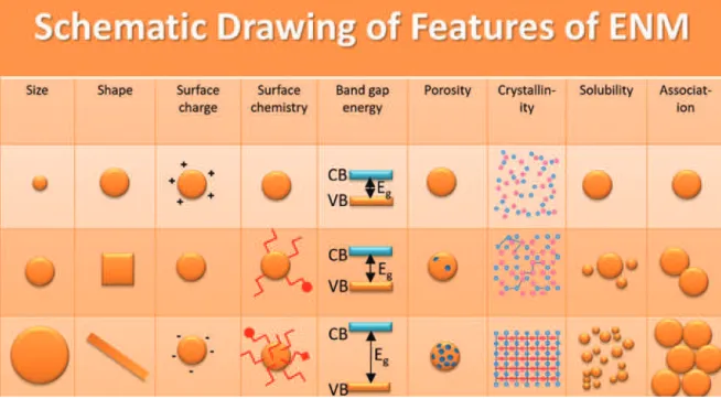
Theranostic nanoparticles are designed to exhibit special features, while being at the same time biocom-patible. Many physicochemical features that are not directly related to the therapeutic or diagnostic aim must be controlled and possibly improved during development (see also [9]). These features include size distribution, shape, surface charge, surface functional groups, porosity, crystallinity, and solubility, as well as beneficial degradation or stability towards association in certain environments, as illustrated in Fig. 1.
Fig. 1: Schematic illustration of the most important features of ENM for a rational design as theranostic material. VB: valence
band; CB: conduction band; Eg: band gap energy (eV). Adapted from [21]. The particle size, shape, surface charge, and deform-ability/degradability of ENM and their effects on overcoming delivery barriers are reviewed in more details by Blanco et al. [22].
2.1.4 Scaffold influences
A variety of materials have been used as scaffolds, with polymers being the most prominent class. Com-mercially available polymers, such as poly(methyl 2-methylpropenoate), more commonly called poly(methyl methacrylate), have been used during the pioneering phase [23]. Currently, priority is given to degradable, and preferably bioresorbable, artificial polymers, such as lactic- and/or glycolic acid-based polymers and copolymers, etc. [24, 25]. Another class of scaffolds is made up of lipids and phospholipids that are able to form liposomes with different surfaces depending on the precursors used [26]. Such engineered particles can mimic the size and lipid composition of endogenous lipoproteins (low-density lipoprotein, high-density lipoprotein, for example ApoB-100 in LDL and Apo-AI and Apo-AII in HDL). They can accommodate lipophilic drugs in the lipid compartment and hydrophilic drugs in the aqueous core [27, 28]. Moreover, new techniques such as electrospinning are versatile methods to fabricate scaffolds. For example, nanofibers of materials such as polymers and ceramics with diameters from 3 nm to several micrometres are used as scaffolds for in vivo and in vitro tissue engineering [29]. Carbon fibres or carbon nanotubes and solid materials such as gold or mesoporous silica are also investigated for these purposes [30, 31]. The possibilities appear unlimited when one considers the number of parent materials and drugs that can be combined. However, in reality, the domain is very much limited, especially if the long list of prerequisites (e.g. reasonable chemistry; possibility of upscaling; easy formulation; sterilization, conditioning, and stability on storage; use-by date; cost; and approval by regulatory agencies) for effective application in humans is taken into account. This very much restricts the number of successful approaches.
2.1.5 Chemical surface modification and hydrophilization
From a physicochemical viewpoint, the particle surface must be compatible with the temperature, pH, ionic strength, and other parameters imposed by the surroundings and must help to prevent particle association. Conventional colloid techniques prevent the association of pristine NP by introducing capping agents (often charged) to the surface to induce steric or electrostatic repulsion between neighbouring NP [3, 32]. This is a
popular means of controlling colloidal stability. Poly(oxyethylene) (the PEO abbreviation comes from the source-based name poly(ethylene oxide) [33]) can also be employed to create a steric barrier that reduces non-specific binding of the NP and has proven a popular surface coating in many drug delivery systems (Fig. 2). PEO is also referred to as poly(ethylene glycol) (PEG), which is a structural variation of PEO with two –OH groups, one at each terminus [34, 35]. The two terms are often used interchangeably, however the systematic term PEO is recommended by IUPAC to describe analogues with repeating fragments of –O–CH2–CH2–. We use the systematic name PEO in this document, but, for describing the process of attaching PEO to surfaces, we use the term ‘PEGylation’, which is widely used in the literature. PEO segments are available in a variety of forms (short or long chains or multiple chains in the same substituent). They are hydrophilic, neutral, and biocompatible, and can be conjugated to biomolecules [36]. Other macromolecules, such as dextran or den-drimers, have also been used to create sterically stabilised NP that are more biocompatible and which can be used for biomolecule attachment [37–39].
Surface PEO segments have been used in liposomal technologies because they prolong in vivo circula-tion lifetimes [40]. Improvements in the pharmacokinetics and bio-distribucircula-tion of dye-doped silica NP upon PEGylation were shown by He et al. [41] in a study comparing 45 nm OH-, COOH- and PEO-coated NP in mice. Wiesner’s ultra-small dye-loaded silica NP, currently in clinical trials, also utilise PEO for enhancing NP cir-culation half-life [42, 43]. The effect of ‘PEGylation’ on these small NP (achieved using a PEO-siloxane) was recently elucidated using fluorescence correlation and cross-correlation spectroscopy [44].
2.1.6 Protein corona
Despite efforts to improve the colloidal stability of nanomaterials through grafting of sterically or electro-statically repelling coatings, additional surface modification often occurs inside the body by adsorption of substances present in body fluids [46, 47]. Such surface ‘fouling’ is primarily attributed to protein adsorption onto the NP surface to form a ‘protein corona’, also referred to as a ‘biomolecular corona’. Two types of protein corona, ‘hard’ and ‘soft’ (Fig. 2), have been shown to form on many types of NP, including gold, polystyrene, titanium, silica, and zinc oxide [48]. A hard corona is a near-monolayer of biomolecules tightly bound (not irreversibly but with possible denaturation effects) to the particle surface. Surrounding this layer is the ‘soft’ corona, which is composed of more loosely associated and rapidly exchanging biomolecules. According to the article ‘What the Cell “Sees” in Bionanoscience’, this corona provides the ‘extrinsic environmentally-derived identity’ that a cell would actually ‘see’ and interact with [49].
The emergence of the protein corona field highlights the importance of performing analytical characteri-zation in complex biological media. For example, when developing assays using NP for biomarker detection, it is crucial to use whole blood, serum, urine, etc., which provide an environment comparable to real-life patient samples. Otherwise, the limits of detection, sensitivity, and signal-to-noise ratio reported may not reflect the true capability of the diagnostic test, as potential NP fouling would not have been considered. From a drug delivery viewpoint, Mahon et al. speculated that, if the nanoparticle corona could be precisely engineered, favourable cell targeting with more efficient and effective NP-based treatments would be possible [50]. A recent report has also shown that the ‘biomolecular’ corona composition may influence how NP are biologically processed and/or cleared. The epitopes of two major proteins in the serum corona, low-density lipoprotein and immunoglobulin G, were presented on the surface of silica NP and could be recognized by their respective receptors. The authors suggested that cells that exhibit such binding receptors may therefore be ‘mistakenly’ recognizing NP (based on their corona composition) as endogenous or exogenous objects, such as lipoproteins or viral infections, respectively [51, 52]. Coating surfaces with an albumin nanolayer was shown to be more efficient than coating them with PLA-PEO di-block copolymers for repelling major pro-teins under modelled physiological conditions [53]. The idea of corona engineering runs in parallel with the concept of ‘cell vision’ [54], where different cell types can elicit significantly different responses to nanoma-terials based on their protein corona composition. Vast improvements in the understanding of nanoparticle-cell interactions are therefore needed for development of biological applications.
2.1.7 Surface modification with biomolecules
When an NP is engineered to target specific cell types, attachment of a targeting biomolecule is usually neces-sary (Fig. 2). The techniques for such functionalization are described in Part 1 [9]. Possible targeting moieties include ligands (usually peptides or proteins) for cell-surface receptors and membrane transporters, antibod-ies that are directed against specific tissues, antibacterial agents, surface components of microorganisms for vaccination, and others. Even after the successful synthesis of such functionalized NP, problems may become evident when they are introduced into physiological environments. Issues may include incorrect orientation of the ligand, unfavourable mobility of the biomolecule, non-specific binding en route to the target, instabil-ity towards degrading enzymes, or premature elimination. Therefore, a stepwise optimization of the surface functionalization method is usually necessary.
2.1.8 Drug release
Many new Nanosized Drug-Delivery Systems (NDDS) are currently focused on applications related to cancer therapy [22, 25, 27, 50, 55–62]. NDDS were also reported to have the potential to significantly accelerate pro-gress in the development of new therapies for other medical conditions, such as neurodegenerative disorders (Alzheimer’s [63] and Parkinson’s [64] disease), type 1 and 2 diabetes [65], and tuberculosis [66, 67], as well as cardiovascular [68] and infectious diseases [69]. In general, NDDS favor a slow release of a therapeutic agent that is either encapsulated in the core of the NP or attached on the surface of the nanocarrier. In some cases, for example in the new, hybrid, or multifunctional nanomaterials, the drug is incorporated inside a shell that surrounds a solid core. Very often, the core and the shell are made of combinations of organic and inorganic materials [70–72]. However, NDDS have little relevance for bioactive species that are very soluble in body fluids and diffuse out very rapidly. They are of more interest for hydrophobic species which usually have longer retention times [73, 74].
The aim of NDDS is to release their load progressively via diffusion or degradation phenomena (or both), and the speed of these processes must be adequate to achieve therapeutic drug concentrations that remain below toxic levels. The choice of whether the drug is encapsulated inside the core of the nanomaterial, inside the shell (usually up to a few tens of nm thick), or even attached on the surface, depends on the desired application, the nature of the drug, the composition of the ENM, whether the drug can diffuse through the shell or not, and whether the core/shell has a different function than transporting and protecting the drug (e.g. photothermal effects, imaging contrast, or magnetic properties). The size of the core and the thickness of the shell must be carefully considered. As a simple illustration, consider a 100 nm (diameter) core/shell nanoparticle with a core diameter of 50 nm and a shell thickness of 25 nm. The core volume is actually 7 times smaller than the volume of the 25 nm shell. Therefore, more drugs could be loaded in a suitable shell for such a nanoparticle, albeit at the expense of an enormous surface area that favors very fast release. On the other hand, although less drug can be loaded inside the core, the 25 nm shell may retard the release of the drug until it is needed. Suitable pharmacokinetic and pharmacodynamic characteristics for NDDS must be considered, as we will explain later.
2.1.9 Drug targeting
Current approaches for targeted drug delivery to diseased tissues rely on two targeting concepts: (i) active and (ii) passive. The active targeting route takes advantage of NDDS that are conjugated to a receptor-targeting ligand. Disease-specific receptors present on tumor cell membranes are reasonably well described in the literature [75–78]. Targeting can result from specific transporter-mediated uptake by target cells or by toxic interactions with target-specific surface markers. There are two major concerns with this strategy: (1) the tar-geting receptors are not specific to diseased cells; although they may be overexpressed on the diseased cell surface, they are also typically available on healthy cells; and (2) the formulation of NP with ligands, such
as antibodies, immobilized in their active form on the surface tends to be difficult due to antibody denatura-tion during long-term storage and challenges with large-scale producdenatura-tion, corona formadenatura-tion and associadenatura-tion, and/or nonspecific ionic interactions. In addition to these concerns, NP conjugated to targeting moieties are chemically complex species and a detailed characterization of their biological activity and their ability to specifically and quantitatively target the desired receptor is often lacking.
Passive targeting utilizes the enhanced permeability and retention (EPR) effect, a phenomenon observed in cancerous tissue [79, 80], including many solid tumors, which have defective blood vessels that exhibit increased vasculature permeability. Such conditions ensure that tumors are sufficiently supplied with nutri-ents and oxygen for rapid proliferation. The leaky blood vessels enable nano-sized material to diffuse into the tumor microenvironment. The EPR effect is believed to be responsible for the clinically successful nanoparti-cle-based therapeutic Doxil® (doxorubicin encapsulated in a liposomal formulation) [81].
Despite the clinical success of Doxil®, the EPR effect is complicated by many variables, such as intersti-tial fluid pressure, irregular blood vessel distribution, the type and location of the tumor, and limited blood flow in the tumor bed. As a result, many NP-based strategies designed to take advantage of the EPR effect were less successful than anticipated. In fact, a controversial 2016 review paper highlighted that only 0.7 % (median) of an injected dose of NP arrive at the tumour site in mouse models and that no improvements on delivery efficiency have been reported for 10 years [82]. Several critical reviews highlight many differences between animal and human tumor models and summarize the principles, anomalies, and challenges of the EPR effect [83–85]. Therefore, the concept of Paul Ehrlich’s ‘Magic Bullet’, which surmises that the ultimate aim of targeted drug delivery is to develop a therapeutic that eliminates disease whilst avoiding interaction with healthy tissue, has started to raise many unanswered questions in the scientific community [86].
2.2 Medical application areas
The literature of the past 10 years shows an explosion of articles that deal with the potential medical appli-cations of nanoparticles [87–89]. This is due to the nearly unlimited possibilities to design new therapeutics and diagnostics using combinations of the various scaffolds, cores, surfaces, and functionalization methods. A few important products have been developed to the clinical stage, and it can be expected that others will follow [90]. It is beyond the scope of this review to describe all successful approaches in detail [91]. Rather, we summarize several areas in which developments have been made or are being investigated (Table 1).
2.3 Challenges related to applications in biology
The future healthcare sector is expected to be increasingly influenced by nanotechnology. However, there are several important considerations that should be taken into account early in the development process for any ENM-based or ENM-enabled products that will be applied inside the human body. One factor is instability in a physiological environment, which may lead to the association of solid nanoparticles, or the formation of complex cross-linked assemblies, with proteins such as opsonins, which label foreign bodies. For soft materi-als (e.g. micelles, liposomes) instability is often an intrinsic property of self-assembled systems that are at equi-librium with monomers. The selection of an appropriate surface functionalization is essential in order to avoid association or interactions with biomolecules and prolong the circulation half-life. PEGylation of the surface is a frequently adopted approach, as summarized in section 3.1. One should also recognize that surface coatings may be unstable in a physiological environment, particularly in cases where they are not covalently attached, and targeting ligands may be neutralized by circulating antigens prior to reaching their target.
ENM must fulfill many other prerequisites to be exploitable in vivo. These are grouped under the terms “biocompatibility” and “biofunctionality” [138]. Biocompatibility includes acceptable levels of acute and chronic toxicity, immunogenicity, carcinogenicity, and thrombogenicity. Biofunctionality includes suitable chemical, physicochemical, and biological characteristics, as well as sterilizability and stability to ensure an adequate use-by period. These requirements must be extended to degradation by-products and possible
biotransformation products formed by enzymes. Finally, the location and release kinetics for drugs contained in nanocarriers and the mechanism by which they will deliver their load to the appropriate cellular location and then be removed from the body must be carefully optimized, as outlined in the Drug Release and Target-ing sections above.
In addition to the above considerations, an ideal nanomedicine should incorporate a means of tracking its progress and its site of accumulation in vivo [139]. Considering the many requirements for a successful therapeutic, designing such nanomaterials is clearly a multifaceted endeavor that is extremely non-trivial. This view was supported by a 2015 review article by Anselmo and Mitragotri [140] that highlighted the oppor-tunities and challenges associated with clinical applications of inorganic nanoparticles.
3 Engineered nanomaterials in consumer goods
3.1 Food
After medical applications, the incorporation of nanomaterials in food is the area that is most likely to impact the human body. For medical treatments, patients will accept the negative side effects associated with a
Table 1: Examples of possible or potential application areas of ENM in diagnostic and therapeutic medicine.
Applications Example
Diagnostics
Improved diagnostic sensitivity
Gold NP: Use of surface optical plasmon effect [92, 93]
Quantum dots: Visualization of targeted tissue e.g. through the skin [94, 95] Fluorescent NP: Similar to quantum dots, often less stable [3, 96]
SPION-NP: superparamagnetism to increase MRI-resolution [97, 98] Gadolinium (and other) NP: Improved MRI-contrast [99–102] Therapy
Encapsulation in tablets To slow down drug release [103, 104]
To give tablets color (i.e. TiO2 in sizes of 200 to 300 nm) Solubilisation of drugs Intravenous: To keep insoluble drugs in solution [105, 106]
Barrier crossing Skin: To enhance absorption of otherwise unabsorbable drugs [107, 108] Eye: To diminish drug absorption before the target is reached [109–111] GI-tract: To improve oral bioavailability [112, 113]
Blood-brain barrier: To achieve required high drug concentration in brain [114, 115] Lung: To carry drug to the lower parts of airways [116, 117]
Placenta: To specifically treat foetus (i.e. below 200 nm) or mother (i.e. above 250 nm), currently in development [118]
Testes, ovaries: To allow uptake of anticancer drugs [119, 120] Liver: To circumvent problems of multi-drug resistance [112] Targeting diseased tissue Surface antibodies that bind to diseased tissue or cells [121]
Surface ligands (e.g. folate, apolipoproteins) that allow receptor-mediated uptake into cells [43, 122]
Damaging diseased tissue Gold: Plasmon effect used to overheat diseased tissue [123, 124]
Quantum dots: Light absorption and production of reactive oxygen species, causing oxidative damage [125, 126]
SPION: Heating of tissue by magnetic resonance [127, 128] Supporting tissue repair Nanocomposite material, e.g. apatite for bone growth [129]
Electrospun nanofibrous scaffolds (e.g. collagen-like) impregnated with growth factors [130, 131]
Improved tissue tolerability Encapsulation (e.g. doxorubicin) in NP with functionalized surface [132, 133] Vaccination Infectious disease: Nanoobjects with immunologically active surfaces are state of
the art [134, 135]
Tumors: Vaccination with nanoobjects carrying tumor markers to induce immunological responses against tumor cells [136, 137]
particular therapy because of the promise of curing certain diseases. The opposite is true for the food sector; here consumers will not accept side effects and demand safe food and ingredients for all products [141]. There is currently a lively discussion on nanoparticles in food or food packaging, because several widely-used engi-neered nanomaterials are allowed for use as food additives with registered numbers [142]. Examples include silver (E 174), titanium dioxide (E 171) (not intentionally nanosized but contains a wide range of sizes includ-ing up to 35 % nanoparticles), and silicon dioxide (E 551). As these materials are mostly allowed only for spe-cific applications, such as colorants or anti-caking additives, the consumer is unable to discriminate between the different uses and tends to judge the ENM in general as “bad”. Nevertheless, the majority of people are not aware of the discussion about food applications [143].
Food additives are strictly regulated. Thus, all materials, nanosized or larger, must be tested for their spe-cific safety in food [144, 145]. The European Commission has regulated the use of “engineered nanomaterials” within the food sector by the Novel Food law [146]. Until 2014, food additives have been handled without any discrimination between nano and non-nano, but the labelling introduced by the European Commission in 2014 [147, 148] makes this difference clear to the consumer. Since 13 December 2014, producers of food products must declare their ingredients by the prefix “(nano)” if an additive fulfils the criteria of a nanomaterial. For food packaging, the situation is less clear, but these applications are normally handled under the law of con-sumer products [149]. During the same time frame, the US FDA published new guidance documents for the use of nanomaterials in cosmetic products, as food ingredients, or as food contact substances, as well as in food for animals [150]. Other countries, such as Japan and the Republic of South Korea, began to implement such considerations or integrate them into their “National Nano-safety Strategic Plans” some years earlier [151].
3.2 Tattoo inks
Tattooing has been practised for centuries, with the oldest recorded human tattoo dating to nearly 5300 years ago. Today, tattoos are increasingly popular and widespread in the developed world. Tattoo inks usually contain a mixture of small organic pigments, water, and isopropyl alcohol. Høgsberg et al. [152] screened coloured pigments of tattoo inks from 13 manufacturers for the presence of nanoparticles. They reported that, with the exception of white pigments, the vast majority of the tested inks contained significant amounts of NP. For the black pigments (mainly carbon black), up to 99.94 % of the volume of the material was made up of particles with diameters smaller than 100 nm, with the smallest diameter 41 nm. Surprisingly, manufactur-ers of tattoo ink are not compelled to reveal the precise ingredients or chemical composition of their prod-ucts, despite their potential systemic absorption. In the literature, the unregulated chemistry as well as the unknown effects and consequences of tattoo ink have been addressed [153, 154], including guidelines on the chemical composition of tattoo inks [155, 156]. No clear relationship between tattoo exposure and skin cancer or cancers in general in humans has been established [157, 158]. However, complications are not uncom-mon and there is increasing concern about safety. Carbon black is classified by the International Agency for Research on Cancer as possibly carcinogenic to humans, based partly on inhalation studies on rats [159], and carbon black nanoparticles can cause inflammation and DNA damage [160]. Titanium dioxide (not neces-sarily the same product as E171 used in food) and carbon black nanoparticles used in tattoo inks are more toxic and generate radicals more efficiently than larger particles of the same materials [161]. The relationship between the shape and size of tattoo ink particles and their biochemical reactivity with cellular and tissue surfaces has been examined [162]. It is evident that more research is needed to fully assess the number of factors that are implicated in the behaviour and potential effects of nanoparticles in tattoo inks, particularly since they include nanomaterials for which concerns have been raised in other applications.
3.3 Textiles
There is a wide variety of possible uses for nanomaterials in the field of textile products. The applications for the optimization of textile products include surface functionalization (e.g. hydrophobicity, dirt and oil
repellent, temperature resistance) and future uses (electric conductivity, light transmission fibres, wearable electronics, sensors, etc.). There are also a high number of novel health-related applications for textiles.
Some examples in this regard are silver-nanoparticle equipment for anti-bacterial activity, specific nanofibres for wound-healing, or even applications within the body, such as 3D-scaffolds of nanofibers or further products [29, 163]. Obviously, direct contact between human skin or tissues and the nano-enabled products is possible. Moreover, there is a huge number of additional applications which may have an impact on animal or human health. To mention only a few, there are suggestions to cover fibres with insecticide repellents or with UV-light filtering systems to be applied directly on the skin, while other projects are exam-ining the direct use of geotextiles for certain environmental applications [164, 165]. Such developments may contribute to the distribution of the nanomaterials used throughout the environment.
3.4 Cosmetics
Body care products are another large application sector where nanomaterials are likely to come in close contact with the human body. A variety of ENM are in use as UV-blockers (TiO2 and ZnO) or carrier systems (liposomes, nanocapsules or fullerenes) [166]. As cosmetics often bridge medical and personal hygiene appli-cations, an increasing overlap in the material families can be observed. Dermal uptake of nanoparticles is very limited [5], although there is evidence that some uptake may occur when particle size is very small or when injured skin or hair follicles are involved. It is unlikely that solid nanomaterials will reach the blood flow [167, 168], with only a few exceptions that are at the borderline between skin care and medical treat-ment [169]. Products in this field are being developed to deliver additional functions, such as pharmaceutical potency (i.e. “Cosmeceuticals”) [170–172].
4 Nanotoxicology
4.1 Toxicological targets
ENM are, obviously, not naturally occurring nanoobjects and are therefore foreign bodies that can potentially elicit toxicological responses in animals or humans. Depending on the type of ENM, toxicological targets and endpoints may occur on various biological levels, as exemplified in Fig. 3.
4.1.1 Molecular level
Different types of ENM have been shown to catalyze the formation of reactive oxygen species, a common first step of toxicity (Fig. 3, a). Metal-containing ENM may induce toxicity by releasing potentially toxic metal ions that bind to functional groups (e.g. thiol groups) on macromolecules (Fig. 3, b), disrupting their structure and function. Cationic ENM tend to be attracted by the negative surface charge of cells, thereby producing deleterious interactions (Fig. 3, c). Any proteins attached non-specifically to ENM (e.g. albumin, opsonin, fibrinogen etc.) will form a nanocoating or ‘corona’ that may disturb the functioning of the designed nano-material (Fig. 3, d).
4.1.2 Cell level
Large particles (about 500 nm) are taken up by phagocytosing cells, such as macrophages and hepatic Kupffer cells. Storage in these cells protects the organism from contact with ENM. However, if the phagocytosing cells
are not able to degrade the ENM at a sufficient speed, then the cells will be overloaded (Fig. 3, e). There is also a risk that ENM-loaded phagocytosing cells will send out signalling molecules that cause local inflammation (Fig. 3, g). When ENM or their components are taken up by antigen presenting cells, a sensitizing immune reaction accompanied by intolerance may result (Fig. 3, f). Small ENM (1 to 10 nm) have been found to enter virtually all cell types, although usually at a low rate. There is some discussion on whether intracellular ENM may interact with the cell nucleus (Fig. 3, h).
4.1.3 Organ level
It is often believed that substances that do not enter cells will not exhibit toxic effects. This, however, is not correct. Extracellular ENM may affect blood flow in the capillaries, clog the renal filter apparatus (Fig. 3, i), disturb regular heartbeat (Fig. 3, j), or initiate chronic disease such as lung fibrosis (Fig. 3, k). There is a pos-sibility that ENM can migrate from the nose to the brain via the olfactory tract (Fig. 3, l), which is otherwise protected by the blood-brain barrier; this route was recognized for environmental ultrafine particles [176, 177] before it was verified for ENM [178, 179]. Although not shown directly in Fig. 3, the liver is another impor-tant organ which must be considered for its uptake and toxicological response to ENM. The physical and chemical properties of ENM have a profound influence on their pharmacokinetic behaviour, which ultimately determines their ability to accumulate in the liver [180]. According to the review published by Zhang et al.
Fig. 3: Toxicological targets and endpoints of ENM at the molecular (a–d), cellular (e–h) and organ level (i–l). Adapted from
[173, 174]. Although not directly shown in this figure, the liver is another important toxicological target. According to a review published by Zhang et al. [175] 30 to 99 % of administered ENM will accumulate and sequester in the liver after administration into the body.
30 to 99 % of administered NP will accumulate and sequester in the liver after administration into the body. The resulting effect is reduced delivery to the targeted diseased tissue and potentially increased toxicity at the hepatic cellular level. The authors review the inter- and intra-cellular interaction between nanoparticles and hepatic cells, the elimination mechanism of nanoparticles through the hepatobiliary system, and current strategies to manipulate liver sequestration [174, 175].
4.2 The four nanotoxicology principles
There are several characteristics of ENM (see sub-sections above) which lead to the assumption that not only the physical and chemical properties, but also their biological interactions, will be different from their bulk congeners. This has been discussed intensely in the past two decades and, as a result, four principles have been defined to describe the extraordinary behaviour of ENM in biological systems [173].
4.2.1 Transport-principle
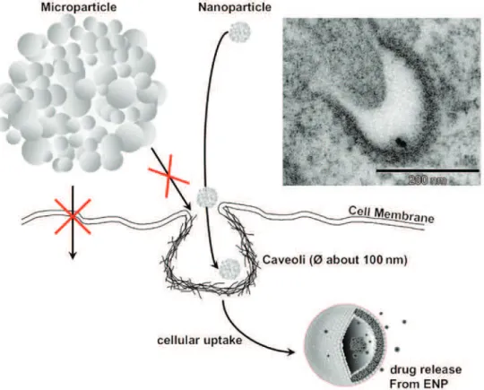
The most important property for the transport of ENM in tissues and cells is their specific small size. Only small particles have access to the vesicular structures on the surface of cells, allowing use of the small vesic-ular transport pathways that are inaccessible to larger particles with sizes above 300 to 500 nm (Fig. 4). Excluding phagocytosis, which can be accomplished only by very specialized “phagocytes” in our body and is a mechanism for larger objects, only ENM can use the small vesicular transport pathways to enter the cell [181, 182].
As an example, in the human lung the alveolar epithelial cells have several thousands of so-called “cave-olae” on their membrane surface at any given time, which means that there is a probability for inhaled ENM to
Fig. 4: Pathways for uptake of nanomaterials into cells. The importance of each pathway differs between cell types. For
example, phagocytosis is characteristic mainly for macrophages and granulocytes. The specific route through which nanomate-rials are internalized or transported inside the cell mostly depends on the material size and surface charge. Adapted from [45, 173].
enter epithelial cells via vesicular transport. Nevertheless, several studies have shown that the lung cleaning system (alveolar macrophages equals phagocytes) takes up most particles, largely independent of their size, and transports them out of the lung [183]. Other pathways of cellular uptake, such as receptor-coupled transport or passive diffusion, have been infrequently described and have not yet been confirmed by the community. The transport through small vesicles has been described as a “Trojan-horse-mechanism” (Fig. 5) [184], because this is an unintended uptake of foreign material which may have adverse effects in the interior of the cell.
4.2.2 Surface-principle
The smaller the particle, the higher is its surface-to-volume ratio (Fig. 6). A one micrometre particle has less than 2 % of its atoms located on the surface, whereas a five nanometer particle has more than 20 % of its atoms in direct contact with the surrounding environment. As all safety rules at workplaces or in the environ-ment are related to a certain mass per cubic meter of air or litre of water or kg of soil, the surface related dose has been often ignored so far. This question was raised in connection with animal studies on the pulmonary toxicity of ultrafine particles many years ago [185, 186] and it is currently more and more within the focus of researchers and regulators [187, 188]. However, it is still an open question if the mass per unit volume, the number, or the specific surface area is the best metric to describe a dose-response relationship for ENM [189].
Reducing the size of particles implies two important consequences for their interaction with the surround-ing environment. First, the surface itself becomes larger and reactions (e.g. catalysis or electron transfer) can occur with a higher probability, making such particles more reactive compared to their larger counterparts.
Fig. 5: Trojan-Horse-like transport of ENM into the interior of cells via vesicular pathways (caveolae). Small particles with a
diameter of less than 100 nm can fit into vesicular structures, such as caveolae, and be transported into the cytoplasm. Within the cells, these vesicles may form a lysosome with an acidic interior, facilitating the dissolution of materials such as ZnO. The TEM image shows two nanoparticles (22 nm) located within caveolae of a lung epithelial cell (A549) in culture. Reproduced with permission from [173].
Second, the smaller the particles, the higher is the potential occurrence of surface defects within the crystal-line structure or interatomic attractive forces facilitating direct interactions of neighbouring molecules with specific surface sites that are not present in the respective bulk material [153, 174]. These two surface proper-ties enhance the possible interaction with biological molecules such as lipids, proteins, or nucleic acids and may induce adverse effects in cells and tissues [152, 190].
4.2.3 Material-principle
Variations in ENM composition (either bulk or surface) lead to differences in their reactivity and behaviour in biological systems [191]. Gold or silver metal particles; metal oxides, such as SiO2, TiO2, ZnO, FexOy, and CeO2;
Fig. 6: An interesting analogy to illustrate the surface-to-volume ratio of engineered nanomaterials. If the particle (e.g. cube)
in the figure has a size a = 1 cm, the surface area of that particle is 6 cm2. By decreasing its size to a = 10 nm, while keeping the
same overall volume and mass, the surface area now grows to 600 m2. By reducing each particle further to a = 1 nm, the surface
and carbon-based materials, such as carbon black, nano-diamonds, carbon nanotubes, or graphene-based materials all have different binding capacities for biological molecules, different reactivity towards biological molecules such as proteins, lipids, or DNA, and different distribution patterns. Moreover, the formation of a molecular corona of biological substances, such as proteins, lipids, or other relevant molecules, is dependent on the material [8, 123] and the tendency of the material to associate strongly affects the behaviour and bio-logical effects of ENM [29, 192–197]. Thus, the material and its composition have a crucial role in determining the biological readout of exposure against ENM.
4.2.4 Fibre-principle
Besides the above described properties of ENM, the shape or aspect ratio is another important attribute with some predictive power for toxicity. There are several examples from the literature that describe the potential pathogenicity of “nanofibres” (HARN: high-aspect ratio nanomaterials [198, 199]) which might conform to the so-called ‘fibre pathogenicity paradigm’. This paradigm states that the pathogenicity of a fibre, espe-cially after inhalation, can be predicted “on a continuum based on its length and biopersistence, as well as its aspect ratio” [200]. The fibre-paradigm applies only to fibres with a key length larger than 5 micrometres where the diameter does not determine the biological effect, as long as it is less than 1 to 3 micrometres and the aspect ratio is a minimum 3:1 (length over diameter) [201]. Carbon nanotubes with a length range that fulfils the nanofibre criteria and that belong to what are termed WHO-fibres, defined by the fibre-paradigm, have been shown to induce similar pathogenicity as asbestos fibres [198, 200–202]. This specific biological effect becomes more and more relevant as carbon nanotubes and other fibre-like nanomaterials are used in an increasing number of technical applications. Medical examples include carbon nanotubes as candidates for drug delivery systems, especially for neuro-regeneration [203], and the use of functionalised carbon nano-tubes as tools for drug targeting, therapeutic applications, or imaging [204, 205].
The ‘fibre pathogenicity paradigm’ seems to be a broadly applicable rule, as it has been shown to apply not only to asbestos and carbon fibres (see above), but also to titanium dioxide nanobelts [206] and nickel nanowires [207]. As long as the aspect ratio is larger than 3:1 and the overall length is longer than 5 microme-tres, either stiff or rigid nanofibres can induce the pathological effects described above and such nanomateri-als should be handled with care during their whole life-cycle.
4.3 Toxicokinetics
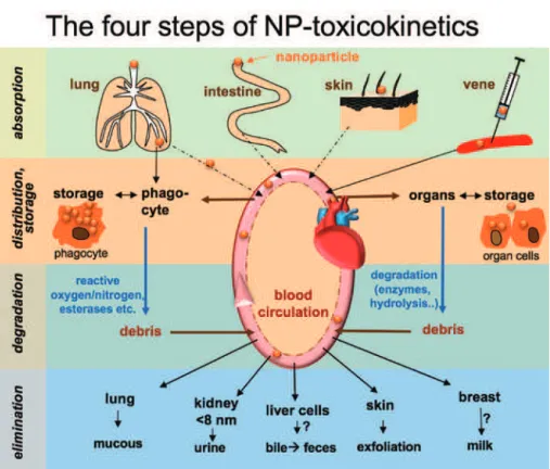
The passage of an ENM through the organism can be divided in four steps: absorption, distribution, degrada-tion, and elimination (Fig. 7). Each of these steps can be extremely variable between different ENMs. Before novel nanosized drugs and body-contacting consumer goods can be considered for applications, their biodis-tribution, biodegradation, and elimination must be carefully assessed [45]. Each type of nanoobject and each of its degradation products will have its own kinetic profile, determined in part by the restrictions imposed by biological barriers.
4.3.1 Biological barriers
Biological barriers are important for the protection of the organism from environmental influences and for its compartmentalization into organs. Barriers largely prevent the uptake of non-nutrient hydrophilic substances and particles, but allow the transport of nutrients (by membrane transporters). Major barriers that protect the organism from environmental influences are the skin, gastrointestinal tract, and the lung (Fig. 8). Some inner organs, such as the placenta and brain, are protected by additional barriers. Barriers commonly involve two structures: (1) a protecting layer of epithelial cells and (2) the adjacent cells that
form the supplying blood capillaries. The barrier-function is achieved by dense tight junctions between neighbouring epithelial cells and/or between the capillary-forming endothelial cells preventing paracellu-lar fluxes. In addition, the skin barrier has multilayered sheets of densely packed dead tissue on top of the
Fig. 7: Schematic illustration of the four steps of nanomaterial toxicokinetics: absorption, distribution/storage, degradation
and elimination.
Fig. 8: Schematic drawing showing that biological barriers may be formed by either densely packed organ cells and/or by
densely closed endothelial cells (forming capillaries). In the Intestinal barrier, the transcellular pathway is open for nutrients. The blood-brain barrier is impenetrable to large hydrophilic molecules and many ENM due to tight junctions between endothe-lial cells and a layer of surrounding pericytes. In the Placental barrier, the huge cell (Syncytiotrophoblast) leaves space for few paracellular pathways. The fetal endothelial cells are tightly connected, which means that particles smaller than 200 nm can cross the placenta, but particles above 300 nm remain in the mother’s body and do not cross the barrier.
living barrier [22]. Biological barriers are very important for three of the four toxicokinetics steps: absorp-tion, distribuabsorp-tion, and elimination.
4.3.2 Absorption
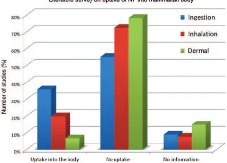
Applications of ENM in theranostics, food technology, cosmetics, or textiles all enable direct body contact. There are different pathways by which the uptake of ENM can occur (Fig. 7, absorption). A literature study evaluating publications between 2000 and May 2013 came to the conclusion that a substantial number of studies observed uptake via ingestion or inhalation, whereas dermal uptake is most unlikely for ENM [5] (Fig. 9).
The small intestine, where the majority of orally taken drugs will be absorbed, constitutes a barrier between the intestinal lumen and the blood formed by densely connected epithelial cells covered by layers of mucus. Nanoparticles are, in general, too large to be absorbed by diffusion through the enterocyte mem-branes. Likewise, permeation through the intact paracellular pathway (between cells) is impossible for mole-cules or ions larger than approximately 1 nm. Additional possible uptake routes involve endocytosis through epithelial cells and passage via M-cells of the immune system, found in the gastrointestinal tract (transcel-lular pathway), or through a diseased and disrupted barrier [208].
For the skin, it has been shown in many different studies and systems that almost none of a given ENM-dose applied to normal or UV-stressed skin reaches living cells in the deeper layers [165, 209–211]. Thus, it is assumed that nanoobjects cannot reach the bloodstream via the skin barrier in any clinically relevant amounts unless they are specifically designed for this purpose [169].
Nanoparticles in the ambient air or in inhalation sprays can be so small that they behave like gas mole-cules in the respiratory system and can even localise in alveoli, the gas exchanging structures of the lungs. Some transition into the bloodstream may occur. However, the overall percentages of the initial dose deliv-ered into the bloodstream are very low [212–214]. Most of the particles will be taken up by alveolar mac-rophages or reticulocytes. The macmac-rophages migrate to the upper airways and transport the ENM out of the lung and the reticulocytes migrate to the adjacent lymph nodes.
Fig. 9: Percentage of publications disclosing the uptake of NP into the mammalian body, classified either via oral (ingestion),
pulmonary (inhalation) or transdermal (dermal) route. The results are from a literature survey for articles published between 2000 and 2013.
4.3.3 Distribution
ENM used in nanomedicine (i.e. drug delivery systems and in vivo imaging probes) enter the body almost exclusively via the parenteral route. Recently, the use of the respiratory route for nanomaterial drug delivery has been explored because of the large, accessible surface area of the lungs and because the air-blood barrier is reasonably thin. Once an ENM crosses the air-blood barrier, the inhaled nanomaterial can enter the circu-lation and accumulate in secondary organs and tissues. Kreyling et al. studied the fate of nanoparticles that enter the lungs and performed a biokinetic analysis (dose-response study) as part of a comprehensive risk assessment (Fig. 10).
Once a small molecule drug or ENM has reached the blood, it will be distributed by the bloodstream to the tissues. However, it should not be assumed that all the ENM will be transported to the target tissue, since
Fig. 10: Biokinetics of nanoparticles after lung exposure. Scheme of translocation of nanoparticles from the lung epithelium to
the regional lymph nodes and to the blood. Thereafter, they may be distributed to and accumulate in various secondary organs, or they may be excreted via the kidneys. AM, Alveolar Macrophage. Reprinted with permission from [213].
adhesion to the walls of the blood vessel may occur. ENM shape and size are major factors governing NP flow in blood and adhesion to blood vessel walls [22, 215]. A literature survey indicated that nearly 100 % of all cell types tested in vitro (i.e. in a closed space) can take up ENM, with only minor exceptions (e.g. some graphene-based materials). Although ENM are taken up by all cell types, the uptake across internal tissue barriers, such as the placenta barrier and the blood-brain-barrier, is usually very restricted. A prominent example is the transport of drugs across the blood-brain barrier to cure brain diseases. Even after more than 30 years of research, it is still a problem to use nanocarriers to deliver a sufficient dose of a hydrophilic drug across this barrier [216, 217]. A somewhat different situation can be found at the placenta barrier. As this organ is spe-cialized to transport various larger molecules, such as proteins in the nanometer size range, ENM can cross this organ after a relatively short time [218–220]. This transport is both size-dependent, as particles smaller than 240 nm can cross but larger particles cannot, and surface dependent, as negatively charged particles are transported much more slowly than neutral or positively charged ENM.
Other, usually less effective, uptake mechanisms may occur [45]. These involve vesicle involution, such as by pinocytosis or receptor mediated endocytosis (clathrin or caveolin-based). Engineered nanoparticle drugs equipped with adequate receptors can make use of endocytosis uptake routes. When particles are larger than 200 nm, they will preferentially be taken up by phagocytosing cells of the immune system, such as macrophages or the hepatic Kupffer cells. If ENM are found to be stored in the liver, they will often be confined to phagocytosing Kupffer cells. It has also been reported that airborne nanoparticles, inhaled via the nose, may travel from the nasal epithelium along the olfactory tract to the otherwise inaccessible brain [178].
4.3.4 Degradation
Except for permanent prostheses, all foreign materials, including ENM, that are introduced into the human body must be eliminated sooner or later via natural pathways to avoid undesired retention. Biochemical processes in phagocyting cells can be highly aggressive and degrade many types of organic compounds by the enzymatic formation of reactive oxygen and nitroxide species, thus releasing ions, molecules, or macro-molecule debris. This is a major and unspecific biodegradation mechanism that often starts at the surface of particles. In parallel, degradation can be caused by abiotic chemical processes, especially hydrolysis and oxi-dation, in various tissues. The particular case of hydrolytically degradable polymers of the aliphatic polyester type, such as glycolic and lactic acid homopolymers and copolymers that are currently used as nano objects for drug delivery, has been extensively discussed, including changes in composition and morphologies [221– 223]. An entrapped drug can play an important role in particle degradation, depending on its hydrophilic-ity or hydrophobichydrophilic-ity. Targeting proteins on the surface will be degraded by proteases and released small molecules, such as drugs, may be subject to classic biotransformation reactions.
Intact ENM will rarely be subject to the classic biotransformation pathways that are known for small molecules, such as phase I (oxidation) and phase II (conjugation) reactions. This is because, first, ENM will not easily enter cells of the drug-metabolizing organs (predominantly liver cells), and second, their constitu-ents are usually not typical substrates for the common biotransformation enzymes. Therefore, various other mechanisms must prevail.
Similarly, inorganic nanoparticles can also be degraded, as elegantly summarised in a review by Feliu et al. The authors reviewed in vivo degeneration and the fate of a variety of inorganic nanoparticles, and concluded that NP in vivo should no longer be considered as homogeneous entities, but rather as inorganic/ organic/biological nano-hybrids with complex and intricately linked distribution and degradation pathways [224]. For insoluble inorganic particles (e.g. gold or magnetic particles) uptake and storage in phagocytosing cells may be followed by slow dissolution due to low pH in lysosomes or other solubility effects. Residue-free dissolution in the extracellular space is possible for soluble inorganic ENM (e.g. mesoporous silica, ZnO, CuO). Some carbohydrate and peptide polymers can be degraded in the extracellular space by amylases, peptidases, and hydrolases. Hydrolytically degradable polymers include poly(lactic acid)s as well as many
other aliphatic polyesters, poly(anhydride)s, and some polyphosphazenes. Some products can enter the intermediary metabolism. ENM containing a mixture of constituents may be disassembled or biodegraded to smaller entities, which may be stored or further degraded by the above-mentioned mechanisms.
4.3.5 Elimination
If the degradation of ENM is not complete, then the remaining particles must be eliminated to avoid accumulation. The most common route of elimination/excretion is filtration in the kidneys. In the healthy kidney, filtration of nanoparticles can be nearly complete when the particle diameter is below 2 nm and is still efficient at a size of 5 nm, but tends to stop at particle sizes above 6 to 8 nm. Surface charge and interac-tions with biomolecules (corona formation and association) may attenuate renal filtration. Pulmonary par-ticles can be excreted by ciliated epithelium of the airways [225]. Biliary excretion, a major route for small drug molecules, has been observed, but it is unclear if this can be a general route for particle elimination. Zhang et al. published the rather alarming finding that small (8 nm) TiO2 nanoparticles were transferred from dams to pups through breastfeeding during lactation, likely through the disrupted blood-milk barrier [226]. However, this was an indirect conclusion and has yet to be unequivocally confirmed.
4.4 Difficulties associated with the study of nanoparticle toxicity
The full characterization of ENM is an indispensable step prior to biological investigations [9]. Furthermore, the concentration, size, and charge of ENM in biological media must be known if we are to understand how their properties in a biological environment contribute to human cell, tissue, organ, and organism toxicity. Much experimental work today is done with isolated, cultured cells [227]. Although this is a reasonable and often quick approach (e.g. using flow cytometry), such results provide only limited information about the situation in the more than 200 other cell types of the organism or about the role of barriers. Influences of blood flow, renal filtration, or extracellular degradation enzymes are largely ignored in cellular test systems. Additionally, ENM degradation in cultured cells may deviate from that in the healthy organism. Cells in vitro tend to divide faster than the respective cells in the healthy organism, possibly concomitant with an unchar-acteristic cellular particle uptake rate. A complete picture of toxicokinetics is only gained in studies in more complex physiological systems, such as perfused organs and whole animals [228]. In other words, and con-trary to frequent affirmation, cytocompatibility does not imply biocompatibility.
Because of their specific physicochemical properties, the behaviour of nanomaterials in a biological environment is different from that of “normal” chemicals. Nanoobjects in biological systems are difficult to analyse compared to their pristine state, since the ENM is present in low concentrations, may be decom-posed or associated, and must be analysed against a complex matrix background. These considerations frequently require sample pre-treatment to remove interfering species or increase the concentration. Even labelling methods do not readily provide clear information when particle association and degradation occur [229, 230]. In addition, the methods which are available for the analytical characterisation of ENM [9] often require specialized equipment and demand an experienced operator. Without adequate characterization of the material under investigation, the results of toxicological studies are more or less worthless, as has been pointed out recently [231].
The next problem for the interpretation of the data from nanotoxicological experiments is the missing comparability. Most researchers do not use reference nanomaterials to verify a specific biological response or to understand how their biological model responds to such stimuli or toxic compounds [232]. Further-more, ENM often interfere with the analytical devices or methods, leading to false results and interpretations [233–235]. Most often, the methods used are not standardised or validated, although several groups have demanded better validation [236, 237]. Finally, the general implementation of measurement uncertainty that is needed to compare results of different studies has yet to be accomplished [238].
The investigation of ENM with cultured cells and animals also has a very specific weakness: the applied dose or concentration! In contrast to dissolved chemicals or toxins in biological assays, nanoparticles may not distribute equally within the suspensions. Depending on their density, they may sediment quickly (100 % of the given amount is directly in contact with the treated cells) or float on the surface (the cells “see” none of the applied nanoparticles in the test). The surface of the nanoparticles may be covered by proteins or other natural molecules, thereby hiding reactive surfaces, or the nanoparticles may agglomerate and change their size during the treatment. All these parameters will influence the biological response. Moreover, the actual literature reports the concentrations of nanoparticles in biological systems as mass per volume, surface or cell, number of particles per cell or volume, or NP surface area per surface area of culture disc, thus prevent-ing direct comparison of experimental results from different studies. Until now there is no agreement on the appropriate dose metrics for nanomaterials, although some approaches have been published [189, 239, 240]. Taking all these aspects together, the difficulties in the interpretation of published results are tremendous. This results in a very controversial situation, as has been demonstrated recently [5]. The domain of protein-surface interactions provides a good illustration of these issues. Most of the in vitro studies reported so far considered individual proteins that were generally far from physiological concentrations, mostly because of limitations imposed by analytical techniques. Different results may be obtained when mixtures of proteins, serum, or plasma are considered. Albumin, the predominant protein in blood, appears to play an important role in reducing the adsorption of the other blood proteins and the activation of platelets [241].
4.5 Case study: quantum dots
Quantum dots (QD) illustrate the challenges associated with the development of a rational, science-based approach to nanotoxicology. The superior optical properties of QD make them promising candidates for applications such as in vivo and in vitro imaging and drug delivery and they are likely to be used in various manufactured products, such as dye-sensitized solar cells, sensors, and light emitting diodes [242–244]. Given the widespread interest in medical applications of QD and the likelihood of their incorporation in a range of consumer products, it is critical to understand the possible health risks associated with exposure to these nanomaterials. There is a common perception that QD are toxic and therefore are unlikely to be approved for clinical applications [242]. This is based primarily on early studies of QD containing cadmium, a metal known for its high chronic toxicity. Cell culture experiments have demonstrated that a combination of the release of toxic Cd ions and the photo-induced production of reactive oxygen species is responsible for the toxicity of Cd-containing QD [242]. Nevertheless, one cannot generalize these conclusions, since later studies have shown that protecting QD with a shell (e.g. ZnS or silica, which are porous) can minimize toxicity. Further-more, there are now numerous QD compositions that exclude Cd, although some include components such as indium, for which limited toxicological data are available, and alternative types of luminescent nanomateri-als, such as carbon nanodots, show considerable promise and are likely to be less toxic.
QD are a heterogeneous family of nanomaterials that can have many different compositions for both core and shell, exist in a range of sizes, and have a diversity of surface chemistries. These factors are recognized to have a significant role in controlling their in vivo biodistribution and activity. In addition to the complex-ity in QD composition and properties, cytotoxiccomplex-ity studies have used different assays and different cell lines and are sometimes confounded by interference between QD and the assay reagents. In some cases, in vitro studies demonstrating toxicity have been shown to be relatively poor models for in vivo toxicity. For example, researchers found that QD-based imaging using monkeys caused no adverse effects, although QD aclated in lymph nodes, bone marrow, liver, and spleen for up to 3 months after injection. The long term cumu-lative effects of degradation in the environment or in biological systems have not been investigated. However, it is possible that cadmium-containing materials can exert genotoxic, epigenetic, and metalloestrogenic effects [243] and that the lack of clearance of QD may potentially lead to effects on reproductive health [245].
A recent critical review has suggested a number of possible solutions to deal with the variability in exper-imental methods that have been used to study QD toxicity [242]. These include the standardization of the dose