Dynamics of vascular normalization during anti-angiogenic
therapy: Implications for combination therapy
by
Ricky T. Tong B.S. Chemical Engineering California Institute of Technology, 2000
SUBMITTED TO THE HARVARD-MIT DIVISION OF HEALTH SCIENCES AND TECHNOLOGY IN PARTIAL FULFILLMENT OF THE REQUIREMENTS FOR THE
DEGREE OF
DOCTOR OF PHILOSOPHY IN MEDICAL ENGINEERING AT THE
MASSACHUSETTS INSTITUTE OF TECHNOLOGY May 2005 [Juis 2 ;
© 2005 Ricky T. Tong. All rights reserved. The author hereby grants to MIT permission to reproduce
and to distribute publicly paper and electronic copies of this thesis document in whole or in part.
Signature of Author:
Certified by:
OFTECHNOLG
JUN 3 2005 LIBRARIES
Harvard-MIT DixNsion otealth Sciences and Technology May 5, 2005
Rakesh K. Jain, Ph.D. Andrew Werk Cook Professor of Tumor Biology Harvard Medical School/Massachusetts General Hospital Harvard-MIT Division of Health Sciences and Technology Thesis Supervisor
N;¢u r
Accepted by:
Cc
v \ |[ Martha L. Gray, Ph.D. Edward Hood Taplin Professor of MediCal and Electrical Engineering >-Director Harvard-MIT Division of Health Sciences and Technology
Abstract
Dynamics of vascular normalization during anti-angiogenic
therapy: implications for combination therapy
Ricky T. Tong
Submitted to the Harvard/MIT Division of Health Sciences and Technology on May 5, 2005 in Partial Fulfillment of the Requirements for the degree of
Doctor of Philosophy in Medical Engineering
Solid tumors require blood vessels for growth, and the goal of anti-angiogenic therapy is to destroy the tumor vasculature. Recent findings suggest that anti-angiogenic therapy enhances radiation and chemotherapy responses. These findings seem paradoxical, since anti-angiogenic therapy prunes tumor vasculature while chemotherapy and radiation therapy rely on the vasculature to transport cancer drugs and oxygen, respectively, to cancer cells. To resolve this paradox, we propose that anti-angiogenic therapy can "normalize" the tumor vasculature transiently, resulting in a more efficient delivery of drugs and oxygen to cancer cells. We first show that DC101, a monoclonal antibody targeting Vascular Endothelial Growth Factor Receptor 2 (VEGFR2), prunes immature blood vessels, reduces vascular diameter and improves pericyte and basement membrane coverages. Functionally, the vascular permeability to macromolecules and interstitial fluid pressure are reduced. By lowering interstitial fluid pressure while maintaining microvascular pressure, DC101 induces a hydrostatic pressure gradient across the vascular wall, which leads to enhanced penetration of macromolecules in tumors. Tumor hypoxia is also reduced, and it is associated with the increased red blood cell velocity after DC101 treatment. Using gene array, real time PCR and Western blot analyses, changes in angiopoietin-2 level during DC101 treatment are identified. To test if similar effects happen in clinical setting, we obtained tumor biopsy samples from rectal adenocarcinoma patients treated with bevacizumab, an anti-VEGF monoclonal antibody. Our analysis shows that after bevacizumab treatment, microvascular density of the tumors decreases while pericyte coverage increases. The level of angiopoietin-2 also decreases, similar to the pre-clinical data. Thus our work shows a potential mechanism that explains the synergism between anti-angiogenic therapy and conventional therapies. These findings should facilitate the design of optimal dose and schedule of anti-angiogenic therapy.
Thesis Supervisor: Rakesh K. Jain
Title: Andrew Werk Cook Professor of Tumor Biology
Harvard Medical School/Department of Radiation Oncology, Massachusetts General Hospital
Biographical Sketch
EDUCATION
2000 California Institute of Technology, Chemical Engineering, B.S.
PROFESSIONAL EXPERIENCE
Engineering Assistant, Project Technical Liaison Associates, Inc., TX
Research Assistant, California Institute of Technology, Department of Chemistry and Chemical Engineering. Advisor: Professor John D. Roberts
Research Assistant, California Institute of Technology, Department of Chemistry and Chemical Engineering. Advisor: Professor Jacqueline K. Barton
Research Assistant, Chinese University of Hong Kong, Department of Chemistry. Advisor: Professor Lee Hung Kay
Summer Intern, Genentech, Protein Engineering Department. Advisor: Dr. Andrea Cochran
Research Assistant, California Institute of Technology, Department of Chemistry and Chemical Engineering. Advisor: Professor Mark E. Davis
Doctoral Candidate, Medical Engineering, Harvard-MIT Division of Health Sciences and Technology
Research Fellow, Massachusetts General Hospital and Harvard Medical School. Advisor: Professor Rakesh K. Jain
6/96-9/96 1/97-6/97 1/97-6/98 6/98-9/98 6/99-9/99 9/98-6/00 9/00-Present 2/01 -Present
HONORS AND AWARDS
Howard Hughes Medical Institute SURF Fellow (1997), Donald S. Clark Award (1998), Caltech Merit Scholar Award (1998 and 1999), Vice President of CCSA Caltech Chinese Student Association (1997-99), Officer of the Entrepreneur Club (1998-00), Treasurer of the Caltech Student Chapter of Tau Beta Pi (1999-00), President of the Caltech Student Chapter of the American Institute of Chemical Engineers (1999-00), President and Founder of the Student Chapter of the Biomedical Engineering Society (1999-00), NSF Graduate Fellowship (2000-03), Angiogenesis Foundation Honorable Mention (2003), Susan Komen Foundation Fellowship (2003-05)
PROFESSIONAL ACTIVITIES
Teaching assistant in Transport Phenomena (2000) Teaching assistant in Tumor Pathophysiology (2003)
Harvard-MIT Division of Health Sciences and Technology Social Chair (2001)
Harvard-MIT Division of Health Sciences and Technology Ph.D. Admissions Committee (2004-2005)
PUBLICATIONS
Holmlin, R. E., Tong, R. T., and Barton, J. K. (1998). Long-range triplet energy transfer between
metallointercalators tethered to DNA: Importance of intercalation, stacking, and distance. Journal of the
American Chemical Society 120, 9724-9725.
Cochran, A. G., Tong, R. T., Starovasnik, M. A., Park, E. J., McDowell, R. S., Theaker, J. E., and Skelton,
N. J. (2001). A minimal peptide scaffold for beta-turn display: Optimizing a strand position in disulfide-cyclized beta-hairpins. Journal of the American Chemical Society 123, 625-632.
Alexandrakis, G., Brown, E. B.*, Tong, R. T.*, McKee, T. D., Campbell, R. B., Boucher, Y., and Jain, R.
K. (2004). Two-photon fluorescence correlation microscopy reveals the two-phase nature of transport in tumors. Nature Medicine 10, 203-207. * These authors contributed equally.
Willett, C. G., Boucher, Y.*, Di Tomaso, E.*, Duda, D. G.*, Munn, L. L.*, Tong, R. T.*, Chung, D. C., Sahani, D. V., Kalva, S. P., Kozin, S. V., Mino, M., Cohen, K. S., Scadden, D. T., Hartford, A.C., Fischman, A. J., Clark, J. W., Ryan, D. P., Zhu, A. X., Blaszkowsky, L. S., Chen, H. X., Shellito, P. C.,
-3-Lauwers, G. Y., and Jain, R. K. (2004). Direct evidence that the VEGF-specific antibody bevacizumab has antivascular effects in human rectal cancer. Nature Medicine 10, 145-147. * These authors contributed equally.
Tong, R. T., Boucher, Y., Kozin, S. V., Winkler, F., Hicklin, D. J., and Jain, R. K. (2004). Vascular
normalization by vascular endothelial growth factor receptor 2 blockade induces a pressure gradient across the vasculature and improves drug penetration in tumors. Cancer Res 64, 3731-3736.
Winkler, F.*, Kozin, S. V.*, Tong, R. T., Chae, S. S., Booth, M. F., Garkavtsev, I., Xu, L., Hicklin, D. J., Fukumura, D., di Tomaso, E., Munn, L. L., and Jain, R. K. (2004). Kinetics of vascular normalization by
VEGFR2 blockade governs brain tumor response to radiation; Role of oxygenation, angiopoietin-1, and matrix metalloproteinases. Cancer Cell 6, 553-563. * These authors contributed equally.
Xu, L., Tong, R. T., Cochran, D. M., and Jain, R. K. (2005). Blocking PDGF-D/PDGFRi signaling inhibits human renal cell carcinoma progression in an orthotopic mouse model. Cancer Res (In press)
INVITED TALKS
Tong, R.T., "Dynamics of vessel normalization following VEGF blockade." 2004 Keystone Symposia:
Angiogenesis, Jan 1, 2004.
Tong, R.T., "Vascular normalization by VEGFR2 blockade improves molecules penetration and radiation response in two tumor models." 2005 American Association for Cancer Research Annual Meeting, April 20, 2005.
ABSTRACTS
Tong, R.T., Jain, R.K., "The effects of anti-angiogenic therapy on the anatomical and functional properties of solid tumors," HST Forum, (2002).
Tong, R.T., Boucher, Y., Kozin, S.V., Jain, R.K., "Anti-VEGFR2 blocking antibody normalizes the tumor vasculature and microenvironment." MGH Clinical Research Day (2003).
Tong, R.T., Jain, R.K., 'The effects of anti-angiogenic therapy on the anatomical and functional properties of solid tumors," HST Forum, (2003).
Tong, R.T., Boucher, Y., Jain, R.K., "Dynamics of vessel normalization following VEGF Blockade," SAC MGH Research Symposium, (2004).
Tong, R.T., Boucher, Y., Hicklin, D.J., Jain, R.K., "Dynamics of vessel normalization following VEGF Blockade," Proceedings of the American Association for Cancer Research, 2556 (2004).
Tong, R.T., Boucher, Y., Kozin, S.V., Hicklin, D.J., Jain, R.K., "Dynamics of vessel normalization following VEGF Blockade," Breast Cancer Research at Harvard, (2004).
Tong, R.T., Jain, R.K., "Dynamics of vessel normalization following VEGF Blockade," HST Forum,
(2004).
Tong, R.T., Boucher, Y., Kozin, S.V., Hicklin, D.J., Jain, R.K., "Vascular normalization by VEGFR2
blockade induces a pressure gradient across the vasculature and improves drug penetration in tumors."
MGH Clinical Research Day (2004).
Tong, R.T., Jain, R.K., "Vascular normalization in tumors by anti-angiogenic therapy: from animal models to clinical trial." HST Forum (2005).
Acknowledgements
"If you think in terms of a year, plant a seed; if in terms of ten years, plant trees; if in terms of 100 years, teach the people."
Confucius, BC 551-479
I would like to start by thanking my thesis advisor, Professor Rakesh Jain for his guidance and advice. He is always inspirational and constantly pushes me to reach my full potential. I am very grateful for all of his help as I learn to become an independent researcher. He has also provided me with numerous opportunities to work with other lab members and outside collaborators and to attend conferences. His attitude towards work and colleagues has created a very friendly and supportive working environment.
I would like to thank the members of my thesis committee, Professors Robert Langer (MIT), Professor William Deen (MIT), and Professor Bruce Zetter (HMS). Their advice, input, and support have been invaluable. I would also like to thank the Harvard-MIT Division of Health Sciences and Technology and the MIT Chemical Engineering Department for opening many educational avenues and building solid and rigorous foundation for my career.
My colleagues and friends in the Steele Lab have always been there for me when I need them the most. I am deeply grateful for their help and time. They have always been very patient with me, as I often have many questions and need lot of assistance. In particular, I would like to thank Frank Winkler and Sergey Kozin as they are part of the "normalization team". I truly enjoy working with them to discover the wonders of DC101 together. I would like to thank Yves Boucher, Dan Duda, Emmanuelle di
5-Tomaso, Lance Munn, Edward Brown, Mike Booth, and Dai Fukumura for their help, which I immensely appreciate. I have also benefited tremendously through working with George Alexandrakis, Lei Xu, Igor Garkavtsev, and Jeroen Hagendoorn. I would also like to thank Patrick Au, Wilson Mok, and Satoshi Kashiwagi for many of our wonderful discussions. I can always expect them to be in lab with me late at night. I would like to thank Sylvie Roberge, Julia Khan, and Peigen Huang for their help with the animal work. I would like to thank Chelsea Swandal, Melanie Berg, Russell Delgiacco, Lucine Petit, and Carolyn Smith for their outstanding technical support and dedication to the projects. I am deeply grateful for the help from Phyllis McNally, Tara Belezos, and Doug Stay throughout the years. I would like to thank David Cochran, Ryan Lanning, Pooja Pathak, Dennis Dolman, Naoto Koike, Tim Padera, Brian Stoll, Josh Tam, Rosemary Jones, and Hide Isaka for their valuable input into my work. A special thank to Marek Ancukiewicz for all the last minute help on statistics. I am very thankful for all of the interesting discussion with James Baish, and for his help on fractal analysis on tumor vasculature. I would also like to thank my UROP students Jennifer Lobo, Jenny Ruan, Margaret Kim, and Sam Kwei. I would not have had the luxury to work on as many projects without their assistance.
I would like to thank Christopher Willett and Tracy Batchelor for providing me the wonderful opportunities to work on clinical trials. They have opened my eyes to the exciting world of clinical research. I would also like to thank my outside collaborators, Peter Carmeliet (University of Leuven), Annelii Ny (University of Leuven), Raju Kucherlapati (HMS), Qingcong Lin (HMS), Badri Roysam (Rensselaer Polytechnic Institute), and Alex Tyrrell (Rensselaer Polytechnic Institute). Working with them led
me to appreciate the broad spectrum of cancer research. I would like to thank Dan Hicklin (ImClone) for DC101, Eugene Renkin (University of California Davis) for nylon wicks, David Jackson (Oxford University) for LYVE-1 antibodies. I am grateful to the NSF and Susan G. Komen Foundation for funding my research.
Mostly I would like to thank my family and friends. My parents, YuShan and Winnie Tong, and my brother, James Tong, provide me the support and encouragement that a child dreams of. Their sacrifice allowed me to pursuit my dream, and has made my PhD study seems completely painless. Finally, I can never thank enough my friends who have been endlessly editing my papers, thesis, and application essays. Chirag, Sophia, Paul, Janice, Jackie, Karen, Clay, Richard, Kevin, Lisa, Sindy thank you so much for all of your help and support.
Thank you to all of the people who have been so patient and helpful in molding my career and future.
-7-Table of contents
A bstract ... 2
Biographical Sketch ... 3
A cknow ledgem ents...5
Table of contents ... 8
List of Figures ... 10
List of Tables ... 13
List of Tables ... 13
Chapter 1: Original Contribution ... 14
Chapter 2: Introduction ... 20
Chapter 3: Specific Aims ... 25
Chapter 4: Background ... 30
Barriers to drug delivery: A need for finding a new therapy ... 31
Tumor Pathophysiology ... 32
Tumor Interstitial Fluid Pressure ... 32
Angiogenesis ... 33
Anti-angiogenic treatment ... 35
Vascular Normalization ... 36
Chapter 5: Material and Methods ... 38
5.1 Animal models and therapeutic agents ... ... 39
5.2 Microscopy ... 41
5.5 Molecular techniques ... 53
Chapter 6: Vascular Structure in Tumors ... 56
Chapter 7: Vascular Function and Tumor Microenvironment ... 71
Chapter 8: Molecular Changes during DC 101 Treatment ... 110
Chapter 9: Bevacizumab Phase I Human Clinical Trial ... .... 122
Chapter 10: Discussion and Future Perspective ... 136
R eferences ... 144
Appendix 1 - Anti-Angiogenic Therapy and Chemotherapy ... 156
Appendix 2 - Anti-Angiogenic Therapy and Radiation ... 161
Appendix 3 - RBC Velocity Analysis ... 163
-9-List of Figures
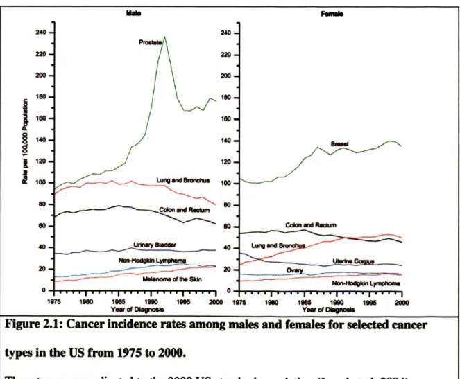
Figure 2.1: Cancer incidence rates among males and females for selected cancer types in
the US from 1975 to 2000 ... 21
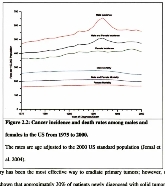
Figure 2.2: Cancer incidence and death rates among males and females in the US from 1975 to 2000 ... 22
Figure 2.3: Schematic of changes in tumor vasculature during the course of anti-angiogenic therapy ... 24
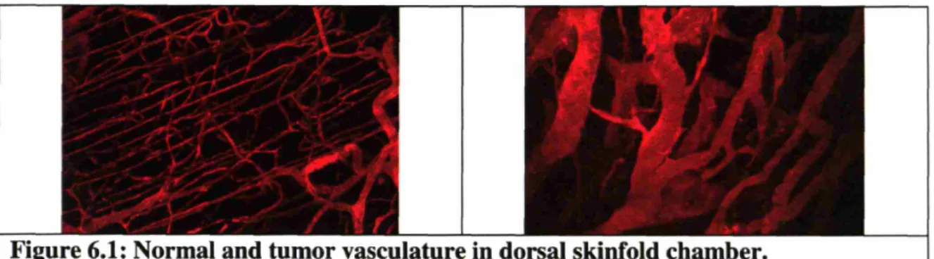
Figure 6.1: Normal and tumor vasculature in dorsal skinfold chamber ... 59
Figure 6.2: Normal and tumor vasculature in cranial window ... 59
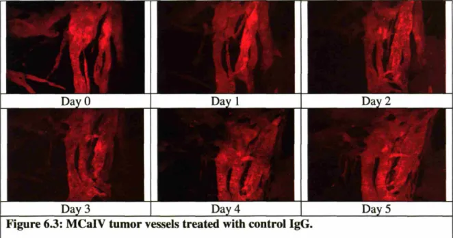
Figure 6.3: MCaIV tumor vessels treated with control IgG ... 60
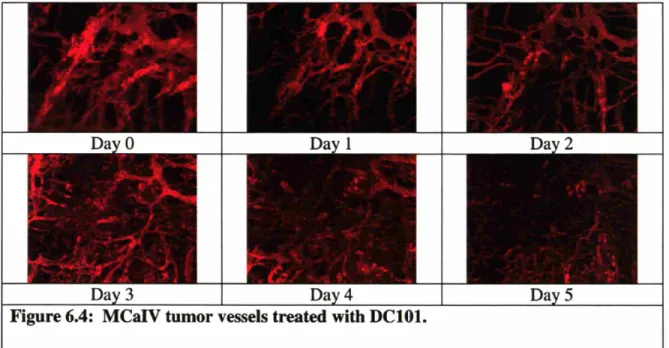
Figure 6.4: MCaIV tumor vessels treated with DC101 ... 61
Figure 6.5: LS174T tumor vessels treated with DC101 ... 61
Figure 6.6: DC101 lowers vascular density and diameter in MCaIV tumors ... 62
Figure 6.7: Vascular diameter distributions in MCaIV tumors ... 63
Figure 6.8: Vessel morphology in U87 tumors... 64
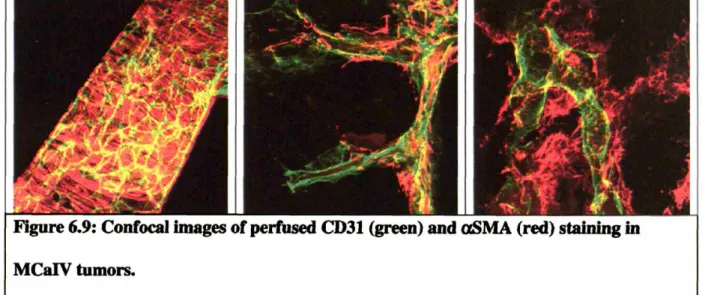
Figure 6.9: Confocal images of perfused CD31 (green) and aSMA (red) staining in M C aIV tum ors ... 65
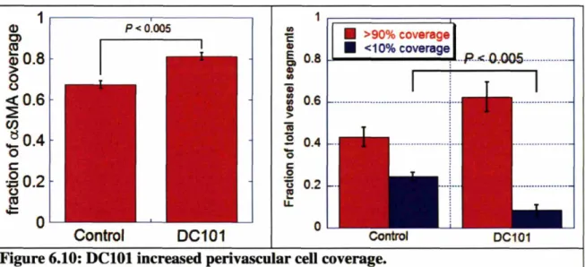
Figure 6.10: DC101 increased perivascular cell coverage. ... 66
Figure 6.11: DC 101 improved basement membrane coverage . ... 67
Figure 6.12: Quantification of collagen IV staining. ... 68
Figure 7.1: DC101 lowered interstitial fluid pressure in U87 tumors in nude mice ... 74
Figure 7.3: DC101 lowered interstitial fluid pressure in spontaneous tumors developed in
aged C 3H m ice ... ... 76
Figure 7.4: Ferritin functional lymphangiography and LYVE-1 staining in MCaIV tumors after D C 101 treatm ent ... ... 77
Figure 7.5: DC101 lowered vascular permeability of albumin in MCaIV tumors ... 78
Figure 7.6: DC101 lowered interstitial oncotic pressure ... 79
Figure 7.7: DC 101 induced a hydrostatic pressure gradient across the vascular wall ... 80
Figure 7.8: DC 101 increased penetration of macromolecules from blood vessels ... 81
Figure 7.9: DC101 reduced tumor hypoxia. ... 83
Figure 7.10: Line scan m ethod... ... 85
Figure 7.11: Normal brain vessels after control IgG treatment ... 86
Figure 7.12: RBC velocities of normal brain vessels. ... 87
Figure 7.13: RBC velocities as a function of time ... 88
Figure 7.14: U87 glioblastoma tumor vasculature during DC101 treatment ... 89
Figure 7.15: RBC velocity and vessel diameter of U87 tumors in the control IgG-treated group ... 90
Figure 7.16: RBC velocity and vessel diameter of U87 tumors in the DC101-treated group ... ... 9 1 Figure 7.17: DC 101 lowered vascular diameter in orthotopic U87 glioblastoma model. 92 Figure 7.18: Effect of DC 101 on RBC velocity of U87 tumor vessels ... 93
Figure 7.19: Computer traces of 3D vascular network. ... 102
Figure 7.20: Calculated pressure and interstitial profiles (Case I) ... 104
Figure 7.21: Calculated pressure and interstitial profiles (Case II) ... 105
-11-Figure 7.22: Combination of radiation and anti-angiogenic therapies was only synergistic
during the normalization time window. ... 107
Figure 8.1: DNA MicroArray ... 113
Figure 8.2: Real time-PCR. DC101 reduced the Ang-2 gene expression ... 115
Figure 8.3: Ang-2 Western Blot ... 116
Figure 8.4: Ang-2 Immunostaining on MCaIV tumors. ... 117
Figure 8.5: Ang-1 protein level increased after DC101 treatment ... 120
Figure 9.1: Microvascular density. ... 124
Figure 9.2: Perivascular cell coverage. ... 126
Figure 9.3: Fraction of vessels with pericyte coverage ... 127
Figure 9.4: Angiopoietin-2 immunostaining ... 128
Figure 9.5: Quantification of Angiopoietin 2 immunostaining ... 129
Figure 9.6: Interstitial fluid pressure pre- and post-bevacizumab treatment ... 132
List of Tables
Table 1.1: Original contribution ... ... 19 Table 8.1: Gene Array Data ... 114 Table 9.1: Summary of Ang- 1 and Ang-2 expression profiles in human colon, breast, and brain tum ors ... 133 Table 10.1: Pre-clinical evidences supporting normalization of the tumor vasculature. 140 Table 10.2: Effects of VEGF blockade in both pre-clinical and clinical data ... 141
Recent pre-clinical and clinical trials suggest that anti-angiogenic therapy should be combined with cytotoxic/radiation therapy for successful treatment of solid tumors. However, there are no generally accepted guidelines for optimal scheduling of these therapies. The synergistic results seen in combined therapy also present a paradox. One would expect that anti-angiogenic therapy, which aims to destroy tumor vasculature, would severely compromise, instead of enhance, the delivery of oxygen and therapeutics to the solid tumors, and thus lead to a less effective chemotherapeutic/radiation response. The primary goal of this thesis is to resolve this paradox by understanding the dynamic changes during anti-angiogenic therapy. We propose that anti-angiogenic therapy will transiently "normalize" the tumor vasculature by passively pruning immature tumor blood vessels and actively remodeling the remaining vasculature. We further hypothesize that the changes in vascular function during this normalization time window will enhance the delivery of molecules in tumors. This work will drive our understanding of general tumor pathophysiology and may improve treatment of solid tumors using anti-angiogenic therapy.
To get a better picture of the effects of anti-angiogenic therapy on tumors, multiple approaches were used to characterize the changes. By implanting tumors in a chronic window model such as the dorsal skinfold chamber window or cranial window in mice, daily three dimensional images of tumor vasculature were captured using two photon laser scanning microscopy (Chapter 6). A few days after DC101 treatment, an anti-murine VEGFR2 antibody, vascular density and diameter significantly decreased. Surprisingly, many of the vessels became less tortuous during the treatment.
15-The cellular structure around the tumor vasculature was analyzed using immunostaining techniques to determine if some tumor blood vessels were more vulnerable to angiogenic therapy than others. The results demonstrated for the first time that anti-angiogenic therapy improves pericyte coverage and basement membrane coverage around tumor blood vessels. Interestingly, by monitoring pericyte coverage throughout the treatment, it became apparent that anti-angiogenic therapy both preferentially pruned tumor blood vessels with no pericyte coverage, and at the same time, indirectly increased the pericyte coverage of the remaining vessels.
Next, the effects of these structural changes in the tumor vasculature on vascular function, tumor microenvironment, and more importantly, the delivery of molecules in tumors were studied. Using intravital microscopy, tumor blood vessels were demonstrated to be less leaky during the treatment (Chapter 7). Of interest, interstitial fluid pressure (IFP) was significantly lowered after the treatment. Using the micropipette technique, microvascular pressure (MVP) and interstitial fluid pressure were measured in one preparation. While interstitial fluid pressure was reduced after DC101 treatment, no changes were observed in microvascular pressure. Thus, anti-angiogenic therapy induced a hydrostatic pressure gradient across the vascular wall.
Fluid movement across the vascular wall is governed by the Starling's equation: Jv = Lp [(MVP - IFP) - (p - rn)]
which states that the rate of fluid movement across a unit area of vascular wall, or fluid flux (Jv), is proportional to both hydrostatic (MVP, IFP) and oncotic pressure (p, 7r) difference across the vascular wall. Interestingly, we found that the modification in hydrostatic pressure was accompanied by a change in interstitial oncotic pressure.
Moreover, by using experimentally measured values and the mathematical model developed by Baxter and Jain (Baxter and Jain 1989), it also showed that DC101 lowered interstitial fluid pressure throughout the entire tumor, and the model further predicted an increase in interstitial fluid velocity. Using two photon line scan method, we showed that red blood cell velocity increased after DC101 treatment, and the increase was associated with a reduced tumor hypoxia.
Next, to determine the molecular changes in tumors during anti-angiogenic therapy, tumor samples were obtained, and mRNA and protein were examined (Chapter 8). Gene array and real time PCR data showed that angiopoietins were modified during the treatment. The change was further confirmed by Western blot analysis.
But are these experiments in mice relevant to cancer patients? Our clinical collaborators began a phase I clinical trial using bevacizumab, an anti-VEGF monoclonal antibody, to characterize its effects on rectal cancer (Chapter 9). Patients with primary and locally advanced adenocarcinoma of the rectum were enrolled in a preoperative treatment protocol of bevacizumab administration alone, followed 2 weeks later by concurrent administration of bevacizumab with 5-fluorouracil and external beam radiation therapy to the pelvis. Surgery was performed to resect remaining disease 7 weeks after treatment completion. Tumor biopsies were obtained before and 12 days after initial bevacizumab administration, and immunohistochemistry was performed on these samples. Confirming the results from the animal studies, the human tumors showed decreased microvascular density with increased pericyte coverage after bevacizumab treatment. Furthermore, angiopoietin-2 level was also lowered after the treatment. These results further
17-confirmed that anti-angiogenic therapy normalized tumor vasculature, and provided a potential mechanism for the synergistic effects seen in combined treatment.
In summary, this dissertation provides a thorough examination of the effects of VEGFR2 blockade on tumors at structural, cellular, functional, and molecular levels. Biopsies from rectal carcinomas taken from patients treated with bevacizumab show similar cellular modification as in animal models. The continued progress in this field will provide clues on multiple avenues, such as designing an optimal schedule for combined therapies and discovering surrogate markers for anti-angiogenic therapy. It will also help establish guidelines for future anti-angiogenic clinical trials.
Table 1.1: Original contribution
Specific Aim Experiment Novelty Contribution*
la Vascular density and diameter RT
lb Pericyte coverage Y RT
lb Basement membrane coverage Y RT
2a Vascular permeability RT
2b Red blood cell velocity Y RT
2c Interstitial fluid pressure measurement in Y RT spontaneous tumors
2c Microvascular and interstitial fluid pressure Y RT measurements
2c Plasma and interstitial oncotic pressures Y RT 2d Macromolecule distribution/penetration Y RT
2d Hypoxia Y RT, FW, SK
3a Gene array RT
3b Real-time PCR RT
3c Western blot RT
4a Microvascular density RT, EdT
4a Pericyte coverage Y RT, YB
4b Angiopoietin 2 staining Y RT
* RT: Ricky Tong; FW: Frank Winkler; SK: Sergey Kozin; EdT: Emmanuelle di Tomaso; YB: Yves Boucher
Cancer is a major health problem in the world. Since 1990, the United States has seen
nearly 15 million new cancer cases diagnosed (Fig. 1). 1.3 million new cases of cancer
were diagnosed in 2004, excluding an estimated 59,000 new cases of breast carcinoma in
situ, and 41,000 new cases of in situ melanoma (lemal et al. 2004). Over half a million Americans will die of cancer this year alone, a daily average of more than 1,500 people.
Men have approximately 45% lifetime risk of developing cancer, whereas for women, the
risk is about 38%. Cancer is clearly an important public health concern in the US and
around the world, and has been for centuries .
...
240 240 220 220 200 200I
180100 180160 ~ 140 140 ~ l 120 120..
~ 100 100 80 80eo 80 Colon and R8cUn 40 Urinary 8ledder 40
Non-Hodgaln LYfl1Phom!I u.m.~
20 20
MeIanome d !he SkIn
NorHiodgIdn L~
o 0
1875 1gee) 11185 1990 19115 2000 1875 1980 11185 1880 Ulll5 2000 Vear ofDI8gnosIs Vear of DIegnc*a
Figure 2.1: Cancer incidence rates among males and females for selected cancer
types in the US from 1975 to 2000.
The rates are age adjusted to the 2000 US standard population (lemal et al. 2004)
As a result, the prevention, detection, and treatment of cancer constitute a significant
portion of the current national health budget. Despite this effort, the number of people
-living with cancer has been predicted to double between the years 2000 and 2050
(Simmonds 2003). Furthermore, the annual percent change for the number of cancer
deaths has not decreased significantly (Figure 2). Thus, a tremendous amount of effort
has been put forth in the search for new and innovative treatments for cancers.
700
1--~ ~300 lJ
200 100 o 1875 1l1llll 1_ 1180 1. 2000 V_ d DIIIgnoIII/DMlhFigure 2.2: Cancer incidence and death rates among males and
females in the US from 1975 to 2000.
The rates are age adjusted to the 2000 US standard population (lemal et
al.2004).
Surgery has been the most effective way to eradiate primary tumors; however, studies
have shown that approximately 30% of patients newly diagnosed with solid tumors have
already developed metastases (Cotran et ale 1999). The presence of metastases is a strong
indication of poor prognosis, and that is what makes cancer so lethal (Ruoslahti 1996).
impossible to remedy with surgery. Thus, systemic therapies such as chemotherapy are often employed to treat metastasized tumors or as post-surgery preventive care.
However, in order for chemotherapy to be effective, therapeutic agents must reach all cancer cells in sufficient quantity to promote anti-cancer activity without causing major systemic toxic effects. A number of cytotoxic drugs have shown potent anti-cancer properties in a laboratory setting; however, existing therapeutic agents have not dramatically reduced the number of deaths caused by solid tumors (Jemal et al. 2003). Due to both drug delivery barriers and the development of drug resistance in cancer cells, many chemotherapy trials have not lived up to expectations (Jain 1989; Jain 1994; Jain 1998). Anti-angiogenic therapy, which targets the genetically stable endothelial cells, may overcome some of the obstacles offered by conventional therapy and has become a promising anti-cancer modality (Folkman 1971; Kerbel and Folkman 2002).
Interestingly, recent clinical studies have shown that anti-angiogenic therapy is more effective when combined with chemotherapy/radiation therapy (Kabbinavar et al. 2003; Hurwitz et al. 2004). However, these results present an apparent paradox: while anti-angiogenic therapy aims to eradicate tumor blood vessels, chemotherapy and radiation therapy rely on tumor vasculature to deliver cancer drugs and oxygen, respectively, to cancer cells. The lack of mechanistic understanding of the effects of anti-angiogenic therapy has been a major challenge to the evaluation of treatment efficacy and the design of optimal treatment schedules. This thesis focused on the effects of anti-angiogenic therapies, DC101 and Avastin, both blocking Vascular Endothelial Growth Factor (VEGF) pathway, on the tumor vasculature and microenvironment. It also rigorously tested the "normalization" hypothesis put forward in 2001 by Dr. Rakesh Jain (Jain 2001)
-(Fig. 2.3). According to this hypothesis, VEGF blockade can normalize tumor
vasculature and enhance drug and oxygen delivery. We identified the structural,
functional, and molecular changes in tumor blood vessels as well as determined the
mechanism responsible for the synergistic effects of anti-angiogenic therapy combined
with radiation therapy.
-
,
B. Abnormal
c.
Normalized D. InadequateFigure 2.3: Schematic of changes in tumor vasculature during the
course of anti-angiogenic therapy.
A. Normal microcirculation with mature vessels. B. Chaotic, irregular
tumor blood vessels composed mostly of immature vessels. C.
Judiciously applied anti-angiogenic therapy might prune immature
vessels, leading to a more normalized tumor vascular network. D.
Excessive pruning leads to inadequate oxygenation and drug delivery
Chapter 3: Specific Aims
-Anti-angiogenic therapy has been shown to enhance the efficacy of cytotoxic and radiation therapies; however, the mechanism responsible for this synergism and the optimal dose or schedule have not been determined. Each of the following specific aims addresses a critical aspect to understand and resolve this issue.
Hypothesis: VEGF blockade by DC101, an anti-VEGFR2 antibody, prunes immature tumor vessels and normalizes the remaining vasculature by modulating genes that stabilize and fortify the vascular wall. The improved vascular function enhances the delivery of therapeutic agents during the normalization time window.
Specific Aim 1: Determine the structural changes of tumor vasculature during DCO1 treatment. Hypothesis: DCIOI prunes immature blood vessels and normalizes the remaining vasculature.
Specific Aim la: Examine the effects of DCIOI on tumor vessel architecture.
Mice with tumors implanted in either the dorsal skinfold chamber or cranial window were treated with DC101. Dynamic changes in vascular density and diameter were measured in vivo by both conventional fluorescence and two photon microscopy. A computer algorithm was used to extract the changes in tumor blood vessel morphology.
Specific Aim lb: Determine the effects of DCIOI on pericyte and basement membrane coverage.
Tumor blood vessels have abnormal and heterogeneous pericyte and basement membrane coverage. Endothelial cell survival is dependent on pericyte coverage and VEGF levels, thus DC101 could target immature blood vessels with limited pericyte coverage.
Immunohistochemistry was used to detect and quantify the changes in pericyte and basement membrane coverage.
Specific Aim 2: Determine the functional changes in the tumor vasculature and the
resulting changes in tumor microenvironment during DCOI treatment. Hypothesis: DCIOI improves vascular function by decreasing vascular permeability to
macromolecules, re-establishing a hydrostatic pressure gradient across the vascular wall, and increasing red blood cell (RBC) velocity.
Specific Aim 2a: Characterize the vascular permeability of the normalized tumor vasculature.
The tumor vasculature has an abnormally high vascular permeability and a slow blood flow rate. The effects of DC101 on macromolecule permeability were measured before and after DC 101 treatment by either conventional fluorescence or two photon microscopy.
Specific Aim 2b: Examine the changes in red blood cell velocity after anti-angiogenic treatment
Lowering vascular permeability by VEGF blockade may prevent flow stasis in vessels and should improve RBC velocity in tumor vessels. The effect of DC101 on RBC velocity was measured before and after DC 101 treatment by two photon microscopy.
Specific Aim 2c: Determine the hydrostatic and oncotic pressure profiles across the vascular wall.
Convective flow across tumor vessels is determined by differences in oncotic (osmotic pressure of proteins) and hydrostatic pressures across the tumor vasculature (Starling's equation). Oncotic and hydrostatic pressures were measured both inside and outside
-tumor blood vessels in -tumors treated with DC101 or control IgG antibody. The changes in oncotic and hydrostatic pressures were related to vascular permeability measurements and lymphatic drainage.
Specific Aim 2d: Measure the distribution/penetration of molecules during the normalization time window.
Modifications in the tumor vasculature and pressure profiles induced by DC101 could affect the distribution of molecules in tumors. Fluorescently labeled macromolecules were injected into the circulation and the distribution/penetration of macromolecules was determined. Hypoxia was also measured during DC101 treatment by immunohistochemistry.
Specific Aim 3: Determine the molecular changes during DCIOI treatment. Hypothesis: DCIOI modulates genes that stabilize the vascular wall.
Specific Aim 3a: Determine the modification of gene expression by DCO11 treatment.
Total RNA was obtained from both the control and DC101 treated groups. Gene array analysis was performed with angiogenic gene array chips.
Specific Aim 3b: Confirm the changes in gene expression level by real time (RT)-PCR.
Several genes identified in specific aim 3a were studied and quantified by RT-PCR. Gene expression was compared between the control and DC 101 treated groups.
Specific Aim 3c: Confirm the changes in protein level by Western blot analysis.
In order to further confirm the RT-PCR findings in specific aim 3b and to determine cell types associated with the changes in gene expression, Western blot and immunostaining were performed for both the control and DC 101 treated groups.
Specific Aim 4: Examine the effects of bevacizumab in rectal adenocarcinomas in patients. Hypothesis: Similar to DCIOI treatment in mice, bevacizumab decreases vascular density, increases perivascular cell coverage and decreases angiopoitein-2 levels in tumors.
Specific Aim 4a: Determine the effects of bevacizumab on pericyte coverage in rectal adenocarcinoma in patients.
Human biopsy samples of rectal adenocarcinomas were obtained before and 12 days after treatment with bevacizumab, an anti-VEGF monoclonal antibody. Perivascular cells and blood vessels were stained with antibodies against aoSMA and CD31, respectively, and the percentage of vessels covered by perivascular cells was measured.
Specific Aim 4b: Determine the changes in Angiopoietin-2 level in rectal
adenocarcinomas in patients treated with bevacizumab.
Angiopoietin-2 expression was identified by immunohistochemistry in biopsy samples of rectal adenocarcinomas obtained before and 12 days after treatment with bevacizumab. A computer algorithm was used to quantify the staining.
Barriers to drug delivery: A need for finding a new therapy
For any anti-cancer therapy to be successful, it must meet at least two criteria: first, the agent must be potent and effective at inhibiting the growth of tumors or destroying tumor cells. Second, it must reach tumor cells in sufficient quantity (Jain 1998). Most cancer research has been focused on the first criteria, and many potent cytotoxic agents are now available to physicians. However, cancer cells rapidly develop drug resistance to anti-cancer drugs due to their inherent genetic instability (Lehne et al. 1998). Chemoresistance is a major clinical problem and ultimately leads to treatment failure in many cancer patients.
Furthermore, physiological barriers impede the delivery of chemotherapeutic drugs to tumor cells (Jain 1998). Tumor vessels are structurally and functionally abnormal, and the chaotic blood supply in tumors limits the delivery of blood-borne agents (Jain 1988; Jain 1989). These abnormalities lead to a heterogeneous tumor blood flow, with some regions having inadequate perfusion. The second barrier is the vascular wall (Jain 1987). The variability in vessel permeability results in variable drug delivery to different parts of the tumor. In addition, the elevated interstitial fluid pressure (IFP) reduces intratumoral transvascular convection, thus limiting extravasation (Baxter and Jain 1989; Boucher et al.
1991; Netti et al. 1999). The third barrier is the tumor interstitium. The interstitial space consists of a matrix of collagens, proteoglycans, and other molecules that hinder the delivery of molecules (Jain 1987; Netti et al. 2000; Brown et al. 2003). Thus, for any therapeutic agent to be effective, it must pass through all these barriers and successfully reach target cells in optimal quantity.
-Tumor Pathophysiology
The tumor vasculature differs from normal vessels in both structure and function. The imbalance in the production of pro-angiogenic factors and anti-angiogenic factors leads to the formation of angiogenic vessels. As a result of the excess angiogenic factors, tumor blood vessels are leaky, dilated, chaotic, and poorly organized (Baish and Jain 2000; Jain 2003). The chaotic and heterogeneous vascular network further prevents blood-borne molecules to be delivered uniformly in sufficient quantities (Baish and Jain 2000). Tumor blood flow is also highly heterogeneous, and this is partly due to the increased leakiness of tumor blood vessels (Chaplin and Hill 1995; Netti et al. 1996; Baish et al. 1997).
In addition, pericytes around tumor vessels are loosely associated with endothelial cells, and some vessels are even completely devoid of perivascular cells (Morikawa et al. 2002). The lack of pericytes leads to endothelial hyperplasia and signs of increased transendothelial permeability (Hellstrom et al. 2001). Proper pericyte coverage has been shown to be essential for vessel maturation (Darland and D'Amore 1999; Jain 2003). Thus, the tumor vasculature is considered immature due to the abnormality in or absence of pericyte coverage along blood vessels. Basement membrane of some tumor blood vessels is often thick and becomes multi-layered, while many of the vessels lack a complete basement coverage (Baluk et al. 2003; Kalluri 2003).
Tumor Interstitial Fluid Pressure
Since 1950, several investigators have demonstrated that interstitial hypertension is a characteristic of solid tumors (Young et al. 1950; Jain 1987; Boucher et al. 1991; Netti et al. 1999). In normal tissues, the excess fluid filtered from blood vessels is drained by
lymphatic vessels to maintain the IFP close to zero (mmHg). In tumors, IFP homeostasis is perturbed due to impaired lymphatic drainage (Leu et al. 2000; Padera et al. 2002) as well as abnormalities in vascular structure and function (Jain 2003). The lack of functional lymphatics in tumors prevents the removal of interstitial fluid from the extracellular space, thereby increasing the IFP. Furthermore, tumor blood vessels generally have a higher permeability than normal vessels (Jain 1987; Jain 1994). As a result, an abnormally high concentration of plasma proteins leaks out of the vessels, which leads to a negligible oncotic pressure difference across the vessel wall (Stohrer et al. 2000). Thus, both the oncotic and hydrostatic transvascular pressure gradients are reduced, which reduces the fluid filtration within solid tumors (Boucher et al. 1990;
Boucher and Jain 1992). In addition, elevated IFP coupled with high vascular permeability can also reduce blood perfusion in solid tumors and further limit the delivery of drugs (Netti et al. 1996). Thus, the combination of inefficient blood vessels and tumor microenvironment hinders the adequate delivery of blood-borne molecules such as oxygen and cytotoxic drugs throughout the tumor tissues during anti-cancer therapy.
Angiogenesis
Researchers have long recognized that tumor growth and metastasis are dependent upon the formation of a vascular network (Folkman 1971). Angiogenesis is the growth of new blood vessels from pre-existing ones, and it is involved in developmental processes, wound healing, tissue regeneration, chronic inflammation, and other pathological states (Folkman 1995; Carmeliet and Jain 2000; Jain and Carmeliet 2001; Kerbel and Folkman 2002; Carmeliet 2003). Without angiogenesis, a solid tumor cannot grow beyond 1-2
-mm in diameter (about 106 cells) due to limitation in the diffusion of oxygen and other nutrients (Carmeliet and Jain 2000; Hlatky et al. 2002). When cancer cells turn on the "angiogenic switch", they recruit vessels from the surrounding normal blood vessels, which permit tumors to continue growth and proliferation.
Because of their crucial role in tumor growth, many angiogenic and anti-angiogenic factors have been studied extensively in the quest for novel anti-cancer treatments. Some angiogenic factors stimulate endothelial cells to proliferate and migrate while others degrade the basement membrane (Cross and Claesson-Welsh 2001). Vascular Endothelial Growth Factors (VEGFs) are the most studied angiogenic factor family (Ferrara 2002). VEGF, also known as Vascular Permeability Factor (VPF), has been shown to induce angiogenesis in solid tumors. VEGF was originally discovered in the context of its ability to increase permeability of microvessels in vivo, and termed vascular permeability factor (Senger et al. 1983). To date, six members of the VEGF family have been identified: VEGF-A (VEGF), VEGF-B, VEGF-C, VEGF-D, VEGF-E (also called Orf virus VEGF), and P1GF (Veikkola et al. 2000). The physiological importance of VEGFs and VEGF receptors (VEGFRs) in blood vessel formation has been demonstrated with knockout mice (Ferrara et al. 2003). Targeted deletions of the VEGF (Carmeliet et al. 1996; Ferrara et al. 1996), VEGFR1 (Fong et al. 1995), and VEGFR2 genes (Shalaby et al. 1995) in mice resulted in embryonic lethal phenotypes due to their inability to form normal vasculature. In humans, VEGF and VEGFRs are abundantly expressed and play significant role in the neovascularization of glioma (Plate et al. 1992), neuroblastoma (Rossler et al. 1999), breast (Yoshiji et al. 1996), bladder (O'Brien et al. 1995), renal (Takahashi et al. 1994), and gastro-intestinal (Brown et al. 1993; Takahashi et al. 1995)
cancers. VEGF can be induced by hypoxia (Shweiki et al. 1992) and inhibit endothelial cell apoptosis through the PI3-kinase/Akt signaling pathway (Gerber et al. 1998). Due to the importance of VEGF in tumor angiogenesis, we selected the inhibition of the VEGF
signaling pathway to test the normalization hypothesis.
Anti-angiogenic treatment
Anti-angiogenic therapy can potentially overcome the two main problems experienced by conventional chemotherapy - drug resistance and barriers to drug delivery. Anti-angiogenic therapy targets tumor endothelial cells, which are presumably derived from normal blood vessels or from circulating endothelial cells. Compared to cancer cells, endothelial cells are considered to have a lower turnover rate and are genetically more stable (Boehm et al. 1997; Kerbel 1997). Thus, patients treated with anti-angiogenic therapy are less likely to develop drug resistance. Furthermore, the target of many anti-angiogenic agents is the vascular lining, and these agents do not face the same transport barriers as conventional chemotherapeutic agents that target cancer cells. Since a large number of tumor cells depend on a small number of endothelial cells to supply nutrients and oxygen, anti-angiogenic treatment might also amplify the therapeutic effect. Finally, compared to chemotherapy or radiation therapy, anti-angiogenic therapies are less toxic (Folkman 1995).
VEGF is an attractive target for anti-angiogenic therapy because its receptors are expressed mostly on endothelial cells, and are upregulated on tumor endothelium compared to normal endothelial cells (McCarty et al. 2003). Several anti-angiogenic therapies that target VEGF/VEGFR activities, such as VEGF antisense (Cheng et al. 1996; Saleh et al. 1996), VEGF-toxin conjugate (Ramakrishnan et al. 1996; Arora et al.
-1999), soluble VEGFR (Kendall and Thomas 1993), anti-VEGF mAb (Kim et al. 1993), anti-VEGFR2 mAb (Prewett et al. 1999), anti-VEGFR2 single-chain antibody (Zhu et al. 1998), VEGFR2 tyrosine kinase inhibitor (Fong et al. 1999), dominant negative anti-VEGFR2 mutant (Millauer et al. 1994), and a DNA vaccine against anti-VEGFR2 (Niethammer et al. 2002) have been shown to inhibit tumor growth. DC101, a neutralizing monoclonal antibody, binds to the murine VEGFR2 receptor with high affinity and blocks ligand-induced receptor activation. It has been demonstrated that DC 101 can inhibit or delay tumor growth in several human cancer xenografts (Prewett et al. 1999; Bruns et al. 2000; Kunkel et al. 2001), and spontaneous sarcomas and adenocarcinomas (Izumi et al. 2003). In this study, the effects of DC101 were investigated extensively as an anti-angiogenic agent. As for the translation part of the thesis, bevacizumab (Avastin) was used to treat patients with rectal adenocarcinoma. Bevacizumab is a monoclonal antibody targeting human VEGF. Various immunostaining studies were performed to test the validity of pre-clinical findings.
Vascular Normalization
While anti-angiogenic treatment alone seems promising, numerous studies also suggest that anti-angiogenic agents can potentiate the effects of radiation therapy and chemotherapy (Appendix 1 and 2). Encouraging results have demonstrated that combining anti-angiogenic therapy with either conventional chemotherapy or radiation therapy has additive or synergistic effects (Teicher 1996; Mauceri et al. 1998; Klement et al. 2000; Kozin et al. 2001). Interestingly, anti-VEGF treatment can enhance tumor oxygenation (Lee et al. 2000) and increase intratumoral uptake of therapeutic agent CPT-11 (Wildiers et al. 2003). In a recent clinical study, patients with colorectal cancers
showed improved response when treated with anti-VEGF antibody combined with cytotoxic agents (Hurwitz et al. 2004).
However, the results appear to be counter-intuitive initially. Anti-angiogenic therapy aims to starve off cancer cells by eliminating the tumor blood vessels. On the other hand, chemotherapy and radiation therapy rely on tumor blood vessels to transport cancer drugs and oxygen, respectively, to cancer cells. Therefore, anti-angiogenic therapy should further impede the delivery of therapeutic agents and reduce the effectiveness of conventional therapies. In 2001, the "normalization" hypothesis was put forward to explain this apparent paradox (Jain 2001). Anti-angiogenic therapy prunes the immature vessels in solid tumors and actively remodels the rest, leaving a more "normal" vasculature (Figure 2.3). It improves the integrity and function of the remaining network, which enhances the delivery of therapeutic agents. Thus, we hypothesize that the normalization of tumor blood vessels by anti-angiogenic agents can enhance drug delivery and the efficacy of cytotoxic agents.
The material and methods section is divided into five parts: i) animal models and therapeutic agents, ii) microscopy and iii) quantitative measurements and procedures, iv) histology, and v) molecular techniques.
5.1 Animal models and therapeutic agents
All animal models were developed using procedures carried out following the Public Health Service Policy on Humane Care of Laboratory Animals and approved by the Massachusetts General Hospital Institutional Review Board Subcommittee on Research Animal Care (MGH SRAC protocol 2004N000050, 2002N000138 and 2004N000002). The experiments were continuously monitored by the MGH veterinary staff. The mice were 8-10 weeks old. Unless otherwise specified, they were bred and maintained in our defined flora- and specific-pathogen-free animal colony.
5.1.1 Harvesting tumor cells
Tumor cells were harvested from either nude, C3H, or severe combined immunodeficient (SCID) mice with either human or murine tumors growing in the subcutaneous space. The mice were euthanized with an intraperitoneal injection of sodium pentobarbital (Fatal-Plus, 200 mg/kg). The incision was made, and the connective tissue surrounding the tumor was severed and the excised tumor was placed on a sterile plate. Tumors were then cut into small pieces (0.01 to 0.03 mm3) and were ready for implantation. If tumor
slurry was required for the particular experiment, the tumor was then minced with 0.05 ml of Hanks solution until the tissue had a paste-like texture. For this thesis work, multiple cell lines were used: MCaIV murine mammary carcinoma, U87 human glioblastoma, and LS 174T human colon adenocarcinoma.
5.1.2 Dorsal skinfold chamber
39-The dorsal skinfold chambers were prepared as previously described (Leunig et al. 1992). The entire preparation was done under anesthesia (100 mg of ketamine hydrochloride/10 mg of xylazine per kg body weight intramuscularly) in aseptic conditions inside the animal colony. Briefly, the back of the mouse was shaved and hair was removed using hair removal cream. Two symmetrical titanium frames were stitched on to fix the extended double layer of dorsal skin between the frames. Roughly 15 mm diameter of skin was removed from one side, leaving the opposite side of the skin, striated muscle and subcutaneous tissue intact. The fascia was carefully removed, and a cover glass was mounted into the frame. The animals were allowed for at least one day of recovery before using them in experiments.
5.1.3 Cranial window
The procedure of cranial window preparation was described in previous studies (Yuan et al. 1994). The entire preparation was done under anesthesia (100 mg of ketamine hydrochloride/10 mg of xylazine per kg body weight intramuscularly) in aseptic conditions inside the animal colony. A stereotactic apparatus was used to fix the head of the mouse. The skin of the frontal and parietal regions of the skull was cleaned and removed in a circular manner on top of the skull. Using a small high speed air-turbine drill, a 6 mm diameter circle was drilled on the skull. Cold saline was applied to the skull during the drilling process to avoid thermal damage to the brain. When the bone flap became loose, a blunt blade was used to remove the bone flap. The dura membrane was cut completely from the surface of both brain hemispheres. Finally, a cover glass was glued on the bone to cover up the brain. A few days after the surgery, the cover glass was removed. With a 23G needle, a path was made under the surface of the brain
adjacent to the sagittal sinus. A piece of tumor tissue was inserted inside the brain through the path. A new cover glass was glued on.
5.1.4 Spontaneous tumor model
C3H mice were maintained in the animal colony and allowed to live their normal life span. Aged C3H mice were screened weekly for tumor development. The experiments were started when tumors became visible and palpable.
5.1.5 Anti-VEGFR2 monoclonal antibody, DC101
Rat anti-mouse VEGFR2 monoclonal antibody DC101 was developed and provided by ImClone Inc., and negative control polyclonal rat IgG was purchased from Jackson ImmunoResearch Laboratories. DC101 or control IgG was given at 40 mg/kg every 3 days i.p., unless otherwise noticed.
5.2 Microscopy
5.2.1 Intravital microscopy
Light from a mercury lamp was directed into the objective lens and then into the sample through a dichroic mirror. Fluorescence emission was collected by the same objective lens. After passing through the appropriate band pass filter, the signal was collected by a CCD camera. The CCD camera was connected to a signal amplifier and a computer. The image was capture using NIH Image (NIH, Bethesda, MD).
5.2.2 Confocal laser scanning microscopy
The confocal laser scanning microscopy allows three-dimensionally resolved imaging using a confocal aperture to reject fluorescence light that originates from outside of the focal plane. Briefly, the confocal microscopy (Olympus) was equipped with three
-different laser sources. The laser beams were focused into the samples through objective lens. A z-stepper motor allowed 3D imaging by taking a stack of 2D optical sections.
5.2.3 Two photon laser scanning microscopy
Two photon microscopy enables one to acquire 3D sub-micron resolution images (Brown et al. 2001). Briefly, a tunable MilleniaX-pumped Tsunami Ti:Sapphire laser (Spectra-Physics, Mountain View, CA) was directed into a Zeiss microscope (Zeiss, Jena, Germany) through a galvanometer-driven x-y scanner (MRC600, Bio-Rad, Hemel Hempstead, England). The laser light entered through the side and was deflected into the objective lens by a dichroic mirror. A piezo-driven stepper motor was used to adjust axial position during data acquisition. Fluorescence emission was collected through the same objective lens using photomultiplier tubes (HC125-02 PMTs, Hamamatsu Photonics, Bridgewater, NJ).
5.3 Quantitative measurement and procedures
5.3.1 Angiography
FITC-dextran 2M MW (Sigma) or Rhodamine-dextran 2M MW was used to trace blood vessels in vivo. Prior to imaging, the mouse was anesthesized using ketamine/xylazine solution, and was injected 0.1 ml of fluorescent dye by tail vein cannulation. Extra care was given to ensure no air bubbles were injected into the blood circulation. The animals with either dorsal skinfold chambers or cranial windows were then fixed and stabilized on specially designed plates for imaging.
Vascular density and diameter calculation were performed using NIH Image. Briefly, vessels were traced and diameter was determined for each individual vessel. Vascular volume density was calculated by assuming the vessels were cylindrical shape.
5.3.3 Permeability measurement
The effective vascular permeability was measured using cyanine-5-labeled BSA (Cy5 Bis NHS Ester, Amersham Biosciences Corp., Piscataway, NJ) or Tetramethylrhodamine (TRITC) BSA (Molecular Probes, A23016), according to published methods (Yuan et al. 1993; Yuan et al. 1994). The extravasated fluorescent BSA signal at a given region in the tumor was measured and quantified every two minutes for 23 minutes immediately after the injection. Measurements were done both before and three days after the injection of DC101 in the same tumor region. A macro computer program was used to quantify the rate of extravasation of BSA normalized by the density of blood vessels in the same region.
5.3.4 Red blood cell velocity measurement
Red blood cell (RBC) velocity was measured using the line-scan method (Brown et al. 2001). Under fluorescent microscopy and two photon microscopy, RBCs appear as dark spots in the vessel because the cells absorb the fluorescent signal. By scanning the same location (same line) repeatedly at a known speed, the rates of RBCs passing through the vessels were calculated.
5.3.5 Interstitial fluid pressure measurement using wick-in-needle technique
Tumors were implanted subcutaneously into the hind legs of mice. Interstitial fluid pressure (IFP) was measured when the tumors reach a mean diameter of 6 mm. FP was measured using the wick-in-needle technique (Fadnes et al. 1977; Boucher et al. 1991)
-before and during DC101 treatment. In brief, 23-gauge needles with a 2-3 mm side-hole at 4-5mm from the tip were filled with surgical sutures (6-0 Ethilon) to increase the contact area and improve fluid communication. The needle was connected to a pressure transducer (Model P23XL; Spectramed Inc., Oxnard, CA) through polyethylene tubing filled with sterile, heparinized (70 units/ml) saline. The pressure transducer was connected to a preamplifier (Model 1 -G4113-01; Gould, Inc., Cleveland, OH), and the signal was sent to an analogue-to-digital converter (Powerlab 4/20; ADInstruments, Colorado Springs, CO). A measurement was acceptable when the pressure measurements following compression and decompression did not differ by more than 15%, as this ensured that the fluid communication was satisfactory. The pressure was determined from the stable values after compression and decompression. For each time point, IFP was measured in two different tumor regions.
5.3.6 Micropipette preparation
Capillary tubing was used (0.86 mm o.d.; 0.38 mm i.d.) to make micropipettes with a horizontal pipette puller (Narishige PN-3; Narishige International USA, Inc., Long Island, NY). The inner tip diameter was grinded to around 2.5 micron. The micropipettes were filled with M NaCl solution.
5.3.7 Microvascular and interstitial fluid pressure measurements using micropipette technique
MCaIV tumors were implanted in the dorsal skinfold chambers of SCID mice. The dorsal skinfold chamber provides a stable and easy access environment for IFP and MVP measurements using the micropipette technique (Boucher and Jain 1992). Since the success rate was quite low for this type of measurement, and the measurement was a
rather invasive one, only one time point (3 days after the injection of DC 101/control IgG) was measured. For each mouse, several measurements were made for both MVP and IFP. Briefly, after anesthetizing the animals, tumors in the dorsal skinfold chambers were continuously flushed with warm saline to keep the tumors at body temperature and wet throughout the measurements. The pressure measurements were performed using micropipettes and a servo-null device (Model 5; Instrumentation for Physiology and Medicine, Inc., San Diego, CA).
A graded micromanipulator (Model 385, Spectra Physics, Mountain View, CA) was used to maneuver the micropipette and to measure the depth of insertion. Micropipettes were inserted inside and outside the tumor blood vessels to measure MVP and IFP, respectively. For IFP measurement, the micropipette was inserted at 0.5 to 1 mm from the surface of the tumor. The micropipette was positioned to penetrate the tumor perpendicularly. The insertion process was aided by the use of a stereomicroscope (Nikon SMZ-1; Charles Seifert Associates, Carnegie, PA). Each day before the measurement, the system was calibrated using a water column that can generate known pressures. The pressure was adjusted to zero in the saline film covering the tumor surface. The IFP measurement was accepted when (a) no visible distortion of the tumor or skin
surface was observed, (b) fluid communication between the micropipette and the interstitial fluid was demonstrated electrically, (c) zero pressure in the saline film on the tumor surface was recorded both before the insertion and after the withdrawal of the micropipette from the tumor.
For the MVP measurement, 1M NaCl solution with Evans blue dye was introduced in the micropipettes by capillary action. Micropipettes were introduced perpendicularly to the
-vascular wall (Boucher and Jain 1992). The pressure measurement was acceptable when the criteria (a) through (c) were satisfied. Furthermore, there was no significant modification of RBC velocity during the insertion of the micropipettes. The injection of Evans blue dye was used to confirm that the micropipettes were inserted inside the vessels, and there was blood flow within the vessels.
5.3.8 Plasma oncotic pressure measurement
For each animal, around 150 gL of blood was drawn using heparinized-coated capillary tubes (Fisher Scientific, Pittsburg, PA) through the venous sinus of the eyes without the use of anesthesia (Stohrer et al. 2000). The blood sample was immediately transferred to a 2-ml centrifuge tube. After centrifugation at a force of 1500g for 10 minutes, 8 gL of plasma fluid was collected for oncotic pressure measurements. To minimize any evaporation of the plasma fluid, the measurement was done immediately after the collection of fluid. A membrane colloid osmometer was used to measure oncotic pressure (Aukland and Johnsen 1974; Stohrer et al. 2000). Ultrafiltration membranes (Amicon PM 10, Millipore Corp., Bedford, MA) with a molecular weight cutoff of 10,000 g/mole were used. Samples were applied on the membrane, and saline was loaded on the other side of the membrane. The osmometer was connected to a pressure transducer, a pre-amplifier, and then to a computer.
5.3.9 Interstitial fluid oncotic pressure measurement
Interstitial fluid was collected using the chronic wick technique (Kramer et al. 1986; Wiig et al. 1991). The wick was washed with acetone and ethanol, and was soaked in saline overnight. A long wick (-5 cm) was implanted together with the tumor in the flank region of SCID mice. After two weeks the wick was completely covered by the tumor
mass. The wick was collected post mortem to prevent bleeding or staining of the wick with blood. The entire process was done as quickly as possible to avoid any evaporation. Any portion of the wick that was stained with blood was cut off, and the remaining portion of the wick was quickly transferred to a centrifuge (Johnsen 1974). Around 8 gL of supernatant was used for the measurement. An osmometer was used to measure interstitial oncotic pressure.
5.3.10 Functional lymphatic assay
To identify functional lymphatic vessels in tumors, ferritin microlymphangiography and LYVE-1 immunostaining were performed according to published methods (Leu et al. 2000; Padera et al. 2002). Briefly, a total of 5 gL of ferritin (F4503, Sigma-Aldrich Corp., St. Louis, MO) was injected slowly into the subcutaneous MCaIV tumors in the hind legs of mice at three different locations over a period of five minutes. One hour after the injection, tumor tissues were fixed and embedded in paraffin sections. Any LYVE-1 positive lymphatic vessels containing ferritin were considered as functional lymphatic vessels.
5.4 Histology
5.4.1 Tissue preparation and sectioning
Mice were perfused and fixed with 4% paraformaldehyde to maintain vessel morphology. Briefly, mice were anesthesized with ketamine/xylazine solution. Other preparations were performed at that time depended on the experiments. For example, functional vasculature was labeled by injecting 0.1 ml of biotinylated lectin (Vector Laboratories). To perfuse the fixative, animals were pinned to a dissection block, and a small incision was made to open up the skin and ribs and to expose the beating heart and lungs. The
-heart was held using forceps, and a small incision was made at the apex of the -heart. A cannula was inserted through the incision, and 4% paraformaldehyde was pumped at 80
-120 mmHg for 5 minutes. The tissues of interest were then prepared in one of the following procedures: i) to prepare for frozen blocks, the tissues were put in 4% paraformaldehyde for 3 hours, and then 30% sucrose overnight at 40C. The tissues were then embedded in OCT solution (Sakura Finetek, California) at -200C ; ii) to prepare for paraffin blocks, the tissues were transferred to formalin, and were embedded in paraffin using standard procedures at the MGH Pathology Department.
5.4.2 IHC protocol for cSMA and lectin double staining on thick frozen section
80 microns thick sections were cut using a cryostat. To remove OCT, the tissues were rinsed in PBS for 3x30 minutes. The tissues were then blocked with 3% BSA + 0.1% Triton-X for 1.5 hour at room temperature. The tissues were stained with streptavidin-conjugated fluorochrome (Alexa 488 or Alex 647, Molecular Probes) to label biotinylated-lectin perfused blood vessels for 90 minutes. The tissues were then rinsed in PBS for 3x30 minutes. The tissues were incubated at 40C overnight with Cy-3 conjugated SMA antibodies (Sigma). The sections were then put on coverslip with VectaShield.
5.4.3 IHC protocol for Collagen IV and lectin double staining on thick frozen section
80 microns thick sections were cut using a cryostat. To remove OCT, the tissues were rinsed in PBS for 3x30 minutes. Then the tissues were blocked with 3% BSA + 0.1% Triton-X for 1.5 hour at room temperature. The tissues were stained with streptavidin-conjugated fluorochrome (Alexa 488 or Alex 647, Molecular Probes) to label biotinylated-lectin perfused blood vessels for 90 minutes. The tissues were then rinsed in