International Immunology, Vol. 5, No. 6, pp. 673 - 680
Identification of T cell stimulatory epitopes
from the 18 kDa protein of Mycobacterium
leprae
T. Mark Doherty
1, B. Thomas Backstrom
2, Steven G. Love
3,
David R. K. Harding
3, and James D. Watson
Department of Molecular Medicine, University of Auckland School of Medicine, Auckland, New Zealand 'DNAX Research Institute for Cellular and Molecular Biology Inc., 901 California Avenue, Palo Alto, CA 94304, USA
2Basel Institute for Immunology, Grenzacherstrasse 487, Postfach, CH-4005 Basel, Switzerland 3Department of Chemistry and Biochemistry, Massey University, Palmerston North, New Zealand
Key words: antigen presentation, leprosy, T helper subsets
Abstract
We have used different mouse strains to examine In vivo and In vitro responses to the 18 kDa protein of Mycobacterium leprae, which appears to be strongly Immunogenlc In both mice and humans. B and T cell stimulatory epitopes recognised by different strains of mice have been mapped using overlapping peptldes that span the entire 18 kDa protein. Previous work established that Immunization of mice with the 18 kDa protein results in specific antibody production to common B cell epitopes and immunization of mice with peptldes containing these B cell epitopes resulted In the induction of specific IgG to only a limited subset of epitopes in each strain. Now we report that T cells purified from mice Immunized with peptides that stimulate antibody production, proliferate In vitro when rechallenged. The proliferating T cells produce levels of IL-2 and IFN-7, that Indicate antigen-specific T helper type 1 cells are present in significant numbers. Thus, a comparison of In vivo and In vitro data suggests that T cells bearing the phenotype associated with potentially protective cell-mediated responses can be primed In vivo by epitopes on small peptldes. Since T cells from both strains of mice are capable of responding to the immunogenic synthetic peptldes In vitro, but give different responses to the same peptldes In vivo, factors other than epltope structure appear to Influence T cell subset activation. This may have Important Implications for diseases such as leprosy where a polarized T cell response appears to develop and for the development of synthetic subunlt vaccines.
Introduction
A current approach to the problem of developing new generation vaccines is to identify dominant epitopes contained within major protein antigens of the infectious agent (1). This method has been particularly informative for research into leprosy, a chronic infectious disease caused by the obligate intracellular parasite
Mycobacterium leprae (2), since viable laboratory models do not
exist (3). The availability of genomic libraries of M. leprae DNA (4), and of a range of mAbs that bind antigens from lysates of whole M. leprae (5) has enabled the cloning of a number of genes that encode protein antigens defined by the immune response to M. leprae (6,7).
In this paper we discuss T cell responses to defined antigenic epitopes of the 18 kDa protein of M. leprae, which appears to
be a significant factor in human immune responses to M. leprae infection. Crude lysates of recombinant expression vectors producing this protein have been shown to stimulate human T cell clones specific for M. leprae (8). Lymphocytes from a large proportion of leprosy contacts also proliferate in response to an 18 kDa fusion protein (9) and some M. fep/ae-specific human T cell clones respond to a synthetic peptide duplicating part of the amino acid sequence of the 18 kDa protein (10). We have previously used antibody responses to synthetic peptides spanning the 18 kDa protein to identify B cell epitopes (11,12). We reasoned that if a peptide contained both B and T cell epitopes, immunization of mice with such a peptide should elicit antibody responses and provide an in vivo assay for the activation
Correspondence to: T. M. Doherty
of T cell help. T helper (T^ cell epitopes identified by this method in B10.BR mice and BALB/cJ mice showed considerable strain-specific variation (12).
T cell proliferation and cytokine ELISA assays have been used to examine the phenotype of responding T cells from both strains of mice to the 18 kDa protein or to peptides. T cell populations purified from mice immunized with 18 kDa protein or peptides recognise and respond in vitro to epitopes on many of the 20-mer peptides and these responding T cells appear to exhibit a predominantly Th1 phenotype. In particular, T cells from both strains of mice respond in vitro to epitopes located on peptides 1 - 20, 16 - 35, 31 - 50, and 61 - 80, which we have previously shown each contains an epitope that only stimulates T cell antibody help in B10.BR mice (12).
It is now well established that the class of Th cell which is preferentially activated can determine whether protective immunity develops in response to immunization with a defined antigen (13,14). It has also been shown that the same antigen can induce a dominant Th1 response under some circumstances, but a Th2 dominated response under others (15). Furthermore, there is increasing evidence that antigen-specific expansion of T cell subsets may mediate the development of pathology in leprosy (16). The data presented in this paper also show that the ability to prime for a proliferative response in vitro is not sufficient to predict immunogenicity in vivo. This means that knowledge of the phenotype of responding T cells is as important as the identity of T cell stimulatory epitopes when considering the construction of synthetic subunit vaccines.
Methods
Animals
All strains of mice were obtained as breeding pairs from the Jackson Laboratory (Bar Harbor, ME) and maintained as inbred strains in the Department of Molecular Medicine at Auckland University, New Zealand. Mice were used in experiments at 6 - 1 0 weeks of age. F, hybrid mice were bred at Auckland University.
Preparation of antigen
The recombinant plasmid pML-3, which contains the gene for the 18 kDa protein of M. leprae, was used to transform the
Escherichia coli strain DH5a (6). Cells were grown to late log
phase, harvested by centrifugation, and the 18 kDa protein extracted and purified as detailed elsewhere (6,7). The purified 18 kDa protein was checked by polyacrylamide gel electro-phoresis and was consistently of greater than 95% purity.
Ten overlapping peptides which spanned the entire 18 kDa protein were synthesized by solid phase techniques (17) and then purified by reverse phase high performance liquid chromato-graphy. These were residues 1 - 2 0 , 16 - 35, 31 - 50, 46 - 65, 61 - 8 0 , 7 6 - 9 5 , 91-110, 106-125, 121-140, and 136-148. Lyophilized, irradiated M. leprae, derived from infected armadillos, was supplied by Dr P. Brennan (Colorado State University, Fort Collins, CO) and M. tuberculosis from the National Tuberculosis Reference Laboratory (Greenlane Hospital, Auckland, New Zealand). All mycobacteria were stored in dry form under sterile conditions at 4°C until used as an antigen for immunization or in assays.
Immunization
Antigens were prepared by dissolving lyophilized antigen in PBS, (150 mM NaCI, 2 mM NaH2PO4, 20 mM Na2HPO4, pH 7.2) or in 0.1% acetic acid, if insoluble in PBS. The protocols used to stimulate antibody production and to detect responses by ELISA have been described previously (12).
To obtain populations of primed T cells, immunizations were given in 100 y\ of 50% Freund's incomplete adjuvant emulsified with 50% PBS. A volume of 25 /J of this emulsion was injected into each of the hind footpads of the mouse and 50 iA was injected at the base of the tail. Draining (popliteal and para-aortic) lymph nodes were removed 7 - 1 4 days after final immunization and restimulated in vitro. Control animals received the same volume of saline emulsified with Freund's incomplete adjuvant.
T cell proliferation assays
Mice were sacrificed 7 - 1 4 days after immunization and the popliteal and para-aortic lymph nodes removed into BSS plus 10% FCS on ice. Adherent fat was removed from the lymph nodes and single lymph node cell suspensions were prepared by forcing the lymph nodes through a fine mesh stainless steel sieve. T cell populations derived from the lymph nodes were enriched by passage over nylon wool, using a modification of the technique of Julius etal. (18). Briefly, the eluted cells were subjected to two cycles of column purification. The recovery of T cells was typically 15 - 20% from lymph node, and the eluted cells contained less than 1 % B220+ and Mac-1 + cells as determined by FACS analysis. Purified T cells did not proliferate significantly in the presence of the immunizing antigen unless exogenous antigen presenting cells (APCs) were added to the culture (data not shown). The T cells were resuspended in complete Dulbecco's modified Eagle's medium (DMEM; Gibco, Grand Island, NY, including 2 mM glutamine, 100 U/ml each of penicillin 6-potassium and streptomycin sulfate, 10% FCS, and 50 fM 2-mercaptoethanol) plus 5 ng/ml IL-1 at a concentration of 2 x 1 06 cells/ml. Aliquots (100 /J) of T cell suspension were dispensed into 96-well flat-bottomed plates containing 100 /tl of medium with antigen titrated over the range of 0 - 1 0 0 /ig/ml and 104 bone marrow-derived macrophages as a source of APCs. Cultures were incubated at 37°C in 10% CO2 for 4 days, before the addition of 10 iA of [3H]thymidine (New England Nuclear, Boston, MA) containing 0.25 /iCi, to each well, and a further 8 h incubation. Plates were then harvested onto glass-fibre filter paper (Whatman, Maidstone, UK) and proliferation was assessed by [3H]thymidine incorporation, determined in a scintillation counter (Beckman model LS 8000, Fullerton, CA).
Phenotype of responding cells
CD4+ or CD8+ cells were removed from cell populations by complement-mediated lysis, using either the anti-CD4 monoclonal antibody GK1.5 (20) or the anti-CD8 monoclonal antibody Lyt 2.2 (21) (diluted 1:50 in DMEM/10% FCS) for 30 min on ice. Cells were washed once and resuspended with the anti-x chain mAb MAR-18.5 (22) (diluted 1:100 in DMEM/10% FCS) of 30 minutes on ice and rewashed. Cells were then resuspended in DMEM, 0.5% FCS, 3.3% fresh rabbit complement, and incubated at 37°C for 30 min. Cells were washed twice, viable cell numbers determined, before use in proliferation assays.
antigen presentation assays and tested for the presence of the cytokines IL-2, IL-3, IL-4, and IFN-7 by ELISA as previously described (19).
Establishment of T cell lines
Long-term T cell lines were established from single cell lymph node suspensions prepared from mice immunized with the 18 kDa protein as detailed above. Briefly, cells were cultured for 4 days with 10 pg/ml 18 kDa protein at 5 x 10⁶ cells/ml in 1.5 ml in 24-well culture plates. Blasts were recovered from the interface of a Histopaque 1083 (Sigma, St Louis, MO) gradient centrifuged at 300 g for 15 min. Blasts were washed twice with medium, and cultured with 1x10* irradiated syngeneic spleen cells and 2 pg/ml 18 kDa protein in 1.5 ml of complete medium. T cell lines were maintained by weekly stimulation with 10s T cells, 1 x 10s irradiated syngeneic spleen cells, 2 ng/ml IL-2, and 2 pg/ml 18 kDa protein in 1.5 ml of complete medium.
Analysis of Tcell line cytokines by the polymerase chain reaction (PCR)
T cell lines were harvested after 10 days stimulation with antigen and thoroughly washed. Cells were incubated with concanavalin A (5 /ig/ml) and PMA (50 ng/ml) at 5 x 10⁵ cells/ml in 2 ml in 24-well plates. Cells were harvested after a further 6 h and total RNA prepared by guanidinium thiocyanate lysis, purification on a CsCI gradient, and phenol - chloroform extraction as previously described (23). The recovered RNA was resuspended at 1 /tg/13 pi in diethyl pyrocarbonate treated distilled, deionized water. cDNA synthesis was performed using 1 pg of RNA in a total volume of 20 pi containing 10 mM Tris- HCI, 8 mM MgCI2, 50 mM KCI, 0.01% gelatin, 500 pM each dNTP, 1 mM dithiothreitol, and 20 U AMV reverse transcriptase (Promega, Madison, Wl). The mixture was incubated at room temperature for 10 min, then 50 min at 45°C. The reaction was stopped at 90°C for 5 min and quenched on ice. RNAse H was added (5 U), the mix incubated for a further 20 minutes at 37°C, and then diluted to 100 pi. PCR reactions were performed on serial dilutions of cDNA (from 10 ng/ml) in a final volume of 25 pi containing 10 mM Tris-HCI, 2 mM MgCI2, 50 mM KCI, 0.01% gelatin, 200 pM each dNTP, 1 pM each primer, 1 mM dithiothreitol, and 1 U AMV Taq polymerase (Perkin-Elmer Cetus, Norwalk, CT). PCR reactions were run for 60 cycles using a solid block thermal cycler 480 (Perkin-Elmer Cetus). Then, 5 pi of each PCR reaction was electrophoresed through a 1.5% agarose gel and visualized with ethidium bromide to determine the size of the amplification products. Negative control including E. coli tRNA for the cDNA synthesis and no cDNA or no enzyme for the PCR reactions. Positive controls for PCR reactions were plasmids containing the appropriate gene. Each pair of primers spanned at least one intron, allowing mRNA to be distinguished from any contaminating genomic DNA, and were synthesized in the Department of Molecular Medicine.
Results
Murine proliferative responses to the 18 kDa protein
To determine whether in vitro T cell proliferative responses were subject to a similar pattern of MHC restriction as previously shown for in vivo IgG responses to the 18 kDa protein, three strains of
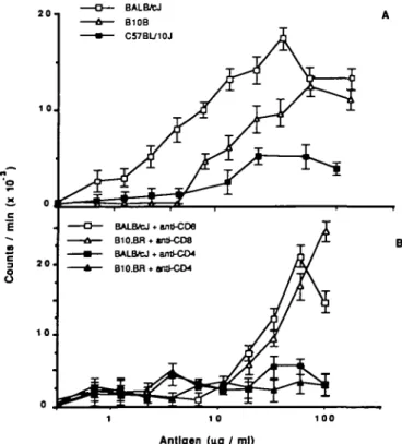
mice, C57BL/10J, BALB/cJ, and B10.BR, were immunized with 18 kDa protein and after 7 days the draining lymph nodes were removed and T cells enriched as described in Methods. The recovered T cells were cultured with activated macrophages, as a source of APCs and 18 kDa protein as antigen. The proliferative responses described in Fig. 1(A) show that T cells derived from C57BL/10J mice immunized with the 18 kDa protein respond less vigorously than those from BALB/cJ or B10.BR mice. Thus, as previously observed for IgG responses to the 18 kDa protein (12), inan/n vitro Tcell proliferative assay, B10. BRand BALB/cJ are also high responder strains, while C57BL/10J is a low responder (24).
Pretreatment of purified T cells with anti-CD4 antibody and complement completely abrogated proliferation to the 18 kDa protein, while identical treatment using anti-CD8 antibody had little effect (Fig. 1B) showing that in all cases, responding T cells were of the CD4+ phenotype. Inclusion of the mAb M5/114 (anti I-Ad, anti I-Edk) at 50 pg/ml in in vitro restimulations of BALB/cJ T cells completely abolished proliferative responses, while inclusion of similar levels of MKD6 (anti-l-Ad) or the mAb 10-3.6 (anti I-A*) had no effect. Proliferation of T cells from B10.BR mice was inhibited by mAb M5/114, but not MKD6 or 10.36, indicating that in both strains the proliferative response to all the peptides examined was I-E restricted (data not shown).
Mice were also immunized with either 18 kDa protein,
M. leprae, M. tuberculosis, or ovalbumin and then rechallenged
o K U|U I — Count a 2 0 ' 1 0 . 2 0 . 1 0 . 0 • — BALB/cJ A— B10B • — C57BL/10J J
O— BALB*J * anll-CDa T A— B10BH • tnS-C08 /£•
• BALflfcJ * antl-CD4 1 /
Jr
1 10 100 Antigen (no / ml)
Fig. 1. (A) In vitro proliferate responses to the 18 kDa protein with T cells from 18 kDa protein-immunized BALB/cJ, B10.BR, and C57BL/10J mice, and syngeneic APCs as described in Methods. Each point represents the arithmetic mean of triplicate cultures. (B) Effect on T cell proliferation to 18 kDa protein of treatment of 18 kDa protein-reactive T cells with anti-CD4 and anti-CD8 mAbs. Each point represents the arithmetic mean of triplicate cultures.
o
X
c o O
4 -, A. 18 kDa primed T calls challenged with peptide :
3 0 10 -0 4 -0 2 0 1 0 -• BIO.BR • BALB/CJ 1-20 16-35 31-50 46-65 61-80 76-95 91-115 106-125 121-140 136-148 B. Peptide primed T calls
challenged wtth 18 kDa
1-20 16-35 31-50 46-65 C. Peptld* prtmad T calls
challenged wtth peptUe
61-80 76-95 91-115 108-125 121-140 136-148
1-20 16-35 31-50 46-65 61-80 76-95 91-115 106-125 121-140 136-148
Peptide Residues
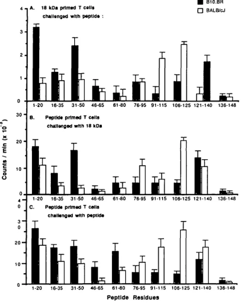
Fig. 2. (A) In vitro proWerative responses of T cells from BALB/cJ and B10.BR mice immunized with the 18 kDa protein, cultured with 20-mer peptides
(at 25 /tg/ml) and syngeneic APCs as descnbed in Methods. Each point represents the arithmetic mean of triplicate cultures. Average background (T cells plus antigen without APCs) was 400 c.p.m. or below; proliferation in culture with 10 fig/ml 18 kDa protein was -25,000 c.p.m., in both strains. (B) In vitro proliferate responses oi T cells from BALB/cJ and B10.BR mice immunized with individual 20-mer peptides, cultured with the 18 kDa protein (at 10 /ig/ml) and syngeneic APCs as described in Methods. Average background (T cells plus antigen without APCs) was 1000 c.p.m. or beiow, in both strains. Each point represents the arithmetic mean of triplicate cultures. (C) In vitro proliferate responses of T cells from BALB/cJ and B10.BR mice immunized with individual 20-mer peptides, cultured with the immunizing 20-mer peptide (at 25 /ig/ml) and syngeneic APCs as described in Methods. Average background (T cells plus antigen without APCs) was 900 c.p.m or below, in both strains. Each point represents the arithmetic mean of triplicate cultures.
with each of these antigens in vitro (data not shown). T cells from mice immunized with the 18 kDa protein responded to rechallenge with the 18 kDa protein, M. leprae, or M. tuberculosis in culture. However, M. fep/ae-primed T cells did not proliferate significantly when restimulated in culture with 18 kDa protein. T cells from ovalbumin-immunized mice only responded when cultured with ovalbumin. Thus, the 18 kDa protein will stimulate T cells reactive with both M. leprae and M. tuberculosis, although responses to this protein constituted only a fraction of the total response to
M. leprae immunization. The ability of the 18 kDa protein to
stimulate T cells that responded to M. tuberculosis was interesting,
in that it suggested that there exist cross-reactive epitopes as yet undefined. The inability of BCG vaccination to consistently prevent the development of leprosy (25) implies that such crossreactive epitopes are not necessarily protective.
Mapping of T cell epitopes on the 18 kDa protein using 20-mer peptides
Groups of BALB/cJ and B10.BR mice were immunized with either the 18 kDa protein or 20-mer peptides. After 7 days the draining lymph nodes were removed, T cells purified, and cultured with exogenous macrophages and either 18 kDa protein or one of
2 0 3 0 4 0 SO -L MLMRTDPFRELDRfAEQVLQTSARPAVHPMPAWqEQEEFWEFOLPGIKA 37.2 11.9 6 0 70 SO 90 1 0 0 DSLDIDIERNVVTVRAERPQVDPDnEMLAAERPROVFNRQLVLGENLDTE 6.4 110
I
1 2 0 130I
140I
RILASYQEGVLKLSIPVAERAKPRKISVDRGNNGHQTINKTAHEIIDA 14.6 23.9Fig. 3. Regions of the 18 kDa gene predicted to be optimal T cell stimulating epitopes by the least squares algorithm (31) are outlined. The score below each epitope reflects the predicted potential of each site, with higher numbers reflecting a closer match to the idealized epitope. The site predicted to be the closest match to the 'ideal' epitope is contained within peptide 1 - 20, which stimulates proliferation in both strains of mice tested. However, peptide 136-148, containing the majority of the second best match, is not an effective antigen, and peptide 91 - 1 1 0 , which contains the third best match, is effective at priming profrferative responses in only one strain (BALB/cJ). Those regions which provided help for dominant antibody responses in B10.BR mice are indicated by shaded boxes and in BALB/cJ mice, by white boxes (12).
each of the 10 overlapping synthetic 20-mer peptides. On day 4, proliferation was assessed by incorporation of [3H]thymidine. The results presented in Fig. 2(A) show that while T cells from both strains of mice immunized with the 18 kDa protein respond to many of the 20-mer peptides in T cell proliferation assays, there were differences in the magnitudes of response. T cells from B10.BR mice responded more strongly to peptides 1 - 2 0 , 3 1 - 5 0 , and 121-140 than those from BALB/cJ mice, while peptides 91 - 1 1 0 and 106-125 stimulated greater responses from BALB/cJ cells. Peptides 1 6 - 3 5 , 4 6 - 6 5 , and 7 6 - 9 5 elicited similar responses in cultures prepared from either strain. It is interesting to observe that these peptides contained the regions of the 18 kDa protein that stimulated the strongest antibody responses in each strain (Fig. 3), suggesting that the region of the protein which is immunodominant, varies in animals with different MHC haplotypes.
20-mer peptides as antigens
BALB/cJ and B10.BR mice were immunized as described with either one of the 20-mer peptides or the 18 kDa protein, T cells purified from the draining lymph nodes after 7 days and cultured with activated macrophages and either the 18 kDa protein or the immunizing peptide. The results presented in Fig. 2(B) show that both strains of mice contained populations of T cells capable of a strong proliferate response to 18 kDa when immunized with a number of peptides. However, the proliferative responses stimulated by immunization with some of these peptides (such as 1 - 2 0 in B10.BR mice or 106-125 in BALB/cJ mice) were of significantly greater magnitude than those induced by others. T cells reactive with these epitopes were therefore likely to dominate early responses to an antigen that contained them.
The proliferative responses observed when T cells from mice immunized with the 18 kDa protein were cultured in vitro with
synthetic peptides encoding parts of the 18 kDa protein were small in magnitude (Fig. 2A) compared to those observed for the whole 18 kDa protein. This suggests that none of these peptides individually contained a discrete epitope or epitopes against which the majority of the response to the whole 18 kDa protein was directed. Either the dominant epitopes were not represented amongst these peptides or the response to the whole 18 kDa protein was comprised of multiple T cells with many specificities. The relative magnitude of responses observed when T cells from peptide-immunized mice were challenged in vitro with the immunizing peptide, correlated with those observed for secondary responses to the whole 18 kDa protein, indicating that the dominant epitopes are conserved on the peptides (Fig. 2B and C).
Phenotype of responding T cells
When T cells from peptide-immunized mice were treated with anti-CD4 antibody and complement, before addition to culture, proliferation was reduced to background levels, while anti-CD8 antibody had no appreciable effect (data not shown) indicating that as with responses to the 18 kDa protein, responding T cells were mostly of the CD4+ phenotype. To determine the nature of CD4+ cells that proliferated in response to challenge with protein or peptides in vitro, supernatants were taken from proliferating cultures after 48, 60, or 72 h. Supernatants were then assayed for the presence of IL-2, IFN-7, or IL-4, as described in Methods. It can be seen from the data presented in Table 1 that a strong proliferative response to challenge in vitro correlated with high levels of IFN-7 in the supernatant. In all cases, cultures with high levels of IFN--y contained high levels of IL-2 (data not shown). Although these supernatants were taken from cultures which contained heterogenous mixtures of T cells, it was clear that little IL-4 (produced by Th2 or Th0 cells) was present. This indicates that the T cells which proliferate the response to in vitro challenge with most 20-mer peptides were predominantly of the Th subset. BALB/cJ and B10.BR mice do make measurable, though low, quantities of IL-4 when immunized and challenged with whole protein, or B10.BR when immunized and challenged with peptide 1 - 2 0 (Table 1), suggesting that Th2 or Th0 cells were primed in vivo by these antigens.
While Th1 cells are doubtlessly primed in vivo, the apparent dominance of Th1 cells in proliferation assays may be an artifact of the culture conditions. To test this T cell lines were developed from 18 kDa immunized BALB/cJ or B10.BR mice. Nine T cell lines were obtained, specific for four different peptides. All were a/3+, CD4+, CD8~. These lines were phenotyped by semi-quantative PCR for IL-2, IL-4, IL-10, granulocyte macrophage colony stimulating factor (GM-CSF), IFN-7, and (as a control) /3-actin. The results presented in Table 2 indicate that the majority of lines did not contain IL-4 producing cells. This is consistent with the data in Table 1, which indicated that a strongly polarized Th1 response developed in response to in vitro rechallenge with peptides or the 18 kDa protein. The epitopes to which the T cell lines responded were those that stimulated the strongest responses from unfractionated cells—T cell lines from BALB/cJ mice responded an epitope (or epitopes) on peptide 106-125, while T cell lines from B10.BR mice responded to epitopes on peptides 1 - 20 and 31 - 50. There is one other interesting point, which was that two T cell lines from BALB/cJ mice were specific for an epitope or epitopes on peptide 46 - 65, which stimulated
Table 1 . Cytokines produced by a secondary response in vitro with purified APCs
Antigen BALB/cJ B10.BR
IL-4 (ng/ml) IFN-7 (ng/ml) IL-4 (ng/ml) IFN-7 (ng/ml) 1 - 2 0 16-35 31-50 46-65 61-80 76-95 91 -110 106-125 121-140 136-148 18kDa <0.5 <0.5 <0.5 <0.5 <0.5 <0.5 <0.5 <0.5 <0.5 <0.5 1.9 (±0.7) 939 (±20.1) <0.5 <0.5 <0.5 <0.5 130.3i 56.8l 302.5 l 123.9 1 <0.5 284.7 (±31 6) i (±30.8) I (±10.6) i (±60.2) I (±21.8) 2.6 (±0.9) <0.5 <0.5 <0.5 <0.5 <0.5 <0.5 <0.5 <0.5 <0.5 1.6 (±0.8) 248.2 (±61.0) 94.9 (±22.4) 226.1 (±48.5) <0.5 42.0 (±13.5) 41.9 (±12.8) 42.8 (±13.7) 41.9 (±14.2) 184 2 (±38.4) <0.5 261.9 (±58.2)
Supernatants were taken at 60 h from the culture, fresh medium added and proliferation assayed by 3H incorporation as described in Methods.
Supernatants were quantified against known recombinant IL-4 or IFN-7 standards. In all cases, a high level of IFN-7 in the supernatant correlated with increased levels of IL-2 and IL-3 (data not shown), which is indicative that the cytokines were produced by actively proliferating Th1 cells in
both strains Values have had background (response to a non-18 kDa peptide) subtracted. Samples where cytokine was not significantly above background are represented by '<0.5'. Values represent the means of triplicate cultures.
Table 2. Cytokines produced by T cell lines in vitro
T cell line Strain Peptide IL-2 II-3 IL-4 IFN-7 Phenotype 1 BC, 5.BC 2.BC 3.BC, 4 BC 1 BR, 5.BR 3.BR, 4.BR BALB/cJ BALB/cJ BALB/cJ B10.BR B10.BR 4 6 - 6 5 106-125 106-125 31/50 1-20 Th0 or mixed Th1/Th2 Th1 Th1 Th1
Cells were taken after 6 h concanavalin A and PMA stimulation, and RNA prepared as described in Methods. PCR products were quantified against /S-actin and plasmids containing the gene in question after 60 cycles of PCR.
an insignificant response from unfractionated cells (Table 1). Immunization of both BALB/cJ and B10.BR mice induced significant levels of antibody reactive with this peptide (12). It is possible that this antibody response was largely supported by
1^2 cells which often proliferate poorly in vitro (unpublished
data) allowing Th1 cells to outgrow and dominate the long-term cultures, while not being obvious in short-term restimulations.
Response to peptide 1-20 in BALB/cJ mice
The data presented in this and a previous paper suggest an apparent paradox. Both B10.BR and BALB/cJ mice have B cells that recognise an epitope, or epitopes, on peptide 1 - 2 0 , as immunization with intact 18 kDa protein led to the production of high titres of IgG that binds to this peptide (12). Immunization of both strains of mice with peptide 1 - 20 leads to the induction of antibody only in B10.BR, not BALB/cJ mice (12). These findings imply that only B10.BR, not BALB/cJ mice, have T cells capable of providing cognate help for B cell responses to epitopes located within peptides 1 - 20. However, the data presented in Fig. 2 and Table 1 show that purified T cells from BALB/cJ mice immunized with either 18 kDa protein or peptide 1 - 20 recognise and respond to an epitope or epitopes contained within peptide 1 - 2 0 .
To investigate whether the differences in the responses shown by these mouse strains were due to different affinities of the peptide for MHC, bone marrow-derived APCs from B10.BR and
20
1 0
o o
BALB* APC + F1 Torts B108RAPC + F1 Trails F1 APC t F1 T cells F I T a f l s a J o n e
6.25 1.0s
Peptide 1-20 Antigen ( n g / m l )
Fig. 4. Proliferative responses of T cells from F, (BALB/cJ xB10.BR)
mice immunized with peptide 1 - 20 cultured with 20-mer peptides at 25 /ig/ml and F, or parental APCs as described in the Methods section Average background (T cells plus antigen without APCs) was 800 c.p.m. or below, in both strains. Each point represents the arithmetic mean of trinlicate cultures.
BALB/cJ mice were used to pfesent peptide 1 - 20 to T cells from F, (BALB/cJ xB10.BR) mice, which had been immunized with this peptide. It was found (Fig. 4) that BALB/cJ- or B10.BR-derived macrophages, pulsed with peptide 1 - 20 before
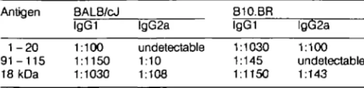
Table 3. Antibody isotypes produced in vivo (midpoint titres) Antigen 1-20 9 1 - 1 1 5 18 kDa BALB/cJ lgG1 1:100 1:1150 1:1030 lgG2a undetectable 1:10 1:108 B10.BR lgG1 1:1030 1:145 1:1150 lgG2a 1:100 undetectable 1:143 IgG midpoints titres to the indicated antigens from BALB/cJ and B10.BR mce immunized muftiple times with peptides 1 - 20,91 -115, and 18 kDa protein peptide. 91 - 1 1 5 was used rather than the 20-mer 91 - 1 1 0 since the former was a more potent stimulator of antibody cross-reactive with the whole 18 kDa protein (12). Results represent the mean of specific antibody in sera titrated from five individuals of each strain. The isotype specific ELISAs used antibodies for lgG1 (G1-6.5) and lgG2a (R12-4) from Pharmingen (San Diego, CA). Immunization with these peptides stimulated antibody production with a low proportion of lgG2a, a pattern associated with Th2 dominated responses (24) In contrast, the ratio of
lgG1 and lgG2a is more even, when mice are immunized with the 18 kDa protein Together with the results shown in Table 1, this suggests that both Th1 and Th2 cells may be activated.
irradiation, and then washed to remove free antigen, stimulated F, T cells with similar efficiency, demonstrating that BALB/cJ-derived APCs process and present this peptide at a similar efficiency to those derived from B10.BR mice.
The strong proliferative response observed to in vitro rechallenge with peptide 1 - 20 by T cells derived from BALB/cJ mice shows that cells can be effectively primed by this peptide
in vivo, but these cells do not provide help for effective antibody
production (Table 3). This appears to be due to selective activation of Th1 cells in vivo (manuscript submitted).
Discussion
The aim of this work has been to map regions within the 18 kDa protein that contain T cell epitopes, using an in vitro T cell proliferative assay and to compare these with T cell epitopes previously mapped by their ability to provide help for B cell responses (12). This approach may give insight into the factors governing activation of pathogenic or protective responses to the leprosy bacillus, given that the 18 kDa protein appears to be an important antigen in the response to M. leprae (8 -10). In the mouse footpad model, immunization with M. habana, which unlike the majority of mycobacteria shares the 18 kDa protein, induced some protection against challenge with M. leprae (31), suggesting that this protein may contain epitopes with potential for protective immunization.
The specific epitopes which dominate an immune response may have a major impact on the progress of disease (27), due to the action of antigen-specific suppressor cells (28) or as a result of the imbalance of subsets of Th cells (29). It has been postulated that the terminal poles of leprosy, which are characterized by either strong cell-mediated immunity or T cell unresponsiveness, may be caused by an imbalance in Th1 and Th2 cells, analogous to similar states studied in other disease models (30). Characterization of the epitopes recognised by T cells and the nature of the T cell response to dominant epitopes is therefore of great importance.
Our results indicate that while different strains of mice see similar B cell epitopes on the 18 kDa protein, they see different,
but overlapping, sets of T cell epitopes. This implies a degree of promiscuity in TCR- antigen - M H C interactions that could enable the construction of composite synthetic immunogens, recognised by divergent, or outbred populations. Moreover, the epitopes which evoked the strongest responses (as assessed by proliferation) in different mouse strains, are the same ones which provided the most potent T cell help for antibody production, (12) suggesting that the same epitopes are seen in vivo and in vitro. The cytokines produced in vitro (Tables 1 and 2) show that Th1 cells are effectively primed by these peptides, although analysis of the antibody isotypes suggest that Th2 responses are a significant part of the in vivo response (Table 3).
A striking feature of the responses described here is the difference between BALB/cJ and B10.BR mice with regard to their responses to peptide as compared with the whole 18 kDa protein. When T cells from BALB/cJ mice immunized and challenged in vitro with individual peptides were assayed for proliferation, responses were seen against those peptides which had stimulated a response with 18 kDa-primed T cells but, in addition, responses to peptides 1 - 2 0 , 7 6 - 9 5 , and 121-140 became apparent (Fig. 2). This indicates that BALB/cJ mice possess T cells reactive with epitopes contained on these peptides, but when immunized with the 18 kDa protein, these T cells are not effectively primed. In contrast, B10.BR mice respond effectively to the same peptides whether immunized with peptide or whole protein. Given that the APCs from BALB/cJ or B10.BR mice appear to present peptide 1 - 2 0 to F, T cells with no significant difference in efficiency (Fig. 4), it seems unlikely that either antigen - MHC binding, or the reaction of the TCR and the antigen - MHC complex is in some way defective in BALB/cJ mice. The epitopes so far defined remain unchanged across a wide range of antigen concentrations, suggesting that T cell activation in these strains is not influenced by minor variations in the concentration of antigen. This leaves two likely explanations for these data. It is possible that variation in avidity of antigen-specific receptors in these two strains could lead to preferential uptake of antigen by different classes of APCs. Results obtained in our laboratory suggest that the nature of the response can be manipulated by influencing the type of APCs which presents to a mixed T cell population (manuscript submitted) with subsequent bias in the development of T cell subsets. Equally, it may be that variation in H-2 I-E molecules could lead to activation in one strain of specific suppressor cells, as suggested for M./eprae-specific HLA-DQ restricted human suppressor clones (28). Alternatively, this result may reflect real differences in the nature of the cellular populations of these mice, perhaps developed during the selection process. The development of systems in which antigenic epitopes have been described allows these questions to be further addressed, since at last we are able to analyse exactly what the effects of different immunization strategies are on defined T cell populations in vivo. This must help our understanding of the molecular processes involved in T cell recognition and activation.
Acknowledgements
This work was supported by the Pacific Leprosy Foundation (formerly the Leprosy Trust Board) and the Health Research Council of New Zealand. T.B. was supported by the Leprosy Mission, Auckland, New
Zealand. The authors wish to thank Dr Roger Booth and Dr Ross Prestidge
Abbreviations
APC DMEM PCR
References
antigen presenting cell
Dulbecco's modified Eagle's medium potymerase chain reaction
T helper cell
1 Young, D., Lathigra, R., Hendrix, R., Sweetser, D., and Young, R. A. 1988. Stress proteins are immune targets in leprosy and tuberculosis.
Proc. Nati Acad. Sci. USA 854267
2 Fine, F. P. E. M. and Rodrigues, L. C. 1990. Modern vaccines Mycobacterial diseases. The Lancet 335:1016.
3 Johnston, P. A. S. 1987. The search for animal models of leprosy.
Int. J. Lep. 55:535.
4 Young, R. A , Mehra, V., Sweetser, D., Buchanan, T. M., Clark-Curtiss, J., Davis, R. W., and Bloom, B. R. 1985. Genes for the major protein antigens of Mycobacterium leprae. Nature 316450 5 Engers, H. D., Abe, M., Bloom, B. R., Mehra, V., Britton, W.,
Buchanan, T. M., Khanolkar, S. K., Young, D. B., Qoss, O., Gillis, T., Harboe, M., Ivanyi, J., Kolk, A. H. J., and Shepard, C. C. 1985. Workshop: results of a Wortd Health Organisation-sponsored workshop on monoclonal antibodies to Mycobacterium leprae. Infect.
Immun. 48:603.
6 Booth, R. J., Harris, D. P., Love, J. M., and Watson, J D. 1988. Antgenic proteins of Mycobacterium leprae. complete sequence of the gene for the 18 kDa protein. J. Immunol. 140:597.
7 Booth, R. J., Grandison, P. M., Prestidge, R. L, and Watson, J. D. 1988. The use of a 'universal' yeast expression vector to produce an antigenic protein of Mycobacterium leprae. Immunol. Lett. 19:65. 8 Mustafa, A. S., Gill, K. H., Norland, A., Britton, W. J , Mehra, V.,
Bloom, B. R., Young, R. A., and Godal.T. 1986. Human T cell clones recognise a major Mycobacterium leprae protein expressed in E.coli.
Nature 319:63
9 Dockrell, H. M., Stoker, N. G., Lee, S. P., Jackson, M., Grant, K. A., Jouy, N. F, Lucas, R., Hasan, S. B., Hussain, R., and McAdam, K. P. W. J. 1989. T cell recognition of the 18 kDa protein of Mycobacterium leprae. Infect. Immun. 57:1979.
10 Oftung, F., Shinnick, T. M., Mustafa, A. S., Lundin, K. E. A., Godal, T., and Nerland, A. H. 1990. Heterogeneity among human T cell clones recognising an HLA-DR4, Dw4-restncted eprtope from the 18 kDa antigen of Mycobacterium leprae defined by synthetic peptides.
J. Immunol. 144:1478.
11 Doherty, T. M., Booth, R. J., Love, S. G., Gibson, J. D., Harding, D. R. K., and Watson, J D. 1989. Characterisation of an antibody-binding epitope from the 18 kDa protein of Mycobacterium
leprae. J. Immunol. 142:1691.
12 Doherty, T. M., Backstrom, B. T., Love, S. G., Harding, D. R. K., and Watson, J. D. 1991. Immune responses to the 18 kDa protein of
Mycobacterium leprae. Similar B cell epitopes but different T cell
epitopes seen by inbred strains of mice. J. Immunol. 146:1934. 13 Scott, P., Natovitz, P., Coffman, R. L , Pearce, E., and Sher, A. 1988.
Immunoregulafon of cutaneous leshmaniasis. T cell lines that transfer protective immunity or exacerbation belong to different T helper subsets and respond to distinct parasite antigens. J. Exp. Med. 168.1675.
14 Haanen, J. B. A. G., de Waal Malefljt, R., Res, P. C. M., Kraakma, E. M., Ottenhoff, T. H. M., de Vries, R. R. P., and Spits, H. 1991. Selection of human T helper type Hike T cell subset by mycobacteria. J. Exp. Med. 174:583.
15 Uew, F. Y., Singleton, A., Qllari, E., and Howard, J. G. 1985.
Prophylactic immunization against experimental leishmaniasis. V. Mechanism of the antiprotective blocking effect induced by subcutaneous immunization against Leishmania major infection.
J. Immunol. 135:2101.
16 Bloom, B. R., Salgame, P , and Diamond, B. 1992. Revisiting and revising suppressor T cells. Immunol. Today 13:131.
17 Merrifield, R B. 1969. Solid phase peptide synthesis. Adv. Enzymol. 32:221.
18 Julius, M. H., Simpson, E., and Herzenberg, L. A. 1973. A rapid method for the isolation of functional thymus-derived murine lymphocytes. Eur. J. Immunol. 3:645.
19 Cherwinski, H. M., Schumacher, J. H., Brown, K. D., and Mosmann, T. R. 1987. Two types of mouse helper T cell clone. Ill Further differences in rymphokine synthesis between TH1 and TH2
clones revealed by RNA hybridization, functionally monospecific bioassays and monoclonal antibodies. J. Exp. Med. 166:1229. 20 Dialynas, D. P., Wilde, D. B., Marrack, P., Pierres, A., Wall, K. A.,
Havran, W., Oflen, G., Loken, M. R., Pierres, M., and Kappler, J. 1983. Characterisation of the murine antigenic determinant, designated L3T4a, recognised by monoclonal antibody GK1.5: expression of L3T4a by functional T cell clones appears to correlate primarily with class II MHC antigen-reactivity. Immunol Rev. 74:29.
21 Bluestone, J A., McKenzie, I F., MetvoW, R. W., Ozata, K., Sandrm, M. S., Sharrow, S. O. and Sachs, D. H. 1984. Serotogteal analysis of H-2 mutations using monoclonal antibodies.
J. Immunogenet. 11 197.
22 Lanier, L L, Gutman, G. A., Lewis, D. E., Griswold, S. T., and Warner, N. L. 1982. Monoclonal antibodies against the immuno-globulin kappa chains. Hybridoma 1:125.
23 Chomczynski, P. and Sacchi, N. 1987. Single step method of RNA isolation by acid guanidinium thiocyanate-phenol-chloroform extraction. Anal. Biochem. 162:156.
24 Harris, D. P., Backstrom, B. T , Booth, R. J., Harding, D. R K., and Watson, J. D. 1989 The mapping of epitopes of the 18 kDa protein of Mycobacterium leprae recognised by murine T cells in a proliferation assay. J. Immunol. 143:1866.
25 Bagshawe, A., Scott, G. C, Russell, D. A., Wigley, S. C, Merianos, A., and Berry, G. 1989. BCG vaccination in Karimui, Papua New Guinea, 1963-1979. Bull WHO. 67:389.
26 Coffman, R. L, Seymour, B. W. P , Lebman, D. A , Hiraki, D. D , Christiansen, J. A., Shrader, B , Cherwinski, H. M., Savelkoul, H. F. J., Finkelman, F. D., Bond, M. W., and Mosmann, T. R. 1988. The role of helper T cell products in mouse B cell differentiation and isotype regulation. Immunol. Rev. 102:1.
27 LJew, F. Y., Millot, S. M., and Schmidt, J. A. 1990. A repetitive peptide of leishmania can activate T helper type 2 cells and enhance disease progression. J. Exp. Med. 172:1359.
28 Salgame, P., Convit, J., and Bloom, B. R 1991. Immunologteal suppression by human CD8+ T cells is receptor dependent and
HLA-DQ restricted. Proc. Natl Acad. Sci. USA 88:2598.
29 Mosmann, T. R. and Coffman, R. L 1989. Heterogeneity of cytokine secretion patterns and functions of helper T cells. Adv. Immunol. 46:111.
30 Mason, D. 1990. Genetic variation in the stress response: susceptibility to experimental allergic encephalomyelitis and implications for human inflammatory disease Immunol. Today 12:57.
31 Lamb, F. I., Singh, N. P., and Couteton, M. J. 1990. The specific 18 kDa protein of Mycobacterium leprae is present in Mycobacterium
habana and functions as a heat shock protein. J. Immunol. 144:1922.
32 Margalit, H., Spouge, J. L., Cornette, J. L, Cease, K. B., DeLisi, C , and Berzofsky, J. A. 1987. Prediction of immunodominant helper T cell antigenic sites from the primary sequence. J. Immunol. 138:2213.