Advanced modular self-inactivating lentiviral
expression vectors for multigene interventions in
mammalian cells and in vivo transduction
Barbara Mitta, Markus Rimann, Markus U. Ehrengruber
1, Martin Ehrbar
2,
Valentin Djonov
3, Jens Kelm and Martin Fussenegger*
Institute of Biotechnology, Swiss Federal Institute of Technology, ETH Zurich, ETH Hoenggerberg, HPT, CH-8093 Zurich, Switzerland,1Brain Research Institute, University of Zurich, CH-8057 Zurich, Switzerland,2Department of
Materials and Institute for Biomedical Research, ETH Zurich and University of Zurich, CH-8044 Zurich, Switzerland and3Institute of Anatomy, University of Bern, CH-3009 Bern, Switzerland
Received as resubmission June 27, 2002; Accepted September 5, 2002 ABSTRACT
In recent years, lentiviral expression systems have gained an unmatched reputation among the gene therapy community for their ability to deliver thera-peutic transgenes into a wide variety of dif®cult-to-transfect/transduce target tissues (brain, hemato-poietic system, liver, lung, retina) without eliciting signi®cant humoral immune responses. We have cloned a construction kit-like self-inactivating lenti-viral expression vector family which is compatible to state-of-the-art packaging and pseudotyping technologies and contains, besides essential cis-acting lentiviral sequences, (i) unparalleled poly-linkers with up to 29 unique sites for restriction endonucleases, many of which recognize 8 bp motifs, (ii) strong promoters derived from the human cytomegalovirus immediate-early promoter (PhCMV) or the human elongation factor 1a (PhEF1a),
(iii) PhCMV± or PPGK± (phosphoglycerate kinase
promoter) driven G418 resistance markers or ¯uorescent protein-based expression tracers and (iv) tricistronic expression cassettes for coordinated expression of up to three transgenes. In addition, we have designed a size-optimized series of highly modular lentiviral expression vectors (pLenti-Module) which contain, besides the extensive cen-tral polylinker, unique restriction sites ¯anking any of the 5¢U3, R-U5-y+-SD, cPPT-RRE-SA and 3¢LTRDU3modules or placed within the 5¢U3 (±78 bp)
and 3¢LTRDU3 (8666 bp). pLentiModule enables
straightforward cassette-type module swapping between lentiviral expression vector family mem-bers and facilitates the design of Tat-independent (replacement of 5¢LTR by heterologous promoter elements), regulated and self-excisable proviruses (insertion of responsive operators or LoxP in the
3¢LTRDU3 element). We have validated our lentiviral
expression vectors by transduction of a variety of insect, chicken, murine and human cell lines as well as adult rat cardiomyocytes, rat hippocampal slices and chicken embryos. The novel multi-purpose con-struction kit-like vector series described here is compatible with itself as well as many other (non-viral) mammalian expression vectors for straightfor-ward exchange of key components (e.g. promoters, LTRs, resistance genes) and will assist the gene therapy and tissue engineering communities in developing lentiviral expression vectors tailored for optimal treatment of prominent human diseases. INTRODUCTION
Gene therapy strategies rely on ef®cient transfer of therapeutic transgenes into desired target cells. A variety of viral and non-viral vectors and expression concepts have been designed and evaluated for their safety, high-level transduction, tropism and sustained expression in a variety of therapeutically relevant cells and tissues (1,2). Retroviral vectors derived from oncoretroviruses such as the murine leukemia virus (MLV) emerged as the most widely used gene therapy tools for transgene delivery currently in the clinics (3). The attractive-ness of oncoretroviral transduction technologies resides in: (i) their ability to mediate stable integration in the target chromosomes likely promoting long-term expression of delivered transgenes; (ii) their large cloning capacity suf®cient for most foreseeable clinical situations; (iii) their compatibil-ity with pseudotyping strategies which extend the tropism of gene delivery; and (iv) the exclusive delivery of therapeutic transgenes in the absence of viral genes which precludes any potent humoral immune response eliminating transduced cells and enables recurring treatments (3,4).
In addition to these characteristics common to all retro-viruses, lentiviruses such as the human immunode®ciency virus type 1 (HIV-1) can replicate in non-mitotic cells owing to their so-called pre-integration complex, a macromolecular *To whom correspondence should be addressed. Tel: +41 1 633 34 48; Fax: +41 1 633 10 51; Email: [email protected]
structure comprising the viral genome, a few structural proteins and the enzymes responsible for reverse transcription and integration (5±7). Transduction of proliferation-incompe-tent cells is a decisive asset for molecular interventions in tissues considered key targets for future gene therapy includ-ing the brain, the heart, the hematopoietic system, the liver, the lungs and the retina (8±14). Like all retroviruses, the HIV-1 genome contains the gag, pol and env regions, which encode the core proteins, the virion-associated enzymes and the envelope (Env) glycoprotein (13,15). This coding region is ¯anked by the long terminal repeats (LTRs; 5¢ and 3¢LTR) and cis-acting sequences essential for integration, transcription and polyadenylation. In addition, HIV-1 contains two regu-latory genes, tat and rev, required for viral replication (LTR transactivation and nuclear export of viral RNA) and four accessory genes, vif, vpr, vpu and nef, which are dispensable for viral growth but critical for in vivo replication and pathogenesis (15). Basal transcription of the HIV-1 provirus' 5¢LTR initially results in small amounts of multiply spliced transcripts encoding Tat, Rev and Nef. Tat transactivates 5¢LTR-mediated transcription until Rev reaches a threshold concentration and mediates cytoplasmic accumulation of unspliced and singly spliced viral transcripts followed by production of the late viral proteins (15).
Capitalizing on a re®ned understanding of HIV-1 molecular biology following decades of intensive research on acquired immunode®ciencies, a group of researchers pioneered the transition of this well-evolved pathogen into a high leverage gene therapy tool (16±18). HIV-1-based vectors have been generated using a multiply attenuated packaging system, which is activated in its most advanced third generation con®guration by transient transfection of four plasmids encoding: (i) gag (coding for the virion main structural proteins) and pol (responsible for lentivirus-speci®c enzymes); (ii) rev (a post-transcriptional regulator for gag and pol expression as well as nuclear RNA export); (iii) vsv-g (required for pseudotyping); and (iv) the desired transgene in a lentiviral expression con®guration (lentiviral expression vector or lentivector) (19). Latest developments include packaging cell lines, which express all packaging components and require exclusive transfection of the lentiviral expression vector (20±23). The lentiviral expression vector, which can be combined with any generation of packaging system, is the only genetic material transferred to the target cell. It typically comprises the transgene cassette ¯anked by cis-acting elements required for encapsidation, reverse transcription and integration including the packaging signal (y+), the polypurine tracts (PPT), 5¢ and 3¢LTRs as well as env-derived sequences encompassing the Rev response element (RRE) (16±19,24). Latest generation HIV-1-based expression tech-nologies include self-inactivating (SIN) vectors, which loose the transcriptional capacity of the LTR once integrated into target cells. SIN vectors show improved performance and are devoid of transcriptional interference and in vivo suppression associated with standard vectors and enable construction of more-stringent tissue-speci®c and/or regulatable expression con®gurations (25,26).
Unfortunately, current lentiviral expression vectors are the result of a progressive development rather than a bottom-up rational design and are therefore often lacking convenient multiple cloning sites (MCS) and a modular set-up to increase
compatibility with existing expression technologies (gene regulation, recombination) (16,17,19). This situation has signi®cantly delayed the dissemination of lentiviral expression technology beyond the pioneering laboratories to a broad scienti®c community. We describe here a size-optimized highly modular construction kit of SIN lentiviral expression vectors containing extended MCS, multicistronic expression cassettes as well as various promoter and resistance elements. MATERIALS AND METHODS
Cell culture and ¯uorescence microscopy
Chinese hamster ovary cells (CHO-K1; ATCC CCL-61), baby hamster kidney cells BHK-21 (ATCC CCL-10), human ®brosarcoma cells HT-1080 (ATCC CCL-121), human cer-vical adenocarcinoma cells HeLa (ATCC CCL-2), human hepatocellular carcinoma cells HepG2 (ATCC HB-8065) and human chronic myelogenous leukemia cell line K-562 (ATCC-243) were cultivated in FMX-8 (Cell Culture Technologies GmbH, Switzerland), Dulbecco's modi®ed Eagle medium (DMEM) (BHK-21, HT-1080, HeLa, HepG2; catalog no. 52100-039; Life Technologies AG, Basel, Switzerland) or Iscove's modi®ed Dulbecco's medium (K-562, catalog no. 4220-022; Life Technologies AG) supple-mented with 10% fetal calf serum (all other cell lines; PAA Vienna, Austria; Lot. no. A01129-242). The chicken bursal cell line DT40 was cultivated as described before (27). Adult rat cardiomyocytes (ARC) were prepared and cultivated as described previously (28). Hippocampal slices prepared from 6-day postnatal rats were cultured using the roller-tube technique (29).
For DRAQ5-mediated DNA-speci®c staining of transduced cells, 4 3 103CHO-K1 cells were seeded into chamber slides
(Lab-Tek, Nalge Nunc International, IL) and grown for 24 h before they were infected with 2 3 106 c.f.u./ml of desired
lentiviruses (pMF365-, pBM40- and pBM45-derived). After 48 h, DRAQ5 (Biostatus Ltd, Leicestershire, UK) was added to the culture at a ®nal concentration of 25 mM for 7 min. The medium was removed and cells washed twice with PBS (Dulbecco's phosphate-buffered saline, Sigma, catalog no. D5773) before they were ®xed with 4% paraformaldehyde in PBS for 8 min and washed another ®ve times with PBS. After removing the PBS, the cells were covered with a drop of Lisbeth's medium [Tris-buffered glycerol: 3:7 mixture of 0.1 M Tris±HCl (pH 9.5) and glycerol plus 50 mg/ml n-propyl-gallat] and sealed with a cover slip for confocal microscopy.
Expression of ¯uorescent proteins was visualized using a Leica DM-RB ¯uorescence microscope (Heerbrugg, Switzerland) or a confocal microscope set-up (Zeiss Axioplan ¯uorescence microscope, Biorad MRC-600 con-focal scanner, Silicon Graphics workstation) equipped with appropriate ®lters. The ¯uorescence of the enhanced green ¯uorescent protein (EGFP) and the enhanced yellow ¯uores-cent protein (EYFP) were visualized using the XF114 ®lter (Omega Optical Inc., Brattleboro, VT), the enhanced cyan ¯uorescent protein (ECFP) was detected using the XF105 ®lter (Omega Optical Inc.) and the red ¯uorescent protein was monitored using the N2.1 ®lter by Leica Inc (Heerbrugg, Switzerland).
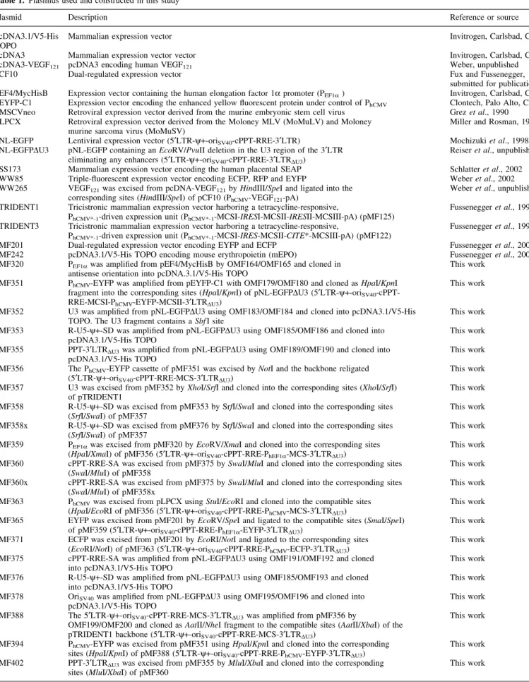
Table 1. Plasmids used and constructed in this study
Plasmid Description Reference or source
pcDNA3.1/V5-His
TOPO Mammalian expression vector Invitrogen, Carlsbad, CA
pcDNA3 Mammalian expression vector vector Invitrogen, Carlsbad, CA
pcDNA3-VEGF121 pcDNA3 encoding human VEGF121 Weber, unpublished
pCF10 Dual-regulated expression vector Fux and Fussenegger,
submitted for publication pEF4/MycHisB Expression vector containing the human elongation factor 1a promoter (PEF1a) Invitrogen, Carlsbad, CA pEYFP-C1 Expression vector encoding the enhanced yellow ¯uorescent protein under control of PhCMV Clontech, Palo Alto, CA PMSCVneo Retroviral expression vector derived from the murine embryonic stem cell virus Grez et al., 1990 PLPCX Retroviral expression vector derived from the Moloney MLV (MoMuLV) and Moloney
murine sarcoma virus (MoMuSV) Miller and Rosman, 1989
pNL-EGFP Lentiviral expression vector (5¢LTR-y+-oriSV40-cPPT-RRE-3¢LTR) Mochizuki et al., 1998 pNL-EGFPDU3 pNL-EGFP containing an EcoRV/PvuII deletion in the U3 region of the 3¢LTR
eliminating any enhancers (5¢LTR-y+-oriSV40-cPPT-RRE-3¢LTRDU3)
Reiser et al., unpublished pSS173 Mammalian expression vector encoding the human placental SEAP Schlatter et al., 2002 pWW85 Triple-¯uorescent expression vector encoding ECFP, RFP and EYFP Weber et al., 2002 pWW265 VEGF121was excised from pcDNA-VEGF121by HindIII/SpeI and ligated into the
corresponding sites (HindIII/SpeI) of pCF10 (PhCMV-VEGF121-pA)
Weber et al., unpublished pTRIDENT1 Tricistronic mammalian expression vector harboring a tetracycline-responsive,
PhCMV*-1-driven expression unit (PhCMV*-1-MCSI-IRESI-MCSII-IRESII-MCSIII-pA) (pMF125)
Fussenegger et al., 1998 pTRIDENT3 Tricistronic mammalian expression vector harboring a tetracycline-responsive,
PhCMV*-1-driven expression unit (PhCMV*-1-MCSI-IRES-MCSII-CITE*-MCSIII-pA) (pMF122)
Fussenegger et al., 1998 pMF201 Dual-regulated expression vector encoding EYFP and ECFP Fussenegger et al., 2000 pMF242 pcDNA3.1/V5-His TOPO encoding mouse erythropoietin (mEPO) Fussenegger et al., 2000 pMF320 PEF1awas ampli®ed from pEF4/MycHisB by OMF164/OMF165 and cloned in
antisense orientation into pcDNA.3.1/V5-His TOPO This work pMF351 PhCMV-EYFP was ampli®ed from pEYFP-C1 with OMF179/OMF180 and cloned as HpaI/KpnI
fragment into the corresponding sites (HpaI/KpnI) of pNL-EGFPDU3 (5¢LTR-y+-oriSV40 -cPPT-RRE-MCSI-PhCMV-EYFP-MCSII-3¢LTRDU3)
This work pMF352 U3 was ampli®ed from pNL-EGFPDU3 using OMF183/OMF184 and cloned into pcDNA3.1/V5-His
TOPO. The U3 fragment contains a SbfI site This work
pMF353 R-U5-y+-SD was ampli®ed from pNL-EGFPDU3 using OMF185/OMF186 and cloned into
pcDNA3.1/V5-His TOPO This work
pMF355 PPT-3¢LTRDU3was ampli®ed from pNL-EGFPDU3 using OMF189/OMF190 and cloned into
pcDNA3.1/V5-His TOPO This work
pMF356 The PhCMV-EYFP cassette of pMF351 was excised by NotI and the backbone religated (5¢LTR-y+-oriSV40-cPPT-RRE-MCS-3¢LTRDU3)
This work pMF357 U3 was excised from pMF352 by XhoI/SrfI and cloned into the corresponding sites (XhoI/SrfI)
of pTRIDENT1 This work
pMF358 R-U5-y+-SD was excised from pMF353 by SrfI/SwaI and cloned into the corresponding sites
(SrfI/SwaI) of pMF357 This work
pMF358x R-U5-y+-SD was excised from pMF376 by SrfI/SwaI and cloned into the corresponding sites
(SrfI/SwaI) of pMF357 This work
pMF359 PEF1awas excised from pMF320 by EcoRV/XmaI and cloned into the corresponding sites (HpaI/XmaI) of pMF356 (5¢LTR-y+-oriSV40-cPPT-RRE-PhEF1a-MCS-3¢LTRDU3)
This work pMF360 cPPT-RRE-SA was excised from pMF375 by SwaI/MluI and cloned into the corresponding sites
(SwaI/MluI) of pMF358 This work
pMF360x cPPT-RRE-SA was excised from pMF375 by SwaI/MluI and cloned into the corresponding sites
(SwaI/MluI) of pMF358x This work
pMF363 PhCMVwas excised from pLPCX using StuI/EcoRI and cloned into the compatible sites (HpaI/EcoRI of pMF356 (5¢LTR-y+-oriSV40-cPPT-RRE-PhCMV-MCS-3¢LTRDU3)
This work pMF365 EYFP was excised from pMF201 by EcoRV/SpeI and ligated to the compatible sites (SmaI/SpeI)
of pMF359 (5¢LTR-y+-oriSV40-cPPT-RRE-PhEF1a-EYFP-3¢LTRDU3)
This work pMF371 ECFP was excised from pMF201 by EcoRI/NotI and ligated to the corresponding sites
(EcoRI/NotI) of pMF363 (5¢LTR-y+-oriSV40-cPPT-RRE-PhCMV-ECFP-3¢LTRDU3)
This work pMF375 cPPT-RRE-SA was ampli®ed from pNL-EGFPDU3 using OMF191/OMF192 and cloned
into pcDNA3.1/V5-His TOPO This work
pMF376 R-U5-y+-SD was ampli®ed from pNL-EGFPDU3 using OMF185/OMF193 and cloned
into pcDNA3.1/V5-His TOPO This work
pMF378 OriSV40was ampli®ed from pNL-EGFPDU3 using OMF195/OMF196 and cloned into
pcDNA3.1/V5-His TOPO This work
pMF388 The 5¢LTR-y+-oriSV40-cPPT-RRE-MCS-3¢LTRDU3was ampli®ed from pMF356 by
OMF199/OMF200 and cloned as AatII/NheI fragment to the compatible sites (AatII/XbaI) of the pTRIDENT1 backbone (5¢LTR-y+-oriSV40-cPPT-RRE-MCS-3¢LTRDU3)
This work pMF394 PhCMV-EYFP was excised from pMF351 using HpaI/KpnI and cloned into the corresponding
sites (HpaI/KpnI) of pMF388 (5¢LTR-y+-oriSV40-cPPT-RRE-PhCMV-EYFP-3¢LTRDU3)
This work pMF402 PPT-3¢LTRDU3was excised from pMF355 by MluI/XbaI and cloned into the corresponding
In vivo transduction of chicken embryos
Experiments on chicken embryos were conducted following the shell-free cultivation protocols by Djonov and co-workers (30). After 3 days of incubation at 37°C, Brown Leghorn eggs were opened, and their contents were carefully poured into plastic Petri dishes, 80 mm in diameter. The chicken embryos
were incubated at 37°C in a humidi®ed atmosphere. Recombinant lentiviruses were applied locally on the top of the growing chorioallontoic membrane (CAM) or injected intravenously at embryonic day 9. On embryonic day 11, the CAMs were examined by in vivo ¯uorescence microscopy following intravenous (i.v.) injection of 0.1 ml 2.5 ¯uor-esceine isothiocyanate dextran (2 000 000; Sigma, St Louis, Table 1. Continued
Plasmid Description Reference or source
pBM1 The ECFP-IRESI-RFP cassette was excised from pWW85 by EcoRI/AscI and cloned into the corresponding sites of pLentiTRIDENT2 (EcoRI/AscI) 5¢LTR-y+-oriSV40-cPPT-RRE-PhCMV -ECFP-IRESI-RFP-IRESII-MCSIII-3¢LTRDU3
This work pBM6 The PPGK-neo cassette was ampli®ed from pMSCVneo using OBM1/OBM2 and cloned in sense
orientation into pcDNA3.1/V5-His TOPO This work
pMB7 PEF1awas excised from pMF320 using EcoRV/XmaI and cloned into the compatible sites (HpaI/XmaI) of pMF388 (5¢LTR-y+-oriSV40-cPPT-RRE-PhEF1a-MCS-3¢LTRDU3)
This work pBM8 The PPGK-neo cassette was excised from pBM6 by PacI/MluI and cloned into the corresponding sites
(PacI/MluI) of pMF356 (5¢LTR-y+-oriSV40-cPPT-RRE-MCSI-PPGK-neo-MCSII-3¢LTRDU3)
This work pBM9 The PPGK-neo cassette was excised from pBM6 by PacI/MluI and inserted into the corresponding sites
(PacI/MluI) of pMF359 (5¢LTR-y+-oriSV40-cPPT-RRE-PEF1a-MCSI-PPGK-neo-MCSII-3¢LTRDU3)
This work pBM13 The PPGK-neo cassette was excised from pBM6 using PacI/MluI and cloned into the corresponding sites
(PacI/MluI) of pMF388 (5¢LTR-y+-oriSV40-cPPT-RRE-MCSI-PPGK-neo-MCSII-3¢LTRDU3)
This work pBM14 The PPGK-neo cassette was excised from pBM6 using PacI/MluI and cloned into the corresponding sites
(PacI/MluI) of pBM7 (5¢LTR-y+-oriSV40-cPPT-RRE-PEF1a-MCSI-PPGK-neo-MCSII-3¢LTRDU3)
This work pBM40 EYFP was excised from pLentiModule4 by NheI/MluI and cloned into the compatible sites (SpeI/MluI)
of pBM7 (5¢LTR-y+-oriSV40-cPPT-RRE-PhEF1a-EYFP-3¢LTRDU3)
This work pBM42 mEPO was excised from pMF242 by EcoRI/EcoRV and cloned into the compatible sites (EcoRI/PmeI)
of pMF359 (5¢LTR-y+-oriSV40-cPPT-RRE-PhEF1a-EPO-3¢LTRDU3)
This work pBM43 VEGF121was excised from pWW265 by SalI/MluI and ligated into the corresponding sites (SalI/MluI)
of pMF359 (5¢LTR-y+-oriSV40-cPPT-RRE-PhEF1a-VEGF121-3¢LTRDU3)
This work pBM44 SEAP was excised from pSS173 by EcoRI/EcoRV and ligated into the compatible sites (EcoRI/PmeI)
of pMF359 (5¢LTR-y+-oriSV40-cPPT-RRE-PhEF1a-SEAP-3¢LTRDU3)
This work pLentiTRIDENT1 The MCSI-IRESI-MCSII-IRESII-MCSIII cassette was excised from pTRIDENT1 by EcoRI/BglII and
cloned into the compatible sites (EcoRI/BamHI) of pMF363 (5¢LTR-y+-oriSV40-cPPT-RRE-PhCMV -MCSI-IRESI-MCSII-IRESII-MCSIII-3¢LTRDU3) (pMF370)
This work pLentiTRIDENT2 The MCSI-IRES-MCSII-CITE*-MCSII cassette was excised from pTRIDENT3 by EcoRI/BglII and
cloned into the compatible sites (EcoRI/BamHI) of pMF363 (5¢LTR-y+-oriSV40-cPPT-RRE-PhCMV -MCSI-IRES-MCSII-CITE*-MCSIII-3¢LTRDU3) (pMF369)
This work pLentiTFT1 The triple-¯uorescent expression cassette ECFP-IRESI-RFP-IRESII-EYFP was excised from pWW85
using EcoRI/MluI and cloned into the corresponding sites (EcoRI/MluI) of pLentiTRIDENT1 (5¢LTR-y+-oriSV40-cPPT-RRE-PhCMV-ECFP-IRESI-RFP-IRESII-EYFP-3¢LTRDU3) (pMB2)
This work pLentiTFT2 EYFP was excised from pWW85 by SpeI/MluI and cloned to the corresponding sites (SpeI/MluI) of
pBM1 (5¢LTR-y+-oriSV40-cPPT-RRE-PhCMV-ECFP-IRES-RFP-CITE*-EYFP-3¢LTRDU3) (pMB3)
This work pLentiModule1 PPT-3¢LTRDU3was excised from pMF355 by MluI/XbaI and cloned into the corresponding sites
(MluI/XbaI) of pMF360x (XhoI/NruI-U31-StuI/SbfI-U32 -SrfI-R-U5-y+-SD-SwaI-cPPT-RRE-SA-MCS-DU31-PmeI/HpaI/BclI-DU32-NdeI/NheI/SalI/XbaI) (pBM4)
This work pLentiModule2 The PhCMV-EYFP cassette was excised from pMF351 by EcoRI/MluI and cloned into the corresponding
sites (EcoRI/MluI) of pLentiModule1 (XhoI/NruI-U31-StuI/SbfI-U32 -SrfI-R-U5-y+-SD-SwaI-cPPT-RRE-SA-PhCMV-EYFP-DU31-PmeI/HpaI/BclI-DU32-NdeI/NheI/SalI/XbaI) (pBM5)
This work pLentiModule3 oriSV40was excised from pMF378 by XbaI/NheI and cloned into the XbaI sites of pLentiModule1
(XhoI/NruI-U31-StuI/SbfI-U32-SrfI-R-U5-y+-SD-SwaI-cPPT-RRE-SA-MCS-DU31-PmeI/HpaI/BclI-DU32 -NdeI/NheI/SalI/XbaI-oriSV40) (pBM15)
This work pLentiModule4 oriSV40was excised from pMF378 by XbaI/NheI and cloned into the XbaI sites of pLentiModule2
(XhoI/NruI-U31-StuI/SbfI-U32-SrfI-R-U5-y+-SD-SwaI-cPPT-RRE-SA-PhCMV-EYFP-DU31 -PmeI/HpaI/BclI-DU32-NdeI/NheI/SalI/XbaI-oriSV40) (pBM12)
This work pLentiModule5 The PhEF1a-EYFP cassette was excised from pMF365 by BamHI and ligated in sense orientation into
the corresponding site (BamHI) of pLentiModule1 (XhoI/NruI-U31-StuI/SbfI-U32 -SrfI-R-U5-y+-SD-SwaI-cPPT-RRE-SA-PhEF1a-EYFP-DU31-PmeI/HpaI/BclI-DU32-NdeI/NheI/SalI/XbaI) (pBM45)
This work pLentiModule6 The PhEF1a-VEGF121cassette was excised from pBM43 by BamHI and ligated in sense oritentation into
the corresponding site (BamHI) of pLentiModule1 (XhoI/NruI-U31-StuI/SbfI-U32 -SrfI-R-U5-y+-SD-SwaI-cPPT-RRE-SA-PhEF1a-VEGF121-DU31-PmeI/HpaI/BclI-DU32-NdeI/NheI/SalI/XbaI) (pBM46)
This work pLentiModule7 The PhEF1a-EPO cassette was excised from pBM42 by BamHI and ligated in sense orientation into the
corresponding site (BamHI) of pLentiModule1 (XhoI/NruI-U31-StuI/SbfI-U32 -SrfI-R-U5-y+-SD-SwaI-cPPT-RRE-SA-PhEF1a-EPO-DU31-PmeI/HpaI/BclI-DU32-NdeI/NheI/SalI/XbaI) (pBM47)
This work pLentiModule8 The PhEF1a-SEAP cassette was excised from pBM44 by BamHI and ligated in sense orientation into the
corresponding site (BamHI) of pLentiModule1 (XhoI/NruI-U31-StuI/SbfI-U32 -SrfI-R-U5-y+-SD-SwaI-cPPT-RRE-SA-PhEF1a-SEAP-DU31-PmeI/HpaI/BclI-DU32-NdeI/NheI/SalI/XbaI) (pBM48)
MO) (31). The developing blood vessels were monitored using a Polyvar-Reichert ¯uorescent microscope at a magni®cation of 253. The microscope was equipped with a custom-built heating table to maintain the temperature of the specimens at 37°C. Blood circulation and microvascular patterns were monitored for ~15 min for every CAM using an LE CCD Optronics video camera (Visitron system, Puchheim, Germany) and a digital video recorder (Sony, DHR-1000 VC). Construction of lentiviral expression vectors
Lentiviral expression vectors (Table 1) containing extended multiple cloning sites. The basic multi-purpose lentiviral
expression vector pMF356 (5¢LTR-y+-oriSV40
-cPPT-RRE-MCS-3¢LTRDU3) is derived from pNL-EGFPDU3, a pNL
series lentiviral vector containing an EcoRV/PvuII deletion in the U3 region (corresponding to 8666±9066 in Genbank accession no. AF033819) of the 3¢LTR [3¢LTRDU3(25,32)].
pMF356 was constructed by a two-step procedure as follows. (i) The PhCMV-EYFP cassette (PhCMV, promoter of
the human cytomegalovirus; EYFP) was ampli®ed from pEYFP-C1 (Clontech, Palo Alto, CA) using oligonucleotides OMF179: GATCGTTAACTCTAGAGGCGCGCCCGGGC-
sequence in lower case, restriction sites HpaI/HincII/XbaI/ AscI/SrfI/SmaI/EcoRI/SalI/SacII/SbfI/FseI/ClaI (GmATC
sen-sitive) /NruI/NotI are underlined] and OMF180: GATC- GGTACCTCGAGGATCCGTTTAAACGCGTATTTAAA- TTAATTAGCGATCGCACTAGTGCATGCTTCGAAGC-GGCCGCttacttgtacagctcgtc (annealing sequence in lower case, restriction sites KpnI/XhoI/BamHI/PmeI/MluI/SwaI/ PacI/SgfI/SpeI/SphI/SfuI/NotI are underlined) and ligated
as HpaI/KpnI fragment into pNL-EGFPDU3 resulting in pMF351 (5¢LTR-y+-oriSV40-cPPT-RRE-MCSI-PhCMV
-EYFP-MCSII-3¢LTRDU3). (ii) The PhCMV-EYFP cassette was
eliminated from pMF351 by NotI restriction and religation, which resulted in pMF356 (5¢LTR-y+-oriSV40
-cPPT-RRE-MCS-3¢LTRDU3).
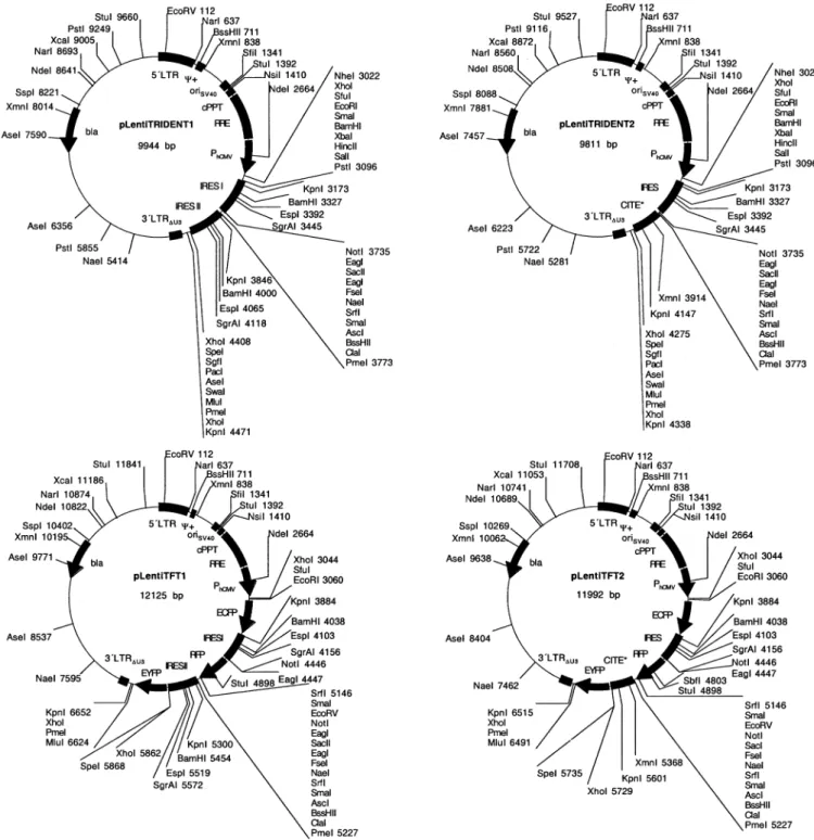
pMF356 was further digested with PacI/MluI and the PacI/ MluI PPGK-neo (PPGK, promoter of the phosphoglycerate Figure 1. (Previous page and above) SIN lentivectors containing optimized MCS. Generic HIV-1-derived lentivectors containing the 5¢LTR, the extended packaging signal (y+), the Simian virus 40 origin of replication (oriSV40), the cPPT, the RRE and the 3¢LTR with deleted enhancer sequences (3¢LTRDU3). The MCS was placed between RRE and 3¢LTRDU3. Besides this basic lentivector con®guration a variety of derivatives containing different mammalian/ human promoters (PPGK, phosphoglycerate kinase promoter; PhEF1a, human elongation factor 1a promoter; PhCMV, human immediate early cytomegalovirus promoter) and reporter (EYFP and ECFP) or resistance genes (neo, G418 resistance gene) have been designed.
kinase; neo, G418 resistance gene) cassette of pBM6 was inserted (pBM8; 5¢LTR-y+-oriSV40-cPPT-RRE-MCSI-PPGK
-neo-MCSII-3¢LTRDU3). pBM6 was constructed by amplifying
PPGK-neo from pMSCVneo (Clontech) using oligonucleotides
OBM1: CGACTAGTTTAATTAAaattctaccgggtagggg (an-nealing sequence in lower case, SpeI/PacI sites are under-lined) and OBM2: CGGGTACCACGCGTcctcagaagaac-tcgtcaag (annealing sequence in lower case, KpnI/MluI sites are underlined) and cloned in sense orientation into pcDNA3.1/V5-His TOPO (Invitrogen, Carlsbad, CA).
pMF356 derivatives containing either PhEF1a (PhEF1a,
promoter of the human elongation factor 1a; pMF359) or PhCMV(pMF363) promoters were generated as follows.
pMF359 (5¢LTR-y+-oriSV40-cPPT-RRE-PhEF1a
-MCS-3¢LTRDU3). (i) PhEF1a was ampli®ed from pEF4/MycHisB
(Invitrogen) using oligonucleotides OMF164: GATC-GGATCCtgcaaagatggataaagt (annealing sequence in lower case, BamHI site is underlined) and OMF165: GATC- AAGCTTTTAATTAAGCGATCGCGCCCGGGCGCGGC-CGCaactagccagcttgggtc (annealing sequence in lower case, HindIII/PacI/SgfI/SrfI/SmaI/NotI sites are underlined) and cloned in antisense orientation into pcDNA3.1/V5-His TOPO (Invitrogen) to result in pMF320. PhEF1a was excised from
pMF320 by EcoRV/XmaI and cloned into the compatible sites (HpaI/XmaI) of pMF356 thereby resulting in pMF359 (5¢LTR-y+-oriSV40-cPPT-RRE-PhEF1a-MCS-3¢LTRDU3). (ii)
EYFP was excised from pMF201 (33) by EcoRV/SpeI and cloned into the compatible sites (SmaI/SpeI) of pMF359 to result in pMF365 (5¢LTR-y+-oriSV40-cPPT-RRE-PhEF1a
-EYFP-3¢LTRDU3). (iii) pMF359 was restricted using PacI/
MluI and the PPGK-neo cassette excised from pBM6 by PacI/
MluI was inserted (pBM9; 5¢LTR-y+-oriSV40
-cPPT-RRE-PhEF1a-MCSI-PPGK-neo-MCSII-3¢LTRDU3). pMF359
deriva-tives include: (i) pBM42: pMF359 was restricted with EcoRI/ PmeI and EPO (erythropoietin) excised from pMF242 (33) by EcoRI/EcoRV was inserted (5¢LTR-y+-oriSV40
-cPPT-RRE-PhEF1a-EPO-3¢LTRDU3). (ii) pBM43: pMF359 was restricted
with SalI/MluI and VEGF121 (variant 121 of the
vascu-lar endothelial growth factor) excised from pWW265 (C.Weber and A.Zisch, unpublished data) by SalI/MluI was inserted (5¢LTR-y+-oriSV40-cPPT-RRE-PhEF1a-VEGF121
-3¢LTRDU3). (iii) pBM44: pMF359 was restricted with
EcoRI/PmeI and SEAP (human placental secreted alkaline phosphatase) excised from pSS173 (34) by EcoRI/EcoRV was inserted (5¢LTR-y+-oriSV40-cPPT-RRE-PhEF1a
-SEAP-3¢LTRDU3).
pMF363 (5¢LTR-y+-oriSV40-cPPT-RRE-PhCMV
-MCS-3¢LTRDU3): (i) PhCMV was excised from pLPCX (Clontech)
with StuI/EcoRI and ligated to the compatible HpaI/EcoRI sites of pMF356 to result in pMF363 (5¢LTR-y+-oriSV40
-cPPT-RRE-PhCMV-MCS-3¢LTRDU3). (ii) ECFP was excised
from pMF201 (33) by EcoRI/NotI and ligated to the corres-ponding sites (EcoRI/NotI) of pMF363 thereby resulting in pMF371 (5¢LTR-y+-oriSV40-cPPT-RRE-PhCMV
-ECFP-3¢LTRDU3).
Tricistronic pLentiTRIDENT expression vectors. pLenti-TRIDENT vectors contain a ppLenti-TRIDENT-derived tricistronic expression cassette inserted in the MCS of pMF363. pLentiTRIDENT1 (pMF370; 5¢LTR-y+-oriSV40
-cPPT-RRE-PhCMV-MCSI-IRESI-MCSII-IRESII-MCSIII-3¢LTRDU3) was
constructed by excising the MCSI-IRESI-MCSII-IRESII-MCSIII cassette from pTRIDENT1 [pMF125 (35)] by EcoRI/BglII and cloning it into the compatible sites (EcoRI/ BamHI) of pMF363. pLentiTRIDENT1 was further restricted using EcoRI/MluI and the triple-¯uorescent expression cassette (ECFP-IRESI-RFP-IRESII-EYFP) excised from pWW85 (36) by EcoRI/MluI was inserted [pLentiTFT1 (pBM2); 5¢LTR-y+-oriSV40-cPPT-RRE-PhCMV
-ECFP-IRESI-RFP-IRESII-EYFP-3¢LTRDU3]. pLentiTRIDENT2 (pMF369;
5¢LTR-y+-oriSV40-cPPT-RRE-PhCMV
-MCSI-IRES-MCSII-CITE*-MCSIII-3¢LTRDU3) was constructed by excising
the MCSI-IRES-MCSII-CITE*-MCSIII cassette from
pTRIDENT3 [pMF122 (35)] by EcoRI/BglII and cloning it into the compatible sites (EcoRI/BamHI) of pMF363. pLentiTRIDENT2 was further restricted using EcoRI/AscI and the ECFP-IRESI-RFP cassette excised from pWW85 (36) by EcoRI/AscI was inserted to result in pBM1 (5¢LTR-y+-oriSV40-cPPT-RRE-PhCMV
-ECFP-IRES-RFP-CITE*-MCSIII-3¢LTRDU3). Subsequently, pBM1 was digested using
SpeI/MluI and the EYFP cassette excised from pWW85 by SpeI/MluI was inserted [pLentiTFT2 (pBM3); 5¢LTR-y+-oriSV40-cPPT-RRE-PhCMV
-ECFP-IRES-RFP-CITE*-EYFP-3¢LTRDU3].
Highly compact lentiviral expression vectors. pNL-derived lentiviral expression vectors contain super¯uous chromosomal sequences ¯anking the lentiviral expression unit (32). In order to eliminate these chromosomal sequences, the 5¢LTR-y+-oriSV40-cPPT-RRE-MCS-3¢LTRDU3 cassette was ampli®ed
from pMF356 using oligonucleotides OMF199:
GATCGACGTCacttacaccaggaaaggc (annealing sequence in lower case, AatII site is underlined) and OMF200: GATCGCTAGCacctctacctcctggggg (annealing sequence in lower case, NheI site is underlined) and cloned as AatII/NheI fragment to the compatible, AatII/XbaI-restricted pTRIDENT1 [pMF125 (35)] backbone thereby resulting in pMF388 (5¢LTR-y+-oriSV40-cPPT-RRE-MCS-3¢LTRDU3).
pMF388 derivatives include: (i) pMF394: pMF388 was restricted with HpaI/KpnI and the PhCMV-EYFP cassette
excised from pMF351 by HpaI/KpnI was inserted (5¢LTR-y+-oriSV40-cPPT-RRE-PhCMV-EYFP-3¢LTRDU3). (ii) pBM7:
PhEF1awas excised from pMF320 by EcoRV/XmaI and cloned
into the compatible HpaI/XmaI sites of pMF388 (5¢LTR-y+-oriSV40-cPPT-RRE-PEF1a-MCS-3¢LTRDU3). (iii) pBM13:
pMF388 was restricted with PacI/MluI and the PacI/MluI PPGK-neo cassette excised from pBM6 was inserted
(5¢LTR-y+-oriSV40-cPPT-RRE-MCSI-PPGK-neo-MCSII-3¢LTRDU3).
(iv) pBM14: pBM7 was restricted with PacI/MluI and the PacI/MluI PPGK-neo cassette excised from pBM6 was
inserted (5¢LTR-y+-oriSV40-cPPT-RRE-PEF1a-MCSI-PPGK
-neo-MCSII-3¢LTRDU3). (v) pBM40: pBM7 was restricted
with SpeI/MluI and EYFP excised from pBM12 by NheI/MluI was inserted (5¢LTR-y+-oriSV40-cPPT-RRE-PhEF1a
-EYFP-3¢LTRDU3).
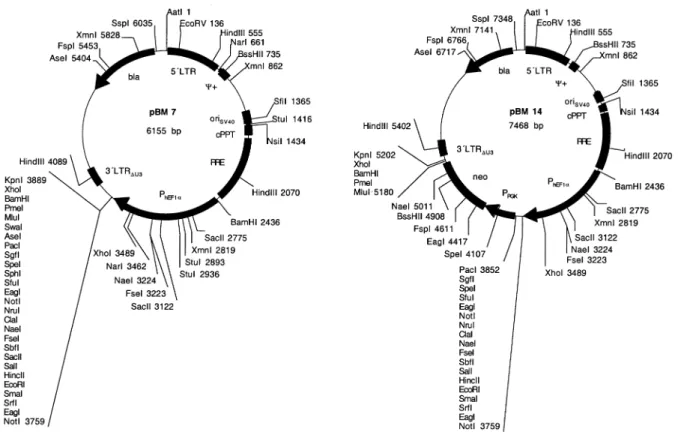
pLentiModule, a modular lentiviral expression vector (all sites indicated correspond to GenBank accession no. AF033819). pLentiModule consists of four minimal lentiviral modules [U3, R-U5-y+-SD, cPPT-RRE-SA, PPT-3¢LTRDU3
(DU3-R-U5)] containing unique restriction sites at their joints. These modules were independently ampli®ed from pNL-EGFPDU3
and cloned into pcDNA3.1/V5-His TOPO (Invitrogen). (i) pMF352: U3 (8631±9085) was ampli®ed using oligonucle-otides OMF183, GATCCTCGAGTCGCGActggaagggctaatttgg (annealing sequence lower case, XhoI/NruI sites are under-lined); and OMF184, GATCGCCCGGGCCAGTACAGGC- AAAAAGCAGCTGCTTATATGTAGCATCTGAG- GGCTCGCCACTCCCCAGTCCCGCCCAGGCCACACCT-CCCTGCAGGcctggaaagtccccagcg (annealing sequence lower case, SrfI and SbfI are underlined; this primer inserts a SbfI at position 9007 of U3) (XhoI/NruI-U31-SbfI-U32-SrfI).
(ii) pMF376: R-U5-y+-SD (1±793) was ampli®ed from pNL-EGFPDU3 using oligonucleotides OMF185, GATCGCC-CGGGCggtctctctggttagacc (annealing sequence lower case, SrfI site is underlined); and OMF193, GATCATT-TAAATtttaaagttctaggtgat (annealing sequence lower case, SwaI site is underlined) (SrfI-R-U5-y+-SD-SwaI). pMF353: a shorter R-U5-y+-SD module (1±377) was ampli®ed using oligos OMF185 and OMF186: GATCATTTAAAT-tcattaatctaattctcc (annealing sequence lower case, SwaI site is underlined). (iii) pMF375: cPPT-RRE-SA [4330±4475 (cPPT), 7200±8024 (RRE)] was ampli®ed using oligonucle-otides OMF191, GATCATTTAAATatgcataaaagaaaaggg (an-nealing sequence lower case, SwaI site is underlined); and OMF192, CTAGGATCACGCGTTTAATTAAGCGATCG- CACTAGTATCGATGGCGCGCCGGCCGGCCAGGCCT-GCGGCCGCGAATTCGACGTCatccgttcactaatcgaa (anneal-ing sequence lower case, MluI/PacI/SgfI/SpeI/ClaI/AscI/FseI/ StuI/NotI/EcoRI/AatII are underlined) (SwaI-cPPT-RRE-SA-MluI/PacI/SgfI/SpeI/ClaI/AscI/FseI/StuI/NotI/EcoRI/AatII). (iv) pMF355: PPT-3¢LTRDU3 (DU3-R-U5) (8615±9181 and
1±181) was ampli®ed using oligonucleotides OMF189, GATCACGCGTGGATCCAAAAGAAAAGGGGGGACT- GGAAGGGCTAATTCACTCCCAAAGAAGACAAGATG-TTTAAACGTTAACTGATCActgctttttgcctgtact [annealing sequence lower case, MluI/BamHI is underlined, PPT is shown in bold; PmeI/HpaI/BclI are underlined and placed between the EcoRV/PvuII (GAT ¼ CTG, italics) ligation sites ¯anking the DU3 deletion (corresponding to 8666±9066) of the 3¢LTR (25,32)]; and OMF190, GATCTCTAG-AGTCGACGCTAGCCATATGctgctagagattttccaca (anneal-ing sequence lower case, XbaI/SalI/NheI/NdeI are underlined) (MluI/BamHI-PPT-DU31-PmeI/HpaI/BclI-DU32-R-U5-NdeI/
NheI/SalI/XbaI).
pLentiModule was assembled on a pTRIDENT1 backbone following a four-step procedure. (i) U3 was excised from pMF352 using XhoI/SrfI and ligated to the corresponding sites (XhoI/SrfI) of pTRIDENT1 to result in pMF357. (ii) R-U5-y+-SD was excised from pMF353 and pMF376 by SrfI/SwaI and ligated into the corresponding sites (SrfI/SwaI) of pMF357 to result in pMF358 and pMF358x. (iii) cPPT-RRE-SA was excised from pMF375 using SwaI/MluI and ligated into the corresponding sites (SwaI/MluI) of pMF358 and pMF358x to result in pMF360 and pMF360x. (iv) PPT-3¢LTRDU3 was
excised from pMF355 by MluI/XbaI and ligated into the corresponding sites (MluI/XbaI) of pMF360 and pMF360x to result in pMF402 and pLentiModule1 (pBM4; XhoI/NruI-U31-StuI / SbfI-U32-SrfI-R-U5-y +
-SD-SwaI-cPPT-RRE-SA-AatII/EcoRI/NotI/EagI/StuI/FseI/EagI/AscI/BssHII/ClaI/SpeI/ SgfI/PacI/MluI/BamHI-DU31-PmeI/HpaI/BclI-DU32-NdeI/NheI/
SalI/XbaI). plentiModule1 derivatives include the following. (i) pLentiModule2: pLentiModule1 was cut with EcoRI/MluI
Table 2. EPO, VEGF121and SEAP production in CHO-K1 cells following lentiviral transduction
Lentivector Production (ng/ml)
EPO VEGF121 SEAP
pBM42 65.4 6 9.2 pBM43 20.0 6 0.5 pBM44 2.1 3 1036 80.6 pLentiModule7 37.1 6 5.5 pLentiModule6 11.3 6 1.1 pLentiModule8 1.1 3 1036 23.6
Figure 2. Confocal microscopy analysis of transduction ef®ciencies of key lentivectors. CHO-K1 were transduced with 2 3 106 EYFP-encoding
pMF365-, pBM40- and pLentiModule-derived lentiviruses. Transduced cells were stained with the DNA-speci®c DRAQ5 dye and analyzed for YFP-and DRAQ5-mediated ¯uorescence by confocal microscopy (magni®cation 2003).
Table 3. Titer and p24 production of various lentivectors
Lentivector Titer (c.f.u./ml)a p24 (ng/ml)
pNL-EGFPb 2 3 107 95.0 6 5.3 pNL-EGFPDU3 4 3 107 165.3 6 5.1 pMF351 2.5 3 107 104.8 6 6.4 pMF365 2 3 107 97.3 6 8.5 pBM2 ND 158.7 6 7.3 pBM40 2 3 107 97.3 6 9.6 pBM42 ND 66.1 6 8.3 pBM43 ND 70.6 6 7.8 pBM44 ND 63.5 6 8.0 pLentiModule1 2.5 3 107 110 6 7.2 pLentiModule2 2.5 3 107 110 6 7.3 pLentiModule4 2 3 107 97.2 6 12.3 pLentiModule5 1.2 3 107 48.6 6 5.4 pLentiModule6 ND 64.9 6 6.7 pLentiModule7 ND 58.8 6 8.6 pLentiModule8 ND 72.3 6 8.1 ND, not determined.
aDetermined as described in Reiser et al. (17). bDetermined on NIH/3T3 cells, see Reiser et al. (17).
and the PhCMV-EYFP cassette excised from pMF351 by
EcoRI/MluI was inserted to result in pLentiModule2 (pBM5) (XhoI/NruI-U31-StuI/SbfI-U32
-SrfI-R-U5-y+-SD-SwaI-cPPT-RRE-SA-PhCMV-EYFP-DU31
-PmeI/HpaI/BclI-DU32-NdeI/NheI/SalI/XbaI). (ii) pLentiModule5:
pLenti-Module1 was cut with BamHI and the PhEF1a-EYFP-encoding
BamHI cassette of pMF365 was inserted (pBM45) (XhoI/ NruI-U31-StuI/SbfI-U32
-SrfI-R-U5-y+-SD-SwaI-cPPT-RRE-SA-PhEF1a-EYFP-DU31-PmeI/HpaI/BclI-DU32-NdeI/
NheI/SalI/XbaI). (iii) pLentiModule6: pLentiModule1 was cut
with BamHI and the PhEF1a-VEGF121-encoding BamHI
cas-sette of pBM43 was inserted (pBM46) (XhoI/NruI-U31-StuI/
SbfI-U32-SrfI-R-U5-y+-SD-SwaI-cPPT-RRE-SA-PhEF1a
-VEGF121-DU31-PmeI/HpaI/BclI-DU32-NdeI/NheI/SalI/XbaI).
(iv) pLentiModule7: pLentiModule1 was cut with BamHI and the PhEF1a-EPO-encoding BamHI cassette of pBM42 was
inserted (pBM47) (XhoI/NruI-U31-StuI/SbfI-U32
-SrfI-R-U5-y+-SD-SwaI-cPPT-RRE-SA-PhEF1a-EPO-DU31-PmeI/HpaI/
BclI-DU32-NdeI/NheI/SalI/XbaI). (v) pLentiModule8:
-SEAP-encoding BamHI cassette of pBM44 was inserted (pBM48) XhoI/NruI-U31-StuI/SbfI-U32
-SrfI-R-U5-y+-SD-SwaI-cPPT-RRE-SA-PhEF1a-SEAP-DU31-PmeI/HpaI/BclI-DU32-NdeI/
NheI/SalI/XbaI).
In order to place the oriSV40on pLentiModule1, this plasmid
was restricted with XbaI and the oriSV40XbaI/NheI fragment
excised from pMF378 was inserted to result in pLentiModule3 (pBM15) (XhoI/NruI-U31-StuI/SbfI-U32
-SrfI-R-U5-y+-SD-SwaI-cPPT-RRE-SA-MCS-DU31-PmeI/HpaI/BclI-DU32
-NdeI/NheI/SalI/XbaI-oriSV40). pMF378 was constructed by
amplifying oriSV40 from pNL-EGFPDU3 using
oligonucle-otides OMF195: GATCTCTAGAtatgactccgcccatccc (anneal-ing sequence lower case, XbaI site is underlined) and OMF196: GATCGCTAGCttgcaaaagcctaggcc (annealing sequence lower case, NheI sequence is underlined) and ligating it into pcDNA3.1/V5-His TOPO. Furthermore, pLentiModule2 was restricted with XbaI and the oriSV40
XbaI/NheI fragment excised from pMF378 was inserted to result in pLentiModule4 (pBM12) (XhoI/NruI-U31
-StuI/SbfI-U32-SrfI-R-U5-y+-SD-SwaI-cPPT-RRE-SA-PhCMV
-EYFP-DU31-PmeI/HpaI/BclI-DU32-NdeI/NheI/SalI/XbaI-oriSV40).
Lentivirus production and infection
For production of replication-incompetent SIN lentiviruses, a mixture containing 94 ml DMEM, 6 ml FUGENE (Roche Diagnostics AG, Rotkreuz, Switzerland), 25 mM chloroquine, 1 mg pLTR-G [encoding the pseudotyping envelope protein VSV-G of the vesicular stomatitis virus (17)], 1 mg of the helper construct pCD/NL-BH* (32) and 1 ml of the desired
transgene-encoding lentiviral expression vector were trans-fected into human embryonic kidney cells (HEK293-T, kindly provided by Andreas Zisch). The medium was replaced after 12±15 h and virus particles were produced for another 48 h. Viral particles were collected from the HEK293-T supernatant by ®ltration through a 0.45 mm ®lter (Schleicher & Schuell GmbH, Dassel, Germany: FP 030/2) yielding typical titers of 2 3 107c.f.u./ml when titrated on CHO-K1 cells. p24 assays
were performed using a commercial kit (NEK-050, Perkin Elmer, Boston, MA) [pMF351, 104.8 6 6.4 ng/ml p24; pLentiModule1, 109.9 6 7.25 ng/ml p24 (triplicate mean values)]. 60 000 target cells per 6-well were infected with 200 ml viral supernatant. Hippocampal slice cultures were infected as described by Ehrengruber and co-workers (37). RESULTS
Design of multi-purpose lentiviral expression vectors containing extensive multiple cloning sites
Most lentiviral expression vectors currently available from the research groups, which have pioneered lentiviral transduction technologies are lacking extensive MCS rendering the straightforward design of HIV-1-based expression vectors complicated (16). We have constructed a versatile lentiviral expression vector family derived from the HIV-1-based SIN pNL-EGFPDU3 (J.Reiser, unpublished data; 38) which con-tains up to 29 unique sites for restriction endonucleases, many of which are rare-cutting 8 bp-recognizing enzymes. pNL-EGFPDU3 has a classical lentiviral expression vector set-up Figure 3. (Previous page and above) Size-optimized SIN lentivectors. These lentivectors are derivatives of the ones shown in Figure 1 and are devoid of any chromosomal sequences ¯anking the lentiviral expression vectors.
consisting of 5¢LTR, y+, splice donor (SD), origin of replication of the simian virus 40 (oriSV40), central polypurine
tract (cPPT), RRE, splice acceptor (SA) and a 3¢LTRDU3. The
PhCMV-EGFP expression cassette is inserted between the SA
and the 3¢LTRDU3 using the unique HpaI and KpnI sites
(5¢LTR-y+-SD-oriSV40-cPPT-RRE-SA-HpaI-PhCMV
-EGFP-KpnI-3¢LTRDU3). The oriSV40 is boosting the vector copy
number in transiently transfected cells producing SV40 large T antigen, such as HEK293-T cells (17). A 400 bp EcoRV/ PvuII deletion within the 3¢LTR (3¢LTRDU3) inactivates the
5¢LTR promoter upon reverse transcription and integration when the DU3 deletion is transferred to the 5¢LTR in a self-suf®cient manner, a process also referred to as SIN (25,26).
In order to extend the MCS of pNL-EGFPDU3, a PCR-ampli®ed PhCMV-EYFP cassette ¯anked by 26 unique
restric-tion sites was cloned into the HpaI/KpnI sites of pNL-EGFPDU3 and resulted in pMF351 (5¢LTR-y+-oriSV40
-cPPT-RRE-MCSI-PhCMV-EYFP-MCSII-3¢LTRDU3; Fig. 1,
Table 1). The PhCMV-EYFP was subsequently excised by a
NotI deletion resulting in pMF356, a lentiviral expression vector containing 28 unique restriction sites (5¢LTR-y+-oriSV40-cPPT-RRE-MCS-3¢LTRDU3; Fig. 1, Table 1). As
non-exhaustive examples for the construction kit-like lentiviral expression vector platform a variety of pMF356 derivatives have been designed which contain: (i) a PPGK-driven
neomycin resistance gene (neo) (pBM8; 5¢LTR-y+-oriSV40
-cPPT-RRE-MCSI-PPGK-neo-MCSII-3¢LTRDU3), (ii) a
strong constitutive human elongation factor 1a promoter (PhEF1a; pMF359; 5¢LTR-y+-oriSV40-cPPT-RRE-PhEF1a
-MCS-3¢LTRDU3), (iii) a PhEF1a-driven EYFP cassette
(pMF365; 5¢LTR-y+-oriSV40-cPPT-RRE-PhEF1a
-MCSI-EYFP-MCSII-3¢LTRDU3), (iv) a PhEF1a promoter plus a
PPGK-neo cassette (pBM9; 5¢LTR-y+-oriSV40
-cPPT-RRE-PhEF1a-MCSI-PPGK-neo-MCSII-3¢LTRDU3), (v) a human
cy-tomegalovirus immediate early promoter (PhCMV; pMF363;
5¢LTR-y+-oriSV40-cPPT-RRE-PhCMV-MCS-3¢LTRDU3) or
(vi) a PhCMV-ECFP cassette (pMF371; 5¢LTR-y+-oriSV40
-cPPT-RRE-PhCMV-MCSI-ECFP-MCSII-3¢LTRDU3) (see Fig. 1
and Table 1 for all plasmids).
In order to validate this class of multi-purpose lentivectors, a variety of pMF359 derivatives were constructed which encode EPO (pBM42), the vascular endothelial growth factor (VEGF121, pBM43) or the SEAP (pBM44) under control of
PhEF1a. pBM42-, pBM43- and pBM44-derived lentiviruses
were able to drive high-level protein secretion following transduction of CHO-K1 (Table 2). Also, pMF351- and pMF365-based lentivectors displayed similar transduction ef®ciencies compared with their parental constructs of the pNL-EGFP series (38) (Fig. 2, Table 3).
Although the basic lentiviral expression units contained in these expression vectors (pMF351, pMF356, pBM8, pMF359, pMF365, pBM9, pBM363, pMF371; Fig. 1) are only ~3.5 kb in size, they are ¯anked by super¯uous chromosomal regions that are remnants of the integration sites of the ®rst HIV proviruses cloned from the human chromosome [molecular clone HXB2 (39)]. As these chromosomal sites are not packaged and transferred to the target cells, they are not limiting the cargo size of the lentiviral expression units. However, the entire expression vectors may reach the replicative limit when ampli®ed in Escherichia coli.
Elimination of any chromosomal sequences was achieved by PCR-mediated ampli®cation of the lentiviral expression units (5¢LTR-y+-oriSV40-cPPT-RRE-MCS-3¢LTRDU3). AatII
and NheI sites engineered in the primer extensions enabled ligation to the pTRIDENT-derived minimal high-copy num-ber vector backbone fragment generated by AatII/XbaI digestion (35,40). This cloning procedure resulted in pMF388, a 4.8 kb vector containing a lentiviral expression module with 29 unique restriction sites for convenient integration of desired transgenes (Fig. 3, Table 1; pMF388, 5¢LTR-y+-oriSV40-cPPT-RRE-MCS-3¢LTRDU3). Several
ex-emplary pMF388 derivatives have been designed which contain in addition to the basic lentiviral expression unit: (i) a PhCMV-driven EYFP cassette (pMF394; 5¢LTR-y+-oriSV40
-cPPT-RRE-MCSI-PhCMV-EYFP-MCSII-3¢LTRDU3), (ii) a
PhEF1a promoter (pBM7; 5¢LTR-y+-oriSV40
-cPPT-RRE-PhEF1a-MCS-3¢LTRDU3), (iii) a PhEF1a-driven EYFP
cas-sette (pBM40; 5¢LTR-y+-oriSV40-cPPT-RRE-PhEF1a -EYFP-Figure 4. Transduction of optimized lentivectors into various insect,
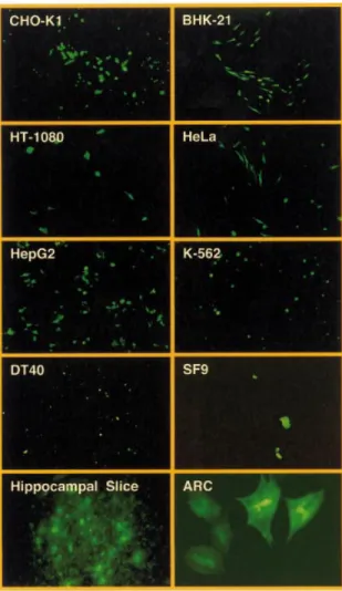
chicken, murine and human cell types as exempli®ed by VSV-G-pseudo-typed pMF351-derived lentiviruses (Fig. 1). The gene encoding the EYFP was successfully transduced into Chinese hamster ovary cells (CHO-K1), baby hamster kidney cells (BHK-21), human ®brosarcoma cells (HT-1080), human cervial adenocarcinoma cells (HeLa), human hepatocellular carcinoma cells (HepG2), human chronic myelogenous leukemia cells (K-562), bursal chicken cells (DT40) as well as insect cells (SF9). pMF351-derived lentiviruses also transduced glial cells in hippocampal slice cultures (hippocampal slice) and primary ARC (magni®cation 1003, hippocampal slice 503; ARC 4003).
3¢LTRDU3), (iv) a PPGK-driven neomycin resistance cassette
(PPGK-neo; pBM13; 5¢LTR-y+-oriSV40
-cPPT-RRE-MCSI-PPGK-neo-MCSII-3¢LTRDU3) or (v) a PhEF1apromoter plus a
PPGK-neo cassette (pBM14; 5¢LTR-y+-oriSV40
-cPPT-RRE-PhEF1a-MCSI-PPGK-neo-MCSII-3¢LTRDU3) (see Fig. 3 and
Table 1 for all plasmids).
Besides the great ¯exibility to accommodate almost any desired transgene, the extended MCS of the lentiviral expres-sion vectors shown in Figures 1 and 3 are fully compatible with pQuattro, pTRIDENT, pRetroTRIDENT, pTWIN, pRetroTWIN and pDuoRex type constructs and enable one-step exchange of multicistronic and regulated expression units among these different expression vector families (35,40±42). In addition, these vectors facilitate straightforward swapping of cis-acting elements and transgene expression units between each other using: (i) remaining polylinker sites; (ii) unique restriction sites within the (5¢) b-lactamase coding region (bla; AseI, FspI, XmnI, SspI, AatII); and/or (iii) corresponding cis-acting elements (EcoRV, NarI, BssHII, XmnI, S®I, StuI, NsiI). We have tested the construction kit-like lentiviral expres-sion vector platform by infecting various insect (SF9), chicken (DT40), murine (BHK-21, CHO-K1) and human (HeLa, HT-1080, HepG2, K-562) cell lines as well as ARC and hippocampal slices using the pMF351-derived lentivirus (5¢LTR-y+-oriSV40-cPPT-RRE-MCS-PhCMV
-EYFP-MCSII-3¢LTRDU3). All cell lines transduced with pMF351-derived
lentiviruses show high-level expression of the EYFP while EYFP expression is absent in mock-transduced control cultures. Even ARCs, which are known to be refractory to a variety of transfection/transduction technologies, were readily
transduced by the pMF351-derived lentivirus providing an alternative to the recently described Sindbis-based transduc-tion systems (28). Also, this lentivirus transduced glial cells in rat hippocampal slices (Fig. 4). In CHO-K1 cells the transduction ef®ciency of pBM4-derived lentiviruses was similar compared with the parental pNL-EGFP and pMF351 vector series (38) (see above and Fig. 2, Table 3).
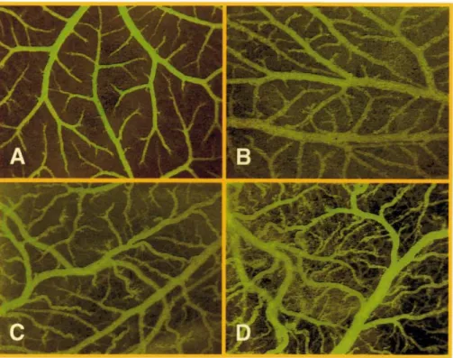
The biological potential of lentiviral transduction was further exempli®ed by expression of VEGF in chicken embryos. Infection of the chick embryos' CAM with increas-ing concentrations of pBM43-derived lentiviruses (5¢LTR-y+-oriSV40-cPPT-RRE-PhEF1a-VEGF121-3¢LTRDU3) resulted
in a dose-dependent VEGF121-mediated local induction of
new blood vessels demonstrated by increased vascular dens-ity, enhanced formation of numerous arterioles and venules as well as by a boost in vessel endpoint and branching density (Fig. 5). When overdosing VEGF121 (5 3 106 c.f.u./ml
producing 35.5 6 3.8 ng/ml VEGF121in 48 h; not shown) or
following i.v. application of pBM43-derived lentivirus (1.5 3 106c.f.u./ml producing 10.6 6 0.9 ng/ml VEGF121in 48 h),
the hierarchical, tree-like structure of the supplying vessels disappears and the multitude of arterioles and venules adopt an irregular tortuous shape associated with atypical delta- or brush-like vessel endpoints (Fig. 5D).
Construction of pLentiTRIDENT vectors for tricistronic expression of up to three transgenes
Multicistronic expression technologies for coordinated expression of several transgenes from a single arti®cial eukaryotic operon have generated unprecedented impact in Figure 5. In vivo examination of microvascular growth in the CAM of 11 day old chicken embryos 48 h after pBM43-based lentiviral VEGF121transduction. Whereas the regular vessel pattern observed on the CAM remained intact in mock transductions (A), dose-dependent angiogenic response resulted in increased vascular density and atypical (brush- and delta-like) endpoint patterns following spotting of 0.5 3 106c.f.u./ml (producing 3.4 6 0.3 ng/ml VEFF121in 48 h)
(B), 3 3 106c.f.u./ml (producing 21.2 6 1.7 ng/ml VEGF121in 48 h) or i.v. injection of 1.5 3 106c.f.u./ml (producing 10.6 6 0.9 ng/ml VEGF121in 48 h)
rational reprogramming of mammalian production cell lines and anti-in¯ammation engineering (43,44). Particularly in a therapeutic setting when the therapeutic proteins consist of multiple subunits or multiple target/strategy approaches involving ribozymes, antisense RNA, transdominant pro-teins and intracellular antibodies, multicistronic expression technologies are highly desirable (45). We have recently
pioneered a multicistronic expression vector family, pTRIDENT, which enables tricistronic expression of up to three transgenes from a single constitutive or regulatable promoter. Whereas the ®rst cistron is translated in a classical cap-dependent manner, translation initiation of the second and third cistrons rely on viral cap-independent internal ribosome entry sites of poliovirus (IRES) or encephalomyocarditis virus Figure 6. pLentiTRIDENT, tricistronic lentiviral expression vectors for simultaneous expression of up to three transgenes. Besides the basic lentiviral cis-act-ing elements (see Fig. 1) pLentiTRIDENT contain a tricistonic expression unit consistcis-act-ing of two IRES or encephalomyocarditis viral origin (CITE*) ¯anked by extensive MCS for integration of desired transgenes, for example the ones encoding the cyan (ECFP), red (RFP) and yellow (EYFP) ¯uorescent genes (pLentiTFT1 and pLentiTFT2).
(CITE) which had been optimized for maximum translation enhancement (CITE*) (35,40,42).
In order to merge multicistronic expression and lentiviral transduction technology, we have designed the pLenti-TRIDENT vector series, which contains a PhCMV-driven
tricistronic eukaryotic operon within a pMF356-derived lentiviral expression cassette (Fig. 6, Table 1). pLenti-TRIDENT1/(pLentiTRIDENT2) was constructed by excising the MCSI-IRESI-MCSII-IRESII/(CITE*)-MCSIII cassette of pTRIDENT1/(pTRIDENT3) by EcoRI/BglII and ligating it into the lentiviral transduction unit of pMF363 (EcoRI/ BamHI) [5¢LTR-y+-oriSV40-cPPT-RRE-PhCMV
-MCSI-IRESI-MCSII-IRESII/(CITE*)-MCSIII-3¢LTRDU3]. The
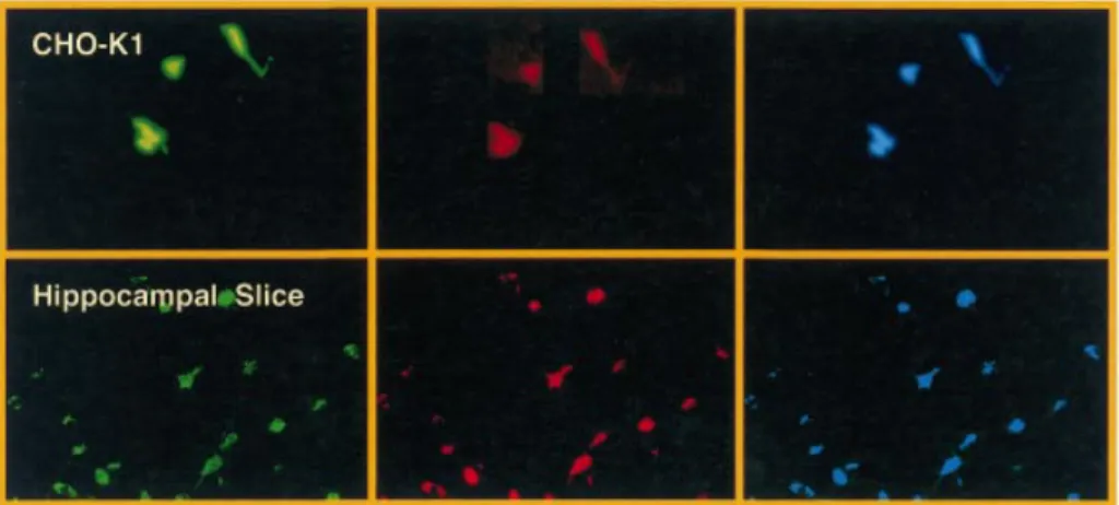
pLenti-TRIDENT vectors were validated by inserting the genes encoding the (enhanced) cyan, red and yellow ¯uorescent proteins into the ®rst, second and third cistrons, respectively, which resulted in the triple-¯uorescent pLentiTRIDENT vectors (pLentiTFT1/(pLentiTFT2). CHO-K1 cells and glial cells in hippocampal slices transduced with pLentiTFT vectors showed simultaneous high-level expression of all three ¯uorescent marker proteins (Fig. 7).
pLentiModule, an advanced highly modular lentiviral expression system
Previous lentiviral expression vectors have been designed for optimal integration of desired transgenes by engineering of extended MCS. However, lentiviral transduction units have been successfully modi®ed in the past years to optimize speci®c cis-acting modules for: (i) Tat-independent produc-tion of lentiviruses by replacement of the 5¢LTR enhancer region (19,23,26,46); (ii) optimization of packaging signal and Rev-responsive elements (47,48); and (iii) replacement of the enhancer sequences of the 3¢LTR by LoxP sequences for conditional site-speci®c excision of transduced expression units (49) or by antibiotic-responsive operator modules for conditional proviral gene expression (46).
In order to combine minimal lentiviral transduction mod-ules with extended polylinkers and the highest ¯exibility for advanced modi®cation of the cis-acting elements we designed a novel modular lentivector (pLentiModule) which consists of four modules: (i) XhoI/NruI-U31-StuI/SbfI/PstI-U32-SrfI, (ii)
SrfI-R-U5-y+-SD-SwaI/NsiI, (iii)
SwaI/NsiI-cPPT-RRE-SA-MCSI and (iv) SwaI/NsiI-cPPT-RRE-SA-MCSI-3¢LTRDU3(1)-MCSII-3¢LTRDU3
(2)-MCSIII. These modules are ¯anked by unique restriction sites and were sequentially assembled into a minimal pTRIDENT-derived prokaryotic vector backbone to result in pLentiModule1 [XhoI/NruI-U31-StuI/SbfI/PstI-U32
-SrfI-R-U5-y+-SD-SwaI/NsiI-cPPT-RRE-SA-MCSI-3¢LTRDU3
(1)-MCSII-3¢LTRDU3(2)-MCSIII; Fig. 8, Table 1]. In contrast to
the previously described lentivectors pLentiModule is devoid of the oriSV40since transfer of this simian virus element into
target cells is incompatible with current therapeutic mod-alities.
pLentiModule1 displays a construction kit-like highly modular structure and enables design of the following lentivector characteristics. (i) Transfer of the entire lentiviral expression cassette using the ¯anking unique sites XhoI/NruI and MCSIII, (ii) exchange of the 5¢U3 region by XhoI/NruI and SrfI, (iii) switching of the 5¢U3 enhancer (Tat-binding) sequences by XhoI/NruI and StuI/SbfI/PstI, (iv) substitution of the R-U5-y+-SD module using SrfI and SwaI/NsiI, (v) replacement of the cPPT-RRE-SA module by modi®ed counterparts using the SwaI/NsiI and MCSI sites, (vi) cloning of desired transgenes, promoters and resistance cassettes into MCSI which is also compatible with the pLentiTRIDENT vectors and all of the previously described lentivectors (Figs 1, 3 and 6), (vii) swapping of the 3¢LTRDU3element using sites
contained in MCSI and MCSIII and (viii) integration of functional units (for example, LoxP, tissue-speci®c or regulatable operators, insulator sequences) into MCSII which are copied into the 5¢LTR upon reverse transcription and integration. In addition, (ix) cassette swapping with all other vectors described here is further facilitated using any of the sites within the bla region (AseI/FspI/XmnI/SspI/AatII) and the R-U5-y+-SD module (NarI/BssHII/XmnI).
pLentiModule1 was tested by insertion of a PhCMV-EYFP
cassette into its MCS (pLentiModule2; Fig. 6, Table 1) and transduction into insect (SF9), chicken (DT40), murine (BHK-21, CHO-K1) and human (HeLa, HT-1080, HepG2, K-562) cell lines as well as into ARCs and glial cells of hippocampal slice cultures (Fig. 9). Furthermore, pLentiModule1-derived lentivectors encoding EPO (pLentiModule7), VEGF121
(pLentiModule6) or SEAP (pLentiModule8) under control Figure 7. Transduction of Chinese hamster ovary (CHO-K1) cell and glial cells in rat hippocampal slice cultures using pLentiTRIDENT1-derived lentiviruses. Simultanous expression of the genes encoding the cyan, red and yellow ¯uorescent proteins could be observed (magni®cation 2003).
PhEF1a display high-level expression of these therapeutic/
reporter proteins in CHO-K1 cells (Tables 2 and 3).
As pLentiModule1 and pLentiModule2 are devoid of oriSV40, the virus titer is signi®cantly reduced to the ones
generated using an intact oriSV40/large T antigen replication
system (Table 3). We have therefore inserted a 168 bp fragment spanning the oriSV40into MCSIII of pLentiModule1
and pLentiModule2, respectively, resulting in pLentiModule3 and pLentiModule4. As quanti®ed from CHO-K1 transduc-tions, oriSV40-containing pLentiModule derivatives achieve Figure 8. Modular SIN lentiviral expression vectors. In order to facilitate the design of advanced lentiviral expression vectors, all cis-acting lentiviral sequences are ¯anked by unique restriction sites in pLentiModule vectors (XhoI/NruI-U3-SrfI; SrfI-R-U5-y+-SD-SwaI/NsiI; SwaI/NsiI-cPPT-RRE-SA-MCS; MCS-3¢LTRDU3-NdeI/NheI/SalI/XbaI). Unique restriction sites have also been placed within U3 (StuI, SbfI, PstI) and at the site of the deletion within 3¢LTRDU3(PmeI, HpaI, BclI). In order to enhance virus production an oriSV40(origin of replication of the Simian virus 40) has been placed 3¢ of the 3¢LTR outside of the lentiviral expression unit. Test lentivectors pLentiModule2 and pLentiModule3 contain a PhCMV-driven EYFP expression cassette in the central MCS.
virus titers, which compares to aforementioned lentivectors (Fig. 2, Table 3).
DISCUSSION
Since their discovery in the early 1980s, HIV-1 lentiviruses were known to cause one of the most devastating epidemics in industrialized countries in recent history (50,51). However, the unique characteristic of HIV-1 to transduce and integrate into the human genome owing to a set of accessory genes managing nuclear import has brought this deadly virus into the limelight of the gene therapy arena as most genetic defects in humans reside in non-proliferating terminally differentiated cells. As the ®rst development of replication-incompetent HIV-1-based vectors to transduce mitotically inert rat neurons, human skin ®broblasts and CD34+ cells (16,17), lentiviral
transduction technologies have witnessed a progress to the top of currently most promising gene therapy initiatives.
Elimination of some 60% of its genome (including all accessory genes) and re-assembly in multiply attenuated split-genome packaging systems successfully addressed legitimate initial safety concerns. The most advanced lentiviral transduc-tion systems include a third-generatransduc-tion packaging system consisting of a (i) gag-pol, (ii) rev and (iii) a vesicular stomatitis virus-derived envelope protein (VSV-G) as well as (iv) a SIN lentiviral expression vector. The split-genome strategy dramatically reduces the emergence of replication-competent recombinants and pseudotyping eliminates targeted infection of CD4+T lymphocytes as well as extension of the
cell tropism for therapeutic interventions (19). However, for successful infection of some target tissues including the liver some accessory proteins such as Vpr and Vif may be essential and should be considered for the packaging set-up (9). Typically, replication-incompetent lentiviral particles are produced by co-transfecting a helper cell line with all packaging and the lentiviral expression vector. However, with pharmaceutical production of therapeutic lentiviruses in mind a variety of different stable packaging cell lines have been designed which produced replication-incompetent trans-genic lentiviruses in a tetracycline-responsive manner (20±23).
The lentiviral expression unit is the only genetic material transferred to the target cells and encodes the desired therapeutic transgene. This expression vector contains only cis-acting sequences and is devoid of any lentiviral coding sequences, which prevents signi®cant humoral immune responses. Major improvements of lentiviral expression vectors include: (i) elimination of enhancer sequences from the 3¢LTR which results in complete loss of the 5¢LTR's transcriptional capacity and the viral mobilization competence upon integration of the provirus in the target chromosome thereby increasing safety and minimizing interferences with internal and chromosomal promoters in vivo and (ii) replace-ment of 5¢LTR promoters by heterologous promoters to achieve Tat-independent and/or tetracycline-responsive virus production (23,25,26,49). Many of these state-of-the-art lentivectors have been successfully used in pioneering gene therapy studies for the treatment of many prominent human diseases in animal models including Parkinson's disease and metachromatic leukodystrophy (8,10).
Despite the dramatic progress in lentivector development in recent years lentiviral expression con®gurations available from the pioneering research groups have been rather specialized and inconvenient for straightforward design of multi-purpose lentivectors by a broad scienti®c community. We have therefore designed a highly modular construction kit-like expression platform based on the latest generation SIN lentivectors for mono- and multicistronic expression in a variety of cell lines and primary cells including, for the ®rst time, ARCs. Also, to our knowledge, we report here the ®rst lentiviral transduction into chicken embryos. This simple-to-use multi-purpose lentivector expression platform, which is compatible with any of the aforementioned packaging concepts is expected to greatly facilitate the use of lentiviral transduction technologies by a wider scienti®c community to accelerate the advancement in gene therapy and tissue engineering.
Figure 9. Transduction of pLentiModule-derived lentiviruses into various insect, chicken, murine and human cell types as exempli®ed by a VSV-G-pseudotyped pLentiModule3-derived lentivirus (pLentiModule4). The gene coding for the EYFP was successfully transduced into Chinese hamster ovary cells (CHO-K1), baby hamster kindney cells (BHK-21), human ®brosarcoma cells (HT-1080), human cervial adenocarcinoma cells (HeLa), human hepatocellular carcinoma cells (HepG2), human chronic myelogenous leukemia cells (K-562), bursal chicken cells (DT40) as well as insect cells (SF9). pLentiModule4-derived lentiviruses also transduced glial cells in hippocampal slice cultures (hippocampal slice) and primary ARC (magni®cation 1003, hippocampal slice 503; ARC 4003).
ACKNOWLEDGEMENTS
We thank H. M. Eppenberger for providing adult rat cardiomyocytes, J. Reiser for plasmid pNL-EGFPDU3 as well as a set of helper vectors, L. Rietschin and A. Nussbaumer for preparation of the hippocampal slice cultures and B. Lennon, S. Schlatter and W. Weber for critical comments on the manuscript. This work was supported by the Novartis Research Foundation, the Roche Research Foundation and the Bundesamt fuÈr Bildung und Wissenschaft within the Framework V Programme of the European Commission. Work in the labs of M.U.E. and M.F. was supported by the Swiss National Science Foundation (grant nos 31-57125.99 and 631-065946, respectively).
REFERENCES
1. Crystal,R.G. (1995) Transfer of genes to humans: early lessons and obstacles to success. Science, 270, 404±410.
2. Nishikawa,M. and Huang,L. (2001) Nonviral vectors in the new millennium: delivery barriers in gene transfer. Hum. Gene Ther., 12, 861±870.
3. Verma,I.M. and Somia,N. (1997) Gene therapyÐpromises, problems and prospects. Nature, 389, 239±242.
4. Williams,R.S. (1995) Human gene therapyÐof tortoises and hares. Nature Med., 1, 1137±1138.
5. Follenzi,A., Ailles,L.E., Bakovic,S., Geuna,M. and Naldini,L. (2000) Gene transfer by lentiviral vectors is limited by nuclear translocation and rescued by HIV-1 pol sequences. Nature Genet., 1, 566±573.
6. Turelli,P., Doucas,V., Craig,E., Mangeat,B., Klages,N., Evans,R., Kalpana,G. and Trono,D. (2001) Cytoplasmic recruitment of INI1 and PML on incoming HIV preintegration complex: interference with early steps of viral replication. Mol. Cell, 7, 1245±1254.
7. Zennou,V., Petit,C., Guetard,D., Nerhbass,U., Montagnier,L. and Charneau,P. (2000) HIV-1 genome nuclear import is mediated by a central DNA ¯ap. Cell, 101, 173±185.
8. Consiglio,A., Quattrini,A., Martino,S., Bensadoun,J.C., Dolcetta,D., Trojani,A., Benaglia,G., Marchesini,S., Cestari,V., Oliverio,A., Bordignon,C. and Naldini,L. (2001) In vivo gene therapy of metachromatic leukodystrophy by lentiviral vectors: correction of neuropathology and protection against learning impairments in affected mice. Nature Med., 7, 310±316.
9. Kafri,T., BloÈmer,U., Peterson,A., Gage,F.H. and Verma,I.M. (1997) Sustained expression of genes delivered directly into liver and muscle by lentiviral vectors. Nature Genet., 17, 314±317.
10. Kordower,J.H., Emborg,M.E., Bloch,J., Ma,S.Y., Chu,Y., Leventhal,L., McBride,J., Chen,E.Y., Pal®,S., Roitberg,B.Z. et al. (2000)
Neurodegeneration prevented by lentiviral vector delivery of GDNF in primate models of Parkinson's disease. Science, 290, 767±773. 11. Miyoshi,H., Takahashi,M., Gage,F.H. and Verma,I.M. (1997) Stable and
ef®cient transfer into the retina using an HIV-based lentiviral vector. Proc. Natl Acad. Sci. USA, 94, 10319±10323.
12. Park,F., Ohashi,K., Chiu,W., Naldini,L. and Kay,M.A. (2000) Ef®cient lentiviral transduction of liver requires cell cycling in vivo. Nature Genet., 24, 49±52.
13. Vigna,E. and Naldini,L. (2000) Lentiviral vectors: excellent tools for experimental gene transfer and promising candidates for gene therapy. J. Gene Med., 2, 308±316.
14. Zennou,V., Serguera,C., Sarkis,C., Colin,P., Perret,E., Mallet,J. and Charneau,P. (2001) The HIV-1 DNA ¯ap stimulates HIV vector-mediated cell transduction in the brain. Nat. Biotechnol., 19, 446±450. 15. Luciw,P.A. (1996) Human immunode®ciency viruses and their
replication. In Fields,B.N., Knipe,D.M., Howley,P.M., Chanock,R.M., Melnick,J.L., Monath,T.P., Roizman,B. and Straus,S.E. (eds), Fields Virology, 3rd Edn. Lippincott-Raven Publishers, Philadelphia, PA, pp. 1881±1975.
16. Naldini,L., BloÈmer,U., Gallay,P., Ory,D., Mulligan,R., Gage,F.H., Verman,I.M. and Trono,D. (1996) In vivo gene delivery and stable transduction of nondividing cells by a lentiviral vector. Science, 272, 263±267.
17. Reiser,J., Harnison,G., Kluepfel-Stahl,S., Brady,R.O., Karlsson,S. and Schubert,M. (1996) Transduction of nondividing cells using pseudotyped defective high-titer HIV type 1 particles. Proc. Natl Acad. Sci. USA, 93, 15266±15271.
18. Zufferey,R., Nagy,D., Mandel,R.J., Naldini,L. and Trono,L. (1997) Multiply attenuated lentiviral vector achieves ef®cient gene delivery in vivo. Nat. Biotechnol., 15, 871±874.
19. Dull,T., Zufferey,R., Kelly,R., Mandel,R.J., Nguyen,M., Trono,D. and Naldini,L. (1998) A third-generation lentiviral vector with a conditional packaging system. J. Virol., 72, 8463±8471.
20. Farson,D., Witt,R., McGuinness,R., Dull,T., Kelly,M., Song,J., Radeke,R., Bukovsky,A., Consiglio,A. and Naldini,L. (2001) A new-generation stable inducible packaging cell line for lentiviral vectors. Hum. Gene Ther., 12, 981±997.
21. Kafri,T., van Praag,H., Zoyang,L., Gage,F.H. and Verma,I.M. (1999) A packaging cell line for lentivirus vectors. J. Virol., 73, 576±584. 22. Klages,N., Zufferey,R. and Trono,D. (2000) A stable system for
high-titer production of multiply attenuated lentiviral vectors. Mol. Ther., 2, 170±176.
23. Xu,K., Ma,H., McCown,T.J., Verma,I.M. and Kafri,T. (2001) Generation of a stable cell line producing high-titer self-inactivating lentiviral vectors. Mol. Ther., 3, 97±104.
24. Park,F. and Kay,M.A. (2001) Modi®ed HIV-1-based lentiviral vectors have an effect on viral transduction ef®ciency and gene expression in vitro and in vivo. Mol. Ther., 4, 164±173.
25. Zufferey,R., Dull,T., Mandel,R.J., Bukovsky,A., Quiroz,D., Naldini,L. and Trono,D. (1998) Self-inactivating lentivirus vector for safe and ef®cient in vivo gene delivery. J. Virol., 72, 9873±9880.
26. Miyoshi,H., Blomer,U., Takahashi,M., Gage,F.H. and Verma,I.M. (1998) Development of a self-inactivating lentivirus vector. J. Virol., 72, 8150±8157.
27. Bezzubova,O., Silbergleit,A., Yamaguchi-Iwai,Y., Takeda,S. and Buerstedde,J.M. (1997) Reduced X-ray resistance and homologous recombination frequencies in a RAD54±/± mutant of the chicken DT40 cell line. Cell, 89, 185±193.
28. DaÈtwyler,D.A., Eppenberger,H.M., Koller,D., Bailey,J.E. and
Magyar,J.P. (1999) Ef®cient gene delivery into adult cardiomyocytes by recombinant Sindbis virus. J. Mol. Med., 77, 859±864.
29. GaÈhwiler,B.H. (1981) Organotypic monolayer cultures of nervous tissue. J. Neurosci. Methods, 4, 329±342.
30. Djonov,V., Schmid,M., Tschanz,S.A. and Burri,P.H. (2000) Intussusceptive angiogenesis: its role in embryonic vascular network formation. Circ. Res., 86, 286±292.
31. Djonov,V.G., Galli,A.B. and Burri,P.H. (2000) Intussusceptive aborization contributes to vascular tree formation in the chick chorio-allantoic membrane. Anat. Embryol., 202, 347±357.
32. Mochizuki,H., Schwartz,J.P., Tanaka,K., Brady,R.O. and Reiser,J. (1998) High-titer human immunode®ciency virus type 1-based vector systems for gene delivery into nondividing cells. J. Virol., 72, 8873±8883. 33. Fussenegger,M., Morris,R.P., Fux,C., Rimann,M., von Stockar,B., Thompson,C.J. and Bailey,J.E. (2000) Streptogramin-based gene regulation systems for mammalian cells. Nat. Biotechnol., 18, 1203±1208.
34. Schlatter,S., Rimann,M., Kelm,J. and Fussenegger M. (2002) SAMY, a novel mammalian reporter gene derived from Bacillus
stearothermophilus a-amylase. Gene, 282, 19±31.
35. Fussenegger,M., Mazur,X. and Bailey,J.E. (1998) pTRIDENT, a novel vector family for tricistronic gene expression in mammalian cells. Biotechnol. Bioeng., 57, 1±10.
36. Weber,W., Marty,R.R., Keller,B., Rimann,M., Kramer,B.P. and Fussenegger,M. (2002) Versatile macrolide-responsive mammalian expression vectors for multiregulated multigene metabolic engineering. Biotechnol. Bioeng., 80, 691±705.
37. Ehrengruber,M.U., Hennou,S., BuÈeler,H., Naim,H.Y., DeÂglon,N. and Lundstrom,K. (2001) Gene transfer into neurons from hyppocampal slices: comparison of recombinant semliki forest virus, adenovirus, adeno-associated virus, lentivirus and measles virus. Mol. Cell. Neurosci., 17, 855±871.
38. Reiser,J., Lai,Z., Zhang,X.Y. and Brady,R.O. (2000) Development of multigene and regulated lentivirus vectors. J. Virol., 74, 10589±10599. 39. Ratner,L., Fisher,A., Jagodzinski,L.L., Mitsuya,H., Liou,R.-S., Gallo,R. and Wong-Staal,F. (1987) Complete nucleotide sequences of functional clones of the AIDS virus. AIDS Res. Hum. Retroviruses, 3, 57±69.
40. Moser,S., Schlatter,S., Fux,C., Rimann,M., Bailey,J.E. and Fussenegger,M. (2000) An update of pTRIDENT multicistronic expression vectors: pTRIDENTs containing novel streptogramin-responsive promoters. Biotechnol. Prog., 16, 724±735.
41. Fussenegger,M., Moser,S. and Bailey,J.E. (1998) pQuattro vectors allow one-step transfection and auto-selection of quattrocistronic arti®cial mammalian operons. Cytotechnology, 28, 229±235.
42. Moser,S., Rimann,M., Fux,C., Schlatter,S., Bailey,J.E. and
Fussenegger,M. (2001) Dual-regulated expression technology: a new era in adjustment of heterologous gene expression in mammalian cells. J. Gene Med., 3, 529±549.
43. Fussenegger,M., Schlatter,S., Daetwyler,D., Mazur,X. and Bailey,J.E. (1998) Higher productivity of growth-arrested Chinese hamster ovary (CHO) cells expressing the cyclin-dependent kinase inhibitor p27. Nat. Biotechnol., 16, 468±472.
44. Prati,E.G.P., Matasci,M., Suter,T.B., Dinter,A., Sburlati,A.R. and Bailey,J.E. (2000) Engineering of coordinated up- and down-regulation of two glycosyltransferases of the O-glycosylation pathway in Chinese hamster ovary (CHO) cells. Biotechnol. Bioeng., 68, 239±244. 45. Dropulic,B. and Jeang,K.T. (1994) Gene therapy for human
immunode®ciency virus infection: genetic antiviral strategies and targets for intervention. Hum. Gene Ther., 5, 927±939.
46. Marzio,G., Verhoef,K., Vink,M. and Berkhout,B. (2001) In vitro evolution of a highly replicating, doxycycline-dependent HIV for applications in vaccine studies. Proc. Natl Acad. Sci. USA, 98, 6342±6347.
47. Lever,A., Gottlinger,H., Haseltine,W. and Sodroski,J. (1989) Identi®cation of a sequence required for ef®cient packaging of human immunode®ciency virus type 1 RNA into virions. J. Virol., 63, 4085±4087.
48. Nam,Y.S., Petrovic,A., Jeong,K.S. and Venkatesan,S. (2001) Exchange of the basic domain of human immunode®ciency virus type 1 Rev for a polyarginine stretch expands the RNA binding speci®city and a minimal arginine cluster is required for optimal RRE RNA binding af®nity, nuclear accumulation and trans-activation. J. Virol., 75, 2957±2971. 49. Salmon,P., Oberholzer,J., Occhiodoro,T., Morel,P., Lou,J. and Trono,D.
(2000) Reversible immortalization of human primary cells by lentivector-mediated transfer of speci®c genes. Mol. Ther., 4, 404±414. 50. Barre-Sinoussi,F., Chermann,J.C., Rey,F., Nugeyre,M.T., Chamaret,S.,
Gruest,J., Dauguet,C., Axler-Blin,C., Vezinet-Brun,F., Rouzioux,C., Rozenbaum,W. and Montagnier,L. (1993) Isolation of a T-lymphotropic retrovirus from a patient at risk for acquired immune de®ciency syndrome (AIDS). Science, 220, 868±871.
51. Gallo,R.C. and Montagnier,L. (1987) The chronology of AIDS research. Nature, 326, 435±436.