Why Transcortical Reflexes?
MARIO WIESENDANGER, DIETER G. RUEGG AND GREGORY E. LUCIER "Between the brain and the muscle there is a circle of nerves; one nerve conveys the influence of the brain to the muscle, another gives the sense of the condition of the muscle to the brain." Charles Bell (1826)
SUMMARY: Experiments in humans and in monkeys have indicated that load perturbations, occurring during volun-tary movements and postural activity, may be automatically compensated for. Overall muscle stiffness opposing load changes is determined by the visco-elastic properties of the muscle, by seg-mental reflex actions and finally by long-loop reflexes. Under certain cir-cumstances, for instance when the sub-ject or the experimental monkey is '
'pre-pared" to counteract perturbations which are unpredictable in time, the long-loop "reflexes" appear to be re-sponsible for most of the corrective mus-cle tension. Experiments in anaes-thetized monkeys revealed that signals from stretch afferents reach neurons of the motor cortex, possibly via a relay in the cortical area 3a. The latencies of these responses to well controlled mus-cle stretches were in the same range as motor cortical cell discharges recorded in alert monkeys subjected to load per-turbations. Furthermore, these re-sponses of cells in the motor cortex also had the appropriate timing to indicate a causal relationship with the long-latency RESUME: Les experiences chez les hu-mains et les singes ont indique que les perturbations survenant durant les
mouvements volontaires et Vactivite posturale peuvent etre compensees auto-matiquement. Dans certaines circon-stances, par exemple quand le sujet ou le singe experimental est "prepare" a neutraliser les perturbations qui sont im-previsibles dans le temps, les "reflexes"
a long trajet semblent etre responsables pour la plus grande partie de la tension correctrice du muscle. Nos resultats experimentaux appuient fortement f'hy-pothese, proposee premierement par
Phillips (1969), d'un servo-mecanisme transcortical ajustant I'efference corti-cate motrice (output) selon les condi-tions de charge dans lesquelles les mouvements sont accomplis.
L'avantage majeur des regulations transcorticales opposees aux regulations
electromyographic responses to load changes referred to above. These ex-perimental results therefore strongly support the hypothesis, first proposed by Phillips (1969), of a transcortical servo-loop adjusting motor cortical output ac-cording to the load conditions in which movements are performed.
The major advantage of transcortical regulations as opposed to segmental regulations, seems to be a powerful gain control acting at the cortical level; it was repeatedly shown that the long-loop re-flexes are strongly modifiable and under
voluntary control. It is suggested that an adaptive gain control at the cortical level is a prerequisite to preserve the complex capabilities of the motor cortex as the chief "executive" for skilled, preprog-rammed movements. A loss of this adap-tive gain control may be, at least partly, the cause of motor disorders such as rigidity in Parkinsonian patients, as re-ported by Tatton and Lee (1975). It is suggested that further investigations of the control of transcortical reflexes may aid in the understanding of the patho-physiology of motor disabilities.
segmentales, semblent etre un controle de gain puissant agissant au niveau cortical; il fut souvent montre que les reflexes a long circuit sont fortement modifiables et sous controle volontaire. II est suggere qu'un controle de gain adaptable au niveau cortical est un pre-requis pour preserver les capacites com-plexes du cortex moteur en tant que directeur des mouvements pre-pro-grammes. Une perte de ce controle de gain adaptable peut etre, au moins par-tiellement, la cause de desordres mo-ieurs, telle la rigidite chez les patients parkinsoniens, comme le rapportait
Tatton et Lee (1975). II est suggere qu'une investigation plus poussee du controle des reflexes transcorticaux pourrait aider dans la comprehension de la pathophysiologic des desordres mo-teurs.
INTRODUCTION
Experiments in human subjects (Marsden, 1973; Marsden, Merton and Morton, 1973) led to the concept that long loop reflexes may play an important role in compensating for sudden load changes interfering with voluntary movements or postural ac-tivity. Similar experiments in monk-eys, which allowed single unit re-cordings in the motor cortex, furth-ermore indicated that the motor cor-tex represents a relay for supraspinal "stretch reflexes" (Evarts, 1973). However, the motor cortex in pri-mates is generally regarded as the chief "executive" of the brain, ad-dressing the motor apparatus of the brainstem and of the spinal cord. The functional importance of this highly complex neural structure would appear to be much reduced if one viewed it merely as a reflex center. Thus, we will make an at-tempt to reconcile the seemingly paradoxical hypotheses of motor cortical function.
It is proposed that motor com-mands, issued from the cortex, may or may not, depending on the con-text, be subjected to feedback mod-ification by afferent signals gener-ated in muscle spindles (and proba-bly also other receptors). In this scheme proprioceptive feedback would have merely a modifying function to reinforce or to suppress, a central command. There is indeed experimental evidence (which will be discussed in detail) indicating that access of peripheral feedback signals
From the Department of Physiology, University of Western Ontario, London, Ontario, Canada N6A 5C1.
Reprint requests to Dr. Mario Wiesendanger, In-stitut de Physiologie, Universite de Fribourg, Perolles, CH-1700 Fribourg, Switzerland.
Intervals (msec.)
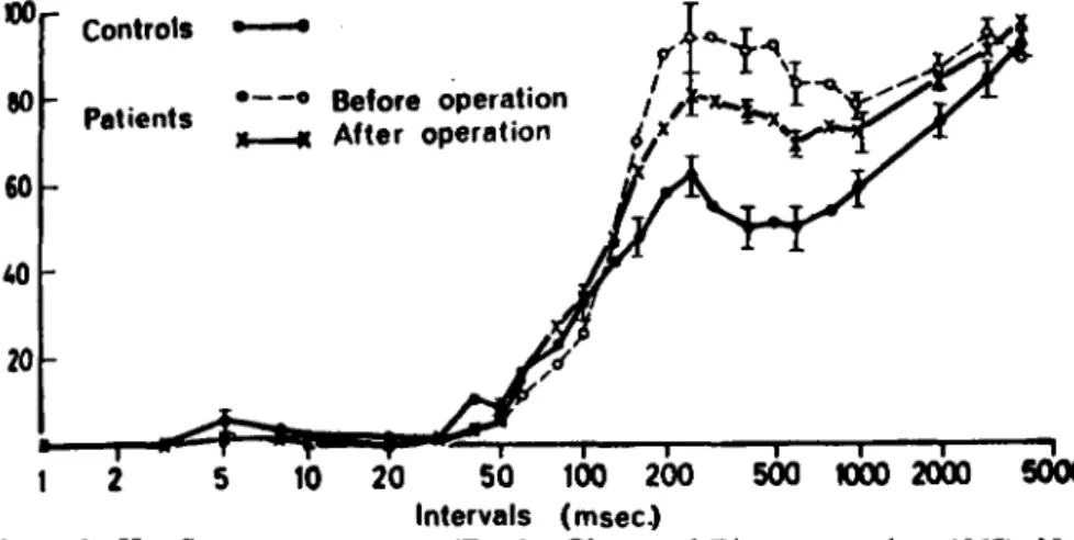
Figure 1—H-reflex recovery curve (Zander Olsen and Diamantopoulos, 1967). Note
"late facilitation", starting at about 100 msec, which may reflect facilitation of motoneurons via a transcortical loop. The "late facilitation" was found to be much more pronounced in Parkinsonian patients than in normal subjects. Stereotaxic surgery in Parkinsonian patients reduced the "late facilitation". Ordinate: percen-tage of test response.
to output cells of the motor cortex is facultative and determined by the context in which a particular motor act is performed. Phillips (1969) was the first to propose that feedback signals from muscle spindles may be used at the cortical level for au-tomatically adjusting the output of cortical neurons. This hypothesis of transcortical load compensation re-quires i, a neural pathway from muscle spindle afferents to the motor cortex representing the affer-ent limb of the 'transcortical reflex'; ii, an adaptive control of the input-output gain, at the cortical level, for signals generated in muscle spindles. This latter requirement appears to be necessary in order to free the motor cortex from a rigid reflex con-notation. The purpose of this paper is thus to summarize experimental evidence for transcortical servoac-tions and to discuss the possible ad-vantages and disadad-vantages of transcortical regulation as compared with segmental regulation by feed-back signals. Furthermore, long-loop stretch reflexes appear to be in-teresting from the clinical point of view: there is growing evidence that an increased gain at the cortical level may explain some aspects of the pa-thophysiology of increased muscle tone.
Long latency responses evoked by stretch afferents
In 1956, Hammond described the
EMG responses to sudden pulls of the forearm flexor muscles in the human subject; this author particu-larly emphasized the presence of a second, temporally dispersed com-ponent which followed the early synchronized EMG potential. The short latency and biphasic form of the first potential identified it easily as the monosynaptic stretch reflex. The latency of the second compo-nent, although around 50 msec, was still much less than a voluntary reac-tion. The nature of this second com-ponent was interpreted much later as a transcortical "reflex" by Evarts (1973) and Marsden, Merton and Morton (1973). Characteristically, the magnitude of the stretch-evoked long-latency response was found to depend on previous instruction given to the human subject or to the monkey: when the instruction was "resist", the response was large and contributed a major part of the reflex tension; but if the instruction was "let go", it was much weaker. Simi-lar observations were made in man by Melvill Jones and Watts (1971) who recorded the stretch response in the ankle extensors to sudden dorsif-lexion of the foot. Impressed by the prominence of the long-latency stretch response they termed it "Functional Stretch Reflex".
The involvement of supraspinal structures upon activation of stretch afferents was also discussed in
studies of the H-reflex in man and animals (reviewed by Wiesendanger, 1972). Most investigators plotting the recovery curve of the H-response noted a "late facilita-tion" starting at about 120 msec. In patients with increased muscle tone, an abnormally large late facilitation was often the most consistent devia-tion from normal curves (Zander Olsen and Diamantopoulos, 1967; Fig. 1). A transcortical reflex loop seems to be the most probable cause, although there are a number of other possibilities to explain the late facilitation (Wiesendanger, 1972).
The evidence for cortical participation in the long-latency stretch response
The evidence that the late EMG response evoked by sudden muscle stretch is mediated by the motor cor-tex has so far been indirect. Evarts (1973) first showed that sudden pas-sive displacements of a handle which a monkey was trained to hold in a narrow zone resulted in a response of cells of the motor cortex govern-ing arm muscles. The motor cortical response preceded the late EMG re-sponse by an interval appropriate to suggest a causal relationship. Similar observations were made by Conrad, Matsunami, Meyer-Lohmann, Wiesendanger and Brooks (1974) and Conrad, Meyer-Lohmann, Mat-sunami and Brooks (1975) when the perturbation was "injected" in the initial phase of a trained arm move-ment. Computer-generated histo-grams furthermore uncovered a tight temporal relation between oscilla-tions of cortical cell discharges and EMG bursts elicited by the perturba-tion, thus providing a further indica-tion of a causal relaindica-tionship between the two. Ablation of the postcentral gyrus abolished the early cortical re-sponse to stretch (Tatton, Forner, Gerstein, Chambers and Liu, 1975), probably by destroying the first cor-tical relay in area 3a (see below). The crucial experiment of ablating or blocking the motor cortex has however not yet been done.
Modification of long-latency muscle responses to stretch
Whether a "perturbation" of a
296 - AUGUST 1975 Why Transcortical Reflexes?
movement causes a prominent long-latency "stretch reflex" or not was found to be highly dependent on the strategy of movement execution. This aspect, first noted by Ham-mond (1956) and by HamHam-mond, Mer-ton and SutMer-ton (1956), was systemat-ically investigated in alert monkeys by Evarts and Tanji (1974). Pertur-bation of a postural contraction re-sulted in an early burst from pyrami-dal tract cells which preceded the secondary EMG burst. Prior instruc-tion modified the motor cortical re-sponse in parallel with the long-latency EMG response, again sug-gesting a causal relationship. It ap-peared that the "instruction" to the monkey (similar to the "resist" — "let g o " strategies used by Ham-mond, 1956) modulated the tonic background activity of pyramidal tract cells. Thus, the reactivity of output cells of the motor cortex crit-ically depended on modulating influ-ences. This gating effect appears to be essentially under voluntary con-trol and obviously depends on past experience (in conditioning experi-ments: feedback by reinforcement) and on the actual context in which the movement is executed. The hypothetical neural circuitry will be discussed below (see The control of transmission . . .).
The recent discovery by Tatton and Lee (1975) that long-latency EMG responses evoked by load pulses are very prominent in Parkin-sonian patients and that these re-sponses are hardly modifiable is of the greatest importance. The results, if confirmed, would indicate that long-loop reflexes would have to be considered in explaining the pathophysiology of rigidity. Milner-Brown, Stein and Lee (1975) have also made the interesting ob-servation that weight lifters may have more prominent long-loop re-flexes than average human subjects. This may signify that the efficiency of weight lifters to cope with heavy loads depends on the progressive development, by training, of long-loop reflexes.
The pathway from muscle afferents to the motor cortex
In previous experiments on ba-boons, electrical stimulation of
mus-<
>1mm
mot. ex.
Figure 2—Entry points of microelectrode tracks in the hand area of the left
peri-Rolandic cortex in a Cebus monkey. In the postcentral gyrus (3a), one electrode track was aimed at area 3a in the depth of the central sulcus. After the recording of stretch-evoked responses, the site was marked by ejection of Fastgreen which was subsequently found in a histological section (right). In this experiment, 5 tracks were made in the precentral gyrus in the area (hatched) from which, upon electrical stimulation, EMG responses in contralateral wrist and finger extensors could be elicited at lowest threshold (about 0.4 mA; above). The deepest point of a microelec-trode penetration in the motor cortex was also marked by Fastgreen (right, mot. ex.). From experiments by Lucier, Riiegg and Wiesendanger; unpublished records.
3a Cell non-PT Cell PT Cel
12 Hz
200 -urn « 3 8 0 0 u m 8 3 0 u m
<
5 0 H z
A / W
5 0 Aim —• 1100 urn
A/VV—
5 5 0 xim —<<(V
300Hz -A/WWWIfr-lOum 170 urn <w\i 2msec W W V W -140 .urn 10 msecFigure 3—Representative response patterns of a neuron in area 3a (3a cell), of a
non-corticospinal neuron (non-PT cell) and of a corticospinal neuron (PT cell). Twenty threshold responses to sinusoidal stretching of the finger extensor muscle at frequencies of 12, 50 and 300 Hz are shown as dot rasters. Each dot represents a spike discharge. The minimal amplitudes required to evoke a cortical response are noted for each frequency. Note that, for each cell type, the threshold amplitudes were lowest for the highest frequencies of sinusoidal stretching. Antidromic re-sponses to a double stimulus applied in the contralateral funiculus is shown for the PT cell as an inset (lower right). Horizontal bar represents 10 msec, except for antidromic response (2 msec). From experiments by Lucier, Riiegg and Wiesen-danger; unpublished records.
THRESHOLD AMPLITUDES 3 a non-PT PT n = 58 n = 109 n = 50 E =i o o E O CD "* Q ^ N I CM O v— • ^
o
CO I I I I I—l_ (O o o CO I I I 1 L_ n 30 20 10 CO o o CO 10 CM CO O •r- rf O CMFigure 4—Division of cortical neurons which responded to
sinusoidal stretching of finger-extensors at minimal amp-litudes > 100 /n m (above horizontal line) and < 100 jLl m (below horizontal line). The numbers are represented as col-umns for each frequency tested. Most "low-threshold" neurons were found among 3a neurons, much lower propor-tions among cells of the motor cortex. Note that, for cor-ticospinal and non-corcor-ticospinal neurons, low thresholds (< 100 Jim) were disclosed only if "dynamic" stimuli of 200-300 Hz were used. From experiments by Lucier, Riiegg and Wiesendanger; unpublished records.
00 $_ CM C O _
3a cell
f
J
^ ^ " " " " ^ 1 1 1 200 Hz i l 50AJy^
i I - 3 2 32 81 96 MSECAA.
128 IGO 192 2 2 1 --^m II , - 3 2 32 6 1 9G MSEC T " 1 128 160 192 221Figure 5—Typical response pattern of a 3a neuron which
re-sponded to sinusoidal stretching of the EDC muscle at a fre-quency of 200 Hz and at a minimal amplitude of 50 /x m. The duration of the vibration train is indicated by a solid bar. Computer generated cumulative distribution function (above) and post-stimulus time histogram (below). Two peaks show up in the histogram. The phasic response is followed by an inhibition lasting about 100 msec; ordinate: number of spikes. From experiments by Lucier, Riiegg and Wiesendanger; un-published records.
cle nerves of the forearm was used to establish the existence of a power-ful projection from low-threshold muscle afferents to area 3a at the bottom of the central sulcus (Phil-lips, Powell and Wiesendanger,
1971). Cytoarchitectonically, this area is transitional between konio-cortex (area 3b) and agranular konio-cortex (area 4) and no evidence was found electrophysiologjcally that this pro-jection area for low-threshold mus-cle afferents has output neurons to the spinal cord. Neurons of the motor cortex were also activated by electrical stimulation of muscle nerves, but less powerfully than 3a neurons (Wiesendanger, 1973). Re-petitive stimulation at intensities above threshold for group II affer-ents had to be used to excite neurons, both corticospinal ("PT-n e u r o ("PT-n s " ) a("PT-nd ("PT-no("PT-n-corticospi("PT-nal ("non-PT neurons"). It was there-fore assumed that the "error sign-als" to the output cells of the motor cortex were provided by secondary
rather than primary muscle spindle endings as originally proposed by Phillips (1969).
In recent experiments on Cebus monkeys, the question was reinves-tigated by using controlled stretches of forearm muscles (Lucier, Riiegg and Wiesendanger, 1975). Since high frequency longitudinal vibration of t e n d o n s r e p r e s e n t s an optimal
stimulus for exciting primary muscle spindle endings (Matthews, 1972), it was hoped that weak effects from primary muscle spindle endings could be disclosed more easily with trains of vibration than with electri-cal stimulation at intensities below group II threshold (and therefore submaximal for the group I volley). The objective was to compare the response pattern of neurons in area 3a with those in the motor cortex, corticospinal and non-corticospinal. Area 3a was first localized by using low intensity (about twitch-threshold) electrical stimulation of the deep radial nerve. Figure 2
illus-trates a location of a typical elec-trode penetration which, in the depth, traversed area 3a receiving a projection from the deep radial nerve. The area of the motor cortex which, upon electrical stimulation with a short train, elicited an EMG response at minimal intensity was the best site for finding stretch-evoked responses. Several penetra-tions made in this area are marked on the photograph of the brain sur-face. An attempt was made to test each cell systematically with sinusoidal stretching of finger and wrist extensor muscles at frequen-cies of 6, 12, 24, 50, 100, 200 and 300 Hz and with step stretches. For each type of stimulus, the responses were photographed first at threshold amp-litudes then at 100 times threshold amplitudes. Figure 3 illustrates rep-resentative response patterns for three selected frequencies and for the three cell types: 3a neurons, non-PT neurons, and PT-neurons. These examples show, and this was typical
298 - AUGUST 1975 Why Transcortical Reflexes?
% 1 2 H Z
/
X/\^-
1 8 m m
i i50msec
I : 6 Hz
f\ / \ j 4 5mm
i 1100 msec
Figure 6—Phasic response of a
corticos-pinal neuron which, at low frequencies of sinusoidal muscle stretch, re-sponded only at high amplitudes of stretching. Note partial driving at 6 Hz and complete suppression of evoked activity to the second cycle at 12 Hz. From experiments by Lucier, Riiegg and Wiesendanger; unpublished re-cords.
for the sample, that minimal amp-litudes, required to activate cortical cells decreased as the applied fre-quency of sinusoidal stretching was increased. With a highly dynamic stimulus such as a 300 Hz vibration, 3a neurons sometimes discharged at amplitudes which were within the range of the thresholds of primary muscle spindle receptors. More sur-prisingly, however, the threshold of neurons of the motor cortex was also drastically decreased when high fre-quency vibration was used. As shown in Figure 4, a small number of cells in the motor cortex, especially of non-PT neurons, were activated with amplitudes below 100 \i m. This (and other criteria described in the original paper) indicated to us that primary muscle spindle endings may, if maximally excited with trains of vibration, contribute to the stretch evoked responses.
Dynamic responses (short bursts) were the dominant pattern for all cell types. Figure 5 is an example of a dynamic response of a 3a neuron; the post-stimulus time histogram re-veals a clustering of the discharges with the formation of two clear peaks followed by a prolonged inhib-ition. Partial driving was observed at frequencies of up to 100 Hz in some
3a neurons and non-PT neurons of the motor cortex. Partial driving at the lowest frequencies (6 Hz) was common for all cell types (Fig. 6). It was thus established in these exper-iments that a fairly restricted input from stretch afferents was indeed capable of activating cells of the motor cortex. Secondary muscle spindle endings, and to some extent also primary muscle spindle endings, were considered to be the most likely receptors involved in produc-ing the motor cortex response. A similar conclusion was reached by Murphy, Wong and Kwan (1975) in similar experiments in cats.
The control of transmission at the cortical level
Electrophysiological studies in anaesthetized monkeys showed that transmission from stretch receptors to area 3a was rapid and secure (Phil-lips et al., 1971; Lucier et al., 1975). Whether area 3a is the first link in a transmission line to the motor cortex has not been established elec-trophysiologically, although latency measurements would be compatible with a transmission of signals from area 3 a to non-PT neurons and fi-nally to PT-neurons. In degeneration studies, it was found that small le-sions in area 3a of Cebus monkeys resulted in degeneration in area 4 (Wiesendanger and Riiegg, in prep-aration). Whatever the pathway for
A . Control before B . S M A
A / V V W
-C -Control after PT stim.
25Hz 4.6mm 50msec 2msec
Figure 7—Effect of electrical stimulation
of the supplementary motor area (SMA) on stretch-evoked discharges of a corticospinal neuron (antidromic invasion to stimulation of contralateral funiculus: PT stim.). A: stretch evoked test response (dot raster as in Fig. 3). B: conditioning stimulus train (15 pulses at 600 Hz, 0.8 mA) suppres-sed the stretch evoked discharge. The SMA stimulus (arrow) produced some dots because of the stimulus artefacts. C: control as in A. From experiments by Lucier, Riiegg and Wiesendanger; unpublished records.
signals from muscle spindles to the motor cortex may be, the elec-trophysiological studies of Lucier et al. (1975) made it clear that the sec-urity of transmission at the level of output cells in the motor cortex is low. All in all the number of output
S M A
STRETCH
120
1 0 0 2 0 0 3 0 0 4 0 0 5 0 0 6 0 0
Figure 8—Time course of inhibition produced by conditioning stimuli applied to the
ipsilateral SMA in five cells of the motor cortex (corticospinal and non-corticospinal neurons). From experiments by Lucier, Ruegg and Wiesendanger; unpublished re-cords.
cells of the motor cortex activated by stretch stimuli was disappoint-ingly small. These findings have led us to the hypothesis that the output cells are "protected" from feedback signals by a powerful inhibitory con-trol, possibly by the ideally located pericellular plexus from basket neurons (Marin-Padilla, 1969) which are presumed to be inhibitory (Sloper, 1973). According to this hypothesis, structures outside the motor cortex would tonically excite inhibitory basket cells. The "gating of transcortical reflexes" in an ap-propriate context (Evarts and Tanji,
1974) may be effected by a disinhibi-tion. Because of its structural rela-tionship and because of some clini-cal observations (reviewed by Wiesendanger, Seguin and Kiinzle, 1973) we decided to test the possi-bility that the supplementary motor area (SMA) represents a gating sys-tem acting on the motor cortex. Pre-liminary studies in anaesthetized monkeys were promising. The effect of the SMA was tested by stimulat-ing this area electrically with a 600 Hz train of 15 pulses at intensities ranging from 0.2 mA to 0.8 mA. These conditioning stimuli preceded a stretch stimulus (step or sinusoidal stretch) by 75 msec. All but two of the 16 units in the motor cortex (7 non-PT and 6 PT-neurons; 5 exper-iments) tested in this way were pro-foundly inhibited. An example is
shown in Figure 7. The effects were long-lasting (100 msec, or more) as illustrated in Figure 8, and may have been mediated directly by fiber con-nections from the SMA to the motor cortex or indirectly via subcortical loops involving the cerebellum or the basal ganglia. These findings in acute experiments may now provide a guide for chronic experiments. The next step in the examination of transcortical reflexes will be to study the role of gating systems, such as the SMA, in the gain control of long-loop reflexes in awake animals. Speculations on the significance of transcortical reflexes
In view of the existence of a seg-mental regulatory mechanism for load compensation, well-documented for instance for
respira-tion (Euler, 1966), it might be ap-propriate to ask what advantage a transcortical servo-loop may have. This question is especially pertinent considering the long time lag in-volved in the circuit. This long delay makes it unlikely that transcortical load compensation plays a role in the performance of very rapid move-ments (see also Grillner, 1973); on the other hand long-loop reflexes may be useful in postural stabiliza-tion in the presence of background perturbations.
In contrast to the segmental stretch reflex, which usually con-sists of a fairly synchronized dis-charge of motoneurons, the long-loop reflex consists of a much more scattered activation of motor units. This temporally dispersed transcor-tical activation may effectively counteract an inherent tendency of the monosynaptic stretch reflex loop to oscillate. It is probable that there are more than one supraspinal "delay lines" for signals from stretch afferents (Murphy et al., 1975). Long-loop reflexes with dif-ferent lag times may have a damping effect and may thus be important to counteract the inherent tendency of oscillations in segmental loops. The most obvious advantage of transcor-tical as compared to segmental load compensation is the fact that the former is much more amenable to adaptive changes than the latter. The existence of a very powerful adaptive gain control, at the cortical level, has now been demonstrated by Evarts and Tanji (1974). This as-pect of transcortical regulation is the most interesting one because an adaptive gain control gives the motor cortex the chance to exert its "higher" role of a chief "executive" of complex motor programs, such as the control of the finger movements during piano playing, simply by shutting off all input from peripheral receptors which, in this context, would only blur the precisely timed dispatch of command signals.
Any control system is of course subject to potential damage. Neurological disorders such as rigid-ity, and the inability to perform rapid alternating movements, may in fact be partially explained by a
patho-logically high gain in intracortical transmission. The experimental ob-servations by Tatton and Lee (1975) referred to above point in this direc-tion. Hopefully, some of these speculations will be tested experi-mentally in patients, and monkeys with experimentally induced abnor-malities of muscle tone.
DISCUSSION
Abrahams (Queens) commented on the possi-ble meaning of supraspinal projections of spindle information from the upper cervical cord that is devoid of monosynaptic reflexes. Since head movements, vision, and audition are integrated in the upper cervical cord, it may be functionally meaningful to have more cortical than spinal integration to better utilize feedback in the development of motor out-puts.
Wiesendanger agreed that this may be so, but that such complexity entailed longer time delays, and therefore spinal loops may still be important as well. In reply to Marshall's (Ot-tawa) question as to whether the inhibition originated only from the supplementary motor area, Wiesendanger replied that pre-liminary work suggested this, because stimu-lation lateral to area 6 produced no inhibi-tion. He allowed however that circuits through the various cortical areas still have to be worked out. He recalled Denny-Brown's demonstration that ablation of SMA causes forced grasping, and that SMA ordinarily seems to hold the grasping reflex in check. It is not known yet whether cortical basket cells are the inhibitory inter-neurons as suggested by Sloper.
Brown (London) was concerned whether the timing of the supposed transcortical reflex was adequately documented. In Evarts' re-cords 10 msec, elapsed between a signal in the postcentral region and PT discharge in the precentral region. In man the time from arrival of a signal from the hand muscles to precentral discharge would be 40 msec, but the latencies of the V2 response revealed an extra 10 msec. Could "gating" in precentral cortex really take 10-15 msec?
Wiesendanger replied in the affirmative: the time for the pathway from area 3a to precen-tral cortex was such that it could be the route taken by responses to natural stimulation. He also commented on the use of the term "re-flex". If "reflex" is defined as a response whose output magnitude is related to that of the input, then this has been demonstrated in the work as reported by Brooks for the rela-tion between load pulses and "early" corti-cal responses.
Hore wanted assurance that input from Pac-cinian corpuscles elicited by vibratory stimuli had been excluded from the cortical re-sponses in the absence of demonstrated pro-jections of muscle spindle la afferents to motor cortex. Wiesendanger thought it reasonable that vibration offers a more con-centrated stimulus for maximal activation of primary spindles to the exclusion of secon-daries, and would therefore be a more opti-mal input to motor cortex than electrical
300 - AUGUST 1975 Why Transcortical Reflexes?
nerve stimulation at strengths restricted to primaries, that had failed to reveal primary input. Participation of Paccinian receptors cannot be excluded entirely, but is unlikely because Mountcastle had found them unre-sponsive to even large amplitude frequencies below 50 Hz: the cortical cells however, did respond even to low amplitudes at 6 Hz. Al-though, as a further safeguard, radial nerve section had been shown to abolish the re-sponses, one cannot be entirely sure that Paccinian corpuscles in the intraosseous membranes could not have been excited. On balance, however one could conclude that the bulk of the signals about load reaching motor cortex were provided by muscle prim-ary and/or secondprim-ary spindle afferents.
ACKNOWLEDGEMENTS
Some of the work described in this paper has been supported by grants of the Medical Research Council of Canada (MA-4992, ME-5092) and of the Multiple Sclerosis Soci-ety of Canada. Dr. J. J. Seguin kindly read and criticized a draft of this paper.
REFERENCES
BELL, C. (1826). On the nervous circle which con-nects the voluntary muscles with the brain. Philosophical Transactions of the Royal Society of London, 116, 163-173.
CONRAD, B., MATSUNAMI, K., MEYER-LOHMANN, J., WIESENDANGER, M. and BROOKS, V. B. (1974). Cortical load compensa-tion during voluntary elbow movements. Brain Research, 71, 507-514.
CONRAD, B. MEYER-LOHMANN, J., MAT-SUNAMI, K. and BROOKS, V. B. (1975). Pre-central unit activity following torque pulse injec-tions into elbow movements. Brain Research, (In press).
EULER, C. V. (1966). Proprioceptive control in re-spiration. In: (Granit, R., ed.). Muscular afferents and motor control, Nobel Symposium I. Almqvist and Wiksell, Stockholm, pp. 197-207.
EVARTS, E. V. (1973). Motor cortex reflexes as-sociated with learned movement. Science, 179, 501-503.
EVARTS, E. V. and TANJI, J. (1974). Gating of motor cortex reflexes by prior instruction. Brain Research, 71, 479-494.
GRILLNER, S. (1973). Muscle stiffness and motor control — forces in the ankle during locomotion and standing. In: (Gydikov, A. A., Tankov, N. T. and Kosavov, D. S., eds.) Motor Control. Plenum Press, New York, pp. 195-215.
HAMMOND, P. H. (1956). The influence of prior instruction to the subject on an apparently neuromuscular response. Journal of Physiology, 132, 17-18P.
HAMMOND, P. H., MERTON, P. A. and SUT-TON, G. G. (1956). Nervous gradation of muscu-lar contraction. British Medical Bulletin, 12, 214-218.
LUCIER, G. E., RUEGG, D. G. and WIESEN-DANGER, M. (1975). Responses of neurons in motor cortex and in area 3a to controlled stretches of forelimb muscles in Cebus monkeys. Journal of Physiology. (In press.)
MARIN-PADILLA, M. (1969). Origin of the pericel-lular baskets of the pyramidal cells of the human motor cortex: A Golgi study. Brain Research, 14, 633-646.
MARSDEN, C. D. (1973). Servo control, the stretch reflex and movement in man. In: (Desmedt, J. E., ed.). New developments in electromyography and clinical neurophysiology. Karger, Basel, Vol. 3, pp. 375-382.
MARSDEN, C. D., MERTON, P. A. and MOR-TON, H. B. (1973). Latency measurements com-patible with a cortical pathway for the stretch re-flex in man. Journal of Physiology, 230, 58-59P. MATTHEWS, P. B. C. (1972). Mammalian muscle
receptors and their central actions. London: Ed-ward Arnold Limited.
MELVILL JONES, G. and WATT, D. G. D. (1971). Observations on the control of stepping and hop-ping movements in man. Journal of Physiology, 219, 709-727.
MILNER-BROWN, H. S., STEIN, R. B. and LEE, R. G. (1975). Synchronization of human motor units: possible roles of exercise and supraspinal
reflexes. Electroencephalography and Clinical Neurophysiology, 38, 245-254.
MURPHY, J. T., WONG, Y. C. and KWAN, H. C. (1975). Afferent-efferent linkages in motor cortex for single forelimb muscles. Journal of Neurophysiology, (In press).
PHILLIPS, C. G. (1969). Motor apparatus of the baboon's hand. Proceedings of the Royal Society of London; Biological Sciences, 173, 183-198. PHILLIPS, C. G., POWELL, T. P. S. and
WIESENDANGER. M. (1971). Projection from low-threshold muscle afferents of hand and forearm to area 3a of baboon's cortex. Journal of Physiology, 217, 419-446.
SLOPER, J. J. (1973). An electron microscope study of the termination of afferent connections to the primate motor cortex. Journal of Neurocytology, 2, 361-368.
TATTON, W. G., FORNER, F. D., GERSTEIN, G. L., CHAMBERS, W. W. and LIU, C. N. (1975). The effect of postcentral cortical lesions on motor responses to sudden upper limb dis-placements in monkeys. Brain Research, (In press).
TATTON, W. G. and LEE, R. G. (1975). Motor responses to sudden upper limb displacements in primates, humans and Parkinsonian patients. Canada Physiology, 6, 60.
WIESENDANGER, M. (1972). Pathophysiology of muscle tone. Neurology Series. Springer, Berlin -Heidelberg - New York, Vol. 9, pp. 17-19. WIESENDANGER, M. (1973). Input from muscle
and cutaneous nerves of the hand and forearm to neurons of the precentral gyrus of baboons and monkeys. Journal of Physiology, 228, 203-219. WIESENDANGER, M. SEGUIN, J. J. and
KUNZLE, H. (1973). The supplementary motor area — a control system for posture? In: (Stein, R. B.. Pearson, K. B., Smith, R. S., and Redford, J. B., eds.). Control of posture and locomotion. Plenum Press, New York, pp. 331-346. ZANDER OLSEN, P. and DIAMANTOPOULOS,
E. (1967). Excitability of spinal neurons in normal subjects and patients with spasticity, Parkinsonian rigidity, and cerebellar hypotonia. Journal of Neurology, Neurosurgery and Psychiatry, 30, 325-331.