O R I G I N A L A R T I C L E
Duration of Methicillin-Resistant Staphylococcus aureus Carriage,
According to Risk Factors for Acquisition
Jonas Marschall, MD; Kathrin Muhlemann, MD, PhD
OBJECTIVE. To examine the duration of methicillin-resistant Staphylococcus aureus (MRSA) carriage and its determinants and the influence of eradication regimens.
DESIGN. Retrospective cohort study.
SETTING. A 1,033-bed tertiary care university hospital in Bern, Switzerland, in which the prevalence of methicillin resistance among S.
aureus isolates is less than 5%.
PATIENTS. A total of 116 patients with first-time MRSA detection identified at University Hospital Bern between January 1, 2000, and December 31, 2003, were followed up for a mean duration of 16.2 months.
RESULTS. Sixty-eight patients (58.6%) cleared colonization, with a median time to clearance of 7.4 months. Independent determinants for shorter carriage duration were the absence of any modifiable risk factor (receipt of antibiotics, use of an indwelling device, or presence of a skin lesion) (hazard ratio [HR], 0.20 [95% confidence interval {CI}, 0.09-0.42]), absence of immunosuppressive therapy (HR, 0.49 [95% CI, 0.23-1.02]), and hemodialysis (HR, 0.08 [95% CI, 0.01-0.66]) at the time MRSA was first MRSA detected and the administration of decolonization regimen in the absence of a modifiable risk factor (HR, 2.22 [95% CI, 1.36-3.64]). Failure of decolonization treatment was associated with the presence of risk factors at the time of treatment (P = .01). Intermittent screenings that were negative for MRSA were frequent (26% of patients), occurred early after first detection of MRSA (median, 31.5 days), and were associated with a lower probability of clearing colonization (HR, 0.34 [95% CI, 0.17-0.67]) and an increased risk of MRSA infection during follow-up.
CONCLUSIONS. Risk factors for MRSA acquisition should be carefully assessed in all MRSA carriers and should be included in infection control policies, such as the timing of decolonization treatment, the definition of MRSA clearance, and the decision of when to suspend isolation measures.
Infect Control Hosp Epidemiol 2006; 27:1206-1212
Methicillin-resistant Staphylococcus aureus (MRSA) is a major meta-analysis concluded that there is no evidence that treat-cause of nosocomial infections worldwide, resulting in sub- ment with antimicrobials can eradicate MRSA carriage.12
stantial morbidity and mortality. Humans are the only sig- In the present study, we sought to determine the duration nificant reservoir of MRSA. Colonization with the microor- of MRSA carriage and how the duration is influenced by risk ganism is a necessary step during the pathogenesis of MRSA factors for MRSA acquisition and receipt of decolonization infection, and it is the source of cross-transmission between treatment. The rationale was to create a basis for the optimal humans.1'2 Placement of MRSA carriers in contact isolation
t i min g for initiating decolonization treatment and suspending
during their stay in healthcare institutions is a current stan- isolation measures for MRSA carriers.
M E T H O D S dard of care.3'4 Although cost-effective overall, isolation
mea-sures are expensive. A recent retrospective study calculated an additional cost of $318 per hospitalization-day per patient
with MRSA, of which approximately 80% was from the use study Setting and Local Infection Control Policy of barrier precautions around beds in multiple-bed rooms.5
Breaks in compliance with isolation policies are a continuous University Hospital Bern (Bern, Switzerland) is a 1,033-bed threat for the dissemination of MRSA. tertiary care center with more than 30,000 admissions per Colonization may persist for months or even years.6 8 How- year. The referral area covers the local region, where ap-ever, data are insufficient about the natural history of MRSA proximately 1 million inhabitants live, and a large number colonization and its determinants. Furthermore, the role of of smaller hospitals within and outside the Bernese region, topical decolonization therapy is still controversial.9"11 A large In this part of Switzerland, the prevalence of methicillin
re-From the Department of Infectious Diseases, University Hospital Bern (J.M., K.M.), and Institute for Infectious Diseases, University of Bern (K.M.), Bern, Switzerland.
Received April 12, 2006; accepted June 14, 2006; electronically published October 4, 2006.
sistance among S. aureus isolates is less than 5%,13 and it ranges from 3% to 5% at University Hospital Bern.
According to local written infection control guidelines, MRSA carriers are put into contact isolation, which involves stay in a single-patient room, use of gloves and gowns by medical personnel during physical contact, and use of masks by medical personnel when exposure to respiratory secretions is expected. In the interdisciplinary intensive care unit, con-tact isolation is implemented in 4-bed cubicles by marking an isolation area of approximately 2 m around the patient's bed with paravents and closing the neighboring bed. Screen-ing for MRSA involves all patients in contact with an MRSA carrier for whom placement in isolation was delayed and patients treated in a foreign healthcare institution during the 6 months before admission. Patients with MRSA are tagged in the hospital's patient administration system. On readmis-sion, tagged MRSA carriers are put into contact isolation.
MRSA decolonization treatment comprises a 5-day course of nasal application of mupirocin ointment, daily skin dis-infection with 4% chlorhexidine soap, and daily gargling with 0.1% chlorhexidine solution. The decision for decolonization treatment is made by the infection control team and requires that systemic antibiotic therapy and indwelling devices have not been used for at least 14 days and that all skin lesions have healed. During the study period, screening for MRSA control was performed intermittently as convenient during ambulatory visits or inpatient treatment, and results were therefore distributed over intervals of weeks to months, in-dependent of whether decolonization treatment had been administered.
Recruitment of Study Patients and Collection of Data The study was performed in accordance with local ethical guidelines. Patients with MRSA carriage detected for the first time between January 1, 2000, and December 31, 2003, were identified retrospectively through the laboratory information system of the Institute for Infectious Diseases, University of Bern, which serves University Hospital Bern. A total of 223 patients met these criteria. Thirty-eight patients were ex-cluded, because they were not treated at University Hospital Bern at the time MRSA was first detected, and information concerning risk factors for MRSA acquisition was not avail-able. An additional 69 patients were lost to follow-up after 1 month and were therefore also excluded. A total of 116 patients remained for analysis.
A standardized questionnaire was used to collect patient data retrospectively from medical records, infection control unit statistics, and the laboratory information system. Follow-up for individual patients started on the date MRSA was first detected and ended on December 31, 2004.
Terms and Definition
The following risk factors for MRSA carriage at the time MRSA was first detected were considered: receipt of systemic antibiotic therapy or performance of a surgical procedure
during the last 30 days, presence of skin lesions (surgical site wounds, ulcers, and/or dermatitis), use of indwelling devices (intravascular central catheters, endotracheal tube or trache-ostoma, urinary catheters, and/or wound drainage devices), receipt of immunosuppressive therapy (steroid treatment equivalent to 7.5 mg/day or more of prednisone, chemo-therapy, or immunomodulators), presence of diabetes mel-litus, presence of renal insufficiency, receipt of hemodialysis, and presence of malignant tumors. Risk factors considered during follow-up were the same as those for the time when MRSA was first detected, with the exception of performance of surgical interventions during the past 30 days. We calcu-lated the sum of risk factors present at the following junctures: the time MRSA was first detected, the time of decolonization treatment (if applicable), and the time of the final MRSA screening during follow-up. For analysis, risk factors were also categorized dichotomously (3 or less vs more than 3). Moreover, we defined modifiable risk factors as receipt of antibiotic treatment within the previous 30 days, presence of skin lesions, and use of indwelling foreign devices.
The total follow-up time was defined as the interval from the first detection of MRSA until the last MRSA screen-ing (positive or negative). Clearance of MRSA carriage was assumed when 2 or more of the final screenings during follow-up were negative for MRSA. Long-term carriage was defined as persistent carriage for more than 12 months dur-ing follow-up.
Detection of and Screening for MRSA Colonization Routine MRSA screening involved testing of specimens from the following anatomical sites: both nares, the groin (2 spec-imens), any skin lesions, tracheal secretions in intubated or tracheostomized patients, and urine in patients with urinary catheter. Samples obtained for suspected MRSA infection were included in the screening samples. Swabbing was per-formed with twisted wire rayon-tipped applicators (Copan Venturi Transystem). One applicator was used for both nares and another for both specimens from the groin, and appli-cators were premoistened with physiological saline. Labora-tory screening for MRSA was performed with the mannitol-oxacillin biplate test, selective culturing for gram-positive organisms, the coagulase tube test (to exclude coagulase-negative staphylococcus), and resistance pattern testing on Muller-Hinton agar. Available consecutive MRSA isolates from the same patient were typed using pulsed-field gel elec-trophoresis (PFGE), as described elsewhere.14
Statistical Analysis
All analyses were performed in StatView, version 5.0 (SAS Institute), or Stata, version 8 (Stata Corporation), using a cutoff of P value of .05 or less (2-tailed). Differences between means were tested by the Student's t test or the Mann-Whit-ney U test, and proportions were compared with the x2 or Fisher's exact test, as appropriate. The duration of MRSA
colonization determinants was analyzed by Kaplan-Meier curves using the log-rank test and Cox regression analysis. RESULTS
Characteristics of Study Patients and Risk Factors for MRSA Acquisition
A total of 116 patients with newly detected MRSA coloni-zation were followed up for a mean duration (±SD) of 496 ± 431 days (median, 316.5 days [range, 31-1,616 days]). The characteristics of these 116 patients at the time MRSA was first detected are listed in Table 1. Almost half of the patients (45.7%) were 65 years or older. Male patients pre-dominated (69.8% of the study population). Most patients were of Swiss nationality (80.2%) and lived in private house-holds (90.5%). Half of the MRSA carriers (48.3%) were iden-tified through the screening of contacts. The predominant anatomical sites of MRSA detection were skin lesions (39.7%) and the nares (35.3%). The most prevalent risk factors for MRSA acquisition were skin lesions (71.6% of patients), past antibiotic therapy (56.9%), and past surgery (54.3%). Almost all patients (96.6%) had been hospitalized within the previous 6 months, and the mean hospitalization duration (±SD) before MRSA detection was 11.9 ± 17.7 days. Almost half of the patients (43.1%) had an MRSA infection at the time MRSA was first detected.
Clearance of MRSA During Follow-up
During follow-up, a mean of 3.8 MRSA screenings were per-formed per patient per year. Sixty-eight patients (58.6%) cleared MRSA colonization. The median time to clearance, based on the Kaplan-Meier estimate, was 226 days (7.4 months). In patients who cleared colonization, the median follow-up time after clearance was 189.5 days (range, 1-1,602 days). Long-term carriage was observed in 24 patients (20.7%), of whom 19 cleared MRSA colonization, and 5 re-mained colonized throughout the follow-up period.
Independent determinants for MRSA clearance were the absence of modifiable risk factors (hazard ratio [HR], 0.20 [95% confidence interval {CI}, 0.09-0.42]; P< .001), receipt of immunosuppressive therapy (HR, 0.49 [95% CI,
0.23-1.02]; P = .05), and hemodialysis (HR, 0.08 [95% CI, 0.01-0.66]; P = .01) at the time MRSA was first detected and the administration of decolonization treatment (HR, 2.22 [95% CI, 1.36-3.64]; P = .01) (Table 2). Thirty-six patients (31.0%) received decolonization treatment, which failed in 13 (36.1%). The median time to initiation of decolonization treatment was 37.5 days (range, 0-833 days). The median time to clear-ance (based on the Kaplan-Meier estimate) for patients with 0-3 risk factors at the time of MRSA detection was 43 days if decolonization treatment was administered and 238 days if it was not administered (Figure). The corresponding in-tervals for patients with more than 3 risk factors were 221 days if decolonization treatment was received and 597 days if it was not received. The 13 patients who did not respond
to decolonization treatment had a significantly greater num-ber of risk factors at the time of decolonization treatment (mean [±SD], 1.30 ± 1.37 [95% CI, 0.47-2.14]) than pa-tients who cleared MRSA immediately after treatment (0.39
± 0.65 risk factors [95% CI, 0.11-0.67]; P = .01).
Intermittent Negative Results of Screenings for MRSA
During follow-up, 30 patients (25.9%) had intermittent screenings with negative results, and 12 patients (10.3%) had 2 or more consecutive intermittent screenings with negative results. Twelve of the 30 patients had PFGE performed on MRSA isolates that were obtained before and after intermit-tent screenings with negative results; for all 12, the PFGE pattern for the first isolate matched the pattern for the second isolate. For 3 of the 12 patients, extended intervals of 33, 39, and 51 months occurred between the times the 2 MRSA isolates with an identical PFGE pattern were obtained. The median interval between the time MRSA was first detected to the time of the first intermittent screening with a negative result in these 30 patients was 31.5 days (range, 1-365 days), compared with the median time to MRSA clearance of 226 days in the whole study population. The mean number of risk factors present at the time MRSA was first detected was higher for patients with negative results of intermittent screenings (mean, 3.86 vs 2.96; P = .002), and they had a lower probability of clearing MRSA colonization (Table 2). In the Cox regression analysis, the only independent factor associated with negative results of intermittent screenings was the number of risk factors present at time MRSA was first detected (HR, 1.65; 95% CI, 1.24-2.20; P = .005).
MRSA Infection During Follow-up
Of 116 patients, 17 (14.7%) developed an MRSA infection during follow-up. Five of the 17 patients also had an MRSA infection at the time MRSA was first detected. Infection oc-curred a median interval of 123 days (range, 4-705 days; mean interval [±SD], 192.5 ± 212.3 days) after MRSA was first detected. Kaplan-Meier estimates could not be obtained, be-cause less than 50% of patients developed MRSA infection. In the Cox regression analysis, only a large number of risk factors present at the time of the last MRSA screening (HR, 1.41 [95% CI, 1.009-1.975]; P = .04) was significantly as-sociated with MRSA infection. None of the other variables reached statistical significance, perhaps in part because of the small number of MRSA infections observed.
DISCUSSION
The time course of MRSA colonization and its determinants was described in a retrospective cohort of 116 patients with MRSA at a tertiary care university hospital. The characteristics of the study population corresponded to known risk factors for MRSA acquisition, such as older age, prior hospitalization (especially in the intensive care unit), previous surgery, pre-vious antibiotic treatment, presence of skin lesions, and use
TABLE i. Characteristics of 116 Patients Colonized With Methicillin-Resistant Staphylococcus aureus (MRSA) at University Hospital Bern (Bern, Switzerland) between January 1, 2000, and December 31, 2003
TABLE I . (Continued)
Characteristic Value Characteristic Value
Age Mean y ± SD 0-16 y 17-44 y 45-64 y ^ 6 5 y Male sex Residence Private household Long-term care facility Immigrant housing facility Method of MRSA detection
Clinical diagnostic workup MRSA screening, by patient group8
Overall
Contacts with an MRSA carrier
Patients admitted from a foreign institution Anatomical site of MRSA detection15
Skin lesion or wound Nose
Urine Groin
Respiratory tract
Risk factor for MRSA acquisition1" Skin lesion(s)
Surgery during past 30 d
Antibiotic therapy during past 12 mo' During past 30 d During past 1-12 mo Indwelling device Immunosuppressive therapy Diabetes mellitus Renal insufficiency Hemodialysis Malignant disease Hospitalization history
Stay of ^ 24 h during past 6 mo Length of stay, mean d ± SD MRSA detected during stay
Length of stay before MRSA detection, mean d ± SD
ICU history
Admitted during current hospitalization Length of stay before MRSA detection,
mean d ± SD
MRSA infection at time of first MRSA detection Absent
Subsequent infection during follow-up Time to subsequent onset, mean d ± SD
55.7 ± 21.4 6 (5.2) 26 (22.4) 31 (26.7) 53 (45.7) 81 (69.8) 105 (90.5) 7 (6.0) 4 (3.4) 60 (51.7) 56 (48.3) 48 (41.4) 8 (6.9) 46 (39.7) 41 (35.3) 15 (12.9) 12 (10.3) 8 (6.9) 83 (71.6) 63 (54.3) 77 (66.4) 66 (56.9) 30 (25.8) 53 (45.7) 23 (19.8) 23 (19.8) 21 (18.1) 7 (6.0) 23 (19.8) 112 (96.6) 21.9 ± 22.2 92 (79.3) 11.9 ± 17.7 46 (39.7) 3.0 ± 6.3 66 (56.9) 12 (18.2) 247.8 ± 227.5
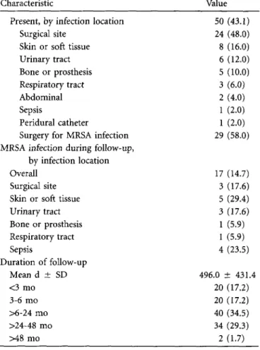
Present, by infection location Surgical site
Skin or soft tissue Urinary tract Bone or prosthesis Respiratory tract Abdominal Sepsis Peridural catheter
Surgery for MRSA infection MRSA infection during follow-up,
by infection location Overall
Surgical site Skin or soft tissue Urinary tract Bone or prosthesis Respiratory tract Sepsis Duration of follow-up Mean d ± SD <3 mo 3-6 mo >6-24 mo >24-48 mo >48 mo 50 (43.1) 24 (48.0) 8 (16.0) 6 (12.0) 5 (10.0) 3 (6.0) 2 (4.0) 1 (2.0) 1 (2.0) 29 (58.0) 17 (14.7) 3 (17.6) 5 (29.4) 3 (17.6) 1 (5.9) 1 (5.9) 4 (23.5) 496.0 ± 431.4 20 (17.2) 20 (17.2) 40 (34.5) 34 (29.3) 2 (1.7) (Continued)
NOTE. Data are no. (%) of patients, unless otherwise indicated. ICU, intensive care unit; MRSA, methicillin-resistant Staphylococcus aureus.
' Includes all patients in contact with an MRSA carrier for whom placement
in isolation was delayed and patients treated in a foreign healthcare institution during the 6 months before admission.
b
This entry totals more than 100%, because patients could have more than one characteristic.
c
Antibiotics exclude perioperative prophylaxis.
of indwelling devices.215"21 Also, the high proportion of pa-tients with MRSA infection at the time MRSA was first de-tected in them (43.1%) was in accordance with earlier re-ports.22 Because of the low prevalence of nosocomial MRSA carriage (3%-5% of patients) and the virtual absence of com-munity-acquired MRSA at this institution, the likelihood of a new and inadvertent colonization during follow-up was minimal. The study has some limitations, however. The ret-rospective study design precluded a strict schedule of follow-up screenings and randomized the administration of decol-onization treatment. Also, we cannot exclude the possibility that study patients may have received MRSA-eradication reg-imens at another healthcare institution during follow-up.
Fifty-nine percent of the study patients cleared MRSA within a median interval of 226 days (7.4 months). Two pre-vious studies found a similar time to clearance of 7-8.5
TABLE 2. Determinants of Clearance of Methicillin-Resistance Staphylococcus aureus (MRSA) Coloni-zation in 116 Patients With Newly Detected MRSA Carriage
Variable Age, y Female sex
No. of risk factors at first MRSA detection Overall
>3 risk factors
^ 1 modifiable risk factor Antibiotic therapy in past 30 d Skin lesion Indwelling device Surgery in past 30 d Immunosuppressive therapy Diabetes mellitus Renal insufficiency Hemodialysis Renal transplantation Tumor MRSA infection At start During follow-up Decolonization treatment
Intermittent screenings with negative results Risk factor at last MRSA screening
Overall no. Antibiotic therapy Immunosuppressive therapy Hemodialysis Clearance of MRSA Yes (n = 68) 53.6 ± 21.1 20 (29.4) 2.86 ± 1.22 25 (36.8) 60 (88.2) 33 (48.5) 49 (72.0) 27 (39.7) 38 (55.9) 8 (11.8) 11 (16.2) 10 (14.7) 1 (13) 5 (7.4) 12 (17.7) 35 (51.4) 5 (7.4) 29 (42.6) 10 (14.7) 1.06 ± 1.17 7 (10.2) 8 (11.8) 1 (1.5) No (n = 48) 58.8 ± 21.9 16 (33.3) 3.69 ± 1.52 27 (56.3) 48 (100) 33 (68.7) 34 (70.8) 26 (54.2) 25 (52.1) 15 (31.3) 12 (25.0) 11 (22.9) 6 (12.5) 5 (10.4) 11 (22.9) 15 (31.3) 12 (25.0) 7 (14.6) 20 (41.7) 2.14 ± 1.54 17 (35.4) 14 (29.2) 6 (12.5) HR (95% CI) 1.00 (0.99-1.01) 0.92 (0.54-1.54) 0.77 (0.65-0.92) 0.62 (0.38-1.01) 0.20a (0.09-0.42) 0.61 (0.37-0.99) 1.13 (0.66-1.94) 0.70 (0.42-1.18) 1.11 (0.68-1.82) 0.49a (0.23-1.02) 0.75 (0.39-1.45) 0.66 (0.33-1.30) 0.08a (0.01-0.66) 0.83 (0.33-2.08) 0.86 (0.46-1.61) 1.53 (0.95-2.46) 0.27 (0.11-0.69) 2.22' (1.36-3.64) 0.34 (0.17-0.67) 0.73 (0.59-0.90) 0.38 (0.18-0.84) 0.45 (0.22-0.95) 0.19 (0.03-1.37) P NS NS .004 .06 < 0 0 1 .04 NS NS NS .05 NS NS .01 NS NS .08 .006 .01 .002 .003 .01 .03 .09 NOTE. Data are no. (%) of patients or mean value ± SD. CI, confidence interval; HR, hazard ratio; NS, nonsignifi-cant (P>.05).
* Adjusted HR estimates are presented for the following independent determinants: modifiable risk factors, receipt of hemodialysis, and receipt of immunosuppressive therapy at time of first MRSA detection and decolonization treatment. Crude estimates are presented for all other variables.
months.7,23 However, longer durations of 14 months8 and 40 months6 have been reported. These differences may be ex-plained by the frequency of infection control screening per-formed in the different studies. For example, Vriens et al.8 screened patients at intervals of 6 months only. A mean of 1.6 screenings per patient per year were performed in the study by Sanford et al.,6 whereas we performed a mean of 3.8 screenings per patient per year. A higher frequency of screenings certainly leads to a more accurate description of the duration of colonization. Estimates of the duration of colonization may also be influenced by the study population's size (49-197 patients were involved in the cited studies) and clinical characteristics. An indication for the latter might be the proportion of long-term carriers. In our study, approx-imately 20% of patients were long-term carriers (defined as carriage of MRSA for more than 1 year), and the longest observed carriage time was 3.3 years. Earlier studies reported a lower frequency of long-term carriage of approximately 10%.6'23'24 Lastly, different definitions used for MRSA
clear-ance may also play a role. As in our study, Sanford et al.6 required 2 consecutive screenings with negative results; else-where, Vriens et al8 required 1 screening with a negative result, and no definition was given in the study by MacKinnon and Allen.23
Thirty of our 116 patients had at least 1 intermittent screen-ing with negative results, and in 10% we observed 2 or more consecutive screenings with a negative result. Data on neg-ative results of intermittent screenings are rare. Vriens et al.8 found 6.7% of their cohort to have negative results of inter-mittent screening, and in the study by Blok et al.25 culture-negative intervals of 8-10 months were observed for 5 of 11 patients with long-term carriage. Low colonization density or an intracellular reservoir may be responsible for negative re-sults of intermittent screenings.26'27 Interestingly, infection during follow-up was more frequent among patients with intermittent carriage, a finding that, to our knowledge, has not been reported up to now. Eventually, negative results of intermittent screenings may lead to a premature suspension
0 ODD 1C0O 1H0O T i m e , days
No decolonization, 0-3 risk factors NodecdonizaJon, 4-7 risk factors Decalaiization, 0-3 risk factors'' Decolonization, 4-7 risk factos"
FIGURE. Kaplan-Meier curves for the time to methicillin-resistant
Staphylococcus aureus (MRSA) clearance, stratified by decolonization
treatment and the presence of risk factors at the time of MRSA detection. "Log-rank test for decolonization treatment versus no de-colonization treatment in patients with 0-3 risk factors (P = .02). b
Log-rank test for decolonization treatment versus no decolonization treatment in patients with 4-7 risk factors (P = .03).
of infection control measures. In our study, however, the median interval to the first intermittent screening with a neg-ative result was considerably shorter (31.5 days) than the observed median time to MRSA clearance (226 days).
Risk factors for MRSA colonization had a significant im-pact on the duration of MRSA colonization. The strength of this study is the detailed analysis of a large number of risk factors. The presence of at least 1 modifiable risk factor (an-tibiotic use, presence of a skin lesion, and use of an indwelling device), receipt of immunosuppressive therapy , and receipt of hemodialysis was independently associated with a longer duration of MRSA carriage. Earlier studies found skin lesions to be the most prominent factor associated with persistent carriage.7'8'23'25,28 Beaujean et al.28 found an association be-tween underlying disease and persistent carriage, albeit in a small study population. Cystic fibrosis was identified as a risk factor for long-term MRSA carriage in 2 studies8'24 but was only present in one of our patients. All these findings suggest that the presence of risk factors should be considered in the decision to suspend isolation precautions. Beaujean et al.28 demanded an absence of risk factors for more than 6 months and 3 screenings with negative results before ending isolation precautions. By comparison, Vriens et al.8 called for 12 months and at least 1 screening with a negative result. On the basis of our findings, with an extended analysis of risk factors for MRSA carriage, we propose that a patient should be considered to have cleared MRSA colonization if at least 2 consecutive screenings have negative results and if there are no other risk factors (modifiable and nonmodifiable), re-gardless of the time that has passed since MRSA colonization was detected. In the presence of long-term, nonmodifiable
risk factors, a minimum follow-up duration (eg, 6 months) should be allowed to elapse, to decrease the risk of false-negative results of intermittent screenings.
Receipt of decolonization treatment was significantly as-sociated with the clearance of MRSA. However, it must be taken into consideration that, in this study, decolonization treatment was only administered when modifiable risk factors for MRSA colonization (antibiotic treatment, use of indwell-ing devices, and presence of skin lesions) were absent. Nev-ertheless, a high failure rate of decolonization treatment was observed (36.1% of patients), and failure was associated with the presence of nonmodifiable risk factors for MRSA colo-nization. Vriens et al.8 reported a much higher success rate for decolonization treatment and concluded that such treat-ment should be administered to all patients, independent of the presence of risk factors. However, the large screening intervals used in their study and the lack of information on nonmodifiable risk factors precludes a comparison with our findings. We suggest that decolonization treatment should
only be given when modifiable risk factors are largely absent and that the success of the treatment should be evaluated more carefully if long-term risk factors are present.
MRSA infection occurred in 14.7% of our patients dur-ing follow-up. This rate is comparable with rates reported elsewhere.1,29 Older age, intensive care unit stay, receipt of hemodialysis, presence of surgical wounds, presence of pres-sure ulcers, and use of intravascular catheters have been as-sociated with an increased progression of MRSA colonization to invasive infection.1,29'30 In our study, a higher number of risk factors at the end of follow-up was associated with an increased risk of MRSA infection.
In conclusion, modifiable and nonmodifiable risk factors for MRSA acquisition are the most important determinants of the duration of MRSA colonization and affect the success rate of decolonization treatments. Moreover, the presence of risk factors may lead to intermittent screenings with negative results. Risk factors should therefore be evaluated carefully in all MRSA carriers and should be considered in infection control policies, such as the timing of decolonization treat-ment and the definition of MRSA clearance, which is the basis for the decision to suspend isolation measures.
Address reprint requests to Kathrin Muhlemann, MD, PhD, Institute for Infectious Diseases, University of Bern, Friedbuhlstrasse 51, 3010 Bern, Swit-zerland (kafhrin.muehlemann@ink.unibe.ch).
A C K N O W L E D G M E N T S
We thank Linda Beul, Susanne Burri, Rolf Kuhn, and Tanja Lohri for help with data collection and for stimulating discussions.
R E F E R E N C E S
1. Davis KA, Stewart JJ, Crouch HK, Florez CE, Hospenthal DR. Methi-cillin-resistant Staphylococcus aureus (MRSA) nares colonization at hos-pital admission and its effect on subsequent MRSA infection. Clin Infect
2. Fishbain JT, Lee JC, Nguyen HD, et al. Nosocomial transmission of meth-icillin-resistant Staphylococcus aureus: a blinded study to establish baseline acquisition rates. Infect Control Hosp Epidemiol 2003; 24:415-421. 3. Muto CA, Jernigan JA, Ostrowsky BE, et al. SHEA guideline for
pre-venting nosocomial transmission of multidrug-resistant strains of
Staph-ylococcus aureus and Enterococcus. Infect Control Hosp Epidemiol 2003;
24:362-386.
4. Garner JS. Guideline for isolation precautions in hospitals: the Hospital Infection Control Practices Advisory Committee. Infect Control Hosp
Epidemiol 1996; 17:53-80.
5. Herr CE, Heckrodt TH, Hofmann FA, Schnettler R, Eikmann TF. Ad-ditional costs for preventing the spread of methicillin-resistant
Staphy-lococcus aureus and a strategy for reducing these costs on a surgical ward. Infect Control Hosp Epidemiol 2003; 24:673-678.
6. Sanford MD, Widmer AF, Bale MJ, Jones RN, Wenzel RP. Efficient de-tection and long-term persistence of the carriage of methicillin-resistant
Staphylococcus aureus. Clin Infect Dis 1994; 19:1123-1128.
7. Scanvic A, Denic L, Gaillon S, Giry P, Andremont A, Lucet JC. Duration of colonization by methicillin-resistant Staphylococcus aureus after hos-pital discharge and risk factors for prolonged carriage. Clin Infect Dis 2001;32:1393-1398.
8. Vriens MR, Blok HE, Gigengack-Baars AC, et al. Methicillin-resistant
Staphylococcus aureus carriage among patients after hospital discharge. Infect Control Hosp Epidemiol 2005; 26:629-633.
9. Harbarth S, Dharan S, Liassine N, Herrault P, Auckenthaler R, Pittet D. Randomized, placebo-controlled, double-blind trial to evaluate the ef-ficacy of mupirocin for eradicating carriage of methicillin-resistant
Staph-ylococcus aureus. Antimicrob Agents Chemother 1999; 43:1412-1416.
10. Parras F, Guerrero MC, Bouza E, et al. Comparative study of mupirocin and oral co-trimoxazole plus topical fusidic acid in eradication of nasal carriage of methicillin-resistant Staphylococcus aureus. Antimicrob Agents
Chemother 1995; 39:175-179.
11. Tomic V, Svetina Sorli P, Trinkaus D, Sorli J, Widmer AF, Trampuz A. Comprehensive strategy to prevent nosocomial spread of methicillin-resistant Staphylococcus aureus in a highly endemic setting. Arch Intern
Med 2004; 164:2038-2043.
12. Loeb M, Main C, Walker-Dilks C, Eady A. Antimicrobial drugs for treat-ing methicillin-resistant Staphylococcus aureus colonization. Cochrane
Database Syst Rev 2003; (4):CD003340.
13. Blanc DS, Pittet D, Ruef C, et al. Epidemiology of methicillin-resistant
Staphylococcus aureus: results of a nation-wide survey in Switzerland. Swiss Med Wkly 2002; 132:223-229.
14. Fux CA, Uehlinger D, Bodmer T, Droz S, Zellweger C, Muhlemann K. Dynamics of hemodialysis catheter colonization by coagulase-negative staphylococci. Infect Control Hosp Epidemiol 2005; 26:567-574. 15. Shimada M, Kamakura T, Itasaka H, Matsumata T, Hashizume M,
Sug-imachi K. The significance of methicillin-resistant Staphylococcus aureus infection in general surgery: a multivariate analysis of risk factors and preventive approaches. Surg Today 1993; 23:880-884.
16. Asensio A, Guerrero A, Quereda A, Lizan M, Martinez-Ferrer M.
Col-onization and infection with methicillin-resistant Staphylococcus aureus: associated factors and eradication. Infect Control Hosp Epidemiol 1996; 17:20-28.
17. Dziekan G, Hahn A, Thune K, et al. Methicillin-resistant Staphylococcus
aureus in a teaching hospital: investigation of nosocomial transmission
using a matched case-control study. / Hosp Infect 2000; 46:263-270. 18. Graffunder EM, Venezia R. Risk factors associated with nosocomial
meth-icillin-resistant Staphylococcus aureus (MRSA) infection including pre-vious use of antimicrobials. } Antimicrob Chemother 2002; 49:999-1005. 19. Von Baum H, Schmidt C, Svoboda D, Bock-Hensley O, Wendt C. Risk factors for methicillin-resistant Staphylococcus aureus carriage in residents of German nursing homes. Infect Control Hosp Epidemiol 2002; 23:511-515. 20. Jernigan JA, Pullen AL, Flowers L, Bell M, Jarvis WR. Prevalence of and
risk factors for colonization with methicillin-resistant Staphylococcus
au-reus at the time of hospital admission. Infect Control Hosp Epidemiol
2003; 24:409-414.
21. Lucet JC, Chevret S, Durand-Zaleski I, Chastang C, Regnier B. Prevalence and risk factors for carriage of methicillin-resistant Staphylococcus aureus at admission to the intensive care unit: results of a multicenter study.
Arch Intern Med 2003; 163:181-188.
22. Coello R, Jimenez J, Garcia M, et al. Prospective study of infection, colonization and carriage of methicillin-resistant Staphylococcus aureus in an outbreak affecting 990 patients. Eur } Clin Microbiol Infect Dis 1994; 13:74-81.
23. MacKinnon MM, Allen KD. Long-term MRSA carriage in hospital pa-tients. / Hosp Infect 2000; 46:216-221.
24. Frenay HME, Vandenbroucke-Grauls CMJE, Molkenboer MJCH, Ver-hoef J. Long-term carriage and transmission of methicillin-resistant
Staphylococcus aureus after discharge from hospital. / Hosp Infect 1992;
22:207-215.
25. Blok HEM, Vriens MR, Weersink AJL, Troelstra A. Carriage of methi-cillin-resistant Staphylococcus aureus (MRSA) after discharge from hos-pital: follow-up for how long? a Dutch multi-centre study. / Hosp Infect 2001; 48:325-327.
26. Crowcroft NS, Ronveaux O, Monnet DL, Mertens R. Methicillin-resistant
Staphylococcus aureus and antimicrobial use in Belgian hospitals. Infect Control Hosp Epidemiol 1999; 20:31-36.
27. Clement S, Vaudaux P, Francois P, et al. Evidence of an intracellular reservoir in the nasal mucosa of patients with recurrent Staphylococcus
aureus rhinosinusitis. / Infect Dis 2005; 192:1023-1028.
28. Beaujean DJMA, Weersink AJL, Blok HEM, Frenay HME, Verhoef J. Determining risk factors for methicillin-resistant Staphylococcus aureus carriage after discharge from hospital. J Hosp Infect 1999; 42:213-218. 29. Coello R, Glynn JR, Gaspar C, Picazo JJ, Fereres J. Risk factors for
developing clinical infection with methicillin-resistant Staphylococcus
au-reus (MRSA) amongst hospital patients initially only colonized with
MRSA. / Hosp Infect 1997; 37:39-46.
30. Muder RR, Brennen C, Wagener MM, et al. Methicillin-resistant staph-ylococcal colonization and infection in a long-term care facility. Ann