CASE REPORT
A balanced complex chromosomal rearrangement (BCCR)
in a family with reproductive failure
J.Lespinasse
1,5, M.O.North
2, C.Paravy
1, M.J.Brunel
1, P.Malzac
3and J.L.Blouin
41Cytogenetic Laboratory, General Hospital, BP 1125, 73011 ChambeÂry cedex,2Cytogenetic Laboratory, Cochin-Saint Vincent de
Paul Hospital, Paris,3Department of Medical Genetics, Hopital d'Enfants de la Timone, Marseille, France and4Division of Medical
Genetics, University of Geneva Medical School, and University Hospitals, 1211 Geneva 4, Switzerland
5To whom correspondence should be addressed at: E-mail: james.lespinasse@ch-chambery.rss.fr
Balanced complex chromosomal rearrangements are very rare events in the human population. Translocations involving three or more chromosomes frequently lead to a severe reproductive impairment secondary to meiotic disturbance in males and to chromosomal imbalance in gametes of females. We report a new familial case of complex chromosome anomaly involving chromosomes 13, 14 and 22. Cytogenetic investigations showed a complex chromosomal chromosome rearrangement involving: (i) a Robertsonian translocation between chromosomes 13 and 14; and (ii) a reciprocal translocation between the long arms of chromosome 14 and the long arm of chromosome 22. The aetiology of the translocation was characterized by conventional ¯uorescence in-situ hybridization (FISH) studies and routine R- and G-banding (RTBG and GBTG) combined with a and b satellite centromeric FISH probes. Predicted con®guration of the hexavalent at pachytene stage of meiosis was used to consider the modes of segregation; only two con®gurations resulted in a normal or balanced gamete karyotype. Reproductive management and genetic counselling are discussed.
Key words: chromosomes 13, 14 and 22/familial balanced complex chromosomal rearrangement/genetic counselling/infertility/meiosis
Introduction
Complex chromosome rearrangements (CCR) are structural abnormalities involving at least three chromosomes with three or more breakpoints. Balanced CCR (BCCR) are infrequent and usually occur de novo (Batavian and Eswara, 1998). Rarely, BCCR are of familial origin and, in these cases, transmission is more frequently from the mother (Farrell et al., 1994). In males, BCCR are thought to lead to severe reproductive impairment through meiotic disturbance or chromosomal imbalance in gametes. We report here a new case of complex chromosomal anomaly involving chromo-somes 13, 14 and 22 with familial transmission.
Family report
The proband (II2-D) is a 40 year old woman who has had nine
®rst-trimester miscarriages (Figure 1). She has a 34 year old sister (II4-A) who has had three miscarriages and a 36 year old
sister (II3-B) who has had two miscarriages and two healthy
children. A 44 year old brother (II1-C) was infertile with
oligoasthenozoospermia. Two other brothers (II5 and II6) of
II2-D could not be studied. The proband's mother (I1-M) had
three miscarriages. She underwent menopause at age 55 years. The proband's father (I2-A) died at 37 years of age.
Cytogenetic analysis
Cytogenetic investigations were carried out using standard methods on lymphocytes from phytohaemagglutinin (PHA)-stimulated peripheral blood cultures and from immortalized B-lymphocytes. Chromosome spreads were processed for RHG, QFQ, GTG, CBG bands and Nuclear Organizer Region (NOR) staining. High resolution banding (RTBG, GBTG) was obtained according to the standard technique (Dutrillaux and Vigas-Pequignot, 1981). RHG, GTG, RTBG and GBTG banded metaphases from lymphocytes were interpreted at
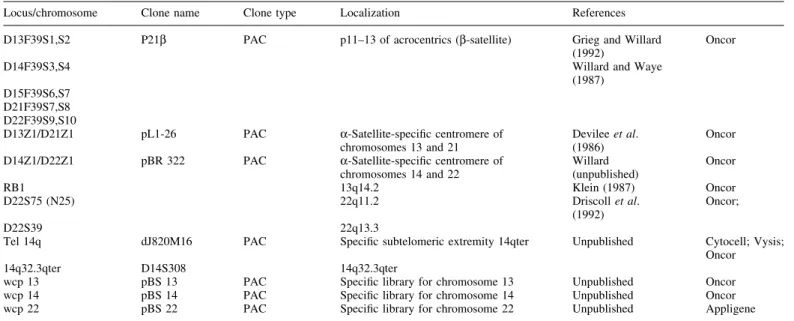
resolution levels of 450 and 650 bands. Conventional ¯uores-cence in-situ hybridization (FISH) was carried out using human probes on metaphases of the transformed lymphoblast cell line according to standard protocols and to the manufacturer's manuals (Table I).
Genotype analysis
Genomic DNA was puri®ed from peripheral blood lympho-cytes according to standard sodium dodecyl sulphate±protei-nase-K and phenol/chloroform extraction methods. DNA polymorphisms in the mother and the four children were analysed by PCR ampli®cation of tandem short sequence repeats. The selected markers on chromosomes 13, 14 and 22 were chosen from the GeÂneÂthon and CHLC collections included in the screening set, version 6.0, distributed by Research Genetics (Buetow et al., 1994; Murray et al., 1994; Dib et al., 1996). One oligonucleotide primer for each marker was labelled with 5 mCi of [g32P]ATP using T4 polynucleotide
kinase. PCR ampli®cations were performed using 60±90 ng of genomic DNA in a total volume of 15 ml mixture per reaction containing 0.4 pmol/l of labelled forward primer, 2.6 pmol/l of unlabelled reverse primer, 1.3 mmol/l of each dNTP, and 0.25 IU Taq polymerase. Radioactive PCR products were separated by electrophoresis on a 6% denaturing polyacrylamide/50 percentage urea gel (Blouin et al., 1995). Two different investigators independently determined genotypes after auto-radiography.
Results
The blood karyotype of II2-D showed apparently homogeneous
BCCR involving chromosomes 13, 14, 22 (Figure 2a, b). This anomaly corresponded to a Robertsonian translocation be-tween one chromosome 13 and one chromosome 14. This Robertsonian translocation was in turn the subject of a
reciprocal translocation with breakpoints situated between the subtelomeric extremity of 14q and the juxta-centromeric part of 22q.
Conventional FISH studies using the chromosome speci®c libraries wcp 13, wcp 14 and wcp 22 con®rmed these results (Figure 3a). RTBG and GBTG combined with a and b-satellite centromeric FISH probes D13Z1, D14Z1, D22Z1, 22q11.2, 22q13.3 and 14q32.3qter allowed to map the breakpoints at 13p11, 14p11, 14q32.33 and 22q11.2 (Figure 3b, c). The mechanism of this CCR can be thus detailed: 13pter® 13p11::14p11®14q32.3::22q11.2®22qter and 14q32.33:: 22q11.1. The chromosomal formula, according to ISCN (1995), is: 45,XX,dic(13;14)(p11;p11)t(14;22)(q32.32;q11.2), der(22)t(14;22)(q32.33;q11.1).ish dic(13;14) t(14;22) (WCP13+, D13Z1+,D13F39S1±;D14F39S3±,D14Z1+, WCP14+; WCP22+, D22S39+, D22S75+, D22F39S9±), der(22)t(14;22)(D13F39S1±, D14F39S3±,D14S308+; D22F39S9+,D22Z1+,WCP13±,WCP14±, WCP22±).
Analysis of the siblings revealed different cytogenetic anomalies (Figure 4). A sister (II4-A) (G3; P0) showed the
same BCCR as that of II2-D, whereas sister II3-B (G4; P2) was
only carrying the Robertsonian translocation; 45,XX, der(13;14)(p11;p11).ish dic(13;14)(WCP13+,D13Z1+,D13F39S1±; D14F39S3±,D14Z1+,WCP14+,Tel 14q+). A brother (II1-C)
inherited an unbalanced form of the reciprocal translocation (between 14q and 22q) according to a 3:1 alternating mode which results in a monosomy of the centromere of 22 and a deletion of subtelomeric extremity 14qter. The chromo-somal formula is 45,XY,der14 t(14;22),± der 22 t(14;22)(q32.32;q11.2).ish der14 t(14;22) (wcp14+,D14Z1+, Tel 14q±; wcp22+,D22F39S9±,D22S39+, Tel 22q+). The mother's I1-M blood karyotype was normal (data not shown).
Study of the product of a spontaneous miscarriage (III9-SA)
from the proband was possible: the chromosomal formula was 47,XX, +13, der(13;14)(p11;p11),+ der(22)t(14;22)
Table I. Fluorescence in-situ hybridization analysis with the following probes used
Locus/chromosome Clone name Clone type Localization References D13F39S1,S2 P21b PAC p11±13 of acrocentrics (b-satellite) Grieg and Willard
(1992) Oncor
D14F39S3,S4 Willard and Waye
(1987) D15F39S6,S7
D21F39S7,S8 D22F39S9,S10
D13Z1/D21Z1 pL1-26 PAC a-Satellite-speci®c centromere of
chromosomes 13 and 21 Devilee et al.(1986) Oncor D14Z1/D22Z1 pBR 322 PAC a-Satellite-speci®c centromere of
chromosomes 14 and 22 Willard(unpublished) Oncor
RB1 13q14.2 Klein (1987) Oncor
D22S75 (N25) 22q11.2 Driscoll et al.
(1992) Oncor;
D22S39 22q13.3
Tel 14q dJ820M16 PAC Speci®c subtelomeric extremity 14qter Unpublished Cytocell; Vysis; Oncor 14q32.3qter D14S308 14q32.3qter
wcp 13 pBS 13 PAC Speci®c library for chromosome 13 Unpublished Oncor wcp 14 pBS 14 PAC Speci®c library for chromosome 14 Unpublished Oncor wcp 22 pBS 22 PAC Speci®c library for chromosome 22 Unpublished Appligene
(q23.33;q11.1) mat. The product of the miscarriage enabled the identi®cation of an unbalanced translocation t(13; 14), which would suggest that a new crossing-over had occurred. DNA genotypes
In order to assess the parent-of-origin of the rearranged chromosome segments and to detect infra-microscopic dupli-cation or deletion that may have arisen from these chromosome
rearrangements, we genotyped individual I1-M and her
children (II2-D, II3-B, II4-A, II1-C). PCR ampli®cations were
performed for a series of microsatellites markers that map in the regions where the translocation breakpoints occur (Table II).
Heterozygous bi-allelic genotypes were obtained for most markers in the four children. A single allele genotype was obtained for only four markers, three of them mapping to
Figure 2. (A) RTBG karyotype of proband II2-D shows normal chromosomes 13, 14 and 22 as well as the derivatives. (B) Partial ideogram
chromosome 22. Interestingly only the most centromeric marker D22S420 shows a single allelic pattern in three out of the four children (II2-D, II4-A, II1-C). However, since the
father's DNA (I2-A) was not available, it is not possible to
discern between a homozygous disomic genotype and a monosomic status for marker D22S420 that could have arisen
Table II. PCR ampli®cations for a series of microsatellite markers that map in the regions where the translocation breakpoints occur
Microsatellites markers I1-M I2-A II1-C II2-D II3-B II4-A Chromosome 13 Pter/CEN D13S787 1 1 2 3 1 3 1 2 1 2 1 2 Pter/CEN D13S1493 1 2 3 ? 1 2 1 3 1 3 1 3 qter D13S285 1 2 3 ? 1 3 1 3 2 3 1 3 Chromosome 14 Pter/CEN D14S742 1 2 3 ? ? ? 2 3 2 3 1 3 qter D14S749 1 2 2 ? 1 2 2 2 1 2 1 2 qter D14S118 2 3 1 ? 1 2 1 3 1 3 1 2 Chromosome 22 1 D22S420 1 2 1 ? 1 1 1 1 1 1 1 2 2 D22S446 1 1 1 2 1 2 1 2 1 2 1 2 3 D22S689 2 3 1 2 2 3 2 3 1 3 2 2 4 D22S685 1 3 2 ? 2 3 1 3 1 (1) 2 1 2 5 D22S683 1 2 2 3 1 2 1 2 2 3 2 2 qter D22S445 1 2 2 ? 1 2 1 2 2 2 1 2 ? = non-informative.
Figure 3. (A) FISH metaphase of proband II2-D stained by WCP probe speci®c for chromosomes 13, 14 and 22. (B) FISH metaphase of
proband II2-D stained by human probe a-satellite centromere 13/21 (FITC) and a-satellite centromeric probe 14/22 (rhodamine). (C) FISH
from a partial deletion of this region of chromosome 22. Other markers also showed single allelic patterns for D22S689 and D22S683 in individual II4-A, D22S445 in individual II3-B, and
D14S749 in individual II2-D. Similarly, a partial duplication of
this region of chromosome 22 cannot be excluded in individual II2-D due to lack of information in the case of single allelic
pattern.
The analysis of these 12 markers did not show any evidence of DNA duplication or deletion in the four children analysed. In addition, all informative markers in the three chromosomes do not provide any evidence of uniparental disomy.
Further analysis of genotypes suggests that the children displaying the same rearrangements preferentially share spe-ci®c haplotypes. For example, the three children having a translocation between chromosomes 13 and 14 show the same alleles on proximal markers of chromosome 13. Moreover, genotypes of chromosome 14 markers are compatible with a common paternal haplotype but not with the maternal haplotype. A compatibility with a common paternal haplotype is also seen with the three most proximal markers of chromosome 22 in the sibs sharing the same chromosomal pattern, whereas sib II4-A does not share.
Discussion
Familial BCCR and segregation
In this family, we propose that the proband's father (I2-A) was
carrying the BCCR, in either all cells or in mosaic, since the complete BCCR is present in at least two of his offspring: II2-D
and II4-A (Figure 5). However, this hypothesis could not be
con®rmed. The normal blood mother's karyotype cannot exclude a maternal germ cell mosaiscism. By using FISH with TTAGGG repeats as the probe, we were able to exclude the possibility of a jumping translocation (JT) in the mother. JT is a rare chromosomal abnormality in which a speci®c chromosomal segment translocates onto the ends of various
Figure 5. (A) Pachytene diagram of proband (II2-D). (B) Pachytene
diagram of father (I2-A). (C) Segregation for II3-B. (D) Segregation
for II1-C. (E) Pachytene diagram of II4-A. (F) Segregation for the
miscarriage III9-SA with a potential crossing-over.
Figure 4. Partial RHG karyotypes of proband's family (II2-D, II4-A,
II3-B, II1-C and III9-SA) show normal chromosomes 13, 14 and 22
chromosomes and may predispose to chromosomal imbalance via non-disjunction. JT mostly involve the acrocentric chromo-somes in the Robertsonian translocations.
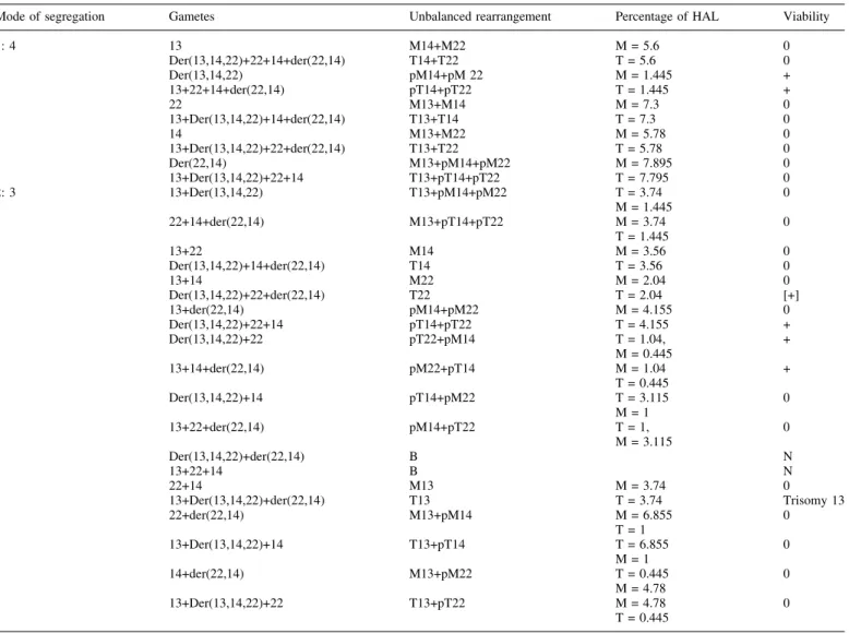
Different classi®cations of CCR have been proposed (Kausch et al., 1988; Lurie et al., 1994). CCR can be divided into two types: a three-way translocation, and a rearrangement with more than one breakpoint per chromosome. This BCCR results from a veritable three-way exchange in which three segments, generated by three chromosome breaks, translocate and unite. Three-way BCCR are rarely transmitted by the father (Farrell et al., 1994). At meiosis, the chromosomes involved in the rearrangement form a hexavalent. This con®guration would allow full synapsis of six homologous segments except for the proximal segments adjacent to the breakpoints. To our knowledge, formal analysis of the breakpoints of cases described in the literature has not been reported. Two gametes arising from alternate segregation would be balanced. The remaining gametes would be unbal-anced to a greater or lesser degree (Table III). A tendency to favour symmetric alternate segregation in the ®rst generation,
probably during male gametogenesis, appears to characterize this family.
We cannot use the `adjacent or alternates' models generally used for translocations between two chromosomes to analyse BCCR. For II3-B and II1-C, there was recombination at the
time of parental meiosis between the chromosome 14 involved in the translocation and the normal chromosome 14. II3-B
received part of the (13;14) with the translocation± recombination±telomeric extremity of normal chromosome 14 and the normal chromosome 22 of her father. II1-C received
the centromeric extremity of chromosome 14 normal± recombination±translocation (14;22) and the 13 normal chromosome of his father.
The result of the microsatellite analysis is not easily interpretable. The molecular analysis of the genotypes shows that the proximal part of chromosome 22 is non-informative. We might surmise the existence of a common haplotype in subjects with a translocation (14;22). However, con®rmation of this is impossible, the mother (I1-M) being homozygote for
marker D22S446, the father's (I2-A) deduced haplotype being Table III. Possible gametic combinations occurring in the 45,XX,dic(13;14)t(14;22), der (22)t(14;22) rearrangement
Mode of segregation Gametes Unbalanced rearrangement Percentage of HAL Viability
1: 4 13 M14+M22 M = 5.6 0 Der(13,14,22)+22+14+der(22,14) T14+T22 T = 5.6 0 Der(13,14,22) pM14+pM 22 M = 1.445 + 13+22+14+der(22,14) pT14+pT22 T = 1.445 + 22 M13+M14 M = 7.3 0 13+Der(13,14,22)+14+der(22,14) T13+T14 T = 7.3 0 14 M13+M22 M = 5.78 0 13+Der(13,14,22)+22+der(22,14) T13+T22 T = 5.78 0 Der(22,14) M13+pM14+pM22 M = 7.895 0 13+Der(13,14,22)+22+14 T13+pT14+pT22 T = 7.795 0 2: 3 13+Der(13,14,22) T13+pM14+pM22 T = 3.74 0 M = 1.445 22+14+der(22,14) M13+pT14+pT22 M = 3.74 0 T = 1.445 13+22 M14 M = 3.56 0 Der(13,14,22)+14+der(22,14) T14 T = 3.56 0 13+14 M22 M = 2.04 0 Der(13,14,22)+22+der(22,14) T22 T = 2.04 [+] 13+der(22,14) pM14+pM22 M = 4.155 0 Der(13,14,22)+22+14 pT14+pT22 T = 4.155 + Der(13,14,22)+22 pT22+pM14 T = 1.04, + M = 0.445 13+14+der(22,14) pM22+pT14 M = 1.04 + T = 0.445 Der(13,14,22)+14 pT14+pM22 T = 3.115 0 M = 1 13+22+der(22,14) pM14+pT22 T = 1, 0 M = 3.115 Der(13,14,22)+der(22,14) B N 13+22+14 B N 22+14 M13 M = 3.74 0 13+Der(13,14,22)+der(22,14) T13 T = 3.74 Trisomy 13 22+der(22,14) M13+pM14 M = 6.855 0 T = 1 13+Der(13,14,22)+14 T13+pT14 T = 6.855 0 M = 1 14+der(22,14) M13+pM22 T = 0.445 0 M = 4.78 13+Der(13,14,22)+22 T13+pT22 M = 4.78 0 T = 0.445
M = monosomy; pM = partial monosomy; T = trisomy; pT = partial trisomy. HAL = haploid autosomal length; B = balanced; N = normal.
non-informative for D22S420. The data for the three sisters (II2-D, II3-B, II4-A) with the Robertsonian translocation
t(13;14) is less ambiguous.
Another hypothesis is possible. I2-A could be a carrier of two
translocations: a Robertsonian translocation der(13;14) (p11;p11) and a reciprocal translocation t(14;22) (q32.3;q11.2). During meiosis, a crossing-over would have occurred between the two chromosomes 14 giving rise to a derivative der(13;14)t(14;22) and another der(22)t(14;22); II3
-B would have received only the translocation t(13q;14q) whereas II1-C would have received the derivative 14 of
translocation t(14;22) without the derivative 22. The other question is the probability of a second crossing-over identical to the index case (II2-D). However, such a relatively simple
mechanism is insuf®cient to explain the identical rearrange-ment in the sib of the proband, which would seem to suggest that the same crossing-over occurred twice in succession.
Review of the literature suggests that severely unbalanced con®gurations often occur in female gametogenesis. Paternal origin was, however, very frequently shown in de-novo CCR informative reports. The probability of such uniparental origin occurring by chance alone is 1/256 (Batista et al., 1993). This low probability leads us to speculate that mechanisms resulting in BCCR occur preferentially during spermatogenesis. BCCR and fertility
BCCR rarely occur in phenotypically normal persons (Fukushima et al., 1986). The impact of CCR on fertility is important. Anomalies of the acrocentric chromosomes increase the risk of sterility (Gabriel-Rodez et al., 1986). The fact that individual I1-M had six children is surprising. The possibility
of a germinal parental mosaicism should be considered. In the female, gametogenesis can accommodate the complexity of CCR. The female may be fertile and have pregnancies that produce phenotypically normal children. In the father's case (I2-A), we suspect that spermatogenesis produces
phenotypi-cally normal children. In contrast, in the literature, male carriers are often subfertile (Johannisson et al., 1985; Saadallah and Hulten, 1985) or sterile due to spermatogenic arrest (Rodriguez et al., 1985).
Studies of the pachytene stage of meiosis have provided clues to the underlying mechanisms responsible for male sterility associated with some autosomal translocations. Three features are regularly observed in such male-sterilizing rearrangements: (i) synaptic failure around breakpoints, (ii) association of the translocation ®gure with the sex chromo-somes, (iii) frequent occurrence of an acrocentric chromosome in the translocation.
Two main models have been proposed to explain gameto-genic failure in the male. Burgoyne and Baker (1994) have argued that impairment of spermatogenesis might be attributed to generalized pairing disruption along the genome, an extension of the earlier hypothesis of Miklos (1974) in which XY-pairing failure was suggested as a primary cause of germ cell failure. Alternatively, the defect could result from XY± multivalent interaction, as originally proposed for the mouse by Forejt (1974) and later suggested by Chandey (1979) to explain human spermatogenic failure. Each mechanism in itself may be
suf®cient to cause spermatogenic failure, but the two could interact, where partial asynapsis between normal and translo-cated chromosomes would favour attraction between the translocation ®gure and the differential segment of the X-autosome (Rosenmann et al., 1985).
Studies of three-way translocation (Johannisson et al., 1985; Saadallah and Hulten, 1985) gave few indications of XY association, all arms of the hexavalents being fully paired during the pachytene stage. Extensive asynapsis around the breakpoints was a feature, but there was very little evidence of spermatogenic depression or arrest, with the sperm count being within normal limits. Our case presents a hexavalent formed by three acrocentric chromosomes (one Robertsonian trans-location and one reciprocal transtrans-location). Meiotic studies on human infertile male carriers of Robertsonian translocation have shown that X-autosome association was attained by the central asynapsis and/or by the terminal chromomere of the acrocentric chromosome involved in the translocation. It was proposed that the acrocentric chromosome favours the contact between the quadrivalent and the sex vesicle, and increases the risk of sterility in male carriers of Robertsonian translocations and of reciprocal translocation involving almost one acro-centric chromosome.
In women, without sex vesicle, an involved cause of infertility does not exist and by itself could explain the different effect on fertility between male and female. Moreover, all the studies on infertile males with a balanced Robertsonian translocation show a slightly reduced number of chiasma. Variations in pattern of maternal recombination have been identi®ed as a risk factor for meiotic chromosome non-disjunction. Recent studies have con®rmed the large difference in recombination frequency between human oocytes and spermatocytes and demonstrate a clear between-sex variation in distribution of crossing-over (Tease et al., 2002). They observed an abnormal pattern of meiotic recombination in abnormal oocytes that showed chromosome-pairing errors. These facts could explain the high rate of conceptuses with presumed severely unbalanced karyotypes (spontaneous mis-carriages) present in women of this family.
BCCR and genetic counselling
The nature of CCR implies that different unbalanced combin-ations might be expected to be viable. By attachment to centromeres, the meiotic spindle ensures attachment at the two poles and thus successful segregation of homologous chromo-somes to opposite poles (Kallio et al., 1998). Therefore, the complex meiotic con®guration disturbs the chromosome orientation and causes abnormal spindle attachment leading to chromosome malsegregation. Moreover, normal meiosis requires crossing-over during homologous chromosome pair-ing at the pachytene stage: these chromatid exchanges, in the case of complex meiotic con®gurations, increase the risk of chromosome rearrangement, as for patient II4-A, and of
segmental aneuploidy, as for III9-SA. A theoretical prediction
of chromosomal segregation in gametes is possible, giving 30 different karyotypes. The empirical estimated risk for spon-taneous abortion is 75±100% for some BCCR (Creasy, 1989). The chance of carrying a pregnancy to term with an abnormal
and developmentally delayed child is possible and has been estimated at 50% (Wang et al., 1993). We think that this risk can be higher depending of the type of BCCR (Ruiz et al., 1996). Viability thresholds for chromosomal imbalances have been estimated at 5% of haploid autosomal length for pure trisomies and 3% for pure monosomies. In a monosomy± trisomy combination, the haploid autosomal length represented by the trisomy should not be >3.6% and should not be >0.6% for the monosomy (Cohen et al., 1994). The resulting viability area has a step shape out of which every chromosomal imbalance is considered as lethal. The risk of serious congeni-tal malformation with de-novo balanced reciprocal trans-location between two chromosomes was estimated at ~7% (3.5% per each break) on the basis of published data (Warburton, 1991). For apparently balanced CCR arising de novo, an empirical risk of up to 90% has been proposed for phenotypic abnormality and mental retardation (Gardner and Sutherland, 1989), although the exact prevalence is impossible to establish. The risk is undoubtedly much smaller. We can speculate on 3.5% per break whatever the number of break-points. These values vary slightly with the segregation mode, the sex of the carrier parent and the genomic content of unbalanced chromosomal segments. An international registry of minimal chromosomal imbalances should be considered in order to assist in the counselling of these patients. Preimplantation genetic diagnosis (PGD) has been used for couples with normal fertility but at high risk of having a child with chromosomal abnormalities. PGD increases the implant-ation rate in human IVF by avoiding the transfer of chromosomally abnormal embryos. Here, the complexity of these BCCR makes the preimplantation diagnosis impossible. Conclusion
We report here a familial case of CCR possibly inherited from the father. In this family, CCR resulted in fertility. Thus, the risk for miscarriages appears to be higher than that of a simple balanced reciprocal translocation carrier. The risk for a liveborn child with an unbalanced rearrangement does not appear to differ signi®cantly. Our data con®rm that it is impossible to predict the risk of unbalanced progeny to carriers of BCCR. In conclusion, cytogenetic analysis is a useful tool to investigate miscarriages, to give adequate genetic counselling and to discuss the choice of an appropriate assisted reproduc-tion technique.
Acknowledgements
We are grateful to Dr J.Michaud and D.Lambert for the revision of the manuscript.
References
Batavian, J.R. and Eswara, M.S. (1998) De novo apparently balanced complex chromosome rearrangement (CCR) involving chromosomes 4, 18 and 21 in a girl with mental retardation: report and review. Am. J. Hum. Genet., 78, 44±51.
Batista, D.A., Tuck-Muller, C.M., Martinez, J.E., Kearns, W.G., Pearon, P.L. and Stetten, G. (1993) Complex chromosomal rearrangement detected
prenatally and studied by ¯uorescence in situ hybridization. Hum. Genet., 92, 117±121.
Blouin, J.L., Christie, D.H., Gos, A., Lynn, A., Morris, M.A., Ledbetter, D.H., Chakravarti, A. et al. (1995) A new dinucleotide repeat polymorphism at the telomere of chromosome 21q reveals a signi®cant difference between male and female rates of recombination. Am. J. Hum. Genet., 57, 388±394. Buetow, K.H., Weber, J.L., Ludwigsen, S., Scherpbier-Heddema, T., Duyk,
G.M., Shef®eld, V.C., Wang, Z. et al. (1994) Integrated human genome-wide maps constructed using the CEPH reference panel. Nature Genet., 6, 391±393.
Burgoyne, P.S. and Baker, T.G. (1984) Meiotic pairing and gametogenic failure. In Evans, C.W. and Dickinson, H.G. (eds), Controlling Events in Meiosis. Proceedings of the 38th Symposium, Reading. Company of Biologists, Cambridge, pp. 349±362.
Chandley, A.C. (1979) The chromosomal basis of human infertility. Br. Med. Bull., 35, 181±186.
Cohen, O., Cans, C., Mermet, M.A., Demongeot, J. and Jalbert, P. (1994) Viability thresholds for partial trisomies and monosomies. A study of 1,159 viable unbalanced reciprocal translocations. Hum. Genet., 93, 188±194. Creasy, M.R. (1989) Complex chromosomal rearrangements. Am. J. Med.
Genet., 32, 560.
Devilee, P., Slagboom, P., Cornelisse, C.J. and Pearson, P.L. (1986) Sequence heterogeneity within the human alphoid repetitive DNA family. Nucleic Acids Res., 14, 2059±2073.
Dib, C., Faure, S., Fizames, C., Samson, D., Drouot, N., Vignal, A., Millasseau. P. et al. (1996) A comprehensive genetic map of the human genome based on 5,264 microsatellites. Nature, 380, 152±154.
Driscoll, D.A., Budarf, M.L. and Emanuel, B.S. (1992) A genetic etiology for DiGeorge syndrome: consistent deletions and microdeletions of 22q11. Am. J. Hum. Genet., 50, 924±933.
Dutrillaux, B. and Vigas-Pequignot, E. (1981) High resolution R- and G-banding on the same preparation. Hum. Genet., 57, 93±95.
Farrell, S.A., Summers, A.M., Gardner, H.A. and Uchida, I.A. (1994) Balanced complex chromosome rearrangement ascertained trough prenatal diagnosis. Am. J. Med. Genet., 52, 360±361.
Forejt, J. (1974) Non-random association between a speci®c autosome and the X chromosome in meiosis of the male mouse: possible consequence of the homologous centromeres separation. Cytogenet. Cell Genet., 13, 369±383. Fukushima, Y., Kuroki, Y. and Ito, T. (1986) Balanced double complex translocations [46,XX t(1p;6p;7p;3q;11p) 11q22p;21q)]. Am. J. Med. Genet., 25, 313±317.
Gabriel-Robez, O., Ratomponirina, C., Dutrillaux, B., Carre-Pigeon, F. and Rumpler, Y. (1986) Meiotic association between the XY chromosomes and the autosomal quadrivalent of a reciprocal translocation in two infertile men, 46,XY,t(19;22) and 46,XY,t(17;21). Cytogenet. Cell Genet., 43, 154± 160.
Gardner, R.J. and Sutherland, G.R. (1989) Chromosome Abnormalities and Genetic Counselling. Oxford University Press, Oxford.
Grieg, G.M. and Willard, H.F. (1992) Beta satellite DNA: characterization and localization of two subfamilies from the distal and proximal short arms of the human acrocentric chromosomes. Genomics, 12, 573±580.
ISCN 95 (1995) An International System for Human Cytogenetic Nomenclature, Mitelman, F. (ed.). Karger, Basel.
Johannisson, R., Lohrs, U. and Passarge, E. (1985) Pachytene analysis in males heterozygous for a familial translocation (9; 12; 13) (q22; q22; q32) ascertained through a child with partial trisomy 9. Cytogenet. Cell Genet., 47, 160±166.
Kallio, M., Mustalahti, T., Yen, T.J. and Lahdetie, J. (1998) Immunolocalization of alpha-tubulin, gamma-tubulin, and CENP-E in male rat and male mouse meiotic divisions: pathway of meiosis I spindle formation in mammalian spermatocytes. Dev. Biol., 195, 29±37.
Kausch, K., Haaf, T., KoÈhler, J. and Schmid, M. (1988) Complex chromosomal rearrangement in woman with multiple miscarriages. Am. J. Med. Genet., 31, 415±420.
Klein, G. (1987) Chromosomal translocations in B-cell derived tumors. Prog. Clin. Biol. Res., 246, 75±91.
Lurie, I.W., Wulfsberg, E.A., Prabahkar, G., Rosenblum-Vos, L.S., Supovitz, K.R. and Cohen, M.M. (1994) Complex chromosomal rearrangement: some breakpoint may have cellular adaptive signi®ance. Clin. Genet., 46, 244± 247.
Miklos, G.L.G. (1974) Sex-chromosome pairing and male infertility. Cytogenet. Cell Genet., 13, 558±577.
Murray, J.C., Buetow, K.H., Weber, J.L., Ludwigsen, S., Scherpbier-Heddema, T., Manion, F., Quillen, J., Shef®eld, V.C., Sunden, S., Duyk,
G.M. et al. (1994) A comprehensive human linkage map with centimorgan density. Cooperative Human Linkage Center (CHLC). Science, 265, 2049± 2054.
Rodriguez, M.T., Martin, M.J. and Abrisqueta, J.A. (1985) A complex balanced rearrangement involving four chromosomes in an azoospermic man. J. Med. Genet., 22, 66±67.
Rosenmann, A., Wahrman, J., Richler, C., Voss, R., Persitz, A. and Goldman, B. (1985) Meiotic association between the XY chromosomes and unpaired autosomal elements as a cause of human male sterility. Cytogenet. Cell Genet., 39, 19±29.
Ruiz, C., Grubs, R.E., Jewett, T., Cox-Jones, K., Abruzzese, E., Pettenati, M.J. and Rao, P.N. (1996) Prenatally diagnosed de novo apparently balanced complex chromosome rearrangements: two new cases and review of the literature. Am. J. Med. Genet., 64, 478±484.
Saadallah, N. and Hulten, M. (1985) A complex three-breakpoint translocation involving 2, 4, and 9 identi®ed by meiotic investigations of a human male ascertained for sub fertility. Hum. Genet., 71, 312±320.
Tease, C., Hartshorne, G.M. and Hulten, M.A. (2002) Patterns of meiotic recombination in human fetal oocytes. Am. J. Hum. Genet., 70, 1469±1479. Wang, H., McLaughlin, M.N., Thompson, C. and Hunter, A.G. (1993) Use of ¯uorescence in situ hybridization to con®rm the interpretation of a balanced complex chromosome rearrangement ascertained through prenatal diagnosis. Am. J. Med. Genet., 46, 559±562.
Warburton, D. (1991) De novo balanced chromosome rearrangements and extra marker chromosomes identi®ed at prenatal diagnosis: clinical and distribution of breakpoints. Am. J. Hum. Genet., 49, 995±1013.
Willard, H.F. and Waye, J.S. (1987) Chromosome-speci®c subsets of human alpha satellite DNA: analysis of sequence divergence within and between chromosomal subsets and evidence for an ancestral pentameric repeat. J. Mol. Evol., 25, 207±214.