Implications of the E-selectin S128R mutation
for drug discovery
Roland C Preston2, Said Rabbani2, Florian P C Binder2,
Suzette Moes3, John L Magnani4, and Beat Ernst1,2
2
Institute of Molecular Pharmacy, Pharmacenter;3Department of Biochemistry, Biocenter, University of Basel, Klingelbergstrasse 50, CH-4056 Basel, Switzerland; and4GlycoMimetics, Inc., Gaithersburg, MD 20879, USA Received on February 28, 2014; revised on February 28, 2014; accepted on March 26, 2014
The C-type lectin E-selectin mediates the rolling of circulat-ing leukocytes on vascular endothelial cells durcirculat-ing the in-flammatory process. In numerous studies, the S128R mutation of the E-selectin was associated with cardiovascu-lar and autoimmune diseases. There is evidence that the S128R E-selectin mutation leads to a loss in ligand speci fi-city, thus increasing leukocyte recruitment. Apart from the natural tetrasaccharide ligand sialyl Lewisx (sLex), it has previously been proposed that non-fucosylated carbohy-drates also bind to S128R E-selectin. To evaluate the thera-peutic potential of the antagonism of the E-selectin mutant, ligand specificity was reinvestigated on a molecular basis. We determined the ligand specificity of wild-type and S128R E-selectin in a target-based competitive assay, a glycan array screen and cell-based binding assays under static andflow conditions. Regarding ligand-specificity, the binding properties of S128R E-selectin were identical to those of wt E-selectin, i.e., no mutant-specific binding of 3′-sialyl-N-acetyllactosamine, heparin, fetuin and K562 cells was observed. Additionally, the binding affinities of glycomimetic E-selectin antagonists were identical for wt and S128R E-selectin. Overall, the previous reports on carbohydrate ligand promiscuity of S128R E-selectin could not be confirmed.
Keywords: E-selectin / selectin antagonists / single-nucleotide polymorphism / sLexmimic / S128R
Introduction
In case of inflammation, the human body possesses a highly sophisticated defense line. At the initial stage, the members of the selectin family, E-, P- and L-selectin, play an important role by recruiting leukocytes to the inflammatory site (Kansas
1996). However, excessive extravasation of leukocytes leads to tissue damage being deleterious for numerous diseases with an inflammatory component (Ernst and Magnani 2009). Blocking selectins with selectin antagonists is, therefore, regarded as a promising anti-inflammatory treatment.
Selectins are C-type (calcium dependent) lectins consisting of a C-terminal cytoplasmic domain followed by a transmem-brane region, variable numbers of short consensus repeats (SCRs), an epidermal growth factor-like (EGF-like) domain and an N-terminal lectin domain. Their physiological ligands such as the P-selectin glycoprotein ligand-1 (PSGL-1) (McEver
and Cummings 1997; McEver 2001) and E-selectin ligand-1
(ESL-1) (Steegmaier et al. 1995) contain the common tetrasac-charide epitope sialyl Lewisx (sLex, NeuNAcα2-3Galβ1-4 (Fucα1-3)GlcNAc; Figure1A) as part of O-glycosylated glyco-proteins. The structural analysis of E- and P-selectin soaked with sLex revealed the central role of the L-fucose moiety,
which coordinates the Ca2+ ion with its 3- and 4-hydroxyl groups (Somers et al. 2000).
In recent years, several case–control studies have suggested a correlation of a single-nucleotide polymorphism (SNP) in exon 4 of the E-selectin encoding gene with an early onset, increased susceptibility and severity of various, mainly cardiovascular and immune diseases. These include myocardial infarction (Yoshida et al. 2003; Stepień et al. 2011), severe recurrent venous thromboembolism (Jilma et al. 2005,2006) and the for-mation of colon metastasis (Alessandro et al. 2007). The SNP causes a mutation of serine 128 to arginine (S128R) and is present in 10–15% of the Caucasian population (Wenzel et al. 1994). The mechanism by which the S128R mutation alters the physiological function of E-selectin at molecular level is still unclear, since the mutation is located in the EGF-like domain and not in the carbohydrate recognition domain (CRD) of the lectin. Based on the crystal structure of E-selectin (lectin and EGF-like domain), a direct or indirect involvement of the Ser128 in ligand binding is unlikely (Figure1B and C) (Graves et al. 1994;Somers et al. 2000).
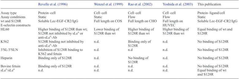
All previous reports on the binding properties of the S128R E-selectin mutant are summarized in TableI. First, Revelle et al. showed a 2- to 3-fold increased adherence of S128R E-selectin to HL60 cells compared with wt E-selectin (Revelle et al. 1996). Furthermore, only S128R E-selectin was capable of binding to heparin and to K562 cells, a cell line lacking fucosyltransferase VII required for sLexbiosynthesis (Snapp et al. 1997). They also showed that the binding of heparin and K562 cell to the S128R E-selectin mutant was reversed by the sLex precursor 3 ′-sialyl-1
To whom correspondence should be addressed: Tel: +41-61-267-1551; Fax: +41-61-267-1552; e-mail: beat.ernst@unibas.ch
Glycobiology vol. 24 no. 7 pp. 592–601, 2014 doi:10.1093/glycob/cwu026
N-acetyllactosamine (3′SLN, NeuNAcα2-3Galβ1-4GlcNAc) or 3′-sialyllactose (3′SL, NeuNAcα2-3Galβ1-4Glc) (Figure1A), in-dicating that the presence of theL-fucose moiety was no longer
required. Furthermore,Revelle et al. (1996)demonstrated that the binding of HL60 cells (which express the sLexepitope) was abol-ished with soluble sLexor an anti-sLexantibody for wt E-selectin, Fig. 1. (A) The natural ligand of E-selectin is sialyl Lewisx(sLex). The proposed S128R E-selectin specific trisaccharide 3′sialyllactose lacks theL-fucose moiety. (B) The crystal structure of the wt E-selectin/sLexcomplex. The residues 135–139 form interactions with the lectin domain (Lec), EGF (epidermal growth factor like domain). (C) The side chain of the Ser128 residue forms a hydrogen bond to the backbone carbonyl of Lys152. This area is rigidified by two disulfide bonds (Cys144-Cys153 and Cys127-Cys142).
Table I. Summary of the reported binding properties of the S128R E-selectin mutant
Revelle et al. (1996) Wenzel et al. (1999) Rao et al. (2002) Yoshida et al. (2003) This publication
Assay type Protein–cell Cell–cell Cell–cell Cell–cell Protein–ligand/cell
Assay conditions Static Static Flow Flow Static
wt and S128R E-selectin constructs
Soluble Lec-EGF-CR2/IgG Full length on COS Full length on CHO Full length on HUVEC
Soluble Lec-EGF-CR2/IgG
HL60 Higher binding of S128R than wt;
S128R not inhibited by sLexor anti-sLex-Ab Lower binding of S128R than wt Higher binding of S128R than wt Higher binding of S128R than wt
Equal binding of wt and S128R
K562 S128R binding not inhibited by
anti-sLex-Ab n.d. Binding only of S128R n.d. No binding of S128R 3′SL/3′SLN Inhibition of S128R binding to K562 and fetuin n.d. n.d n.d. No binding of S128R
Heparin Binding only of S128R n.d. No binding of
S128R
n.d. No binding of S128R
Bovine fetuin Binding only of S128R n.d. n.d. n.d. No binding of S128R
sLex/sLea n.d. n.d. n.d. n.d. Equal binding of wt
and S128R n.d., not determined.
but did not affect binding of S128R E-selectin. Finally, the S128R E-selectin mutant showed specific binding to bovine fetuin, a glycoprotein lacking fucosylated glycans (Green et al. 1988), further suggesting a shift in ligand specificity.
In contrast, Wenzel et al. (1999) reported cell-based assays under static conditions that showed diminished adhesion of HL60 cells to a COS cell monolayer bearing the S128R E-selectin mutant compared with the wt E-selectin counterpart.
Later, Rao et al. (2002) observed that only chinese hamster ovarian cells (CHO) cells displaying the S128R E-selectin mutant were able to tether K562 cells under flow conditions, an effect that was not observed with wt E-selectin. However, no inhibitory effect of heparin for S128R E-selectin as suggested by Revelle et al. (1996) was observed. Since neuraminidase or O-sialogly coprotein endopeptidase treatment of the K562 cells was not deleterious for their tethering toward S128R E-selectin expres-sing CHO cells, Neu5Ac- as well as O-linked glycoprotein-independent binding to the S128R mutant was implied.
Finally, Yoshida et al. (2003) reported increased attachment of HL60 cells toward human umbilical vein cells (HUVECs) displaying S128R E-selectin compared with those displaying wt E-selectin.
Since the S128R E-selectin polymorphism is correlated with numerous diseases, we aimed to re-evaluate its binding speci fi-city at molecular level and additionally determine the potency of E-selectin antagonists which were originally designed for the wt E-selectin. We used a cell-free, target-based assay, which is routinely applied for screening of E-selectin antagonists (Weitz-Schmidt et al. 1996;Thoma et al. 1997). In addition, in a cell-based assay setup, we investigated the binding of HL60 and K562 cells under static andflow conditions.
Results
Characterization of wt and S128R E-selectin
For the affinity determination of three E-selectin antagonists and for further functional analyses, we recombinantly expressed a wt E-selectin construct containing the lectin domain, the EGF-like domain and thefirst two SCRs and the corresponding S128R mutant as IgG chimera. The binding properties of the proteins were evaluated in a cell-free, target-based assay and additionally in a cell-based assay under static andflow conditions.
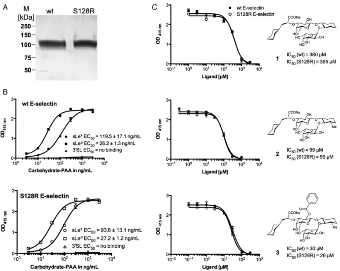
The proteins were expressed in CHO cells followed by functional purification with the functional monoclonal anti-E-selectin antibody 7A9 (see experimental part). The fact that the 7A9-antibody bound S128R E-selectin suggests correct folding of the protein. The sodium dodecyl sulfate polyacrylamide gel electrophoresis (SDS-PAGE) analysis showed a similar appar-ent molecular weight of 100 kDa, suggesting equal glycosyla-tion of the wt and the mutant E-selectin (Figure 2A). The presence of the mutation was confirmed by DNA sequencing and at protein level by liquid chromatography-tandem mass spectrometry analysis (data not shown).
For the evaluation of protein activity, we determined the binding affinities of the natural carbohydrate ligands sLex and sLeato wt and S128R E-selectin as shown in Figure2B. For wt E-selectin, the EC50values for biotinylated sLex- and sLea
-poly-acrylamide polymer (sLex- PAA, sLea-PAA) were 119.5 ± 17.1 ng/mL and 26.2 ± 1.3 ng/mL, respectively. The higher affinity of
sLeacompared with sLexfor recombinant E-selectin has previous-ly been reported with a comparable assay (Weitz-Schmidt et al. 1996;Thoma et al. 1997). Similar affinities were also obtained with S128R E-selectin, with EC50 values of 93.8 ± 13.1 ng/mL
for sLex-PAA and 27.2 ± 1.2 ng/mL for sLea-PAA. Using concen-trations of the biotinylated 3′SL-polyacrylamide polymer (3′ SL-PAA) of up to 3 µg/mL, no binding was observed for both wt and S128R E-selectin, suggesting no or very low affinity. The evaluation of higher concentrations of 3′SL-PAAwas not possible, as the background signal increased significantly.
SLexglycomimetics are efficacious for S128R E-selectin The binding affinities of three previously reported E-selectin antagonists for wt and S128R E-selectin are shown in Figure2C. In all the three of these sLexmimics, the Neu5Ac moiety has been exchanged with (S)-cyclohexyl lactic acid. For antagonist 1 (Norman et al. 1998), GlcNAc has been replaced with (1R,2R)-cyclohexane-1,2-diol and for antagonist 2 and 3 (Schwizer et al. 2012), with (1R,2R,3S)-3-methylcyclohexane-1,2-diol. For an-tagonist 3, the 2-hydroxyl group ofD-galactose is additionally
benzoylated. The half-maximal inhibitory concentration (IC50)
values of all three ligands were similar for the wt as well as S128R E-selectin. In agreement with previously reported results (Schwizer et al. 2012), the affinities of compounds 2 and 3 were approximately 4- and 14-fold higher than that of antagonist 1, re-spectively. The absolute IC50values obtained here are, however,
weaker than previously reported due to the presence of only two SCR domains instead of six SCR domains, which is known to reduce E-selectin affinity (Li et al. 1994).
Evaluation of putative S128R E-selectin ligands
According to Revelle et al. (1996), bovine fetuin and heparin dir-ectly interact with S128R E-selectin, and 3′SLN interferes with the mutant binding to K562 cells. For the evaluation of their af fin-ities (see experimental part) single concentrations of the carbohy-drate ligands (3′SLN: 10 mM, bovine fetuin: 10 mg/mL, heparin; 10 mg/mL) were used to enable clear inhibition. As a result, the binding of sLex-PAA to wt and S128R E-selectin was not inhib-ited by any of the three substances (Figure3A). In a control ex-periment, the assay was also performed with wt P-selectin, for which heparin is a known ligand (Nelson et al. 1993;Wang and Geng 2003). As shown in Figure3A, sLex-PAA binding to wt P-selectin was completely abolished in the presence of heparin.
In addition to the target-based assays, further investigations of the binding properties of S128R E-selectin were performed with a cell-based assay under static and flow conditions. In the static assay, the adhesion of HL60 and K562 cells to wt and S128R E-selectin was determined. Revelle et al. (1996) reported that under static conditions the sLex-presenting HL60 cells adhere to both wt and mutant E-selectin, whereas the sLex-deficient K562 cells selectively bind S128R E-selectin. In our assay, fluorescently labeled HL60 and K562 cells were allowed to bind to E-selectin coated on microtiter plates (see Figure 3B). HL60 cells bind to both forms of E-selectin and since the addition of the functional anti-E-selectin antibody 7A9 inhibited binding entirely, both interactions are clearly E-selectin dependent. For the K562 cells, however, no binding to either form of E-selectin was observed, albeit the high number of applied cells. In theflow assay, adhesion of HL60 RC Preston et al.
cells was studied at physiologic shear stress (Figure3C). Again, the number of initially tethered and consequently rolling cells was similar for wt and S128R E-selectin. While at low shear stress (1 dyne/cm2) the highest number of cells tethered and rolled on wt and mutant E-selectin coated surfaces, decreased tethering was observed when higher shear forces were applied (2 and 3 dynes/cm2). At 5 dynes/cm2, tethering was entirely abolished. Addition of EDTA completely abolished HL60 cells from binding to both forms of E-selectin, thus confirming the calcium-dependent binding (data not shown).
Glycan array screening
To identify potential novel ligands of S128R E-selectin, a glycan array screening was performed at the Core H facilities of the Con-sortium for Functional Glycomics. In this analysis, 465 glycan structures (version 4.1) were tested, including the tetrasaccharides sLea and sLex and derivatives thereof such as 3′SLN, Lea and Lex. For comparison, wt and S128R E-selectin were analyzed in parallel. Binding of the E-selectin constructs was detected with
Alexa Fluor®488 labeled anti-IgG antibody and was quantified by measuringfluorescence intensities as shown in Figure4.
The results clearly indicate that both proteins preferentially bind to glycan structures bearing sLeaand sLex, thus con firm-ing the result of the competitive bindfirm-ing assays. In addition, binding to 3-sulfated Lewisaand Lewisxwas also observed for both E-selectin constructs. Finally, the S128R mutant did not exhibit any affinity for 3′SLN or any other non-fucosylated glycan. For both proteins, the ten best binding glycan structures contain a fucose moiety. This result underlines the importance of the fucose moiety for the binding of wt E-selectin and the corresponding mutant S128R E-selectin.
Discussion
A SNP of E-selectin in which the serine residue at position 128 is substituted by arginine was shown to correlate with various cardiovascular and immune diseases (Yoshida et al. 2003; Fig. 2. (A) SDS-PAGE analysis of recombinant wt and S128R E-selectin purified by affinity chromatography on a 7A9-Sepharose column as described in the Materials and methods section. M, molecular weight marker. (B) Determination of the EC50values for sLex-, sLea- and 3′SL-PAAwith wt E-selectin (B, upper panel) and S128R mutant (B, lower panel). For this assay microtiter plates were coated overnight with either wt or S128R E-selectin (3 µg/mL), blocked with BSA (2%) and incubated with increasing concentrations of sLex-PAA. The bound polymer was detected by colorimetric reaction at 415 nm. The assay was repeated three times, each in duplicate as described in the Materials and methods section. (C) Competitive binding assay for the determination of IC50for antagonists 1-3 with wt and S128R E-selectin. The assay was performed as described in the Materials and methods section.
Jilma et al. 2006; Alessandro et al. 2007). The mutation is located in the EGF-like domain of E-selectin, which is neces-sary for, but not directly involved in, ligand binding (Erbe et al. 1992).Revelle et al. (1996) reported that the regular physio-logical E-selectin ligand, the tetrasaccharide sLex was no longer required for S128R E-selectin to interact with HL60 and K562 cells.
In competition assays with K562 cells or heparin, 3′SL and 3′SLN were identified as ligands for S128R E-selectin. Furthermore, the glycoproteins heparin and bovine fetuin were shown to interact only with S128R E-selectin and not with wt E-selectin. Interestingly, the E-selectin R84A mutation, which is located adjacent to the carbohydrate-binding site, showed identical binding behavior as the S128R mutant. Binding of E-selectin-R84A to fetuin, however, was not observed in a pre-vious publication, notably with the identical assay (Kogan et al. 1995). In contrast, Rao et al. (2002) reported that heparin did not inhibit S128R E-selectin binding to K562 cells. As stated above, the adverse effects of the S128R mutation were
investigated in a number of case–control studies for a variety of diseases, indicating that the loss of ligand specificity is possibly correlated with an increased leukocyte recruitment which in turn leads to pathophysiological situations.
Selectin antagonists have unequivocally been shown to inter-fere with excessive recruitment of leukocytes upon inflammation in vivo (Mulligan et al. 1993;Norman et al. 1998;Ridger et al. 2005). For the development of such antagonists, functional and structural information on the target protein are a prerequisite. For this purpose and due to the controversial reports on the binding properties of the S128R mutant, we decided to express the S128R mutant and the corresponding wt E-selectin as secreted IgG forms. Using cell-free target-based and cell-based binding assays, we assessed the binding properties of S128R E-selectin in comparison with the wild-type (wt). For wt and mutant E-selectin, no significant difference in the affinity for sLeaand sLexwas observed, indicating that the carbohydrate-binding site of the E-selectin mutant is not altered.
Fig. 3. Evaluation of the binding of 3′SLN (10 mM), heparin (Hep) (10 mg/mL) and bovine fetuin (Fet) (10 mg/mL) to wt E-selectin and S128R mutant (A). Microtiter plates were coated with either wt or S128R E-selectin (3 µg/mL), blocked with BSA (2%) and incubated with afixed concentration of sLex-PAA (20 ng/ mL), either in the presence or in the absence of Hep and Fet. As positive control wt P-selectin was co-incubated with sLex-PAA (20 ng/mL) and heparin (10 mg/mL). (B) Cell-based static binding assay with HL60 and K562 cell lines. Microtiter plates were coated with either wt or S128R E-selectin (10 µg/mL), blocked with BSA (2%) and incubated with thefluorescently labeled cells (2 × 10⁵cells/well), in the presence or absence of anti-E-selectin antibody 7A9 (20 µg/mL). Bound cells were measured byfluorescence measurements at 492 nm excitation and 517 nm emission wavelengths. The assay was repeated twice, each in duplicate as described in the Materials and methods section. (C) Cell-basedflow binding assay with HL60 cells at physiological shear stress. Flow chambers were coated with protein A (50 µg/mL) and subsequently with wt or S128R E-selectin (50 µg/mL). The HL60 cells (1 × 10⁶cells/mL) were perfused at given shear stress (1–5 dynes/cm2). Cells were counted after 10 min at three points along theflow chamber. The error bars correspond to the standard error of the mean of the three points.
To investigate a potential treatment of patients carrying the S128R E-selectin polymorphism, binding affinities of three E-selectin antagonists were determined (Figure2C). We dem-onstrate that regardless of the S128R mutation, binding af fin-ities for all antagonists were similar when compared with wt E-selectin. This further confirmed that carbohydrate-binding
site of S128R E-selectin was intact and suggests a possible ap-plication of such sLex mimics in the treatment of affected patients. Regarding the specificity of S128R E-selectin, direct binding of the 3′SL polymer was not observed even at high concentrations. To further evaluate the promoted specific ligands for S128R E-selectin, inhibition assays with sLex Fig. 4. Glycan array screening (version 4.1) of wt and S128R E-selectin. Binding was detected by a second incubation with Alexa Fluor®labeled anti-IgG antibody as described in the Materials and methods section. The values of the RFUs are given the average of 6 measurements of which the lowest and highest values were removed.
polymer in competition with the 3′SLN trisaccharide, heparin and bovine fetuin were performed. Even at high concentrations, none of these three compounds showed inhibition of the sLex polymer binding to S128R E-selectin (Figure3A). These results suggest similar binding phenotypes of wt and S128R E-selectin. Finally, to investigate the binding of S128R E-selectin to leuko-cytes, we performed cell-based assays (Figure3B and C). The sLexbearing HL60 cell line bound wt and S128R E-selectin to the same degree under static and physiologicalflow conditions and in an E-selectin dependent manner. Again, no difference in binding specificity was observed for S128R E-selectin. Furthermore, unlike previously reported, no specific binding of K562 cells was measured. Therefore, no interaction between S128R E-selectin and an unknown carbohydrate or non-carbohydrate ligand on K562 occurred. Finally, to characterize the binding phenotype with another cell-free assay, a glycan array screening was performed at the Consortium for Functional Glycomics (Figure4). The binding of wt and S128R E-selectin to 465 different mammalian glycan structures was evaluated. The results confirmed the similar binding profiles for wt and S128R E-selectin. The 10 best binding glycan structures all con-tained a fucose moiety. The RFU values of glycans lacking the fucose moiety such as 3′SLN showed signals on background level for both proteins. Additionally, no specific ligand for the S128R E-selectin mutant was detected, and thus, its binding properties do not differ from those of the wt. As in the EC50
determinations of sLeaand sLex, an augmented binding of sLea over sLex was also observed which is reflected by the higher RFU values for sLea. Although high E-selectin concentrations (1 mg/mL) were applied for the glycan array, the resulting RFU values were rather low. This was most likely due to the low af fin-ity of E-selectin for its tetrasaccharide ligands (Schwizer et al. 2012). The reason why S128R E-selectin showed eightfold lower maximal RFU values compared with wt E-selectin is not clear, since in the polymer-based activity assays, the affinity (EC50) of both proteins for sLex- and sLea-PAAwere comparable
(equal maximal OD415nmvalues). However, glycan array
screen-ings provide for qualitative and not quantitative measurements. Our results suggest similar binding specificities of wt and S128R E-selectin under static andflow conditions. This is to be expected from a visual inspection of the E-selectin crystal struc-ture in complex with sLex(Somers et al. 2000). It was shown that only the amino acids 135–139 from the EGF-like domain form a direct contact with the lectin domain by hydrogen bonding and van der Waals interactions (Graves et al. 1994). The distance between the Ser128 Cα atom and Ca2+ in the binding site is 43 Å. The Ser128 side chain forms a hydrogen-bond with the backbone carbonyl of Lys152, an interaction found in all selectin crystal structures. This hydrogen bond would be lost in case of the S128R mutation. The fact that the amino acid sequence Ser126-Cys127-Ser128 is conserved in all the three selectins and throughout several species implies that this region is indeed important for the proper function of the selectins. However, the region surrounding the mutation is ri-gidified by three disulfide bonds (Cys122–Cys133, Cys127– Cys142 and Cys144–Cys153) within a 10 Å radius of Ser128 Cα atom (Figure1C).
In conclusion, as the S128R E-selectin displayed unaltered binding specificity under static and flow conditions as wt E-selectin, we conclude that glycomimetic E-selectin antagonists
are also effective antagonists of the mutant. According to our studies, the reported correlation of a SNP in the E-selectin en-coding gene with an increased susceptibility and severity of various, mainly cardiovascular and immune, diseases is there-fore not related to altered binding properties of wt and mutant E-selectin. Further investigations, e.g., structural and biophysic-al protein characterization, as well as functionbiophysic-al in vivo assays are necessary to shed light on possible therapeutic opportunities based on the S128R E-selectin mutant.
Materials and methods Materials and reagents
The E-selectin primers lec-fw (5′-GGCC GAATTC GTG GTC TTA CAA CAC CTC CAC GGA A-3′) and cr2_rev (5′-GGCC
GATATC GAA GCT TTA CAC GTT GGC TTC TCG TT-3′)
containing the restriction sites EcoRI and EcoRV (underlined), respectively, and the mutation primers egf_s128r_fw (5′-AAT ACA TCC TGC AGA GGC CAC GGT-3′) and egf_s128r_rev (5′-ACC GTG GCC TCT GCA GGA TGT ATT-3′) were pur-chased from Microsynth (Balgach, Switzerland). The restriction enzymes EcoRI and EcoRV and the glycosidase PNGaseF were obtained from New England BioLabs (Allschwil, Switzerland). The pFUSE-hIgG2-Fc2 expression vector and the antibiotic Zeocin™ were purchased from Invivogen (Toulouse, France). Tissue cultureflasks and MaxiSorp™ 96-well microtiter plates were from Nunc (Roskilde, Denmark). Ham’s Nutrient Mixture F-12 medium, Roswell Park Memorial Institute (RPMI-15) medium, fetal calf serum (FCS) and the horseradish peroxidase substrate 2,2′-azino-di(3-ethylbenzthiazoline-6-sulfonic acid) (ABTS) were purchased from Invitrogen (Lucerne, Switzer-land). Penicillin (10,000 U/mL)/streptomycin (10 mg/mL), cyanogen bromide-activated-Sepharose®4B, bovine fetuin and heparin sodium salt were obtained from Sigma-Aldrich (Basel, Switzerland). The FuGENE®HD transfection reagent and the streptavidin-peroxidase (streptavidin-POD) were purchased from Roche Applied Science (Rotkreuz, Switzerland). Amicon® ultrafiltration tubes (50 kDa cut-off) were obtained from Millipore (Zug, Switzerland). The ion exchange column UnoQ6 was purchased from Bio-Rad (Reinach BL, Switzer-land). Protein A-Sepharose® was obtained from BioVision (Mountain View, CA). Protein G-Sepharose® was purchased from Amersham Pharmacia (GE Healthcare, Pistcataway, NJ). 3-SL, sLex- and sLea-polyacrylamide (PAA)-biotin were from Gly-coTech (Gaithersburg, MD). The Alexa Fluor® 488 labeled anti-human-IgG(Fc) antibody and the Microscanarray XL4000 scanner were from Invitrogen (Carlsbad, CA) and PerkinElmer Life Sciences (Waltham, MA), respectively. Trypsin sequen-cing grade was obtained from Promega (Dübendorf, Switzer-land). The trapping 300SB C-18 column (0.3 × 50 mm) was purchased from Agilent Technologies (Basel, Switzerland), the Magic 300 Å C18 reverse-phase material (5 µm particle size) from Michrom Bioresources, Inc. (Auburn, CA), the Orbitrap FT hybrid instrument from Thermo Finnigan (San Jose, CA) and the Rheos 2200 pump from (Flux Instruments, Basel, Switzerland). The cell line HL60 was purchased from LGC Standards (Molsheim, France) and K562 cells from the German Collection of Microorganisms and Cell Cultures (DSMZ, Braunschweig, Germany). The BioFlux™ system was purchased from Fluxion (San Francisco, CA).
Molecular cloning
Genomic DNA from wild-type human E-selectin [Lec-EGF-CR6/hIgG1(Fc)] expressing CHO-K1 cells (Scheffler et al. 1995) was used for polymerase chain reaction (PCR) amplification of the cDNA encoding for the human E-selectin lectin domain (bp 1–360), the EGF-like domain (bp 361–471) and the first two con-sensus repeat domains (bp 472–840). The forward primer lec_fw and the reverse primer cr2_rev containing EcoRI and EcoRV re-striction sites were used. The restricted insert was ligated into the corresponding cloning site of the pFUSE-hIgG2-Fc2 expres-sion vector. The resulting construct containing the N-terminal interleukine-2 secretion signal and the Fc part of human IgG2 at C-terminus was transformed into chemocompetent Escherichia coli DH5α. After single clone selection and DNA-minipreparation, the correctness of the construct was confirmed by DNA double-strand sequencing (Microsynth, Balgach, Switzerland).
Site-directed mutagenesis was employed for the thymine-to-adenine substitution at cDNA position 384, shifting the ex-pression from a serine to an arginine at amino acid position 128. The standard overlap extension PCR method was used (Ho et al. 1989). In afirst step, two overlapping DNA fragments were gen-erated separately, using wild-type E-selectin cDNA as template. Thefirst fragment was amplified with the lec_fw primer and the internal reverse primer egf_s128r_rev and the second fragment was amplified using cr2_rev and the internal forward primer egf_s128r_fw. The two internal primers contained a mismatch for the site-directed base substitution. In a second step, both overlapping DNA fragments were elongated to the full-length insert, containing the single point mutation. The cloning into the pFUSE-hIgG2-Fc2 expression vector and sequence verification were performed as described above for the wt construct.
Cell transfection and expression of wt E-selectin, wt P-selectin and S128R E-selectin
CHO-K1 cells (American Type Culture Collection No. CCL-61™) were cultivated in tissue culture flasks in Ham’s Nutrient Mixture F-12 medium supplemented with 2 mM L-glutamine, 10% (v/v) FCS, 100 U/mL penicillin and 100 µg/ mL streptomycin. The cells were transfected with either the wt or the S128R construct using the FuGENE® HD transfection reagent according to the instructions of the supplier (Roche). The selection of stably transfected CHO-K1 cells was achieved by treatment with 500 µg/mL Zeocin™ and single clones were obtained by limiting dilution. Culture medium containing the secreted E-selectin [Lec-EGF-CR2/hIgG2(Fc)] chimera was harvested weekly and sterilefiltered (0.22 µm). The medium was used immediately for protein purification or stored at −20°C.
For control experiments P-selectin [Lec-EGF-CR6/hIgG1 (Fc)] wt expressed in CHO-K1 cells was used (Fritz et al. 1998). Expression and purification of anti-E-selectin antibody 7A9 Mouse hybridoma cells expressing the mouse monoclonal anti-E-selectin antibody 7A9 (American Type Culture Collection No. HB-10135™) were cultivated in RPMI-15 medium supplemen-ted with 15% (v/v) FCS, 100 U/mL penicillin and 100 µg/mL streptomycin. Antibodies were purified from the culture medium by protein G-Sepharose®. The column was attached to a fast protein liquid chromatography apparatus (Bio-Rad, Reinach BL,
Switzerland) and equilibrated with loading buffer (50 mM Tris– HCl ( pH 7.6), 150 mM NaCl and 0.05% v/v Tween-20). The culture medium was filtrated (0.22 µm) diluted with loading buffer and then loaded onto the protein G-Sepharose® column followed by two washing steps,first with loading buffer and then with washing buffer (5 mM NH4OAc, pH 5.0). The protein was
finally eluted with elution buffer (0.5 M NH4OAc, pH 3.4) and
the eluate was immediately neutralized to pH 7–7.5 with a 2.5 M Tris solution. The protein concentration was determined by high-performance liquid chromatography (HPLC) (Mesch et al. 2012). Approximately 5 mg of purified 7A9 antibody were coupled to 1 mL of cyanogen bromide-activated-Sepharose®4B according to the manufacturer’s protocol.
Purification of wt E-selectin, wt P-selectin and S128R E-selectin
The culture medium containing E-selectin was diluted (1:1, v/v) with loading buffer, filtrated (0.22 µm) and purified with the 7A9-Sepharose®column using the same protocol and buffers as for the purification of the 7A9 antibody (see above). The purified protein was concentrated by ultrafiltration (50 kDa cut-off).
P-selectin was purified in two steps. The first step was per-formed on a protein A-Sepharose®column using the same pro-cedure and buffers as described above. The second step was performed with anion exchange chromatography using a UnoQ6 column and the buffer A (20 mM Tris–HCl, pH 7.4) for column equilibration and loading and buffer B (20 mM Tris–HCl, pH 7.4, 1 M NaCl) for protein elution.
Protein purity was confirmed by SDS-PAGE analysis fol-lowed by Coomassie Brilliant Blue G-250 staining. The protein concentration was determined by HPLC as previously reported (Mesch et al. 2012).
Activity binding assay
Biotinylated sLex-, sLea- and 3′SL-polyacrylamide polymers (sLex-PAA, sLea-PAA and 3′SL-PAA, purchased from Glyco-Tech, Gaithersburg, MD 20879) were coupled to streptavidin-β-peroxidase (POD) by incubation of 20 µL (100 µg/mL) polymer, 80 µL (500 U/mL streptavidin-POD conjugate), 10 µL FCS and 80 µL assay buffer (20 mM HEPES pH 7.4, 150 mM NaCl, and 10 mM CaCl2) for 2 h at 37°C. The complexes were
stable for several weeks when stored at 4°C.
MaxiSorp™ 96-well microtiter plates were coated with 100 µL/well of wt or S128R E-selectin at 3 µg/mL in assay buffer overnight at 4°C in a humidified chamber. Control wells were coated with assay buffer without protein for subsequent background signal subtraction. The wells were washed three times with 150 µL/well of assay buffer and blocked with 150 µL/ well of 3% w/v bovine serum albumin (BSA) in assay buffer for 3 h at 4°C. The wells were then washed three times with 150 µL/ well assay buffer before adding 100 µL/well of a serial dilution (0–3 µg/mL) of a sLex-, sLea- or 3′SL-PAA solution. Each con-centration was applied in duplicate. After incubation for 3 h at 25°C and 250 rpm, the wells were carefully washed with 150 µL/ well assay buffer. Carbohydrate-PAA binding was detected by addition of 100 µL/well of ABTS-substrate. The colorimetric re-action was allowed to develop for 10 min and then stopped by the addition of 2% aqueous oxalic acid before the optical density (OD) was measured at 415 nm on a microplate reader. The
OD415 nm values were background subtracted and the EC50
values were calculated using the Prism software (GraphPad Software, Inc., La Jolla). The assays were repeated three times on separate days. The EC50value defines the glycopolymer
concen-tration corresponding to 50% of the maximum binding to the protein.
Competitive binding assay
For the evaluation of the proposed ligands for S128R E-selectin and the affinity measurements of the sLex mimics, polyacryl-amide polymer-based competitive binding assay was performed. The trisaccharide 3′-sialyl-N-acetyllactosamine (3′SLN) was synthesized in-house as reported in the Supplementary data. Antagonists 1-3 were prepared as previously described ( Schwi-zer et al. 2012). MaxiSorp™ 96-well microtiter plates were coated with a solution of E- or P-selectin (3 µg/mL, 100 µL/ well) and blocked for 3 h with 150 µL/well of BSA (3%, w/v). The wells were subsequently co-incubated with 50 µL of sLex-PAA (40 ng/mL) and 50 µL of 3′SLN (10 mM final con-centration), bovine fetuin (10 mg/mL) or heparin sodium salt (10 mg/mL). Inhibition of sLex-PAA binding to wt P-selectin by heparin sodium salt served as positive control. For compounds 1-3, 25 µL of serial dilutions were mixed with 25 µL of sLea-PAA polymer (40 ng/mL). All other assay steps were per-formed similar to the activity binding assay. For 3′SLN, the assay was repeated twice on separate days and for bovine fetuin and heparin three times on separate days. The sLexmimics were tested a single time in duplicate. IC50 values were calculated
using the Prism software. The IC50defines the molar
concentra-tion of the test compound that reduces the maximal specific binding of sLex/a-PAA to E/P-selectin by 50%.
Cell-based static assay
The protein coating, washing and blocking steps of the cell-based binding assay were performed similar to the activity binding assay, with an increase in wt or S128R E-selectin concentration to 10 µg/mL. HL60 and K562 cells were cultivated in suspension in RPMI 1640 culture medium supplemented with 10% FCS, 2 mM L-glutamine, 10% (v/v) FCS, 100 U/mL penicillin and 100 µg/mL streptomycin. The cell suspension was harvested at 5 × 10⁵–1 × 10⁶cells/mL by centrifugation at 1500 rpm and 4°C. The cells were washed twice with PBS, resuspended to 1 × 10⁶ cells/mL and labeled with 5 µM carboxyfluorescein succinimidyl ester) for 8 min at room temperature. The reaction was stopped with FCS (2%, v/v) and the cells were washed twice with assay buffer containing FCS (2%, v/v). Final concentrations of 2 × 10⁵cells/well and 20 µg/mL 7A9 antibody were used. After incubation for 1 h at 37°C and 250 rpm, the wells were washed three times with assay buffer. Fluorescent intensities were mea-sured at 492 nm excitation and 517 nm emission with a fluores-cence plate reader. The cell-based assays were performed in duplicate and repeated twice on separate days. Cell viability was >95% at all times as judged by trypan blue staining.
Cell-basedflow assay
Flow chamber assays were performed with a BioFlux™ system. Theflow chambers were coated over night at 4°C with 50 µg/mL soluble protein A in bicarbonate buffer ( pH 9.6).
After washing with assay buffer, the chambers were perfused with wt or S128R E-selectin (50 µg/mL) and incubated for 2 h at 4°C. The chambers were subsequently blocked with 2% w/v BSA in assay buffer. HL60 cells were prepared as described above, resuspended to 1 × 10⁶cells/mL and perfused at physio-logical shear forces between 1 and 5 dynes/cm2. Rolling cells were determined at three points along the chamber after 10 min perfusion with a Nikon inverted-stage phase-contrast micro-scope with 20 × magnification. Shutter speed was set according to shear stress for correct distinction of adherent and free flowing cells.
Glycan array screening
The glycan array screening (version 4.1) was performed at the Core H facilities of the Consortium for Functional Glycomics as previously described (Blixt et al. 2004). Briefly, the wt and S128R E-selectin samples were diluted to 1 mg/mL in Tris-buffer [20 mM Tris–HCl (pH 7.4), 150 mM NaCl, 2 mM CaCl2, 2 mM MgCl2] supplemented with 0.05% Tween 20
and 1% BSA. An aliquot of 70 µL was applied to the slides containing the covalently linked glycans, each glycan in repli-cate of six. The slides were coverslipped and incubated in a humidified chamber protected from light for 1 h at room tem-perature and subsequently incubated with Alexa Fluor® 488 labeled anti-human-IgG(Fc) antibody at a concentration of 5 µg/mL in Tris-buffer. After washing and drying the slides, the fluorescence was measured with the Microscanarray XL4000 scanner. The relativefluorescence units (RFUs) were then cal-culated as the average of the six replicates of each glycan structure, of which the lowest and the highest values were ignored.
Supplementary data
Supplementary data for this article is available online at http:// glycob.oxfordjournals.org/.
Funding
We thank the Swiss National Science Foundation for the finan-cial support (200020_129935/1).
Acknowledgements
We acknowledge the Protein-Glycan Interaction Core (H) of the Consortium for Functional Glycomics funded by the NIGMS GM62116 for the glycan array analysis.
Conflict of interest None declared Abbreviations
3′SL-PAA, biotinylated 3′SL-polyacrylamide polymer; CRD, carbohydrate recognition domain; EGF-like, epidermal growth factor-like; HL-60, human promyelocytic leukemia cells; K562, human immortalized myelogenous leukemia cell line; Lec, lectin domain; PAA, polyacrylamide; POD,β-peroxidase; RC Preston et al.
PSGL-1, P-selectin glycoprotein ligand-1; RFU, relative fluor-escent unit; SCR, short consensus repeat; sLea-PAA, biotiny-lated sLea-polyacrylamide polymer; sLex-PAA, biotinylated sLex-polyacrylamide polymer; SNP, single-nucleotide poly-morphism, wt, wild type.
References
Alessandro R, Seidita G, Flugy AM, Damiani F, Russo A, Corrado C, Colomba P, Gullotti L, Buettner R, Bruno L, et al. 2007. Role of S128R polymorphism of E-selectin in colon metastasis formation. Int J Cancer. 121:528–535. Blixt O, Head S, Mondala T, Scanlan C, Huflejt ME, Alvarez R, Bryan MC,
Fazio F, Calarese D, Stevens J, et al. 2004. Printed covalent glycan array for ligand profiling of diverse glycan binding proteins. Proc Natl Acad Sci USA. 101:17033–17038.
Erbe DV, Wolitzky BA, Presta LG, Norton CR, Ramos RJ, Burns DK, Rumberger JM, Rao BNN, Foxall C, Brandley BK, et al. 1992. Identification of an E-selectin region critical for carbohydrate recognition and cell-adhesion. J Cell Biol. 119:215–227.
Ernst B, Magnani JL. 2009. From carbohydrate leads to glycomimetic drugs. Nat Rev Drug Discov. 8:661–677.
Fritz J, Katopodis AG, Kolbinger F, Anselmetti D. 1998. Force-mediated kinetics of single P-selectin ligand complexes observed by atomic force microscopy. Proc Natl Acad Sci USA. 95:12283–12288.
Graves BJ, Crowther RL, Chandran C, Rumberger JM, Li S, Huang KS, Presky DH, Familletti PC, Wolitzky BA, Burns DK. 1994. Insight into E-selectin/ ligand interaction from the crystal-structure and mutagenesis of the lec/EGF domains. Nature. 367:532–538.
Green ED, Adelt G, Baenziger JU, Wilson S, Vanhalbeek H. 1988. The asparagine-linked oligosaccharides on bovine fetuin. Structural analysis of N-glycanase-related oligosaccharides by 500-megahertz H-1-NMR spectros-copy. J Biol Chem. 263:18253–18268.
Ho SN, Hunt HD, Horton RM, Pullen JK, Pease LR. 1989. Site-directed muta-genesis by overlap extension using the polymerase chain-reaction. Gene. 77:51–59.
Jilma B, Kovar FM, Hron G, Endler G, Marsik CL, Eichinger S, Kyrle PA. 2006. Homozygosity in the single nucleotide polymorphism Ser128Arg in the E-selectin gene associated with recurrent venous thromboembolism. Arch Intern Med. 166:1655–1659.
Jilma B, Marsik C, Kovar F, Wagner OF, Jilma-Stohlawetz P, Endler G. 2005. The single nucleotide polymorphism Ser128Arg in the E-selectin gene is associated with enhanced coagulation during human endotoxemia. Blood. 105:2380–2383.
Kansas GS. 1996. Selectins and their ligands: Current concepts and controver-sies. Blood. 88:3259–3287.
Kogan TP, Revelle BM, Tapp S, Scott D, Beck PJ. 1995. A single amino acid residue can determine the ligand specificity of E-selectin. J Biol Chem. 270:14047–14055.
Li SH, Burns DK, Rumberger JM, Presky DH, Wilkinson VL, Anostario M, Jr, Wolitzky BA, Norton CR, Familletti PC, Kim KJ, et al. 1994. Consensus repeat domains of E-selectin enhance ligand binding. J Biol Chem. 269:4431–4437.
McEver RP. 2001. Adhesive interactions of leukocytes, platelets, and the vessel wall during hemostasis and inflammation. Thromb Haemost. 86:746–756. McEver RP, Cummings RD. 1997. Role of PSGL-1 binding to selectins in
leukocyte recruitment. J Clin Invest. 100:485–492.
Mesch S, Lemme K, Wittwer M, Koliwer-Brandl H, Schwardt O, Kelm S, Ernst B. 2012. From a library of MAG antagonists to nanomolar CD22 ligands. ChemMedChem. 7:134–143.
Mulligan MS, Paulson JC, Defrees S, Zheng ZL, Lowe JB, Ward PA. 1993. Protective effects of oligosaccharides in P-selectin-dependent lung injury. Nature. 364:149–151.
Nelson RM, Cecconi O, Roberts WG, Aruffo A, Linhardt RJ, Bevilacqua MP. 1993. Heparin oligosaccharides bind L-selectin and P-selectin and inhibit acute-inflammation. Blood. 82:3253–3258.
Norman KE, Anderson GP, Kolb HC, Ley K, Ernst B. 1998. Sialyl Lewis(x) (sLe(x)) and an sLe(x) mimetic, CGP69669A, disrupt E-selectin-dependent leukocyte rolling in vivo. Blood. 91:475–483.
Rao RM, Clarke JL, Ortlepp S, Robinson MK, Landis RC, Haskard DO. 2002. The S128R polymorphism of E-selectin mediates neuraminidase-resistant tethering of myeloid cells under shearflow. Eur. Immunol. 32:251–260. Revelle BM, Scott D, Beck PJ. 1996. Single amino acid residues in the E- and
P-selectin epidermal growth factor domains can determine carbohydrate binding specificity. J Biol Chem. 271:16160–16170.
Ridger VC, Hellewell PG, Norman KE. 2005. L- and P-selectins collaborate to support leukocyte rolling in vivo when high-affinity P-selectin-P-selectin glycoprotein ligand-1 interaction is inhibited. Am J Pathol. 166:945–952. Scheffler K, Ernst B, Katopodis A, Magnani JL, Wang WT, Weisemann R,
Peters T. 1995. Determination of the bioactive conformation of the carbo-hydrte ligand in the E-selectin sialyl Lewis(x) complex. Angew Chem Int Ed Engl. 34:1841–1844.
Schwizer D, Patton JT, Cutting B, Smiesko M, Wagner B, Kato A, Weckerle C, Binder FP, Rabbani S, Schwardt O, et al. 2012. Pre-organization of the core structure of E-selectin antagonists. Chemistry. 18:1342–1351.
Snapp KR, Wagers AJ, Craig R, Stoolman LM, Kansas GS. 1997. P-selectin glycoprotein ligand-1 is essential for adhesion to P-selectin but not E-selectin in stably transfected hematopoietic cell lines. Blood. 89:896–901. Somers WS, Tang J, Shaw GD, Camphausen RT. 2000. Insights into the
molecular basis of leukocyte tethering and rolling revealed by structures of P- and E-selectin bound to SLe(X) and PSGL-1. Cell. 103:467–479. Steegmaier M, Levinovitz A, Isenmann S, Borges E, Lenter M, Kocher HP,
Kleuser B, Vestweber D. 1995. The E-selectin-ligand ESL-1 is a variant of a receptor forfibroblast growth factor. Nature. 373:615–620.
Stepień E, Krawczyk S, Kapelak B, Sobczyński R, Stoliński J, Wypasek E, Undas A, Sadowski J. 2011. Effect of the E-selectin Gene Polymorphism (S149R) on Platelet Activation and Adverse Events After Coronary Artery Surgery. Arch Med Res. 42:375–381.
Thoma G, Magnani JL, Ohrlein R, Ernst B, Schwarzenbach F, Duthaler RO. 1997. Synthesis of oligosaccharide-polylysine conjugates: A well character-ized sialyl Lewis(a) polymer for ELISA. J Am Chem Soc. 119:7414–7415. Wang JG, Geng JG. 2003. Affinity and kinetics of P-selectin binding to
heparin. Thromb Haemost. 90:309–316.
Weitz-Schmidt G, Stokmaier D, Scheel G, Nifantev NE, Tuzikov AB, Bovin NV. 1996. An E-selectin binding assay based on a polyacrylamide-type gly-coconjugate. Anal Biochem. 238:184–190.
Wenzel K, Hanke R, Speer A. 1994. Polymorphism in the human E-selectin gene detected by PCR-SSCP. Hum Genet. 94:452–453.
Wenzel K, Stahn R, Speer A, Denner K, Glaser C, Affeldt M, Moobed M, Scheer A, Baumann G, Felix SB. 1999. Functional characterization of atherosclerosis-associated Ser128Arg and Leu554Phe E-selectin mutations. Biol Chem. 380:661–667.
Yoshida M, Takano Y, Sasaoka T, Izumi T, Kimura A. 2003. E-selectin poly-morphism associated with myocardial infarction causes enhanced leukocyte-endothelial interactions under flow conditions. Arterioscler Thromb Vasc Biol. 23:783–788.