HAL Id: hal-02321905
https://hal.archives-ouvertes.fr/hal-02321905
Submitted on 21 Oct 2019
HAL is a multi-disciplinary open access archive for the deposit and dissemination of sci-entific research documents, whether they are pub-lished or not. The documents may come from teaching and research institutions in France or abroad, or from public or private research centers.
L’archive ouverte pluridisciplinaire HAL, est destinée au dépôt et à la diffusion de documents scientifiques de niveau recherche, publiés ou non, émanant des établissements d’enseignement et de recherche français ou étrangers, des laboratoires publics ou privés.
XCR1 + dendritic cells promote memory CD8 + T cell
recall upon secondary infections with Listeria
monocytogenes or certain viruses
Yannick Alexandre, Sonia Ghilas, Cindy Sanchez, Agnès Le Bon, Karine
Crozat, Marc Dalod
To cite this version:
Yannick Alexandre, Sonia Ghilas, Cindy Sanchez, Agnès Le Bon, Karine Crozat, et al.. XCR1 + dendritic cells promote memory CD8 + T cell recall upon secondary infections with Listeria monocy-togenes or certain viruses. Journal of Experimental Medicine, Rockefeller University Press, 2016, 213 (1), pp.75-92. �10.1084/jem.20142350�. �hal-02321905�
Ar ticle
The Rockefeller University Press $30.00
Conventional DCs constitute a rare and heterogeneous population of cells with unique antigen (Ag)-presenting functions. In mice, conventional DCs are divided into two subsets: CD11b-like and CD8α-like DCs. The division of conventional DCs into these two subsets is conserved across mammalian species, including human (Vu Manh et al., 2015a,b). All CD8α-like DCs specifically express the
chemo-kine receptor XCR1. Therefore, CD8α-like DCs can be
referred to as XCR1+ DCs.
The function of DCs in priming naive T cells for pro-moting protective antiviral or antitumoral immunity is un-disputable, as demonstrated by studies using mouse models to specifically deplete or conditionally inactivate genes in DCs (Bar-On and Jung, 2010a). In mice, XCR1+ DCs
en-compass CD8α+ DCs, which reside in lymphoid organs, and
CD11b−CD103+ DCs, which reside in nonlymphoid tissues
but migrate into draining lymph nodes to convey peripheral Ag to naive T cells. XCR1+ DCs excel at priming effector
CD8+ T cells, including through uptake and processing of
ex-ogenous Ag for their presentation in association with major histocompatibility class (MHC) I molecules, a process called Ag cross-presentation (Vu Manh et al., 2015a). Upon primary response resolution, activated CD8+ T cells contract and give
rise to a pool of long-lasting memory CD8+ T lymphocytes
(mCTLs), that can swiftly and efficiently up-regulate effector functions upon antigen reexposure (Whitmire et al., 2008). Although this has been disputed (Belz et al., 2007; Mehl-hop-Williams and Bevan, 2014), the antigen threshold nec-essary for the activation of memory or previously stimulated (Ag-experienced) T cells is thought to be lower than that for naive T cells. Indeed, in Ag-experienced T cells, including mCTLs, the signaling downstream of the TCR has been re-wired for enhanced sensitivity to Ag binding (Farber, 2009; Kumar et al., 2011) and lower requirement on co-stimula-tion (Pihlgren et al., 1996; Curtsinger et al., 1998; London
Naive CD8+ T cell priming during tumor development or many primary infections requires cross-presentation by XCR1+
den-dritic cells (DCs). Memory CD8+ T lymphocytes (mCTLs) harbor a lower activation threshold as compared with naive cells.
However, whether their recall responses depend on XCR1+ DCs is unknown. By using a new mouse model allowing fluorescent
tracking and conditional depletion of XCR1+ DCs, we demonstrate a differential requirement of these cells for mCTL recall
during secondary infections by different pathogens. XCR1+ DCs were instrumental to promote this function upon secondary
challenges with Listeria monocytogenes, vesicular stomatitis virus, or Vaccinia virus, but dispensable in the case of mouse cy-tomegalovirus. We deciphered how XCR1+ DCs promote mCTL recall upon secondary infections with Listeria. By visualizing for
the first time the in vivo choreography of XCR1+ DCs, NK cells and mCTLs during secondary immune responses, and by
neutral-izing in vivo candidate molecules, we demonstrate that, very early after infection, mCTLs are activated, and attracted in a CXCR3-dependent manner, by NK cell–boosted, IL-12–, and CXCL9-producing XCR1+ DCs. Hence, depending on the infectious
agent, strong recall of mCTLs during secondary challenges can require cytokine- and chemokine-dependent cross-talk with XCR1+ DCs and NK cells.
XCR1
+
dendritic cells promote memory CD8
+
T cell
recall upon secondary infections with Listeria monocytogenes
or certain viruses
Yannick O. Alexandre,
1Sonia Ghilas,
1Cindy Sanchez,
1Agnès Le Bon,
2Karine Crozat,
1*
and Marc Dalod
1*
1Centre d’Immunologie de Marseille-Luminy, Aix Marseille Université UM2, Institut National de la Santé et de la Recherche Médicale, U1104, Centre National de la Recherche Scientifique UMR7280, 13288 Marseille, France
2Institut Cochin, Institut National de la Santé et de la Recherche Médicale, U1016, Centre National de la Recherche Scientifique UMR8104, Université Paris Descartes, Sorbonne Paris Cité, 75014 Paris, France
© 2016 Alexandre et al. This article is distributed under the terms of an Attribution–Noncommercial–Share Alike–No Mirror Sites license for the first six months after the publication date (see http ://www .rupress .org /terms). After six months it is available under a Creative Commons License (Attribution–Noncommercial– Share Alike 3.0 Unported license, as described at http ://creativecommons .org /licenses /by -nc -sa /3 .0 /).
*K. Crozat and M. Dalod contributed equally to this paper.
Correspondence to Karine Crozat: crozat@ciml.univ-mrs.fr; or Marc Dalod: dalod@ ciml.univ-mrs.fr
Y.O. Alexandre’s present address is Dept. of Microbiology and Immunology, The Uni-versity of Melbourne, Peter Doherty Institute for Infection and Immunity, Parkville, VIC 3010, Australia.
Abbreviations used: Ag, antigen; CLN, cutaneous lymph nodes; DT, diphteria toxin; hDTR, human diphtheria toxin receptor; LCMV, lymphocytic choriomeningitis virus;
Lm, Listeria monocytogenes; MCMV, mouse cytomegalovirus; mCTLs, memory CD8+ T lymphocytes; MHC, major histocompatibility complex; RP, red pulp; tdTomato, tan-dem dimer Tomato; VSV, vesicular stomatitis virus; VV, Vaccinia virus; WP, white pulp.
The Journal of Experimental Medicine
on October 21, 2019
jem.rupress.org
Downloaded from
http://doi.org/10.1084/jem.20142350
et al., 2000; Rogers et al., 2000). Hence, contrary to naive T cells, Ag-experienced T cells can respond to Ag presenta-tion by cells other than DCs, not only by other professional Ag-presenting cells, such as B cells or monocytes/macro-phages, but also (and most importantly) directly by their tar-get cells in nonlymphoid tissues, i.e., infected nonimmune cells or tumor cells (Croft et al., 1994; Dengler and Pober, 2000; Macleod et al., 2014). However, the cell types, signals, and molecular cascades driving optimal recall effector re-sponses of mCTLs remain ill defined. In vivo, CD11c+ cells
help in activating mCTLs after secondary infections (Zammit et al., 2005). However, these cells include not only DCs but many other cell types (Probst et al., 2005; Bar-On and Jung, 2010b). In vitro, isolated splenic XCR1+ DCs and CD11b+
DCs induce mCTL proliferation with the same efficiency (Mehlhop-Williams and Bevan, 2014). When comparing the capacity of tumor-isolated myeloid cell types to activate naive or Ag-experienced T cells, XCR1+ DCs are more proficient
(Broz et al., 2014). Therefore, whether XCR1+ DCs or other
antigen-presenting cells are necessary for optimal reactivation of memory T cells in vivo remains unknown.
Several mutant mouse models have been developed to dissect the contribution of XCR1+ DCs to immune
re-sponses in vivo. Some of these models were based on genetic inactivation of transcription factors critical for the ontogeny of XCR1+ DCs such as ID2 (inhibitor of DNA binding),
NFIL3 (nuclear factor, IL-3 regulated), IRF8 (IFN regulatory factor 8), and BATF3. Mice deficient for ID2, NFIL3, and IRF8 are affected in the ontogeny or functions of many cell types besides XCR1+ DCs (Seillet and Belz, 2013). The
pene-trance of BATF3 mutation with regard to XCR1+ DC
devel-opment is incomplete on the C57BL/6J genetic background (Edelson et al., 2011b; Seillet et al., 2013; Mott et al., 2015). Other mouse models were designed to target XCR1+ DCs
based on the expression of the human diphtheria toxin (DT) receptor (hDTR) under the control of the Langerin, Clec9a, Cd205, or Xcr1 genes. However, Langerin, Clec9a, and Cd205 are not specifically expressed in XCR1+ DCs (Jiang
et al., 1995; Sancho et al., 2008; Schraml et al., 2013). Hence, administration of DT in Clec9a-hDTR, Langerin-hDTR, or Cd205-hDTR mice depletes other cell types, including other DC subsets (Kissenpfennig et al., 2005; Fukaya et al., 2012; Piva et al., 2012). The only mutant mouse model reported so far that specifically targets XCR1+ DCs is the
Xcr1-hDTR-venus mouse (Yamazaki et al., 2013). We present an alternative mutant mouse model, named Karma, which allows specific in vivo fluorescent tracking and ablation of XCR1+ DCs.
Here, we used Karma memory mice to transiently elim-inate XCR1+ DCs, and investigate the involvement of these
cells in the reactivation of mCTLs upon secondary infections with several pathogens. We found that XCR1+ DCs are
neces-sary for optimal expansion of mCTLs upon secondary infec-tions with Listeria monocytogenes (Lm), Vaccinia virus (VV), or vesicular stomatitis virus (VSV), but not in the case of mu-rine cytomegalovirus (MCMV). XCR1+ DCs promote recall
of mCTLs as early as a few hours after infection with Lm, in-dependently of the role of these cells as bacterial vehicles. Our results show that reactivation of mCTLs in this context relies on their interactions with XCR1+ DCs, a mechanism that
de-pends on IFN-γ–dependent induction of IL-12 and CXCL9 in XCR1+ DCs, and in part on CXCR3 engagement on mCTLs.
In addition, NK cells are very rapidly activated upon secondary infection and serve as an early IFN-γ source, increasing the ability of XCR1+ DCs to promote the reactivation of mCTLs.
RES ULTS
Specific tracking and efficient conditional depletion of XCR1+ DCs in Karma mice
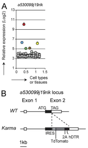
Comparative gene expression profiling of mouse immune cells identified several genes as specifically expressed by XCR1+ DCs, in particular the a530099j19rik gene (Fig. 1 A;
Robbins et al., 2008; Crozat et al., 2011; Miller et al., 2012). We used this gene for knock-in of a construct encoding both the fluorescent tandem dimer Tomato (tdTomato) and the hDTR (Fig. 1 B) to generate a mouse model, named hereaf-ter Karma, allowing specific tracking and conditional deple-tion of XCR1+ DCs in vivo.
In Karma mice, all the tdTomato-positive splenocytes fell exclusively into the XCR1+ subset of DCs as they
ex-pressed high level of CD11c and XCR1 (Fig. 2 A). More than 95% of splenic XCR1+ DCs stained positive for
tdTo-mato (Fig. S1 A and Fig. 2 B). In the dermis (Fig. S1 B) and lungs (unpublished data), tdTomato expression was the high-est in the XCR1+ subset of DCs (defined as CD24+CD103+
DCs; Fig. 2 C). In cutaneous lymph nodes (CLN; Fig. S1 C), tdTomato expression was the highest in both lymphoid tis-sue-resident and dermis-derived XCR1+ DCs, and was low
on migratory LCs (Fig. 2 D). Hence, the expression pattern of tdTomato in the Karma mouse model confirmed efficient tar-geting of all migratory and lymphoid-resident XCR1+ DCs.
We next evaluated the specificity and efficiency of XCR1+ DC conditional depletion in Karma mice. The
ad-ministration of a single dose of DT was sufficient to eliminate >95% of splenic XCR1+ DCs within 6 h without affecting
other immune cells (Fig. 2, E and F). In the spleen, the com-partment of XCR1+ DCs was emptied for at least 2 d and
fully recovered by day 4 after DT treatment (Fig. 2 F). DT ad-ministration also induced an efficient elimination of XCR1+
DCs found in the dermis and in the CLNs (unpublished data). The efficiency of XCR1+ DC depletion in DT-treated
Karma mice was functionally confirmed by two types of as-says. First, CD11c+ cells purified from the spleens of
OVA-in-jected and DT-treated Karma mice failed to cross-present OVA to naive CD8+ T cells in vitro (Fig. 2 G). Second,
DT-treated Karma mice failed to produce bioactive IL-12 upon administration of Toxoplasma gondii–soluble tachyzoïte an-tigen (STAg; Fig. 2 H), which specifically triggers IL-12 pro-duction in XCR1+ DCs through TLR11 (Yarovinsky et al.,
2005). Hence, the Karma mouse model is a robust in vivo system that allows a selective depletion of XCR1+ DCs.
XCR1+ DCs promote the expansion of mCTLs upon secondary
infections with several intracellular pathogens
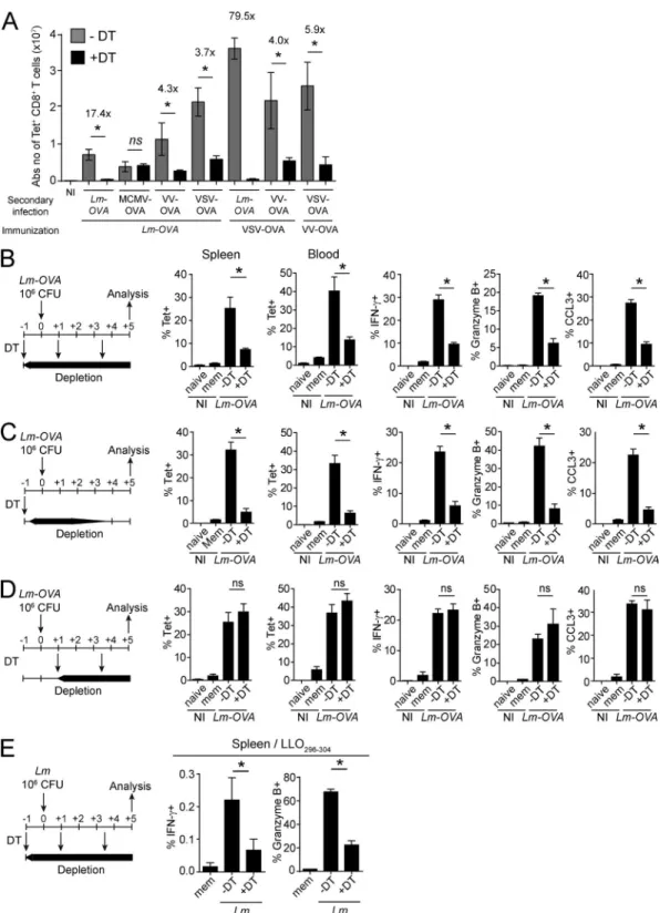
We used Karma mice to investigate whether XCR1+ DCs are
required to promote the recall of mCTLs. Upon immuniza-tion with Lm-OVA, Karma mice generated a pool of long-lived mCTLs quantitatively and qualitatively comparable to those of WT mice (unpublished data). Memory DT-treated Karma mice were then secondary challenged with different OVA-expressing recombinant microbes: the bacteria model Lm (Lm-OVA), mouse cytomegalovirus (MCMV-OVA), VV (VV-OVA), or VSV (VSV-OVA). Upon secondary infection with Lm-OVA, VV-OVA, and VSV-OVA, a sustained deple-tion of splenic XCR1+ DCs strongly decreased the
num-bers of Ag-specific mCTLs found in the spleen of Karma mice 5 d after rechallenge (Fig. 3, A and B; and Fig. S2 A). XCR1+ DCs also promoted the expansion of OVA-specific
mCTLs when VSV-OVA or VV-OVA were used as immuniz-ing agents (Fig. 3 A). Hence, the XCR1+ DC-mediated recall
response of mCTLs is not specific to primary or secondary infections by Lm, but also occurs with several other infectious agents. Because the effect of XCR1+ DC depletion was more
drastic for Lm-OVA than for VSV-OVA or VV-OVA second-ary infections, we focused on Lm-OVA infection to define the critical time frame during which XCR1+ DCs mediate
the reactivation of mCTLs. We therefore administered DT to memory Karma mice at different times around second-ary challenge. An early and transient depletion of XCR1+
DCs, starting before secondary infection and lasting for ∼1.5 d after, significantly prevented expansion and effector functions of mCTLs (Fig. 3 C). In contrast, a late and sustained de-pletion of XCR1+ DCs starting 1 d after infection had no
effect on the recall of mCTL responses (Fig. 3 D). Because the intensity of the immune response to OVA-recombinant microbes may not reflect the immune response to Ag nat-urally existing in the pathogen, we analyzed the activation of mCTLs specific of an endogenous epitope of Lm (Liste-riolysin peptide LLO296-304) in a context of an autologous
secondary infection (Fig. 3 E). We also examined responses against the nucleoprotein NP396 epitope endogenous to the
lymphocytic choriomeningitis virus (LCMV) immunizing agent upon a heterologous secondary infection with VV-NP (unpublished data). In both cases, XCR1+ DCs promoted the
recall of mCTLs specific to endogenous Ag (Fig. 3 E and not depicted). Hence, the role of XCR1+ DCs for promoting the
recall of antigen-specific mCTLs is not limited to the OVA model epitope but applies to different antigens, including en-dogenous epitopes of the immunizing or challenging infec-tious agent. We then studied how XCR1+ DCs are required
to promote mCTL recall within the first day after rechallenge using OVA as a model of Ag and OVA-expressing Lm for primary and secondary infections.
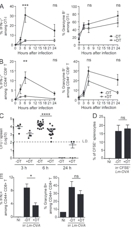
XCR1+ DCs promote the early activation of mCTLs upon
secondary infection with Listeria
As early as 8 h after Lm rechallenge, mCTLs produce IFN-γ, in an Ag-independent fashion (Berg et al., 2003; Bajénoff et al., 2010; Soudja et al., 2012). We therefore analyzed whether depletion of XCR1+ DCs influenced this first, early phase
of mCTL reactivation. To follow the behavior of Ag-specific CD8+ T cells in our experimental model, we injected low
numbers (500–1,000) of naive GFP-expressing OT-I cells before each primary infection, to mimic the preimmune frequency of individual antigen-specific CD8+ T
precur-sors present at steady state in a mouse (Obar et al., 2008). Upon secondary infections, depletion of splenic XCR1+ DCs
abrogated the early induction of IFN-γ in both OVA-spe-cific OT-I mCTLs (Fig. 4 A and Fig. S2 B) and endogenous polyclonal Ag-experienced CD44hi CD8+ T cells (CD44hi
mCTLs; Fig. 4 B and Fig. S2 B) without significantly affect-ing their expression of Granzyme B.
Upon primary infection with Lm, XCR1+ DCs convey
bacteria from the red pulp (RP) to the white pulp (WP) in the spleen, which conditions the delivery of Lm-derived Ag Figure 1. Expression analysis of the a530099j19rik gene and
gen-eration of Karma mice. (A) Microarray analysis of the expression of the
a530099j19rik gene in 96 different cell types or tissues in mouse. pDCs (green), CD11b+ (blue), and XCR1+ (red) DCs, spleen (brown), and lymph
nodes (yellow) are highlighted among all other cell types and tissues (gray). (B) Schematic representation of the Karma mouse genetic construction. An IRES-tdTomato-2A-DTR cassette was inserted downstream of the stop codon in the 3′ untranslated region of exon 2 of the a530099j19rik gene.
Figure 2. In Karma mice, all XCR1+ DCs express the tdTomato, and are specifically and efficiently depleted upon DT administration. (A)
Anal-ysis of the tdTomato expression among total splenocytes. After dead cell exclusion, tdTomato-positive cells were analyzed for lineage (CD3ε/CD19/NK1.1), CD11c, SiglecH, XCR1, and CD11b expression. The percentage of cells among the gate is shown. (top) Gating strategy using control splenocytes; (bottom) staining of Karma splenocytes. (B–D) Analysis of tdTomato expression by DCs in spleen (B), epidermis and dermis (C), and CLNs (D) of Karma mice. See Fig. S1 (A–C) for details about the gating strategy used. WT cells (dotted histogram) were included in overlays to set the tdTomato background signal for
com-to naive T cells and subsequent induction of protective CD8+
T cell responses (Neuenhahn et al., 2006; Aoshi et al., 2008; Campisi et al., 2011; Edelson et al., 2011a). Upon secondary in-fection, Lm may also use XCR1+ DCs to disseminate into the
spleen, which may strongly decrease antigen load in mice de-pleted of XCR1+ DCs and thus explain their strongly
dimin-ished mCTL responses. Bacterial loads were 6.6 times lower in XCR1+ DC-depleted animals than in controls 6 h after
infec-tion (Fig. 4 C), confirming that XCR1+ DCs promote
bacte-rial spreading in the spleen. Yet thousands of colony-forming units were readily detected in the spleen of XCR1+
DC-de-pleted mice, indicating that the amount of replicative bacteria was unlikely to be a limiting factor for the reactivation of mCTLs in the absence of XCR1+ DCs. To test this
hypoth-esis, we used CFSE-stained UV-irradiated Lm-OVA, which lack replicative capacity because of UV-induced DNA dam-ages, but which retain intact structural components preserving their immunogenicity, unlike heat-killed bacteria (Muraille et al., 2005). Although similar bacterial loads were observed in both memory DT-treated and untreated Karma mice injected with UV-irradiated Lm-OVA (Fig. 4 D), the induction of IFN-γ in polyclonal CD44hi mCTLs was significantly lower
in DT-treated Karma mice (Fig. 4 E). Therefore, independent of shuttling live bacteria, XCR1+ DCs must exert specific
immune functions, which promote mCTL responses within the first hours after secondary challenges. This prompted us to investigate the mechanisms by which XCR1+ DCs promoted
the reactivation of mCTLs early after secondary infection. XCR1+ DCs promote mCTL clustering in the RP
early after Lm infection
We first investigated whether the depletion of XCR1+ DCs
impacted the migration of mCTLs, which occurs very early after secondary Lm infection (Bajénoff et al., 2010). Under steady-state conditions, mCTLs mostly resided in the RP of the spleen (Fig. 5 A, NI). Within the first hours after second-ary infection, they transiently formed clusters around B cell follicles (Fig. 5 A, 6 h −DT) before migrating into the T cell zone (Fig. 5 A, 24 h −DT). The depletion of XCR1+ DCs
before secondary infection significantly decreased but did not completely abrogate the early clustering of mCTLs (Fig. 5, A [6 h +DT] and B) and their later migration into the T cell zone (Fig. 5, A [24 h +DT] and C).
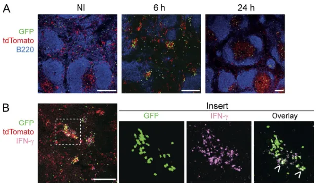
We then examined the microanatomical localization of XCR1+ DCs in the spleen upon secondary infection. At
steady-state in Karma mice, XCR1+ DCs were located in
the RP and in the T cell zone (Fig. 6 A). 6 h after rechal-lenge, XCR1+ DCs formed clear clusters together with OT-I
mCTLs around the marginal zone (Fig. 6 A). IFN- γ–pro-ducing cells, which encompassed a significant proportion of memory OT-I cells, were strictly confined within these clusters (Fig. 6 B). 24 h after infection, the proportion of tdTomato+ cells was more prominent in the T cell zone
as compared with the RP, suggesting that a proportion of XCR1+ DCs that initially resided in the RP, most
proba-bly those clustering with mCTLs at 6 h, entered the WP at 24 h (Fig. 6 A). Altogether, these results showed that the re-sponses of XCR1+ DCs during Lm-OVA secondary
infec-tion were tightly regulated in time and space and promoted mCTL clustering and IFN-γ production in the RP, at the edges of the marginal zone.
XCR1+ DCs secrete IL-12 and CXCL9, which respectively
promote the activation and mobilization of mCTLs
To gain insights into the mechanisms through which XCR1+
DCs promoted the recall of mCTLs early after secondary in-fection, we compared the expression of genes encoding can-didate inflammatory cytokines and chemokines in the spleen of DT-treated versus untreated Karma mice after Lm rechal-lenge. The expressions of Il12b and Cxcl9 genes were signifi-cantly reduced in DT-treated mice as early as 3 h after infection (unpublished data). Upon secondary infection of memory mice, XCR1+ DCs were a major source of IL-12p40/70 as
compared with CD11b+ DCs, neutrophils, and monocytes
(Fig. 7 A and Fig. S3, A and B). The blockade of IL-12 before secondary inoculation with Lm-OVA drastically impaired IFN-γ induction in OT-I (Fig. 7 B) and polyclonal CD44hi
(Fig. 7 C) mCTLs, suggesting that IL-12 provides the very first signal that initiates their reactivation. XCR1+ DCs were
also the major producers of CXCL9 6 h after secondary chal-lenge (Fig. 7 D and Fig. S3 C). CXCL9 staining colocalized strongly with clusters of XCR1+ DCs in the spleen of
mem-ory mice (Fig. 7 E). Therefore, early after secondary infection, XCR1+ DCs are the major source of IL-12 and CXCL9.
In C57BL/6J mice, CXCL9 and CXCL10 specifically bind the chemokine receptor CXCR3, which is mostly ex-pressed on Ag-experienced effector and memory T cells (Qin et al., 1998; Rabin et al., 1999). Upon Lm secondary infec-tion, CXCR3 expression was down-regulated on CD44hi
T lymphocytes, and more profoundly on the CD8+ subset
parison with Karma cells (black histogram). For the spleen, one experiment representative of at least four with three mice per group is shown. For the skin and CLNs, one representative experiment out of three with three mice per group is shown. (E and F) Specific depletion and recovery of XCR1+ DCs in Karma
mice upon DT administration. Splenocytes of DT-injected mice were analyzed by flow cytometry 24 h (E) or several days after treatment (F). The absolute numbers of the analyzed cell population are represented. In these experiments, XCR1+ DCs were gated using CD8α staining in place of XCR1. Data are shown
for one experiment representative of two independent ones, with three mice per group. (G) Antigen cross-presentation is abolished in XCR1+ DC-depleted
mice. Data are shown for one experiment representative of two with three mice per group. Data are represented as mean ± SEM. **, P < 0.01. (H) IL-12p70 induction is reduced in XCR1+ DC-depleted mice upon STAg administration. The experiment was performed with two noninjected (NI) control mice, and with
Figure 3. Splenic XCR1+ DCs promote mCTL responses upon secondary infection with various intracellular pathogens. Memory mice were used 30
d after primary infections. (A) The DT was injected 1 d before, and 1 and 3.5 d after secondary infections with Lm-OVA, MCMV-OVA, VV-OVA, or VSV-OVA. Tetramer-positive cells were measured by flow cytometry 5 d after infections. See Fig. S2 for the detailed gating strategy. Graph shows pooled data (mean ± SEM) from two independent experiments each with two to three mice per group. *, P < 0.05; ns, not significant. NI, noninfected. (B–D) Different protocols of DT treatment were applied as illustrated. DT was injected 1 d before, and 1 and 3.5 d after (B), or 1 d before (C), or 1 and 3.5 d after (D) secondary infection with 106 CFU of Lm-OVA. Splenocytes and blood leukocytes were analyzed by flow cytometry 5 d after infection. IFN-γ, GzmB, and CCL3 induction was
measured after in vitro restimulation with SIINFEKL. Data (mean ± SEM) are shown for one experiment representative of two with three to four mice per group (*, P < 0.05; ns, not significant). NI, noninfected. (E) Analysis of IFN-γ and GzmB induction in CD8+ T cells from the spleen of DT-treated memory Karma
than on the CD4+ one (unpublished data). CD44hi CD8+
T cells also migrated more efficiently than CD44hi CD4+ T
cells in response to CXCL9 in vitro (unpublished data). We therefore hypothesized that the clusters of CXCL9-produc-ing XCR1+ DCs observed in the spleen early after
second-ary infection recruited mCTLs through engagement of their CXCR3 receptors. In vivo blockade of CXCR3 before sec-ondary inoculation with Lm-OVA significantly decreased the early production of IFN-γ by mCTLs (Fig. 7, F and G) and their clustering (Fig. 7 H).
Collectively, these results indicated that, within a few hours after secondary infection, XCR1+ DCs secreted IL-12,
which triggered IFN-γ production by mCTLs, and CXCL9, which participated in the attraction of mCTLs toward XCR1+ DCs, and therefore in their activation.
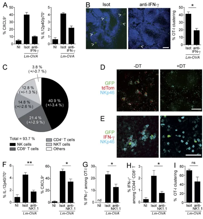
Early IFN-γ production and NK cells promote the induction of CXCL9 and IL-12 in XCR1+ DCs
One of the most potent inducers of CXCL9 expression is IFN-γ. In vivo neutralization of IFN-γ before Lm-OVA sec-ondary infection reduced IL-12 and abrogated CXCL9 pro-duction by XCR1+ DCs (Fig. 8 A), resulting in a decrease
in the clustering of mCTLs (Fig. 8 B). Therefore, IFN-γ constitutes one of the first signals initiating the activation of mCTLs upon Lm-OVA secondary infection by (a) acting in a positive-feedback loop on XCR1+ DCs to amplify IL-12
induction, and (b) inducing CXCL9 expression by XCR1+
DCs, and the redistribution of mCTLs at the edges of mar-ginal zones of the spleen.
Early after secondary infection, NK cells accounted for 40% of IFN-γ–expressing splenocytes (Fig. 8 C). At that time, IFN-γ–producing NK cells localized into cell clusters with XCR1+ DCs and mCTLs around B cell follicles (Fig. 8, D
and E, −DT). NK cell redistribution upon secondary infec-tion depended on XCR1+ DCs, as DT treatment of Karma
mice affected their activation and clustering (Fig. 8, D and E, +DT). We therefore investigated the impact of NK cell de-pletion on the responses of XCR1+ DCs and of mCTLs. NK
cell-depleted memory mice harbored an almost complete loss of IL-12 induction in XCR1+ DCs and a significant
de-crease in the expression of CXCL9 (Fig. 8 F). The induction of IFN-γ in mCTLs was also significantly decreased in these mice (Fig. 8, G and H). However, although slightly reduced, the number of clusters of mCTLs was not significantly al-tered in mice depleted of NK cells (Fig. 8 I). Thus, NK cells contact XCR1+ DCs very rapidly during secondary
infec-tion and deliver signals that amplify the ability of XCR1+
DCs to reactivate mCTLs through IL-12.
Impact of the depletion of XCR1+ DCs on early mCTL
responses during secondary challenge with VV-OVA
During secondary challenge with VV-OVA, the depletion of XCR1+ DCs significantly decreased the early clustering of
mCTLs around the marginal zone (Fig. 9 A) and NK cell IFN-γ (Fig. 9 B), similarly to, but less strongly than, what was observed upon autologous secondary challenges with Lm-OVA. Consistently with these observations, XCR1+ DCs
were a major source of IL-12 and CXCL9 a few hours after secondary infection (Fig. 9 C). These data suggest that the recall of mCTL responses to secondary infections with Lm and VV are promoted by XCR1+ DCs through overlapping
molecular and cellular mechanisms. DIS CUSSI ON
In this study, we described a new mouse model named Karma, which allows specific fluorescent tracking and effi-cient depletion of all XCR1+ DCs. The Karma mouse was
engineered using the a530099j19Rik gene to drive the ex-pression of the tdTomato-hDTR cassette. This gene codes for a predicted G protein–coupled receptor of unknown function. Its expression is highly specific to XCR1+ DCs
throughout the body. Hence, the Karma mouse model is a good alternative to the recently described Xcr1-hDTR-ve-nus mouse model (Yamazaki et al., 2013). We took advan-tage of the ability to specifically and conditionally deplete XCR1+ DCs in the Karma mouse model to investigate the
implication of these cells in mCTL responses to secondary infections by intracellular pathogens.
We established for the first time that XCR1+ DCs are
instrumental for the expansion of mCTLs upon VV-OVA, VSV-OVA, and Lm-OVA secondary challenges. The use of the CD11c-DTR mouse model has previously shown that mCTL recall responses to VSV, influenza, or Lm secondary infections were dependent on CD11c+ cells (Zammit et al.,
2005). Here, we have identified XCR1+ DCs as the major cell
subset involved in the reactivation of mCTLs upon VSV, VV, and Lm secondary infections. The amplitude of the defect pre-viously observed in CD11c+ cell-depleted animals was more
drastic than what we report here in XCR1+ DC-depleted
in-dividuals. This might be a result of differences in experimental conditions, specifically, the fact that we examined responses to endogenously induced mCTLs, whereas the study by Zammit et al. (2005) used adoptive transfer of mCTLs in naive animals before their challenge with autologous or heterologous patho-gens. Alternatively, or in addition, numerous other CD11c+
cell subsets than XCR1+ DCs are known to be depleted in the
CD11c-DTR model and some of these may also contribute to the secondary activation of mCTLs, including through pro-duction of IL-12, IL-18, IL-15, CXCL9, or CXCL10 (Sung et
mice 5 d after secondary challenge, after in vitro restimulation with LLO296-304 peptide. Lm-WT was used for the primary (3 × 10³ CFU) and secondary (106
CFU) infections. The protocol of DT administration applied was the same as in B. See Fig. S2 for the detailed gating strategy. Data (mean ± SEM) are shown for one experiment representative of two with three to four mice per group. *, P < 0.05; ns, not significant. NI, noninfected.
al., 2012; Kastenmüller et al., 2013). During Lm-OVA second-ary infection, XCR1+ DCs were required for the early
pro-duction of IFN-γ by mCTLs, which was previously reported to be independent of cognate antigen–specific interactions. We further deciphered the mechanisms by which XCR1+
DCs promote mCTL recall during secondary Lm-OVA infec-tion. As during primary infection, the first cells in the spleen that host live bacteria upon Lm secondary challenge are most likely XCR1+ DCs. In our study, we used a high dose of
Lm-OVA (106 CFU) for secondary challenge such that live
bacteria could be readily detected in the spleen of XCR1+
DC-depleted animals. This implies that other cell types hosted
the pathogen, such as monocytes that can be infected by Lm (Muraille et al., 2007; Edelson et al., 2011a). We also made use of UV-irradiated bacteria, which failed to actively replicate, to obtain the same level of infection in both XCR1+
DC-de-pleted and control animals. The loss of XCR1+ DCs led to
a significant reduction of mCTL recall under conditions of similar antigenic loads between treated and control animals. Altogether, our results support a mechanism whereby the acti-vation of mCTLs during Lm secondary infection is promoted by their involvement in a ménage à trois with XCR1+ DCs
and NK cells acting as an amplification loop for the activation of all three partners (Fig. S4).
Figure 4. XCR1+ DCs promote the early production of
IFN-γ by mCTLs. Karma mice received 500–1000 naive GFP+
OT-I cells 1 d before primary infection with Lm-OVA. 30 d later, mice were treated with DT (open circle) or left untreated (black circle), and secondary infected 1 d later with 106 CFU of
Lm-OVA. Splenocytes were harvested, incubated with brefel-din A before intracellular staining of IFN-γ and GzmB. Fig. S2B contains details of the gating strategy. (A) Analysis of the activation of OT-I GFP+ mCTLs. ***, P < 0.001. (B) Analysis
of IFN-γ and Granzyme B induction in all endogenous poly-clonal mCTLs as identified by their CD44hi phenotype. Graph
shows pooled data (mean ± SEM) from three independent experiments each with three mice per group. **, P < 0.01. (C) Bacterial load in DT-treated Karma mice after secondary in-fection with Lm. Enumeration of Lm in spleens of DT-treated and untreated memory Karma mice 3, 6, and 24 h after sec-ondary infection; L.D., limit of detection. Data are represented as mean ± SEM. ****, P < 0.0001. Data at 6 h are pooled from five independent experiments each with at least three mice per group. Data at 3 and 24 h are pooled from two indepen-dent experiments each with two to three mice per group. (D and E) Memory DT-treated Karma mice were infected with 108
CFU of UV-C–treated CFSE-stained Lm-OVA. Analysis of CFSE+
splenocytes (D) and of IFN-γ and Granzyme B induction in all polyclonal CD44hi mCTLs in spleens (E) of mice 6 h after
sec-ondary infection with UV-C–irradiated bacteria. Data (mean ± SEM) are shown for one experiment representative of two with three to four mice per group. *, P < 0.05; ns, not signifi-cant. NI, noninfected.
Besides being involved in the early activation of mCTLs
upon Lm-OVA secondary infection through IL-12 and
CXCL9 production, XCR1+ DCs also have a critical role in
the expansion of mCTLs, which occurs later during the recall
response. We demonstrated that XCR1+ DCs were critical
within the first days after infection to promote this phenom-enon. How XCR1+ DC early functions influence the late
expansion of mCTLs remains unknown. Several scenarios Figure 5. XCR1+ DCs are required for the
cluster-ing of mCTLs around the marginal zone, and promote their subsequent migration into the T cell zone.
Mem-ory Karma mice were generated as described in Fig. 3. (A) Spleen sections from untreated or DT-treated memory mice were stained with anti-B220 (dark blue) and anti- GFP (green) to define B cell follicles and OT-I mCTLs, re-spectively, and analyzed by confocal microscopy. Bars, 200 µm. Dotted lines delineate T cell zones. One representative experiment of three, each with three mice per group, is shown. (B) Statistical analysis of the frequency of OT-I mCTLs found clustered around the follicles at 6 h. Graph shows pooled data (mean ± SEM) from three independent experiments each with three mice per group. **, P < 0.01. (C) Statistical analysis of the frequency of OT-I mCTLs pres-ent in the T cell zones at 24 h. Graph shows pooled data (mean ± SEM) from two independent experiments each with three mice per group. **, P < 0.01; ns, nonsignificant.
are possible. First, XCR1+ DCs may promote mCTL
expan-sion through the secretion of soluble factors. We found that IL-12 and CXCL9 did not seem to be required (unpublished data). However, other molecules, such as the IL-15–IL-15Rα complex, which has strong proliferative properties on mem-ory cells (Richer et al., 2015), may be important. Second,
XCR1+ DCs may promote mCTL expansion through MHC
class I–restricted Ag presentation. Third, XCR1+ DCs may
promote mCTL expansion by recruiting the help of a third cell partner. Although, in our experimental setting, NK cells clustered with XCR1+ DCs and mCTLs, the expansion of
mCTLs still occurred when NK cells were depleted (un-published data). However, we cannot exclude that other cells may also form clusters with XCR1+ DCs and mCTLs and
contribute to promoting the expansion of the latter. Two re-cent studies have shown that tripartite interactions between XCR1+ DCs, CD4+ T cells, and primed CD8+ T cells are
critical during primary viral infections to promote the ex-pansion of the latter and their differentiation in protective mCTLs (Eickhoff et al., 2015; Hor et al., 2015). XCR1+ DCs
physically interact with both CD4+ and CD8+ T cells, either
sequentially when the infectious agent does not reach the lymphoid organs and remains confined in the peripheral tis-sues (Hor et al., 2015), or concomitantly, forming tripartite clusters when the pathogen directly infects cells within the lymphoid tissues (Eickhoff et al., 2015). In our experimen-tal model, upon secondary infection with Lm-OVA, CD4+ T
cell activation was attenuated when XCR1+ DCs were absent
(unpublished data). Therefore, as for primary infections, CD4+
T cells could form clusters with XCR1+ DCs and mCTLs to
deliver a help signal necessary to promote the reactivation and expansion of mCTLs.
Recently, in an experimental system similar to ours, Soudja et al. (2012) reported that Ly6C+CCR2+
mono-cytes were also required for this function in Lm-OVA sec-ondary-infected animals. Combining these two studies thus raises the question of the respective contributions of XCR1+
DCs and monocytes in the promotion of mCTL responses upon secondary infection with Lm. To deplete monocytes, they used Ccr2-hDTR mice (Soudja et al., 2012). Although particularly high on classical monocytes, CCR2 is also ex-pressed on cDCs, plasmacytoid DCs, nonclassical monocytes, activated CD4+ and CD8+ T cells, and NK cells (Mack et al.,
2001; Heng and Painter, 2008; Elpek et al., 2011; van Hel-den et al., 2012). Indeed, DT treatment of Ccr2-hDTR mice eliminates not only classical monocytes but also a proportion of plasmacytoid DCs, XCR1+ DCs, and CD11b+ DCs (Shi
et al., 2011; Dutertre et al., 2014). Hence, the alteration of mCTL responses observed in CCR2+ cell-depleted animals
could result from the combined depletion not only of classical monocytes but also of other immune cells, including a frac-tion of XCR1+ DCs. Clusters of CCR2+ cells and mCTLs are
formed in the RP of the spleen as soon as 8 h after infection (Soudja et al., 2012). Interestingly, upon Lm-OVA secondary Figure 6. RP-associated XCR1+ DCs form clusters around the marginal zone and activate mCTLs. Memory Karma mice were generated as described
in Fig. 3. Anti-GFP (green) and anti-dsRed (red) antibodies were used to detect memory OT-I cells and tdTomato+ XCR1+ DCs, respectively, on spleen sections
imaged by confocal microscopy. (A) Spleen sections were stained with anti-B220 (dark blue) to define B cell follicles. (B) IFN-γ staining (pink) of spleen sections 6 h after secondary Lm-OVA infection. Overlay of IFN-γ staining with GFP gave a white color (arrows in insert). One representative experiment out of three with three mice per group is shown. Bars, 200 µm.
Figure 7. Role of IL-12 and CXCL9 production by XCR1+ DCs in the early recruitment and activation of mCTLs after secondary infection. The
induction of IL-12p40/70 (A) and CXCL9 (D) was analyzed by flow cytometry 6 h after Lm-OVA rechallenge in several splenocyte populations (see Fig. S3 for the detailed gating of cells). IL-12p40/70 and CXCL9 were not detected in any other populations than cDC subsets, neutrophils, and monocytes. Intracellular staining was performed directly ex vivo without prior incubation with brefeldin A. Data (mean ± SEM) are shown from one experiment representative of three independent ones, each with at least three mice per group. *, P < 0.05. (B and C) IL-12 neutralization abrogates IFN-γ induction in mCTLs upon second-ary infection. Memory mice were treated with IL-12–blocking antibody (anti–IL-12) or isotype control (Isot) 18h before secondsecond-ary infection. Spleens were harvested at 6 h after secondary Lm-OVA infection. Frequency of OT-I mCTLs (B) and of polyclonal CD44hi mCTLs (C) that produce IFN-γ 6 h post-secondary
infection was measured as in Fig. 4. Data (mean ± SEM) are from one experiment with at least four mice per group. *, P < 0.05. (E) Spleen sections of mem-ory Karma mice either noninfected (NI) or infected for 6 h with Lm-OVA (6 h), were stained with anti-CXCL9 (green) and anti-dsRed (red). Merged image shows overlay of anti-CXCL9 with anti-dsRed. Bar, 200 µm. One representative experiment of two, each with two mice per group, is shown. (F–H) Memory mice were treated with CXCR3-blocking antibody (anti-CXCR3) or isotype control (Isot) 18 h before secondary infection. Spleens were harvested at 6 h after
infection, XCR1+ DCs promote the induction of the CCR2
ligands CCL2 and CCL7 (unpublished data). Therefore, XCR1+ DCs may participate in the attraction of monocytes
in their closed vicinity, favoring cell–cell contacts and pro-moting an optimal inflammatory milieu to mCTLs. Func-tionally, XCR1+ DCs and classical monocytes likely mediate
distinct and complementary functions for the reactivation of
mCTLs. XCR1+ DCs are the main producers of CXCL9
and IL-12, which promotes the local recruitment of mCTLs and their early IFN-γ production but does not affect their expression of the cytolytic molecule Granzyme B. Classical monocytes are the main producers of IL-15/IL15Rα, which promotes the acquisition of cytolytic functions by mCTLs (Soudja et al., 2012). The later expansion of mCTLs upon secondary infection might also depend on complementary functions of classical monocytes and XCR1+ DCs, for
exam-ple IL-15 transpresentation by the former and cross-presenta-tion of Lm antigens by the later.
We showed that upon secondary challenge NK cells ac-count for a large amount of early IFN-γ, which promoted
IL-12 and CXCL9 production by XCR1+ DCs and their
downstream capacity to activate mCTLs. To our knowledge, a role of NK cells in the promotion of the recall of mCTLs has never been reported. Upon primary inoculation with Lm, NK cells participate to a higher cytokine response 24 h after infection by delivering IFN-γ to XCR1+ DCs and
mono-cytes to sustain the production of IL-12 and the expression of inducible nitric oxide synthase, respectively (Kang et al., 2008; Lee et al., 2013). Interestingly, the pattern of NK cell migra-tion during secondary infecmigra-tion differs in terms of time and localization in the spleen as compared with primary infection. We found that, as soon as 6 h after secondary challenge, NK cells, mCTLs, and XCR1+ DCs form clusters. These
clus-ters localize at the edges of the marginal zone around B cell follicles in the RP through which the bacteria colonizes the spleen. The signals triggering the recruitment of NK cells into these clusters are not yet known but CXCR3 engage-ment was not necessary (unpublished data). NK cells express multiple chemokine receptors, including CCR2, CCR5, and CXCR3 (Mack et al., 2001; Bernardini et al., 2012). Hence, multiple chemokine receptors may exert redundant roles for NK cell recruitment into clusters of XCR1+ DCs upon Lm
secondary infection. How does the attraction of NK cells by XCR1+ DCs influence the recall of mCTL responses? Here,
we show that NK cells and XCR1+ DCs interact in
well-or-ganized clusters in the RP early after secondary infection by Lm and that this process amplifies IFN-γ and IL-12 produc-tion by NK cells and XCR1+ DCs, respectively, promoting
the downstream activation of mCTLs. Several reports focused on primary immune responses have demonstrated that the effectiveness of NK cell activation influences CD8+ T cell
responses such as occurring during acute infections with mouse cytomegalovirus (Robbins et al., 2007; Mitrović et al., 2012), during chronic infections with LCMV (Whitmire et al., 2008), or in response to tumor engraftments (Adam et al., 2005). Altogether, these studies suggest that early interactions between DCs and NK cells contribute to orchestrate adaptive immunity against infections or tumors (Walzer et al., 2005), not only during acute primary immune responses but also in chronic infections or upon secondary challenges, by mod-ulating the kinetic and intensity of CD4+ and CD8+ T cell
responses depending on nature of the threat faced by the host. CXCL9/CXCR3 signaling is implicated in the redis-tribution of central memory T cells in interfollicular area and subcapsular regions in LN near pathogen entry site upon viral infections (Sung et al., 2012; Kastenmüller et al., 2013). In these studies, CXCL9 was secreted by macrophages, mono-cytes, and CD11c+ DCs, which reside in the outermost
areas in LN, to be presented on stromal cells, and to pro-vide a guidance signal allowing a zone to zone relocalization of central memory T cells. Our study extends this concept to the immune response against another type of pathogen, an intracellular bacterium, and in another organ, the spleen, by demonstrating that CXCR3 signaling contributes to the microanatomical relocation of splenic RP mCTLs around
CXCL9-producing XCR1+ DCs. Together with CXCL9,
CXCL10 and CXCL11 are also high-affinity ligands for CXCR3. CXCL11 is not functional in C57BL/6J mice as a result of a single-point mutation within the coding se-quence resulting in a frame shift (Carter et al., 2007). CXCL9 and CXCL10 expressions, which are strongly induced in spleen upon Lm-OVA secondary infection, were decreased in XCR1+ DC-depleted mice (Fig. 7 D and not depicted).
Therefore, CXCL9 and CXCL10 might play redundant roles in the attraction of mCTLs in our experimental settings. Al-together, these results strongly suggest that the engagement of CXCR3 on mCTLs by CXCL9 and CXCL10 produced by myeloid cells upon sensing of danger signals may be a general and critical mechanism orchestrating in time and space the recall of adaptive cellular immunity against secondary infec-tions by intracellular pathogens or against tumor resurgence.
In conclusion, by using the Karma mouse model, we discovered that XCR1+ DCs promote the reactivation of
mCTLs upon Lm, VSV, and VV secondary infections. We de-fined some of the underlying cellular and molecular mech-anisms leading to the early activation of mCTLs upon Lm
secondary Lm-OVA infection, cut in equal halves and processed either for flow cytometry or confocal imaging. Frequency of OT-I mCTLs (F) and of polyclonal CD44hi mCTLs (G) that produce IFN-γ 6 h post-secondary infection was measured as in Fig. 3. (H) Spleen sections were stained with anti-B220 (dark blue)
and anti-GFP (green), and analyzed by confocal microscopy. Bar, 500 µm. Arrows show clusters. One representative experiment of three, each with at least three mice per group, is shown. Graph represents percentage of OT-I mCTLs that form clusters in anti–CXCR3-treated memory mice as mathematically determined. Data (mean ± SEM) are pooled from two independent experiments, each including at least three mice per group. *, P < 0.05; **, P < 0.01.
Figure 8. The early IFN-γ production participates in the clustering of mCTLs after secondary infection. IFN-γ–blocking antibodies (anti–IFN-γ) or
isotype control (Isot) were injected to memory mice 18 h before secondary infection. Spleens were harvested at 6 h after, cut in equal halves, and processed either for flow cytometry or confocal imaging. (A) Frequency of IL-12p40/70 and CXCL9-producing XCR1+ DCs in spleens. (B) Spleen sections were stained
for GFP (green) and B220 (dark blue). Bar, 200 µm. Arrows show clusters. Graph represents percentage of OT-I mCTLs that form clusters in anti–IFN-γ-treated memory mice as mathematically determined. Data (mean ± SEM) are shown for one experiment representative of two independent ones, each with at least three mice per group. *, P < 0.05; ns, nonsignificant. (C) Spleens of memory mice were harvested 6 h after secondary infection. Circle diagram encompasses all cells that stained positive for IFN-γ. One representative experiment out of four with three mice per group is shown. (D and E) Spleens from memory mice were fixed 6 h after the infection, and sections were stained with anti-GFP (green), anti-dsRed (red), and anti-NKp46 (cyan; bar, 200 µm; D), or with anti-GFP (green), anti–IFN-γ (red) and anti-NKp46 (cyan; bar, 50 µm; E). One representative experiment out of two, each with three mice per group, is shown. (F-I) Impact of NK cell depletion. (F) Frequency of IL-12p40/70 and CXCL9-producing XCR1+ DCs in spleens 6 h after Lm-OVA secondary infection.
Data (mean ± SEM) are pooled from two independent experiments, each with at least three mice per group. *, P < 0.05; **, P < 0.01. (G and H) Frequency of OT-I mCTLs (G) and of polyclonal CD44hi mCTLs (H) that produce IFN-γ 6 h post-secondary infection was measured as in Fig. 3. One experiment
representa-tive of three independent ones, each with five mice per group is shown. Data are represented as mean ± SEM. *, P < 0.05. (I) Statistical analysis of clustered OT-I mCTLs. Data (mean ± SEM) are pooled from two independent experiments, each with at least three mice per group.
infection. This study revealed that, besides their classical func-tion as professional antigen cross-presenting cells critical for the priming of naive CD8+ T cells against many
intracellu-lar pathogens and against tumors, XCR1+ DCs are also
en-dowed with immunoregulatory functions, such as IL-12 and CXCL9 production, which contribute to orchestrate in time and space the reactivation of mCTLs.
MAT ERIALS AND MET HODS
Ethics statement regarding care and use of animals for ex-perimentation. The animal care and use protocols (authoriza-tion ID no. 11–09/09/2011) were designed in accordance with national and international laws for laboratory animal welfare and experimentation (EEC Council Directive 2010/63/EU, September 2010), and approved by the Mar-seille Ethical Committee for Animal Experimentation (regis-tered by the Comité National de Réflexion Ethique sur l’Expérimentation Animale under no. 14).
Mice and in vivo treatments. The Karma mice
(a530099j19rik-tm1Ciphe) were made according to a standard gene targeting
ap-proach in C57BL/6N-derived ES cells. They were constructed by inserting the IRES-tdTomato-2A-hDTR cassette into the
3′-UTR of a530099j19rik gene. Karma mice were outcrossed for three generations with WT C57BL/6J mice, and Karma littermates were used as DT-untreated controls. For neutraliz-ing antibody experiments, WT C57BL/6J mice were pur-chased from Charles River Laboratories. TgTcraTcrb1100Mjb;
Rag1−/−; Rag2−/− (OT-I) mice (gift from A.-M.
Schmidt-Ver-hulst, Centre d’Immunologie de Marseille-Luminy [CIML], Marseille, France) were bred with C57BL/6 ubiquitinC-GFP mice (C57BL/6-Tg(UBC-GFP)30Scha/J; gift from Marc Bajénoff, CIML, Marseille, France) to obtain TgTcraTcrb1100Mjb;
Rag2−/−; Ubc-GFP+/+ mice, from which we routinely
iso-lated GFP-expressing OT-I cells. For complete XCR1+ DC
depletion, a dose of 32 ng/g of body weight of DT (Merck) was administrated at least 6 h before the experiment. For sus-tained depletion, mice were first injected with 32 ng/g of DT, then received one injection of 16 ng/g every 60 h. This was efficient to deplete XCR1+ DCs for at least 10 d. DT was
diluted in cold endotoxin free PBS. For in vivo antibody-de-pendent neutralization or antibody-mediated cell depletion, Karma memory mice were injected i.p. with purified anti-bodies or their corresponding isotypes (anti-NK1.1, 200 µg; anti-CXCR3, anti–IL-12, and anti–IFN-γ, 500 µg) 1 d before secondary infection. Antibodies are listed in Table S1.
Figure 9. XCR1+ DCs are the major
pro-ducers of IL-12 and CXCL9, and promote mCTL clustering upon secondary infection with VV-OVA. Karma mice received 500–
1,000 naive GFP+ OT-I cells 1 d before primary
infection with Lm-OVA. 30 d later, mice were treated with DT or left untreated, and sec-ondary infected with VV-OVA. Spleens were harvested 6 h after secondary challenge. (A) Spleen sections from untreated or DT-treated memory Karma mice were stained with an-ti-B220 (dark blue) and anti-GFP (green), and analyzed by confocal microscopy. Bar, 200 µm. Statistical analysis of the frequency of OT-I mCTLs found clustered around the follicles. Graph shows data (mean ± SEM) from one rep-resentative of two independent experiments each with at least three mice per group. *, P < 0.05. (B) Analysis of IFN-γ induction in NK cells 6 h after VV-OVA secondary challenge. Spleno-cytes were harvested, incubated with brefeldin A before intracellular staining of IFN-γ. Data (mean ± SEM) are shown from one represen-tative of two independent experiments, each with at least three mice per group. *, P < 0.05. (C) The induction of IL-12p40/70 and CXCL9 was analyzed by flow cytometry in several splenocyte populations (see Fig. S3 for the detailed gating of cells). Intracellular staining was performed directly ex vivo without prior incubation with brefeldin A. Data (mean ± SEM) shown are pooled from two indepen-dent experiments, each with at least three mice per group. **, P < 0.01; ***, P < 0.001.
Antigen cross-presentation assay and cytokine response to STAg. For cross-presentation assay, Karma mice received a sin-gle injection of DT or PBS (control) 1 d before i.v. injection of 2.5 mg of soluble OVA (Sigma-Aldrich). Splenic CD11c+
cells from Karma mice were enriched to >95% purity using CD11c microbeads (Miltenyi Biotec). Splenic OT-I cells were purified with Dynal mouse CD8 cell negative isolation kit (Invitrogen), and labeled with CFSE. CD11c+ and OT-I
cells were co-cultured for 3 d at different ratio in complete medium supplemented with IL-2. OT-I cell proliferation rep-resented by CFSE dilution was assessed by flow cytometry, and absolute numbers of OT-I cells were determined using Flow-Count Fluorospheres (Beckman Coulter).
For cytokine response to STAg, Karma mice received a single injection of DT or PBS (control) 1d before i.v. injec-tion of 6 µg of STAg (gift from G.S. Yap, Rutgers New Jersey Medical School, Newark, NJ). Sera were collected 12 h after to detect systemic IL-12p70 induction by ELI SA (eBioscience). Microbes. Unless stated otherwise in the figure legend, mice were immunized with the following OVA-expressing recom-binant microbes: 5 × 10³ colony-forming-units (CFU) of
Listeria-OVA (Lm-OVA), 105 PFU of Vaccinia-OVA
(VV-OVA) or 2 × 10⁴ PFU of VSV-OVA (from T. Lawrence,
CIML, Marseille, France). Memory mice were secondary challenged with 106 CFU of Lm-OVA, 2 × 10⁶ PFU of
VV-OVA or of VSV-VV-OVA, and 3 × 10⁴ PFU of MCMV-OVA
(from E. Vivier, CIML, Marseille, France). Splenocytes were analyzed by flow cytometry 5 d later.
Lm infection and quantification of bacterial titers. The 10403s strain, either WT Lm or Lm-OVA, was obtained from G. Lauvau (Albert Einstein College of Medicine, New York, NY). Frozen aliquots of Lm were grown in brain-heart infu-sion (BHI) medium to a logarithmic phase (OD600 = 0.05– 0.17) and diluted in PBS before i.v. inoculation. After each infection, inocula were routinely plated on BHI agar plates to confirm their titers. For UV-C irradiation of Lm-OVA, fresh bacterium culture was washed twice in PBS. Suspen-sion of 109–1010 bacteria/ml in PBS was exposed to an UVC
lamp (254 nm; 5 J/cm2) before being stained for 10 min at
37°C with 50 µM of CFSE. Lack of growth of UV-C–inacti-vated Lm-OVA was confirmed by culturing 2 × 10⁹ bacteria
for 48 h at 37°C on BHI agar. Mice were inoculated with freshly prepared 108 CFU of UV-C–irradiated CFSE-stained
Lm-OVA. To measure bacterial titers, spleens were dissociated on nylon mesh in 0.2% NP-40 (Igepal CA630; Sigma-Al-drich). Serial dilutions were performed in the same buffer and were plated onto BHI agar plates. CFU numbers were counted at least 24 h later.
Spleen preparation and flow cytometry analysis. Cell suspen-sions from spleens and CLNs were prepared and stained as described previously (Crozat et al., 2011). The antibodies used for flow cytometry are listed in the Table S1. Stained cell
acquisition was performed on a FAC SCanto II, or on a LSR-II upgraded with a 561nm laser when detection of td-Tomato expression was required.
Analysis of memory CD8+ T cell responses. 500–1,000 naive
GFP-expressing OT-I cells were i.v. transferred in Karma mice 1 d before Lm primary infection. 30 d later, DT or neutralizing antibodies was administrated to the mice be-fore secondary infection with Lm-OVA. When spleens and blood were harvested 5 d after secondary infections, cells were stained with H2-Kb SII NFEKL tetramers (Beckman
Coulter) to identify OVA-specific CD8+ T cells. Splenocytes
were also incubated for 4 h with 1 µM of SII NFEKL and Brefeldin A to detect intracellular IFN-γ, CCL3, and Gran-zyme B. When spleens were harvested 6 h after secondary infection, splenocytes were incubated for 3 h with Brefeldin A in complete RPMI medium to detect spontaneous IFN-γ production in lymphocytes.
Immunofluorescence. Spleens were harvested, cut in 5-mm pieces, and fixed for at least 1 h in Antigenfix (DiaPath), then washed in phosphate buffer (PB1X: 0.02 M NaH2PO4 and
0.08 M Na2HPO4) for 1 h, dehydrated in 30% sucrose
over-night at 4°C, and embedded in OCT freezing media (Sakura Fineteck). 16-µm frozen sections of spleens were blocked in PB1X containing 0.2% saponin, 2% BSA, and 2% 2.4G2 supernatant, and stained in PB1X, 0.2% saponin. When the staining required, further blocking steps with normal goat or rabbit sera were added. Stained sections were mounted in ProLong Gold antifade reagent (Invitrogen), and acquired on a LSM780 confocal microscope (Carl Zeiss). TdTomato expression was amplified by using an anti-Red Fluorescent Protein (RFP) antibody (Rockland). Antibodies used in im-munofluorescence are detailed in Table S1.
Quantification of memory OT-I cell clustering and migration into the T cell zone. OT-I cell clustering and migration into T cell zones were determined computationally using ImageJ and R graphical user interface (GUI) software. We used mo-saic images representing a complete section of spleen to per-form this analysis. For OT-I cell clustering: a macro program developed for Image J allowed us to spot individual GFP+
OT-I cells present on a frozen spleen section. When several GFP+ OT-I cells were too close to be detected as individual
cells by the macro program due to strong signal intensity, we manually separated them using the pencil tool from ImageJ. Coordinates of each distinct GFP+ OT-I cell were retrieved.
OT-I cell locations were then represented graphically with R software after installing the Fixed Point Cluster (FPC) analysis package. The DBS CAN algorithm was set to define a true cluster as a group of three or more proximate cells. Percent of clustered cells were then calculated according to the total number of cells present on the whole section. For OT-I local-ization in the T cell zone: a macro program developed for Image J allowed us to enumerate all individual GFP+ OT-I
cells, as well as GFP+ OT-I cells found in T cell zones. As
pre-viously, merged GFP+ OT-I cells were manually separated. T
cell regions were manually delimited according to B220 staining of B cell follicles and nuclear density.
Microarray data. The data were downloaded from the BioGPS public database (http ://biogps .gnf .org; Gene Expression Omnibus dataset number GSE10246).
Statistical analyses. Statistical analyses were performed using nonparametric Mann-Whitney tests in all experiments. Online supplemental material. Table S1 lists the antibodies used in this study. Fig. S1 shows the gating strategy used to identify DC subsets in spleen, skin, and CLNs. Fig. S2 shows the gating strategy used to analyze tetramer-positive mCTLs and the early activation of mCTLs in secondary infected animals. Fig. S3 shows IL-12 and CXCL9 expression analysis on DCs, neutrophils and monocytes from secondary infected animals. Fig. S4 shows a schematic model of the recall of
mCTLs by XCR1+ DCs during secondary Lm infection.
Online supplemental material is available at http ://www .jem .org /cgi /content /full /jem .20142350 /DC1.
ACKNOWLEDgMENTS
We thank Bruno Estebe, Frédéric Fiore, and Bernard Malissen (Centre d'Immuno-phénomique [CIP HE], UM2 Aix-Marseille Université, Institut National de la Santé et de la Recherche Médicale US012, Centre National de la Recherche Scientifique UMS3367, Marseille, France) for generating the Karma mice; Marilyn Boyron, and the
cytometry and ImagImm core facilities (CIML, UMR7280, France) for technical assis-tance; Mathieu Fallet (CIML, UMR7280, France) for programming macros to quantify memory OT-I cell clustering and migration into the T cell zone; and Toby Lawrence and Elena Tomasello for critical reading of the manuscript. We acknowledge France-BioImaging infrastructure supported by the Agence Nationale de la Recher-che (ANR-10-INSB-04-01, call “Investissements d'Avenir”).
This work was supported by institutional funding from CNRS and Inserm, by the Innate Immunocytes in Health and Disease (I2HD) collaborative project between CIML, AVI ESAN, and SAN OFI, and by grants from Association pour la recherche sur le cancer (ARC), from Fondation pour la Recherche Médicale (label “Equipe FRM 2011”, project number DEQ20110421284) and from the European Research Council under the European Community’s Seventh Framework Program (FP7/2007–2013 grant agreement number 281225). Y.O. Alexandre was supported by doctoral fellowships from the French Ministère de l’Enseignement Supérieur et de la Recherche, and from ARC. S. Ghilas was laureate from a “University President” Excellence Ph.D. Fellowship from Aix Marseille Université and from ARC. C. Sanchez was supported by the I2HD CIML-SAN OFI project.
The authors declare no competing financial interests.
Submitted: 17 December 2014 Accepted: 20 November 2015
REFERENCES
Adam, C., S. King, T. Allgeier, H. Braumüller, C. Lüking, J. Mysliwietz, A. Kriegeskorte, D.H. Busch, M. Röcken, and R. Mocikat. 2005. DC-NK cell cross talk as a novel CD4+ T-cell-independent pathway for antitumor CTL induction. Blood. 106:338–344. http ://dx .doi .org /10 .1182 /blood -2004 -09 -3775
Aoshi, T., B.H. Zinselmeyer, V. Konjufca, J.N. Lynch, X. Zhang, Y. Koide, and M.J. Miller. 2008. Bacterial entry to the splenic white pulp initiates
antigen presentation to CD8+ T cells. Immunity. 29:476–486. http ://dx .doi .org /10 .1016 /j .immuni .2008 .06 .013
Bajénoff, M., E. Narni-Mancinelli, F. Brau, and G. Lauvau. 2010. Visualizing early splenic memory CD8+ T cells reactivation against intracellular bacteria in the mouse. PLoS One. 5:e11524. http ://dx .doi .org /10 .1371 /journal .pone .0011524
Bar-On, L., and S. Jung. 2010a. Defining dendritic cells by conditional and constitutive cell ablation. Immunol. Rev. 234:76–89. http ://dx .doi .org /10 .1111 /j .0105 -2896 .2009 .00875 .x
Bar-On, L., and S. Jung. 2010b. Defining in vivo dendritic cell functions using CD11c-DTR transgenic mice. Methods Mol. Biol. 595:429–442. http ://dx .doi .org /10 .1007 /978 -1 -60761 -421 -0 _28
Belz, G.T., S. Bedoui, F. Kupresanin, F.R. Carbone, and W.R. Heath. 2007. Minimal activation of memory CD8+ T cell by tissue-derived dendritic cells favors the stimulation of naive CD8+ T cells. Nat. Immunol. 8:1060– 1066. http ://dx .doi .org /10 .1038 /ni1505
Berg, R.E., E. Crossley, S. Murray, and J. Forman. 2003. Memory CD8+ T cells provide innate immune protection against Listeria monocytogenes in the absence of cognate antigen. J. Exp. Med. 198:1583–1593. http :// dx .doi .org /10 .1084 /jem .20031051
Bernardini, G., A. Gismondi, and A. Santoni. 2012. Chemokines and NK cells: regulators of development, trafficking and functions. Immunol. Lett. 145:39–46. http ://dx .doi .org /10 .1016 /j .imlet .2012 .04 .014
Broz, M.L., M. Binnewies, B. Boldajipour, A.E. Nelson, J.L. Pollack, D.J. Erle, A. Barczak, M.D. Rosenblum, A. Daud, D.L. Barber, et al. 2014. Dissecting the tumor myeloid compartment reveals rare activating antigen-presenting cells critical for T cell immunity. Cancer Cell. 26:638– 652. http ://dx .doi .org /10 .1016 /j .ccell .2014 .09 .007
Campisi, L., S.M. Soudja, J. Cazareth, D. Bassand, A. Lazzari, F. Brau, E. Narni-Mancinelli, N. Glaichenhaus, F. Geissmann, and G. Lauvau. 2011. Splenic CD8α+ dendritic cells undergo rapid programming by cytosolic bacteria and inflammation to induce protective CD8+ T-cell memory. Eur. J.
Immunol. 41:1594–1605. http ://dx .doi .org /10 .1002 /eji .201041036
Carter, S.L., M. Müller, P.M. Manders, and I.L. Campbell. 2007. Induction of the genes for Cxcl9 and Cxcl10 is dependent on IFN-gamma but shows differential cellular expression in experimental autoimmune encephalomyelitis and by astrocytes and microglia in vitro. Glia. 55:1728–1739. http ://dx .doi .org /10 .1002 /glia .20587
Croft, M., L.M. Bradley, and S.L. Swain. 1994. Naive versus memory CD4 T cell response to antigen. Memory cells are less dependent on accessory cell costimulation and can respond to many antigen-presenting cell types including resting B cells. J. Immunol. 152:2675–2685.
Crozat, K., S. Tamoutounour, T.P. Vu Manh, E. Fossum, H. Luche, L. Ardouin, M. Guilliams, H. Azukizawa, B. Bogen, B. Malissen, et al. 2011. Cutting edge: expression of XCR1 defines mouse lymphoid-tissue resident and migratory dendritic cells of the CD8α+ type. J. Immunol. 187:4411–4415. http ://dx .doi .org /10 .4049 /jimmunol .1101717
Curtsinger, J.M., D.C. Lins, and M.F. Mescher. 1998. CD8+ memory T cells (CD44high, Ly-6C+) are more sensitive than naive cells to (CD44low, Ly-6C-) to TCR/CD8 signaling in response to antigen. J. Immunol. 160:3236–3243.
Dengler, T.J., and J.S. Pober. 2000. Human vascular endothelial cells stimulate memory but not naive CD8+ T cells to differentiate into CTL retaining an early activation phenotype. J. Immunol. 164:5146–5155. http ://dx .doi .org /10 .4049 /jimmunol .164 .10 .5146
Dutertre, C.A., L.F. Wang, and F. Ginhoux. 2014. Aligning bona fide dendritic cell populations across species. Cell. Immunol. 291:3–10. http ://dx .doi .org /10 .1016 /j .cellimm .2014 .08 .006
Edelson, B.T., T.R. Bradstreet, K. Hildner, J.A. Carrero, K.E. Frederick, W. Kc, R. Belizaire, T. Aoshi, R.D. Schreiber, M.J. Miller, et al. 2011a. CD8α+ dendritic cells are an obligate cellular entry point for productive infection by Listeria monocytogenes. Immunity. 35:236–248. http ://dx .doi .org /10 .1016 /j .immuni .2011 .06 .012