HAL Id: hal-00589430
https://hal.archives-ouvertes.fr/hal-00589430
Submitted on 29 Apr 2011
HAL is a multi-disciplinary open access
archive for the deposit and dissemination of
sci-entific research documents, whether they are
pub-lished or not. The documents may come from
teaching and research institutions in France or
abroad, or from public or private research centers.
L’archive ouverte pluridisciplinaire HAL, est
destinée au dépôt et à la diffusion de documents
scientifiques de niveau recherche, publiés ou non,
émanant des établissements d’enseignement et de
recherche français ou étrangers, des laboratoires
publics ou privés.
Circulating procoagulant microparticles in cancer
patients
Johannes Thaler, Cihan Ay, Harald Weinstabl, Daniela Dunkler, Ralph
Simanek, Rainer Vormittag, Jean-Marie Freyssinet, Christoph Zielinski, Ingrid
Pabinger
To cite this version:
Johannes Thaler, Cihan Ay, Harald Weinstabl, Daniela Dunkler, Ralph Simanek, et al.. Circulating
procoagulant microparticles in cancer patients. Annals of Hematology, Springer Verlag, 2010, 90 (4),
pp.447-453. �10.1007/s00277-010-1111-1�. �hal-00589430�
Circulating procoagulant microparticles in cancer
patients
Johannes Thaler
1, Cihan Ay
1, Harald Weinstabl
1, Daniela Dunkler
2, Ralph
Simanek
1, Rainer Vormittag
1, Jean-Marie Freyssinet
4, Christoph Zielinski
3, Ingrid
Pabinger
11
Clinical Division of Haematology and Haemostaseology, Department of
Medicine I,
2
Center for Medical Statistics, Informatics and Intelligent Systems, Section for
Clinical Biometrics,
3
Clinical Division of Oncology, Department of Medicine I,
Medical University of Vienna, Austria
4
U. 770 INSERM Hôpital de Bicêtre; Université Paris-Sud, Le Kremlin-Bicêtre;
Université Louis Pasteur, Faculté de Médecine, Institut d'Hématologie &
Immunologie, Strasbourg, France
Correspondence: Univ.Prof. Dr. Ingrid Pabinger
Clinical Division of Haematology and Haemostaseology
Department of Medicine I, Medical University of Vienna
Waehringer Guertel 18-20, A-1090 Vienna, Austria
Phone number: +43 1 40400 4448Fax number: +43 1 40400 4030
E-mail: [email protected]
Abstract
Accumulating evidence indicates that microparticles (MPs) are important mediators of the interaction between cancer and the hemostatic system. We conducted a large prospective cohort study to determine whether the number of circulating procoagulant MPs is elevated in cancer patients and whether the elevated MP levels are predictive of occurrence of venous
thrombembolism (VTE). We analyzed plasma samples of 728 cancer patients from the ongoing prospective observational Vienna Cancer and Thrombosis Study (CATS). Study endpoint was the occurrence of symptomatic VTE. Sixty-five age- and sex-matched healthy controls were recruited for definingthe cut-off point for elevated MPs (4.62 nanomolar phosphatidylserine nM PS ), which was set at the 95th percentile of MP levels in healthy controls. The measurement of MPs was performed after capture onto immobilized annexin-V, and determination of their procoagulant activity was quantified with a prothrombinase assay. During a median observation period of 710 days 53 patients developed VTE. MP levels (nM PS) were significantly higher in cancer patients than in healthy controls (median 25th-75thpercentile : 3.95 1.74-7.96 vs. 1.19 0.81-1.67 , p<0.001). Multivariate analysis including age, sex, surgery, chemo- and radiotherapy showed no statistically significant association of the hazard ratio of elevated MPs with VTE (0.95 [95% CI: 0.55-1.64], p=0.856). In conclusion, MP levels were elevated in cancer patients compared to healthy individuals in this study. However, elevated MP levels were not predictive of VTE.
Keywords: microparticles, cancer, venous thromboembolism, prothrombinase
assay
Abbreviations: CATS, Cancer and Thrombosis Study; CI, confidence
interval; MPs, microparticles; HR, hazard ratio; PFP, platelet-free plasma; nM PS, nanomolar
phosphatidylserine equivalent; PS, phosphatidylserine; PSGL-1, P-selectin glycoprotein ligand; TF, tissue factor; VTE, venous thromboembolism;
Introduction
Circulating procoagulant microparticles (MPs) are phosphatidylserine (PS)-rich, small vesicles with a diameter of 0.1 m to 1 m, which are emitted from cell membranes into the blood when cells are activated or undergo apoptosis. The majority of MPs in plasma are derived from platelets [1], erythrocytes[2], endothelial cells [3], lymphocytes[4], monocytes[5] and smooth muscle cells [6]. An important role of MPs in the hemostatic system has been proposed, because MPs provide a negatively charged surface for the aggregation and subsequent activation of coagulation factors, and promote coagulation via the expression of coagulation proteins and adhesion molecules on their surface [7-10].Experimental studies have demonstrated that MPs are involved in the pathophysiology of thrombosis [11]. It has been shown that a subgroup of MPs are guided to a developing thrombus through the interaction of P-selectin glycoprotein ligand-1 (PSGL-1), which is present on MPs, and P-selectin, which is expressed on activated platelets and endothelial cells. The accumulation of MP-bound tissue factor (TF) further supports thrombus growth [12]. Clinical studies have reported elevated MP levels in different thrombotic disorders [11]. Yet, results are conflicting regarding the clinical relevance of MPs in venous thromboembolism (VTE). While highly elevated MP levels were measured shortly after an acute VTE event in cancer and non-cancer patients [13,14] MP levels were not elevated 3 months after a VTE event in a high-risk population of non-cancer patients with a history of recurrent VTE [15]. Although cancer patients have a 4- to 7-fold increased risk of VTE and VTE is the second leading cause of death in this population, after cancer itself [16-18] large prospective clinical studies investigating the predictive role of MPs in cancer-associated VTE are still lacking.
As the pathophysiology of cancer-associated VTE is not fully understood and laboratory
parameters to predict cancer-associated VTE are scarce, attention in recent research activities has been focused on MPs as mediators of the interaction between cancer and the hemostatic system. Accumulating evidence indicates that MPs contribute to the development of VTE in cancer patients [7]. High levels of circulating MPs were found in breast cancer [19], gastric cancer [20], and pancreatic cancer patients [13] compared to healthy individuals, and TF-positive MPs were postulated as candidate biomarkers for the development of cancer-associated VTE
[21,22].Therefore, the aim of this prospective observational cohort study was to determine, whether baseline levels of MPs are elevated in cancer patients with different tumour types compared to sex- and age-matched healthy individuals and whether elevated MP levels have a predictive role in the occurrence of VTE in prospectively followed cancer patients.
Design and Methods
Study population
The study protocol was approved by our local Ethics Committee. Informed consent was signed before patients and healthy individuals were included in this study. This study was designed as a case-control study as part of the ongoing observational Vienna Cancer and Thrombosis Study (CATS), which was initiated in 2003. First data of this study were published in 2008 and 2009 [23-25]. Plasma samples of 728 cancer patients with newly diagnosed cancer or progression of disease after complete or partial remission, including malignancies of the brain, breast, lung, stomach, colon, pancreas, kidney, prostate, hematological malignancies (multiple myeloma, high and low grade lymphoma) or other sites (mainly of the gynecologic system and sarcoma) were analyzed. Main patients’ characteristics are listed in table 1. At study inclusion, patients underwent a structured interview on their medical history and data on the site of tumor, histology and tumor-stage were documented. Patients were followed prospectively and information on the course of disease and on the occurrence of VTE was collected for every patient at three-month intervals. Patients were instructed to contact the Division of Haematology and Haemostaseology (Vienna General Hospital), if they perceived symptoms indicative of VTE (deep vein thrombosis or pulmonary embolism), stroke, myocardial infarction, or peripheral arterial thrombosis. In case of death, data on the time and the cause of death including autopsy protocols were collected. Each patient was followed over a two-year period or until the occurrence of VTE, death or loss of follow-up.
We recruited 65 age- and sex-matched healthy control subjects without a history of arterial or venous thrombosis and cancer from the same geographic region and ethnic backgrounds as our patients. Based on their MP levels we defined the cut-off point for elevated MPs, which was set at the 95th percentile of the MP levels in our healthy control group.
Blood sampling and laboratory methods
Venous blood samples were drawn into citrate vacuum tubes (Vacuette, Greiner-Bio One, Kremsmuenster, Austria) by atraumatic and sterile antecubital venepuncture on the day of study entry. The citrated blood was centrifuged (Rotanta/TRC, Hettich, Tuttlingen, Germany) at 1500 g for 15 minutes to get platelet-poor plasma. Platelet-free plasma (PFP) was obtained by a second centrifugation step (Eppendorf, Hamburg, Germany) at 13400 g for two minutes. The
centrifugation of each sample was performed within 1 hour after blood sampling and the freezing of each sample within 1 hour after centrifugation. Plasma aliquots were stored at minus 80°C until assays were performed in series.
MPs were captured by immobilized annexin V as previously described [26]. In brief, biotinylated
streptavidin-coated microtitration plates (Roche Diagnostics, Mannheim, Germany). The plates
were washed 3 times with TBS-Ca2+ (50 mM Tris buffer, pH 7.5, containing 120 mM NaCl, 2.7
mM KCl, 1 mM CaCl2). 300 l aliquots of PFP were thawed and thrombin inhibitor (30 M
PPACK; Merck, Darmstadt, Germany) and factor Xa inhibitor (30 M 1.5-Dns-GGACK; Merck, Darmstadt, Germany) were added. We then incubated 100 l PFP/well for 30 minutes at 37 C. Each sample was measured in duplicate. After 4 washing steps, the anionic phospholipid content was determined by a prothrombinase assay. In this assay immobilized MPs were incubated in a final volume of 150 l with factor Va (250 pM; American Diagnostica, Stamford, USA), Xa (9.3 pM; Hyphen BioMed, Andresy, France), prothrombin (0.7 M; Hyphen BioMed, Andresy,
France) and CaCl2 (2.3 mM) for 15 minutes at 37 C. Then Chromozym TH (1.5 mM; Roche
Diagnostics, Mannheim, Germany), a chromogenic substrate for thrombin, was added and the solution was incubated for 4 minutes. The chromogenic substrate was cleaved by thrombin. Change of colour was measured photometrically. Linear absorbance changes were recorded at 405 nm using a microtitration plate reader (MR 7000, Dynatech, Guernsey, Great Britain).
In the method first described by Aupeix et al [26] a kinetic software was used for data analysis. To make this method applicable on a standard laboratory photometer, the colour reaction was stopped after 4 minutes by the addition of EDTA (5 mM). As the colour development reaction followed a Michaelis Menten kinetic, we compared our measurements to standard curves by means of non-linear regression analysis. Results were expressed as nanomolar phosphatidylserine equivalent (nM PS) by reference to a standard curve that we had constructed by using phospholipids of defined composition (33% PS and 67% phosphatidylcholine, PC) as described by Pigault et al [27]. Phospholipids were quantified by the Steward-assay [28]. When MPs were present, the reaction was significantly accelerated because of the high amount of PS present on MPs.
Statistical analysis
Continuous variables were described with the median and the 25th–75th percentile. The median follow-up time was calculated with the reverse Kaplan Meier method. A Wilcoxon test was applied to test differences in the distribution of MPs between cancer patients and controls. Elevated levels of MPs were defined as those exceeding the 95th percentile of healthy controls. Kaplan Meier analysis and a log-rank test were used to compare the cumulative probability of VTE between cancer patients with elevated or non-elevated MPs.
Univariate and multivariable Cox regression analysis were applied to calculate the risk of VTE from study inclusion until first thrombosis. Data were censored at last follow-up, patient’s death or at end of follow up after 2 years. The multivariable Cox regression analysis comprised the
following parameters: MPs, age, sex, surgery, chemo- and radiotherapy. The covariate of main interest was elevated MPs. We assumed that surgery, chemotherapy and radiotherapy would not only modify the VTE risk at the exact point of the procedure, but also for a certain time-period beyond: 6 consecutive weeks for surgery and 4 for chemo- or radiotherapy (from the first day of a treatment cycle until the last day plus 4 weeks). Time-dependent effects on the VTE risk
were modelled by inclusion of these three time-dependent dichotomous variables into the multivariable Cox regression model. Furthermore, the analysis was adjusted for age at study inclusion and sex. The multivariable Cox regression model was tested for all pair-wise interactions and interactions with log(time) by means of candidate variables. As no significant interaction was found (p-value smaller than 0.01), no interaction was added.
A p-value smaller than 0.05 was regarded as statistically significant. All calculations were conducted with SAS 9.2.
Results
Circulating procoagulant MP in patients and controls
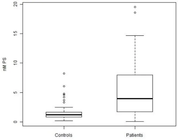
In this study, plasma levels of circulating procoagulant MPs were measured in 728 patients with solid and hematological malignancies and in 65 healthy individuals. The baseline characteristics of patients and controls are given in table 1. MP levels (nanomolar phosphatidylserine nM PS ) were significantly higher in cancer patients than in healthy controls (3.95 nM PS 1.74-7.96 vs. 1.19 nM PS 0.81-1.67 , p<0.001, figure 1). The cut-off point for elevated MPs was set at 4.62 nM PS, which represented the 95th percentile of MP levels in the control individuals of our study.
Elevated MPs were found in 327 (44.9%) cancer patients.
Association of circulating MPs with the occurrence of VTE in cancer
patients
During a median observation period of 710 days (25th-75th precentile: 262-731) 53 (7.3%) patients developed VTE. The overall cumulative probability of VTE in cancer patients was 5.9% after 6 months and 7.3% after one year. The characteristics of patients who developed VTE are given in table 2.
There was no statistically significant difference of MP levels between cancer patients with localized disease and cancer patients with distant metastasis at study inclusion (4.21 nM PS 1.93-7.71 vs. 3.67 1.61-4.30 , p=0.255). MP levels did also not differ significantly between cancer patients who had developed VTE compared to those who had not developed VTE by the time of follow-up (3.56 nM PS 1.86-7.42 vs. 3.97 nM PS 1.73-7.97 , p=0.846). For further statistical analysis cancer patients were dichotomized into those with elevated and those with non-elevated MP levels (cut-off point 4.62 nM PS, representing the 95th percentile of MP levels in healthy controls). In univariate Cox regression analysis the hazard ratio (HR) of elevated circulating MPs ( 4.62 nM PS) was not associated with VTE (HR 0.99 [95% CI: 0.58-1.70], p=0.968). In multivariable analysis, after additionally adjusting for age, sex, surgery, chemo- and radiotherapy the HR of elevated MPs was 0.95 ([95% CI: 0.55-1.64], p=0.856). Additional multivariable analyses including different tumour subgroups, soluble P-selectin, platelet and leukocyte counts
did not reveal a significant association of elevated circulating MPs with the occurrence of VTE (data not shown).
In patients with elevated MP levels cumulative probabilities of VTE were 5.2% after 6 months and 6.8% after 1 year and in those with non-elevated MP levels they were 6.4% after 6 months and 7.7% after 1 year, respectively (Log-rank test: p=0.968).
Discussion
In this study we measured circulating procoagulant MPs in 728 cancer patients, who were followed prospectively for a median of 710 days, and in a control group of age- and sex-matched healthy individuals. In line with previous reports MP levels at study inclusion were significantly higher in the group of cancer patients than in healthy controls without cancer [20,19,13,29]. MPs were also analyzed for their potential of being a predictive parameter for the occurrence of VTE in cancer patients. However, MP levels were not found to be predictive of VTE in cancer patients. Consistent with previous studies, in our study we observed the highest VTE rates in patients with brain tumors (high grade glioma) [30]. However, we did not find higher MP levels in the subgroup of patients with brain tumors compared to other tumor entities. The high risk of VTE in brain tumors is not fully understood. VTE is a multifactorial disease and in cancer patients several risk factors such as immobilization, tumor subtype, tumor size, chemotherapy or radiotherapy and many other acquired or inherited VTE risk factors contribute to the very high VTE risk. This may be especially true for brain tumor patients in whom the high VTE risk exceeds the postoperative period and stays high throughout the course of disease. [31-34].
Up to date, several methods for the measurement of MPs have been developed in order to quantify circulating MPs. Most studies used either flow cytometry to measure the number of MPs, or functional chromogenic assays were deployed to determine the procoagulant potential of MPs [35]. In our large prospective study we used a functional chromogenic assay in a solid phase for MP detection. For the construction of standard curves phospholipids of defined composition were used (33% PS and 67% PC). Interestingly, recent studies showed that the phospholipid
composition of circulating procoagulant MPs varies depending on type of originating cell and type of stimulation [36-38]. Therefore, great variations of the PS and PC distribution on the surface of circulating MPs of different origin may be possible. Especially tumor derived MPs may have a completely different phospholipid composition as the phospholipids used in our standard curves. Nevertheless, this prothrombinase assay is a well established method that had previously been used in several different clinical studies for the assessment of circulating procoagulant MPs in patients with a variety of diseases, such as sepsis [39], HIV[26], type 1 and type 2 diabetes [40], acute coronary syndrome [41], VTE [14,42], and preeclampsia [43]. Yet, up to date there are no studies applying this functional prothrombinase assay in a solid phase in cancer. Using this method we detected higher MP levels in cancer patients than in healthy individuals, but the difference between MP levels of cancer patients who developed future VTE and those who did not was not statistically significant.
As freeze-thawing cycles might increase the number of circulating MPs [44], a limitation of the study might be that we used frozen plasma samples for determination of MPs. However, samples of cancer patients and controls were freshly thawed and treated equally and different preanalytical conditions can be excluded.
Recently, new assays for the measurement of TF-positive MPs or MP-associated TF activity have been established that so far have been predominantly performed in cancer patients. Hron et al measured two-fold higher levels of TF positive MPs in colorectal cancer patients compared to healthy controls and reported that TF positive MPs correlated with elevated D-dimer levels, which is a marker for activation of coagulation [29]. In two small studies functional chromogenic assays were used to compare MP-associated TF activity in cancer patients with and without acute VTE. Tesselaar et al measured an elevated MP-associated TF activity in metastatic breast cancer patients and metastatic pancreatic cancer patients who presented with acute VTE compared to cancer patients without VTE [13]. Strikingly, no MP-associated TF activity was found in non-cancer patients who had suffered from acute VTE. Also Manly et al detected higher MP-associated TF activity levels in 53 cancer patients with different tumour types and acute VTE than in 13 cancer patients without VTE [42]. Furthermore, Zwicker et al applied a newly developed method, the impedance flow cytometry, and showed elevated numbers of TF-positive MPs in 60% of cancer patients with acute VTE compared to 27% of cancer patients without VTE [10].In contrast to the above-mentioned studies, in our large prospective study we measured baseline MP levels of cancer patients and followed them over a 2-year period or until occurrence of VTE or death or loss of follow-up, in order to find parameters that might be predictive of cancer-associated VTE. According to our study protocol, cancer patients with acute VTE at least 3 months prior to study inclusion were not enrolled in order to exclude the effect of an acute VTE event on MP levels and other laboratory parameters. In our study MPs were quantified by measuring the PS content on the MP surface after capture onto immobilized annexin V and by determining their procoagulant activity with a prothrombinase assay. PS was reported to be a highly procoagulant phospholipid [11], but our results do not support a predictive role of PS-exposing MPs for the occurrence of VTE in cancer patients. Whether MPs that additionally express high levels of TF on their surface might be predictive of cancer-associated VTE, remains to be further elucidated in large
prospective studies.
In conclusion, we found that MP levels in cancer patients were elevated compared to healthy individuals. However, with the prothrombinase assay we did not find a significant association between circulating procoagulant MPs and the risk of developing VTE in cancer patients. It has to be considered, that VTE is a complex multifactorial process and that a plethora of pathologic changes occurs in the hemostatic system of cancer patients. The role of procoagulant MPs in cancer-associated VTE still needs to be addressed in other large prospective studies. These studies should also focus on the measurement of TF-positive MPs as a predictive parameter, because recent experimental and clinical research indicates a potential role of this specific MP subpopulation in the pathogenesis of cancer-associated VTE.
Acknowledgements: We thank all persons that supported us in patient recruitment for the Vienna Cancer and Thrombosis Study (CATS), Silvia Koder for skillful technical assistance and Tanja Altreiter for proof-reading of the manuscript.
Financial support: This study was supported by grants from the “Jubiläumsfonds” of the Austrian National Bank, by an unrestricted grant from Pfizer Austria, and the “Fellinger Krebsforschung”.
References
1. Siljander P, Carpen O, Lassila R (1996) Platelet-derived microparticles associate with fibrin during thrombosis. Blood 87I:4651-4663
2. Willekens FL, Werre JM, Groenen-Dopp YA et al. (2008) Erythrocyte vesiculation: A self-protective mechanism? Br J Haematol 141I:549-556
3. Sabatier F, Roux V, Anfosso F et al. (2002) Interaction of endothelial microparticles with monocytic cells in vitro induces tissue factor-dependent procoagulant activity. Blood 99I:3962-3970
4. Diamant M, Tushuizen ME, Sturk A et al. (2004) Cellular microparticles: New players in the field of vascular disease? Eur J Clin Invest 34I:392-401
5. Satta N, Freyssinet JM, Toti F (1997) The significance of human monocyte thrombomodulin during membrane vesiculation and after stimulation by lipopolysaccharide. Br J Haematol 96I:534-542
6. Schecter AD, Spirn B, Rossikhina M et al. (2000) Release of active tissue factor by human arterial smooth muscle cells. Circ Res 87I:126-132
7. Hugel B, Martinez MC, Kunzelmann C et al. (2005) Membrane microparticles: Two sides of the coin. Physiology (Bethesda) 20I:22-27
8. Falati S, Liu Q, Gross P et al. (2003) Accumulation of tissue factor into developing thrombi in vivo is dependent upon microparticle p-selectin glycoprotein ligand 1 and platelet p-selectin. J Exp Med 197I:1585-1598
9. Del Conde I, Bharwani LD, Dietzen DJ et al. (2007) Microvesicle-associated tissue factor and trousseau's syndrome. J Thromb Haemost 5I:70-74
10. Zwicker JI, Liebman HA, Neuberg D et al. (2009) Tumor-derived tissue factor-bearing microparticles are associated with venous thromboembolic events in malignancy. Clin Cancer Res 15I:6830-6840
11. Morel O, Toti F, Hugel B et al. (2006) Procoagulant microparticles: Disrupting the vascular homeostasis equation? Arterioscler Thromb Vasc Biol 26I:2594-2604
12. Polgar J, Matuskova J, Wagner DD (2005) The p-selectin, tissue factor, coagulation triad. J Thromb Haemost 3I:1590-1596
13. Tesselaar ME, Romijn FP, Van Der Linden IK et al. (2007) Microparticle-associated tissue factor activity: A link between cancer and thrombosis? J Thromb Haemost 5I:520-527
14. Chirinos JA, Heresi GA, Velasquez H et al. (2005) Elevation of endothelial microparticles, platelets, and leukocyte activation in patients with venous thromboembolism. J Am Coll Cardiol 45I:1467-1471
15. Ay C, Freyssinet JM, Sailer T et al. (2009) Circulating procoagulant microparticles in patients with venous thromboembolism. Thromb Res 123I:724-726
16. Heit JA, Silverstein MD, Mohr DN et al. (2000) Risk factors for deep vein thrombosis and pulmonary embolism: A population-based case-control study. Arch Intern Med 160I:809-815 17. Blom JW, Doggen CJ, Osanto S et al. (2005) Malignancies, prothrombotic mutations, and the risk of venous thrombosis. Jama 293I:715-722
18. Sousou T, Khorana A. (2009) Identifying cancer patients at risk for venous thromboembolism. Hämostaseologie 29I:121-124
19. Toth B, Liebhardt S, Steinig K et al. (2008) Platelet-derived microparticles and coagulation activation in breast cancer patients. Thromb Haemost 100I:663-669
20. Kim HK, Song KS, Park YS et al. (2003) Elevated levels of circulating platelet microparticles, vegf, il-6 and rantes in patients with gastric cancer: Possible role of a metastasis predictor. Eur J Cancer 39I:184-191
21. Sud R, Khorana AA (2009) Cancer-associated thrombosis: Risk factors, candidate biomarkers and a risk model. Thromb Res 123 Suppl 4I:S18-21
22. Langer FS, B. Haubold, K. Marx, G. Wierecky, J. Brümmendorf, TH. Dierlamm, J. Bokemeyer, C. Eifrig, B. (2008) Tissue factor procoagulant activity of plasma microparticles in patients with cancer-associated disseminated intravascular coagulation. Ann Hematol 87I:451-457 23. Ay C, Simanek R, Vormittag R et al. (2008) High plasma levels of soluble p-selectin are predictive of venous thromboembolism in cancer patients: Results from the vienna cancer and thrombosis study (cats). Blood 112I:2703-2708
24. Vormittag R, Simanek R, Ay C et al. (2009) High factor viii levels independently predict venous thromboembolism in cancer patients: The cancer and thrombosis study. Arterioscler Thromb Vasc Biol 29I:2176-2181
25. Ay C, Vormittag R, Dunkler D et al. (2009) D-dimer and prothrombin fragment 1 + 2 predict venous thromboembolism in patients with cancer: Results from the vienna cancer and thrombosis study. J Clin Oncol 27I:4124-4129
26. Aupeix K, Hugel B, Martin T et al. (1997) The significance of shed membrane particles during programmed cell death in vitro, and in vivo, in hiv-1 infection. J Clin Invest 99I:1546-1554 27. Pigault C, Follenius-Wund A, Schmutz M et al. (1994) Formation of two-dimensional arrays of annexin v on phosphatidylserine-containing liposomes. J Mol Biol 236I:199-208
28. Stewart JC (1980) Colorimetric determination of phospholipids with ammonium ferrothiocyanate. Anal Biochem 104I:10-14
29. Hron G, Kollars M, Weber H et al. (2007) Tissue factor-positive microparticles: Cellular origin and association with coagulation activation in patients with colorectal cancer. Thromb Haemost 97I:119-123
30. Kakkar AK, Levine M, Pinedo HM et al. (2003) Venous thrombosis in cancer patients: Insights from the frontline survey. Oncologist 8I:381-388
31. Jenkins EO, Schiff D, Mackman N et al. Venous thromboembolism in malignant gliomas. J Thromb Haemost 8I:221-227
32. Simanek R, Vormittag R, Hassler M et al. (2007) Venous thromboembolism and survival in patients with high-grade glioma. Neuro Oncol 9I:89-95
33. Semrad TJ, O'Donnell R, Wun T et al. (2007) Epidemiology of venous thromboembolism in 9489 patients with malignant glioma. J Neurosurg 106I:601-608
34. Rong Y, Post DE, Pieper RO et al. (2005) Pten and hypoxia regulate tissue factor expression and plasma coagulation by glioblastoma. Cancer Res 65I:1406-1413
35. Jy W, Horstman LL, Jimenez JJ et al. (2004) Measuring circulating cell-derived microparticles. J Thromb Haemost 2I:1842-1851
36. Abid Hussein MN, Boing AN, Biro E et al. (2008) Phospholipid composition of in vitro endothelial microparticles and their in vivo thrombogenic properties. Thromb Res 121I:865-871 37. Biro E, Akkerman JW, Hoek FJ et al. (2005) The phospholipid composition and cholesterol content of platelet-derived microparticles: A comparison with platelet membrane fractions. J Thromb Haemost 3I:2754-2763
38. Weerheim AM, Kolb AM, Sturk A et al. (2002) Phospholipid composition of cell-derived microparticles determined by one-dimensional high-performance thin-layer chromatography. Anal Biochem 302I:191-198
39. Nieuwland R, Berckmans RJ, McGregor S et al. (2000) Cellular origin and procoagulant properties of microparticles in meningococcal sepsis. Blood 95I:930-935
40. Sabatier F, Darmon P, Hugel B et al. (2002) Type 1 and type 2 diabetic patients display different patterns of cellular microparticles. Diabetes 51I:2840-2845
41. Mallat Z, Benamer H, Hugel B et al. (2000) Elevated levels of shed membrane microparticles with procoagulant potential in the peripheral circulating blood of patients with acute coronary syndromes. Circulation 101I:841-843
42. Manly DA, Wang J, Glover SL et al. (2009) Increased microparticle tissue factor activity in cancer patients with venous thromboembolism. Thromb Res 125I:511-512
43. Gonzalez-Quintero VH, Jimenez JJ, Jy W et al. (2003) Elevated plasma endothelial microparticles in preeclampsia. Am J Obstet Gynecol 189I:589-593
44. Valeri CR, Ragno G, Khuri S (2005) Freezing human platelets with 6 percent dimethyl sulfoxide with removal of the supernatant solution before freezing and storage at -80 degrees c without postthaw processing. Transfusion 45I:1890-1898
Table 1. Baseline characteristics of total study population
Characteristics of all patients (n=728)
Age at study entry, median; (IQR), yrs 62 (53-68)
Gender Female, n (%) 341 (46.84) Male, n (%) 387 (53.16) Site of cancer n (%) Brain 86 (11.8) Breast 125 (17.2) Lung 102 (14.0) Stomach 32 (4.4) Colon 102 (14.0) Pancreas 43 (5.9) Kidney 21 (2.9) Prostate 80 (11.0) Multiple Myeloma 17 (2.3) Lymphoma 74 (10.2) Others 46 (6.3)
Characteristics of all controls (n=65)
Age at study entry, median; (IQR), yrs 60 (58-66)
Gender
Female, n (%) 30 (46.2)
Male, n (%) 35 (53.8)
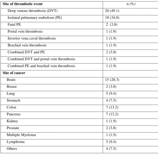
Table 2. Event characteristics of cancer patients with VTE n=53
Site of thrombotic event n (%)
Deep venous thrombosis (DVT) 26 (49.1)
Isolated pulmonary embolism (PE) 18 (34.0)
Fatal PE 2 (3.8)
Portal vein thrombosis 1 (1.9)
Inverior vena caval thrombosis 1 (1.9)
Brachial vein thrombosis 1 (1.9)
Combined DVT and PE 2 (3.8)
Combined DVT and portal vein thrombosis 1 (1.9)
Combined PE and brachial vein thrombosis 1 (1.9)
Site of cancer Brain 15 (28.3) Breast 2 (3.8) Lung 5 (9.4) Stomach 4 (7.5) Colon 7 (13.2) Pancreas 7 (13.2) Kidney 1 (1.9) Prostate 2 (3.8) Multiple Myeloma 1 (1.9) Lymphoma 5 (9.4) Others 4 (7.5)
Figure 1. Box plots: Microparticles in healthy controls and cancer patients
Legend for Figure 1. Box plot analysis of MP levels (nM PS) in healthy controls and cancer patients. The boundaries of the box represent the 25th and the 75th percentiles, respectively. The line inside the box marks the median. The 10% and the 90% percentiles were used for whiskers. Open circles represent outliers.