doi: 10.3389/fmicb.2019.00664
Edited by: Thomas Dandekar, University of Würzburg, Germany Reviewed by: Rajneesh Rana, Indian Veterinary Research Institute (IVRI), India Chih-Horng Kuo, Academia Sinica, Taiwan *Correspondence: Joerg Jores [email protected] †These authors have contributed equally to this work
Specialty section: This article was submitted to Infectious Diseases, a section of the journal Frontiers in Microbiology Received: 22 January 2019 Accepted: 18 March 2019 Published: 03 April 2019 Citation: Jores J, Ma L, Ssajjakambwe P, Schieck E, Liljander A, Chandran S, Stoffel MH, Cippa V, Arfi Y, Assad-Garcia N, Falquet L, Sirand-Pugnet P, Blanchard A, Lartigue C, Posthaus H, Labroussaa F and Vashee S (2019) Removal of a Subset of Non-essential Genes Fully Attenuates a Highly Virulent Mycoplasma Strain. Front. Microbiol. 10:664. doi: 10.3389/fmicb.2019.00664
Removal of a Subset of
Non-essential Genes Fully
Attenuates a Highly Virulent
Mycoplasma Strain
Joerg Jores1,2* , Li Ma3†, Paul Ssajjakambwe2,4†, Elise Schieck2, Anne Liljander2,
Suchismita Chandran3, Michael H. Stoffel5, Valentina Cippa1, Yonathan Arfi6,7,
Nacyra Assad-Garcia3, Laurent Falquet8, Pascal Sirand-Pugnet6,7, Alain Blanchard6,7,
Carole Lartigue6,7, Horst Posthaus9, Fabien Labroussaa1and Sanjay Vashee3
1Department of Infectious Diseases and Pathobiology, Institute of Veterinary Bacteriology, University of Bern, Bern,
Switzerland,2International Livestock Research Institute, Nairobi, Kenya,3J. Craig Venter Institute, Rockville, MD,
United States,4College of Veterinary Medicine, Animal Resources and Biosecurity, Makerere University, Kampala, Uganda, 5Division of Veterinary Anatomy, Department of Clinical Research and Veterinary Public Health, University of Bern, Bern,
Switzerland,6UMR 1332 – Biologie du Fruit et Pathologie, Institut National de la Recherche Agronomique,
Villenave-d’Ornon, France,7UMR 1332 – Biologie du Fruit et Pathologie, Université de Bordeaux, Villenave-d’Ornon, France, 8Biochemistry Unit, Swiss Institute of Bioinformatics, University of Fribourg, Fribourg, Switzerland,9Department for
Infectious Diseases and Pathobiology, Institute of Animal Pathology (COMPATH), University of Bern, Bern, Switzerland
Mycoplasmas are the smallest free-living organisms and cause a number of economically important diseases affecting humans, animals, insects, and plants. Here, we demonstrate that highly virulent Mycoplasma mycoides subspecies capri (Mmc) can be fully attenuated via targeted deletion of non-essential genes encoding, among others, potential virulence traits. Five genomic regions, representing approximately 10% of the original Mmc genome, were successively deleted using Saccharomyces cerevisiae as an engineering platform. Specifically, a total of 68 genes out of the 432 genes verified to be individually non-essential in the JCVI-Syn3.0 minimal cell, were excised from the genome. In vitro characterization showed that this mutant was similar to its parental strain in terms of its doubling time, even though 10% of the genome content were removed. A novel in vivo challenge model in goats revealed that the wild-type parental strain caused marked necrotizing inflammation at the site of inoculation, septicemia and all animals reached endpoint criteria within 6 days after experimental infection. This is in contrast to the mutant strain, which caused no clinical signs nor pathomorphological lesions. These results highlight, for the first time, the rational design, construction and complete attenuation of a Mycoplasma strain via synthetic genomics tools. Trait addition using the yeast-based genome engineering platform and subsequent in vitro or in vivo trials employing the Mycoplasma chassis will allow us to dissect the role of individual candidate Mycoplasma virulence factors and lead the way for the development of an attenuated designer vaccine.
Keywords: Mycoplasma mycoides subsp. capri, attenuation, genome engineering, in vivo challenge, virulence traits
INTRODUCTION
Bacteria belonging to the genus Mycoplasma are
wall-less bacteria that cause massive economic losses in the
livestock sector (chickens, ruminants and pigs) and
are responsible for human pneumonia and sexually
transmitted diseases (STDs). Currently, there is an absence of commercial vaccines against infections with the human
pathogens Mycoplasma pneumoniae and Mycoplasma
genitalium (Linchevski et al., 2009). In contrast, many
livestock vaccines are commercialized, which rely either on adjuvanted killed bacteria or on attenuated strains
obtained after successive rounds of sub-culturing or
chemical mutagenesis (Browning et al., 2005). Due to
these empirical approaches, the exact mechanism triggering the attenuation is unknown for many of the previously
developed live attenuated Mycoplasma vaccines. Strikingly,
these vaccines are far from being optimal since they often display short durations of immunity and limited efficacy (Maes et al., 2008; Nicholas and Churchward, 2012; Jores et al., 2013). A better understanding of pathogenicity and the identification of virulence traits would foster next generation vaccines.
For many years, the lack of genetic tools has limited
our basic understanding of Mycoplasma pathogenicity. Due
to their regressive evolution by gene loss, mycoplasmas appear to lack many of the common bacterial effectors and toxins used to interact with their hosts or to escape
the hosts’ immune systems (Citti et al., 2010;
Chopra-Dewasthaly et al., 2017). Lipoproteins have been proposed to be involved in both aspects by using their cytoadherent properties and allowing antigenic variability through phase
or sequence variation (Chambaud et al., 1999). Other
candidate virulence traits, such as the Mycoplasma Ig
binding protein-Mycoplasma Ig protease (MIB-MIP) system (Arfi et al., 2016) and the hydrogen peroxide production system (Blotz and Stulke, 2017) have been suggested, but not confirmedin vivo.
The availability of a genome engineering platform that
allows directed and precise mutagenesis for Mycoplasma
mycoides is undoubtedly a new starting point toward better understanding of host–pathogen interactions. The species M. mycoides consists of the two subspecies M. mycoides
subsp. mycoides (Mmm) and M. mycoides subsp. capri
(Mmc), which are the causative agents of contagious bovine
pleuropneumonia and a caprine MAKePS syndrome
(comprising mastitis, arthritis, keratitis, pneumonia, and
septicemia), respectively. In this work, we engineered
a Mmc strain by deleting approximately one tenth of
the genome, including candidate virulence traits. The
resulting mutant retains almost wild-type like growth
characteristics and was attenuated both in vitro and
in vivo. The construction of this fully attenuated and safe
laboratory Mycoplasma strain paves the way for research
into host–pathogen interactions and is a good starting point to revisit the actual role of suggested virulence
determinants inMycoplasma.
MATERIALS AND METHODS
Mycoplasma Strains
TheM. mycoides subsp. capri outbreak strain GM12 was used as positive control in thein vivo experiment (DaMassa et al., 1983).
A modifiedMycoplasma capricolum subsp. capricolum strain CK
was used as recipient strain in genome transplantation protocols (Lartigue et al., 2009).
Yeast Strain and Media
The yeastSaccharomyces cerevisiae, strain VL6-48N (MAT αhis3-1200 trp1-11 KlURA3-11 lys2 ade2-101 met14) containing the 1.08 Mb genome ofM. mycoides subsp. capri (Mmc) strain GM12 with an integrated yeast centromeric plasmid (YCp) (Lartigue et al., 2009) was used for construction of the mutants. Yeast cells were grown and maintained in either synthetic minimal medium containing dextrose (SD, Takara Bio) (Lartigue et al., 2009), or in standard rich medium containing glucose (YPD, Takara Bio) or galactose (YPG, Takara Bio) (Noskov et al., 2010). SD medium was supplemented with 5-fluoroorotic acid (5-FOA), for KlURA3 counter-selection (Boeke et al., 1984;Lartigue et al., 2009).
Preparation of Mutagenesis Cassettes
Sixty eight genes that encode candidate virulence traits
were seamlessly deleted from the genome of Mmc
GM12::YCpMmyc1.1 in five consecutive cycles (D1, D3, D4, and D5) using Tandem Repeat coupled with Endonuclease Cleavage [TREC] as described (Noskov et al., 2010; Chandran et al., 2014) or a variation of TREC involving the Cre-lox system for the D2 deletion, see below. Primer sequences to target and confirm the insertion of the mutagenesis cassette into each target site and to verify seamless deletion of the targeted genes are shown in Supplementary Table S1.
The gts gene cluster (D2) was targeted and deleted in the
Mmc genome in the back-ground of the glpFKO deletion strain
by employing a derivative of the Mmc synthetic cell
JCVI-syn1.0 (Gibson et al., 2010). Primers RC0905 and RC0906
(Supplementary Table S1) were used to amplify the mutagenesis cassette from the synthetic cell derivative and was targeted to the gts region. Specific primers were used to confirm correct insertion at the target site by amplifying the junctions between the GM12::YCpMmyc1.1 genome and the inserted cassette. Galactose induction resulted in the Cre-mediated deletion of the gts region, leaving 13 bp of the 50
end of thegtsA region, the 34 bp loxP site, and 27 bp of the 30
end of thelppB gene. Specific primers were used to verify the knock-out.
Transformation and PCR Analysis
Transformation of the CORE3 cassette was performed by the lithium acetate method as described previously (Gietz et al., 1992). Transformed yeast were plated on appropriate selection media [SD medium minus His (Teknova, CA) or SD medium
(minus His and minus Ura)] and incubated at 30◦
C for 48 h. Yeast colonies were patched on appropriate selective media and total DNA was isolated for PCR screening (Noskov et al., 2002). The correct insertion of the mutagenesis cassette was
verified by PCR amplification using upstream and downstream specific primers (Integrated DNA Technologies, Coralville, IA, United States) (Supplementary Table S1).
Transplantation
The modified GM12::YCpMmyc1.1 genomes (D1–D5) were
transplanted into M. capricolum subsp. capricolum (Mcc)
recipient cells with polyethylene glycol and selected for tetracycline resistance as described previously (Noskov et al., 2002; Lartigue et al., 2009). The resulting mutant strains were subjected to multiplex PCRs and pulsed-field gel electrophoresis as described elsewhere (Labroussaa et al., 2016) to confirm integrity of the genome.
Confirmation of the Mutants Using Next
Generation Sequencing and Mapping
Assembly
Total DNA of the strains GM12, GM12::YCpMmyc1.1 and
GM12::YCpMmyc1.1-168 was isolated as described before
(Fischer et al., 2015). DNA was sheared using sonication and subjected to Illumina sequencing using a MiSeq machine by University of California Santa Cruz, Santa Cruz, CA (United States). Reads were mapped to the designed genome sequences based on the parental strains GM12 and
GM12::YCpMmyc1.1 and GM12::YCpMmyc1.1-168. The
raw reads (300 bp PE) were QC with FastQC1. The corrected
reads were mapped onto the reference genome WT-YCP.fa
with bwa mem (Li and Durbin, 2010) and converted to sorted
bam with samtools (Li, 2011). The bam files were analyzed for deletions using Delly2 (Rausch et al., 2012) and Sprites (Zhang et al., 2016), and the predictions validated visually using IGV (Thorvaldsdottir et al., 2013). The list of strains and their deleted regions is summarized in Table 1.
Scanning Electron Microscopy of
Mycoplasma
Unless stated otherwise, chemicals were obtained from Merck (Schaffhausen, Switzerland). Mycoplasmas were washed with
distilled water (dH2O) and fixed with 4% para-formaldehyde
(Life Technologies, Thermo Fisher, Zug, Switzerland; Cat. No.
28906) in dH2O for 5 days at 4◦C. Thereafter, samples of
40 µl of cell suspension were centrifuged onto gold-sputtered poly-L-lysine coated coverslips (high molecular poly-L-Lysine
hydrobromide) at 125 rcf for 4 min. Coverslips were washed
once with PBS and twice with 0.1% bovine serum albumin in PBS (BSA/PBS). Free aldehydes were blocked with 0.05 M glycine in 0.1% BSA-c/PBS (Aurion, ANAWA Trading, Wangen, Switzerland) for 15 min at room temperature. After 3 washes with 0.1% BSA/PBS, cells were fixed with 2.5% glutaraldehyde (Merck 104239) in 0.1 M cacodylate buffer (dimethylarsinic acid sodium salt trihydrate), washed 3 times with dH2O and postfixed
with 1% OsO4 (Polysciences, Warrington, PA, United States)
in 0.1 M cacodylate buffer for 15 min. at room temperature. Five additional washes with dH2O were followed by dehydration
1https://www.bioinformatics.babraham.ac.uk/projects/fastqc/
in an ascending ethanol series. Samples were then transferred to hexamethyldisilazane (Merck 814051) for 10 min, air-dried, mounted onto aluminum stubs with carbon conductive adhesive tabs (Ted Pella Inc., Redding, CA, United States) and coated with approximately 25 nm of gold in an SCD004 (Leica Microsystems, Heerbrugg, Switzerland). Secondary electron micrographs and corresponding backscattered images were obtained with a fully digital field emission scanning electron microscope DSM 982 Gemini (Zeiss, Oberkochen, Germany) at an accelerating voltage of 5 kV, a working distance of 6–8 mm and primary magnifications ranging from 30,000 to 50,000×.
Growth Assay
Overnight cultures of Mycoplasma strains were grown at
37◦
C in SP4 medium containing streptomycin (GM12) or
tetracycline (GM12::YCpMmyc1.1, GM12::YCpMmyc1.1-168)
for about 16 h. Doubling times of theMycoplasma strains were then determined as described elsewhere (Hutchison et al., 2016), except that time interval samples were collected and processed at 0, 1, 2, 3, 4, 5, 6, 7, 9, 12, 15, and 24 h.
In vitro Hydrogen Peroxide Assay
Overnight cultures of Mycoplasma strains were grown as
described above. When the pH of overnight cultures reached 6.0– 6.5, they were inoculated into fresh SP4 medium at 1:200 dilution and incubated at 37◦
C for different time intervals of 0, 5, 7, and 24 h. At each time interval, an aliquot of culture was taken for DNA extraction (Hutchison et al., 2016) and another aliquot was taken to determine hydrogen peroxide levels.
To determine hydrogen peroxide levels, the aliquots were
spun at 14,000 rpm for 10 min at 4◦
C. The pellets were washed with 1 ml of cold PBS, pH 7.5 to remove traces
of media, then resuspended in 400 µl of cold PBS and
stored at 4◦
C. Hydrogen peroxide levels were determined using the Amplex Red Hydrogen Peroxide Assay Kit (Life Technologies, NY) according to the manufacturer’s instructions.
Briefly, 50 µl of diluted samples (1:5 in PBS) was aliquoted
onto 96-well plates and warmed to 37◦
C for 1 h prior to
starting the assay. 100 µM final concentration of glycerol
(Sigma-Aldrich, MO) or GPC (Sigma-Aldrich, MO) was then
added to the diluted sample and incubated at 37◦
C for
1 h. 50 µl of the Amplex Red reagent was added to the
samples, incubated at room temperature in the dark for 30 min and fluorescence was measured using a spectrophotometer (SpectraMax M5, Molecular Devices, CA). Three technical replicates were performed for each sample and normalized to their respective DNA concentrations.
Detection of Immunoglobulin
Degradation by Mycoplasma in vitro and
in vivo
In vitro functionality of the MIB-MIP system was tested using the strains GM12, GM12::YCpMmyc1.1 and GM12::YCpMmyc1.1-168. Each strain was grown overnight at 37◦
C in 3 mL modified
SP5 medium (containing 5% FBS). 250µL of each culture was
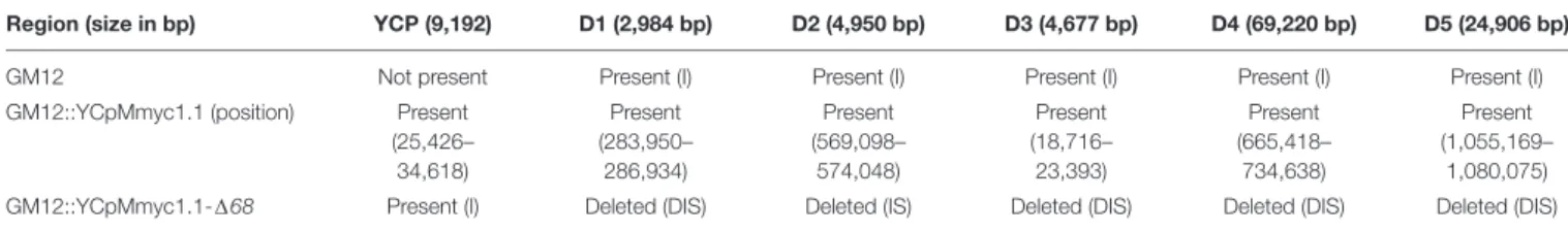
TABLE 1 | Table showing the results of the Illumina sequencing-based mapping assemble of the strains used in this study.
Region (size in bp) YCP (9,192) D1 (2,984 bp) D2 (4,950 bp) D3 (4,677 bp) D4 (69,220 bp) D5 (24,906 bp)
GM12 Not present Present (I) Present (I) Present (I) Present (I) Present (I)
GM12::YCpMmyc1.1 (position) Present (25,426– 34,618) Present (283,950– 286,934) Present (569,098– 574,048) Present (18,716– 23,393) Present (665,418– 734,638) Present (1,055,169– 1,080,075)
GM12::YCpMmyc1.1-168 Present (I) Deleted (DIS) Deleted (IS) Deleted (DIS) Deleted (DIS) Deleted (DIS)
D, detected by Delley; I, validated visually by IGV; S, detected by sprites.
then resuspended in 15µL modified SP5 medium (containing
5% FBS) and incubated with 5 µg of purified caprine IgG
(Sigma) at 37◦
C for 3 h. Bacterial CFUs were estimated for each strain by serial dilutions and were 3.2∗
109 CFUsMmc GM12,
8.5∗
108 CFUs Mmc GM12::YCpMmyc1.1, and 3.5∗
108 CFUs
Mmc GM12::YCpMmyc1.1-168. A sample consisting of 5 µg caprine IgG in dH2O was included as a control. The incubated
samples were mixed with 2× Laemmli Sample Buffer (Bio-Rad) at a 1:1 ratio, boiled for 10 min at 98◦
C and separated onto a 12% SDS-PAGE gel. They were subsequently transferred onto
a 0.2 µm nitrocellulose membrane (Bio-Rad) using a Bio-Rad
Trans-Blot R
TurboTMTransfer System (25 volts, 1.0 A, 30 min). Next, a Western Blot was performed using PBS supplemented with 0.1% Tween-20 and 2% BSA as a blocking buffer, and mouse anti-goat IgG (H+L) (Jackson ImmunoResearch, 205-005-108) and goat anti-mouse (Fc) labeled with horseradish peroxidase (Sigma, A0168) as primary and secondary antibodies. The antibodies were diluted in blocking buffer at 1:2000 and 1:70,000, respectively, and incubated with the membrane for 1 h each. In between the antibody incubations, the membrane was washed once with PBS − 0.1% Tween-20 + 3.2% NaCl and twice with PBS − 0.1% Tween-20, for 10 min each time. The results were visualized using the Fujifilm LAS-3000 Luminescent Image Analyzer.
Serum samples derived from thein vivo challenge were diluted
in water to achieve a total load of 10–20 µg of protein per
sample. Western Blots were performed and analyzed using the same protocol as described above.
Animal Experiment Setup
All protocols of this study were designed and performed in strict accordance with the Kenyan and United States American legislation for animal experimentation and were approved by the institutional animal care and use committees at both institutions (JCVI and ILRI, IACUC reference number 2014.08).
Sixteen male outbred goats (Capra aegagrus hircus), 1–2 years of age and randomly selected in Naivasha, were transferred to the ILRI campus in Nairobi and kept under quarantine for 6 months. After arrival at the campus, all animals were dewormed twice using levamisole and treated prophylactically against babesiosis and anaplasmosis using imidocarb. Upon entry to ILRI, the goats were vaccinated against anthrax and blackleg (Blanthax R
, Cooper), Foot and Mouth Disease (FOTIVAX R ) and Peste des Petits Ruminants (Live attenuated strain Nig. 75/1). All animals were tested negative for presence of antibodies against contagious caprine pleuropneumoniae (CCPP), using a
competitive ELISA (IDEXX). Two weeks before experimental infection, all animals were transferred to the animal biosafety
level two (ABSL2) unit. Mycoplasma cells were cultivated in
PPLO medium supplemented with horse serum (Sacchini et al.,
2011) to early logarithmic phase, aliquoted and stored at
−80◦
C. Afterwards, we determined the CFU using two aliquots. Just before infection we thawed the vials and adjusted the concentration ofMycoplasma to 109CFU per mL−1using broth.
All 16 goats were infected transtracheally by needle puncture 5– 10 cm distal to the larynx. Each animal received 1 mL ofMmc
GM12 or GM12::YCpMmyc1.1-168 liquid culture (equivalent
to 109 colony forming units per animal), followed by 5 mL of phosphate buffered saline (PBS). The animals were allowed to
move freely within the ABSL2 unit and had ad libitum access
to water. They were fedad libitum with hay and received pellets each morning. Three veterinarians monitored the health status of the animals throughout the experiment. Rectal temperature, oxygen blood saturation, heart rate and breathing frequency were measured daily in the morning hours using the GLA M750 thermometer (GLA Agricultural Electronics, United States), VE-H100B oximeter (Edan, United States), and a stethoscope classic II (Littmann, United States) with a water-resistant wrist watch Seamaster (Omega, Switzerland), respectively. Blood samples for subsequent analysis were taken twice a week by jugular vein puncture. Goats were euthanized when they developed severe disease associated with unwarranted moderate to severe pain. Therefore, they received an intravenous injection of Lethabarb Euthanasia Injection (Virbac, United States) of 200 mg.kg−1body
weight. Severe disease and pain were determined by a fever of ≥41◦
C for>3 consecutive days, an oxygen saturation of ≤92% and a lateral recumbency of ≥1 day without the ability to feed or intake water. Goats that were not put down because of ethical reasons were euthanized on 28 dpi.
Pathomorphological and
Histology Analysis
A complete necropsy was performed on all animals. Tissue samples of the neck region around the inoculation site and all internal organs were fixed in 10% buffered formalin for 72 h and subsequently routinely processed for paraffin embedding. Tissue sections were cut at 3µm and stained routinely with hematoxylin and eosin (H&E) and evaluated by a board-certified pathologist.
Microbiology
Venous blood samples, lung samples, carpal joint fluid, and pleural fluid specimens taken at necropsy were used for isolation
of Mmc as described elsewhere (Liljander et al., 2015) using Mycoplasma liquid medium (Mycoplasma Experience Ltd., United Kingdom). Lung samples and pleural fluid were used for
screening of Pasteurella and Mannheimia spp. using standard
methods (Carter and Cole, 1990).
Statistical Analysis
Exact and normal approximation binomial tests were used to
compare the two groups using GenStat 12th Edition (Payne
et al., 2012).P-values for differences in parameters were estimated using a 2-sided 2-samplet-test comparing average levels between both groups at 5% level of significance.
RESULTS
Generation of the Mutant Strain
GM12::YCpMmyc1.1-
168
To demonstrate attenuation of Mmc by rational design, five
genomic regions were targeted in this study. These modifications were done on the genome GM12::YCpMmyc1.1 cloned in S. cerevisiae. This genome has been obtained after the insertion of genetic elements (i.e., ARSH4, CEN6, and HIS3) in the genome ofMmc GM12 allowing its maintenance and selection in the yeast (Lartigue et al., 2009). The precise localizations of each deletion are shown in Figure 1. The first two target deletion regions
FIGURE 1 | Design of mutant GM12::YCpMmyc1.1-168. Cartoon displaying the genomes of the parental strain GM12::YCpMmyc1.1 and its derivative, the deletion mutant GM12::YCpMmyc1.1-168.
contained genes encoding the glycerol-dependent hydrogen peroxide metabolic pathway and its suggested ABC transporter encoded by thegtsABCD operon (Pilo et al., 2007). This pathway has been suggested to be a main virulence mechanism for M. mycoides (Pilo et al., 2007), butin vivo confirmation is still missing and inMycoplasma gallisepticum the pathway does not seem to be linked to virulence (Szczepanek et al., 2014). Thus,
the genesglpF, glpK, and glpO (MMCAP2_0217-0219; 2,984-bp
region; D1) and thegts gene region that includes the gene lppB (MMCAP2_0456-0459; 4,950-bp region; D2) were deleted in the
Mmc genome by the yeast-based engineering method (Lartigue
et al., 2009). As previously mentioned, lipoproteins were another target of interest since they likely trigger not only host–pathogen interactions but also, overwhelming immune reactions that result in inflammation (Browning et al., 2011). Three lipoproteins encoded in the D3 region (MMCAP2_0014-0016; 4,677-bp) as well as six lipoproteins in the D5 region (lppQ, MMCAP2_0889-0904; 24,906-bp) were also excised employing again the yeast-based engineering method. On top of that we deleted a large genomic region that encoded theMycoplasma-specific F1-likeX0 ATPase (Beven et al., 2012), the MIB-MIP system (Arfi et al., 2016), an integrative and conjugative element (ICE) (Tardy et al., 2015) and eight lipoproteins. The ICE was targeted in an effort to reduce mobile elements from theMmc genome. In this case, about 70 kbp (MMCAP2_0550-0591; 69,220-bp region; D4) were targeted and deleted from theMmc genome using the yeast-based engineering method in one stretch.
After each cycle of deletions, the modified Mmc genome
was isolated from yeast cells and transplanted back into M. capricolum subsp. capricolum (Mcc) recipient cells to confirm the viability of each mutant Mmc strain. Overall, the
final mutant strain, named Mmc GM12::YCpMmyc1.1-168,
was generated in five sequential deletion cycles (Figure 1). The gene knock-outs were verified by amplifying across each
deleted region (Supplementary Figure S1). Genomic DNA from
the GM12::YCpMmyc1.1-168 was isolated and analyzed by
sequencing to confirm the deletions (Table 2). The genome
sequence of GM12::YCpMmyc1.1-1168 was deposited at the
ENA database under the accession number LS483503.
The Mutant Strain
GM12::YCpMmyc1.1-
168 Is Viable and
Unaffected in Its Morphology or Growth
in Axenic Medium
The colonies of Mmc GM12::YCpMmyc1.1-168 were of
similar size to those of GM12::YCpMmyc1.1 and GM12. Cell morphology of the GM12, the isogenic parental strain
GM12::YCpMmyc1.1 and GM12::YCpMmyc1.1-168 strains was
evaluated using scanning electron microscopy (Figure 2A). All strains tested were globular in shape and lacked any special morphological features. The diameter of the microorganisms was
in the range of 500 nm, as expected for a Mycoplasma cell.
The mutant GM12::YCpMmyc1.1-168 grew with a doubling
time somewhat similar to that of the parental strains GM12 and GM12::YCpMmyc1.1 (Figure 2B). Together, these results strongly suggest that the deletion of approximately 100 kbp
of genomic content from the Mmc genome did not adversely
affect structural integrity or in vitro growth of the mutant
GM12::YCpMmyc1.1-168.
Inability of the Mutant Strain
GM12::YCpMmyc1.1-168 to Produce
Hydrogen Peroxide in the Presence of
Glycerol in vitro
This pathway was completely deleted in the construction
of the mutant strain GM12::YCpMmyc1.1-168. Therefore
to phenotypically confirm the deletion, we measured and
TABLE 2 | Summary of post mortem records of goats. Animal ID. Date of
euthanasia Bacteremia ccu/ml Inflammation of the neck around the injection site Pulmonary congestion Pulmonary oedema Congested kidneys Mucoid enteritis and congestion Pleural fluid in thoracic cavity Adherent lung to rib cage Liver abscess CK032 6 dpi 107 X X X X X X CK034 5 dpi 107 X X X X X CK040 5 dpi 108 X X X X X X CK043 5 dpi 108 X X X X X CK046 5 dpi 108 X X X X X CK048 5 dpi 109 X X X X X CK051 5 dpi 109 X X X X X CL002 5 dpi 108 X X X X X X CK035 29 dpi CK045 28 dpi CK047 29 dpi CK049 28 dpi CL001 28 dpi CL003 28 dpi
FIGURE 2 | In vitro characteristics of the parental strains GM12, GM12::YCpMmyc1.1 and its deletion mutant GM12::YCpMmyc1.1-168: (A) morphology revealed by scanning electron microscopy. The white size bar displays 500 nm; (B) in vitro doubling time; (C) production of hydrogen peroxide in the presence of glycerol; bars display the standard deviations in (B) and the SEM in (C).
compared hydrogen peroxide production levels between the control GM12, GM12::YCpMmyc1.1 and GM12::YCpMmyc1.1-168 in vitro. In the presence of the glycerol substrate,
the mutant GM12::YCpMmyc1.1-168 shows a significant
decrease in hydrogen peroxide production when compared to its parental strains (Figure 2C). Indeed, while GM12 and GM12::YCpMmyc1.1 produced>0.3 µM of H2O2, the mutant
strain produced very low amounts of H2O2 (0.01µM), at least
30-fold lower under these conditions.
Inability of the Mutant Strain
GM12::YCpMmyc1.1-
168 to Degrade
Immunoglobulin in vitro
Another potential virulence trait encoded by mycoplasmas is the MIB-MIP system, which may play a role in immune evasion by cleavage of immunoglobulins (Figure 3A) (Arfi et al., 2016). Incubation of caprine IgG with GM12, GM12::YCpMmyc1.1
and GM12::YCpMmyc1.1-168 showed a clear difference in the
strains’ abilities to degrade IgG (Figure 3B). The two bands at 26 and 55 kDa corresponds to the IgG light and heavy
chains. The mutant strain GM12::YCpMmyc1.1-168 exhibited
no degradation of IgG, as noted by the lack of the 44 kDa band (Lane 3 of Figure 3B, black asterisk). This band, clearly visible in the other two strains, is indicative of proteolytic cleavage of the IgG heavy chain. Another pattern of degradation, with a
band at a size of about 30 kDa, is visible in the three strains. It was previously reported that this IgG cleavage is not specific or directly linked to the MIB-MIP system (Arfi et al., 2016).
The Mutant Strain
GM12::YCpMmyc1.1-
168 Is Fully
Attenuated in vivo
We next tested whether GM12::YCpMmyc1.1-168 was able to
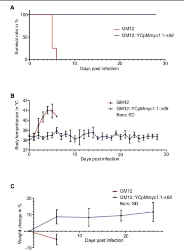
cause disease in its native host. Sixteen male outbred goats (Capra aegagrus hircus) were used in this animal infection trial. The animals were separated into two groups of equal numbers. After the infection, no immediate clinical signs of disease were observed. Two animals in the GM12::YCpMmyc1.1-168 group had to be removed from the experiment, because of acquired wounds unrelated to the infectious agent. Animal euthanasia was planned 28 days post infection (dpi). However, all eight animals inoculated with the GM12 strain developed severe clinical signs, with pyrexia starting 2–3 dpi (Figure 4B). Their body temperature continued to increase, up to 41–41.5◦
C, during the following days (Supplementary Table S2). The animals stopped feeding, were apathetic and showed signs of pain. According to the endpoint criteria stated in the animal experiment protocol, they had to be euthanized between 5–6 dpi (Figure 4A, red line). In sharp contrast, animals inoculated with
FIGURE 3 | In vitro and in vivo ability of the Mmc MIB-MIP system to degrade caprine IgG. (A) Cartoon displaying the proteolytic cleavage between the conserved CH1 and the variable VHdomain of the IgG heavy chain. (B) Immunoblot showing the in vitro ability of GM12 (lane 1), GM12::YCpMmyc1.1 (lane 2) to degrade IgG in comparison to GM12::YCpMmyc1.1-168 (lane 3). (C) Immunoblot showing the in vivo IgG degradation present in serum of septicaemic goat CK51 (GM12 group) after infection in contrast to the healthy goat CK45 (GM12::YCpMmyc1.1-168 group): 4-caprine IgG, 5-goat CK51 at –1 dpi, 6-goat CK51 at 4 dpi, 7-goat CK45 at –1 dpi, 8-goat CK45 at 4 dpi; the black asterisk marks the 44 kDa cleaved fragment.
clinical signs of disease and were all monitored until the end of the trial (Figure 4A). Their body temperature fluctuated within the normal physiological range throughout the study period (Figure 4B and Supplementary Table S2). The animals remained healthy and gained weight during the experiment (Figure 4C). Their heartbeat and respiratory rates, between 80–110 beats/min and 20–30 breaths/min, respectively, remained constant over the course of experimentation.
In all animals infected with the GM12 strain, the main pathological lesions were similar, with a severe and extensive inflammation of the soft tissues of the neck around the
site of inoculation. Additional macroscopic findings were severe pulmonary edema and congestion. Histologically, there was extensive coagulation necrosis of the connective tissue (Figure 5B, thick arrows) and musculature surrounding the trachea, in the vicinity of the inoculation site (Figures 5B,D, diamonds). A marked infiltration of mainly degenerate neutrophilic granulocytes (Figures 5B,D, asterisks) was always found associated with the necrosis. The necrotizing process extended to the trachea, the subcutis and skin. In addition, all animals showed multifocal acute necrosis with infiltration of neutrophilic granulocytes in liver and
FIGURE 4 | Comparison of the clinical parameters monitored during the in vivo challenge trial between the animals that received the GM12 and its derivative GM12::YCpMmyc1.1-168. (A) Kaplan–Meier survival curve based on animals reaching endpoint criteria. (B) Average body temperatures during experimental infection. Values were generated using daily rectal temperatures from the two groups. (C) Average weight gain/loss during experimental infection. Values were generated using interval measures from the two groups. The standard deviations are displayed as bars in (B,C).
kidney and for three animals, in the lung. These lesions were indicative of an acute septicemia. Among all animals
infected with the mutant strain GM12::YCpMmyc1.1-168,
and upon euthanasia 28–29 dpi, no pathological lesions were found around the inoculation site. The soft tissue around the inoculation site was within normal limits. Additionally, neither inflammation nor necrosis associated with the epithelium, the submucosa or any cartilage tissues was histologically observed (Figures 5A,C).
Mmc GM12 was re-isolated from the blood of all animals experimentally infected with the wild-type parental strain. Bacteremia was characterized by 106 up to 109 CCU.ml−1 of
blood, as measured by serial dilutions (Table 2). We have not been able to re-isolate the deletion mutant from all blood samples
collected from goats infected with GM12::YCpMmyc1.1-168.
Additionally, we were not able to re-isolate the mutant strain from tissue samples collected post mortem.
The MIB-MIP System in Mmc GM12 Is
Functional in vivo
The MIB-MIP system was shown to be active in vitro, and to
be present in large amounts at the cell surface (Krasteva et al., 2014) during infection (Weldearegay et al., 2016). Two animal sera were selected from the in vivo infection trial: CK51 from the GM12 group and CK45 from the GM12::YCpMmyc1.1-168 group and tested for cleavage of IgG. The pre-infection sera from both animals demonstrated no proteolytic cleavage of IgG when compared to the IgG control (Figure 3C). Conversely, the post-infection serum of CK51, which had succumbed to disease and had a high titer of bacteria in the venous blood, clearly exhibited the typical band at 44 kDa, consistent with the size of a cleaved IgG heavy chain by the MIB-MIP system. As expected, no band at 44 kDa was seen in the post-infection serum of CK45. This clearly demonstrates, for the first time, that the MIB-MIP system is functional within the caprine host and that its proteolytic activity is triggered during anMmc infection.
DISCUSSION
The first aim of this work was to fully attenuate a highly pathogenic strain of M. mycoides following a rational deletion design. The second aim was to verify this attenuation in vivo using the native host, since no rodent animal models for highly virulent M. mycoides exist (Jores et al., 2013). Many candidate virulence factors ofM. mycoides have been suggested but, none except the capsular polysaccharide (Jores et al., 2018) have ever been confirmed according to Falkow’s postulates in vivo (Falkow, 1988).
In order to generate an attenuated strain, we relied on previous knowledge and selected five genomic regions that encode candidate virulence traits. These regions, distributed around the Mmc genome, comprised of 68 genes. The first two regions (D1 and D2) encode enzymes and putative glycerol transporters involved in the production of hydrogen peroxide using the
glycerol-dependent metabolism of mycoplasmas (Blotz and
Stulke, 2017). The region D3 encodes major antigens (LppA/P72)
in M. mycoides (Monnerat et al., 1999) that induced T cell
responses early in infection (Dedieu et al., 2010). The region D4 includes an integrative conjugative element (ICE), the MIB-MIP system (Arfi et al., 2016) and the F1-likeX0 ATPase (Beven et al., 2012). The last region (D5) encodes six lipoproteins including the lipoprotein Q (LppQ), which has been suspected to be involved in exacerbating immune responses and other virulence determinants (Mulongo et al., 2015). Interestingly, among these 68 genes deleted in our mutant genome, initially comprising 944 genes and annotated RNAs, 67 were defined as non-essential in the JCVI-Syn3.0 minimal cell, a study in which 432 genes were classified as non-essential to sustain the life of a minimal Mycoplasma cell only supported at the end by 438 proteins and
35 annotated RNAs (Hutchison et al., 2016). Only one gene
encoding the glycerol phosphate kinase (GlpK) (MMCAP2_0218, region D1) was retained in the minimal JCVI-Syn3.0 cell and classified as a “quasi-essential” gene. This means that the function encoded by the glycerol kinase, i.e., the transfer of a phosphate
FIGURE 5 | Composite figure displaying representative histological results from tracheal tissue at the inoculation site. Tissues were stained with hematoxylin and eosin. (A,C) Low (40×) and high (200×) magnification of trachea of a goat inoculated with Mmc GM12::YCP1.1-168 depicting unaffected epithelium (dot), submucosa (diamond) and cartilage (asterisk). (B,D) Low (40×) and high (200×) magnification of trachea of a goat inoculated with Mmc GM12, depicting ulceration of the epithelium in (B) (thick arrow), massive extension of the submucosa (diamond) due to extensive areas of necrosis (arrowhead) and infiltration with large numbers of degenerate neutrophilic granulocytes (arrow). Size standards are displayed in each picture. Scale bars: (A,B) 500µm; (C,D) 100 µm.
group on the glycerol molecule to produce glycerol-3-phosphate, could be compensated for by another gene which encodes a similar function in the full-length genome. The compensatory gene forglpK is probably absent in the minimal JCVI-Syn3.0 cell,
but present in our mutant GM12::YCpMmyc1.1-168, allowing
the deletion of glpK without any defect in growth. Therefore, our results are consistent with the quasi-essentiality of glpK. Altogether we retained 69 out of the 87 lipoproteins present in GM12 by deleting 1, 3, 8, and 6 lipoproteins in D2, D3, D4, and D5, respectively, while the minimal cell retained only 15 lipoproteins (Hutchison et al., 2016).
It was paramount for us that the mutant strain
GM12::YCpMmyc1.1-168 maintains a doubling time similar to
its parental strain, since we wanted to create a ‘K12-like’ strain that can be further used as a cellular platform to introduce antigen-encoding genes or large stretches of DNA. It was interesting for us to observe that, despite complete removal of the glycerol pathway, which may be important for cell metabolism, there was no compelling impact onin vitro growth. It is known that there is a trade-off between genome size and growth rate.
The drastic deletions in the genome of JCVI-syn3.0 strain led to a substantial increase in the generation time, from ∼60 to ∼180 min (Hutchison et al., 2016). Recently, we also have shown that the deletion of a gene encoding an enzyme important for synthesis of carbohydrates can subsequently lead to an increase in the generation time (Schieck et al., 2016). However, in this work, we significantly reduced the genome of GM12 by more than 100 kbp (i.e., 106,737 bp) without seeing any compelling
difference in the growth rate of GM12::YCpMmyc1.1-168
in comparison to the wild-type strain. Still, this reduction represents ∼10% of the initial genome size confirming that, in addition to the size of the deletions, the nature of the genes deleted is also very likely to influence the generation time of mycoplasmas.
The main goal of this work was to construct a fully attenuated Mmc strain, that is safe to handle in the laboratory. The first confirmation of the attenuation of the GM12::YCpMmyc1.1-168 strain was obtained in vitro. The production of hydrogen peroxide was almost completely abolished in the mutant
gtsABCD pathways in the metabolism of glycerol. In addition, the loss of the IgG specific cleavage band at 44 kDa in GM12::YCpMmyc1.1-168 confirmed the role of the MIB-MIP system in the degradation of the host immunoglobulins.
The mutant GM12::YCpMmyc1.1-168 is essentially a
quasi-intermediate to the minimal cell for which a high degree of genome stability was reported. The stable reproduction of the in vitro characteristics of GM12::YCpMmyc1.1-168 also strongly supports stability of this mutant.
To confirm the mutant strain’s attenuation in vivo, we
developed an animal challenge model using Kenyan goats, outbred animals derived from different herds. The use of such animals increased variability to get a better idea of reproducibility and significance of the results (Richter et al.,
2010). Similar challenge models using either 7.3 × 10⁷ or
1012 CFUs of M. mycoides subsp. capri have been reported
from previous challenge experiments (Sunder et al., 2002;
Manimaran et al., 2006).
Animals infected with the GM12 strain developed specific clinical signs (fever, heavy breathing, septicemia, etc.) and were all euthanized by 6 dpi. Strikingly, none of the goats
infected with GM12::YCpMmyc1.1-168 developed such signs
and were healthy for the entire course of the experimentation. These results exceeded our expectations and confirmed the complete abolishment of pathogenicity of the mutant strain
GM12::YCpMmyc1.1-168. In addition, the massive septicemia
associated with very high titers of Mycoplasma observed in
animals infected with the GM12 strain prompted us to investigate whether the MIB-MIP system would leave signatures of its action on immunoglobulins (Ig) in the serum of an animal (CK51) that had a titer of 109 CFU/ml. Specific IgG cleavage, characteristic of the MIB-MIP system (Arfi et al., 2016), was observed. No such cleavage was observed in the serum of animals infected with the mutant strain. This work shows, for the first time, that the MIB-MIP system of M. mycoides is functional in vivo. Mycoplasmas have been viewed as stealth pathogenic organisms because they lack most of the immune activators or PAMPs found in other bacteria (Mogensen, 2009). Indeed, the lack of a cell wall or the capacity to produce either LPS or flagellins likely contribute to the chronicity of infection. The only PAMP that has been described for several Mycoplasma species is the surface lipoproteins, abundant components of their membrane (Chambaud et al., 1999). In the present study, we suggest another mechanism that could contribute to activating the immune system. Indeed, Ig cleaved by several bacteria, including those
generated by Mycoplasma hyorhinis, have been described as
ligands of the innate immune receptor LILRA2 (Hirayasu et al., 2016). Once bound to this receptor, it triggers the activation of the innate immune system. It is also possible that this cleavage is in line with the ‘nutritional virulence’ of the parasite (Abu Kwaik and Bumann, 2013). The exact significance of the Ig cleavage regardingMycoplasma infection of mucosal surfaces remains to be studied. We did not include a Sham group in our study but recent results obtained from a transtracheal challenge of goats withM. capricolum subsp. capripneumoniae did clearly show no effect ofMycoplasma medium and PBS injected transtracheally as expected (Liljander et al., 2019).
Interestingly, we observed severe inflammation around the site of injection in the animals that received GM12 whereas animals that were injected with the strain GM12::YCpMmyc1.1-168 developed no such pathomorphological lesions. Overwhelming immune reactions at the site of vaccination have been reported from immunizations against contagious bovine pleuropneumonia using liveM. mycoides subsp. mycoides based vaccines such as T1/44, which is the closest relative of Mmc (Fischer et al., 2012). Therefore, it is likely that any one or several of the deleted genes encode proteins that drive this overwhelming immune reaction in the GM12.
To conclude, we confirmed,in vitro and in vivo, our ability to design a fully attenuated strainvia the precise reduction of ∼10%
of the Mmc genome. However, we cannot currently pinpoint
the weight of each deletion on the observed attenuation. The total clearance of the pathogen and the absence of a compelling humoral immune response, even at the inoculation point, is surprising and supports the total abolishment of pathogenicity. Now it is necessary to test more defined mutants such as aglpOKF mutant strain to get clarity about its real role in pathogenicity.
In addition, the design of next generation vaccines for, but not restricted to, Mycoplasma diseases will benefit from this study since a chassis that is fully attenuated and able to accommodate antigens for vaccine delivery that can be constructed based on our deletion mutant is now within reach. To induce a proper immune response via such a chassis, we have the option to add genes that appropriately stimulate an inflammatory immune response or alternatively, we can construct different chassis that direct responses toward Th1 or Th2 using TLR agonists. We
consider a genetically modified Mycoplasma less problematic
than other potential chassis since the survival time ofMycoplasma in general in the environment is very short. In addition, the unconventional codon usage (where UGA encodes tryptophan) and high AT content ofMycoplasma minimizes the risk of spread of genes to other bacteria. Additional experiments are necessary to decipher the role of individual virulence traits to understand these minimal bacterial pathogens better and to develop next generation rationale vaccines. Regardless, this study provides an attractive blueprint toward these goals, especially for those that are needed in low and middle-income countries.
AUTHOR CONTRIBUTIONS
JJ and SV designed the research. LM, NA-G, SC, JJ, AL, PS, MS, ES, VC, YA, HP, and FL performed the research. JJ, FL, SC, YA, LF, PS-P, CL, AB, and SV analyzed the data. JJ and SV wrote the manuscript.
FUNDING
This research was supported by National Science
Foundation [Grant Number IOS-1110151 (to SV, CL,
and JJ)]. Additional support was received from the
University of Bern and the CGIAR research program on Livestock and Fish. ES was supported by the German
Federal Ministry for Economic Cooperation and Development (Project No. 09.7860.1-001.00 Contract No. 81136800). AL was supported by the Centrum for International Migration and Development in Germany.
ACKNOWLEDGMENTS
We thank Ray-Yuan Chuang, Vladimir Noskov, Steven Weber, Pamela Nicholson, Helga Mogel, Elisabeth Cook, and the ILRI
animal caretakers for their excellent advice and help. This manuscript has been released as a preprint at bioRxiv (DOI: https://doi.org/10.1101/508978).
SUPPLEMENTARY MATERIAL
The Supplementary Material for this article can be found online at: https://www.frontiersin.org/articles/10.3389/fmicb. 2019.00664/full#supplementary-material
REFERENCES
Abu Kwaik, Y., and Bumann, D. (2013). Microbial quest for food in vivo:
’nutritional virulence’ as an emerging paradigm.Cell Microbiol. 15, 882–890. doi: 10.1111/cmi.12138
Arfi, Y., Minder, L., Di Primo, C., Le Roy, A., Ebel, C., Coquet, L., et al. (2016). MIB-MIP is a mycoplasma system that captures and cleaves immunoglobulin G.Proc. Natl. Acad. Sci. U.S.A. 113, 5406–5411. doi: 10.1073/pnas.1600546113 Beven, L., Charenton, C., Dautant, A., Bouyssou, G., Labroussaa, F., Skollermo, A.,
et al. (2012). Specific evolution of F1-like ATPases in mycoplasmas.PLoS One 7:e38793. doi: 10.1371/journal.pone.0038793
Blotz, C., and Stulke, J. (2017). Glycerol metabolism and its implication in virulence inMycoplasma. FEMS Microbiol. Rev. 41, 640–652. doi: 10.1093/femsre/fux033
Boeke, J. D., Lacroute, F., and Fink, G. R. (1984). A positive
selection for mutants lacking orotidine-50-phosphate decarboxylase
activity in yeast: 5-fluoro-orotic acid resistance. The origin
of the Mol. Gen. Genet. 197, 345–346. doi: 10.1007/BF0033
0984
Browning, G. F., Marenda, M. S., Noormohammadi, A. H., and Markham, P. F. (2011). The central role of lipoproteins in the pathogenesis of mycoplasmoses. Vet. Microbiol. 153, 44–50. doi: 10.1016/j.vetmic.2011.05.031
Browning, G. F., Whithear, K. G., and Geary, S. J. (2005). “Vaccines to control Mycoplasmosis,” in Mycoplasmas : Molecular Biology, Pathogenicity and Strategies for Control, eds A. Blanchard and G. Browning (Wymondham: Horizon Bioscience), 569–597.
Carter, G. R., and Cole, J. R. (eds) (1990).Diagnostic Procedures in Veterinary Bacteriology and Mycology. San Diego, CA: Academic Press.
Chambaud, I., Wroblewski, H., and Blanchard, A. (1999). Interactions between mycoplasma lipoproteins and the host immune system.Trends Microbiol. 7, 493–499. doi: 10.1016/S0966-842X(99)01641-8
Chandran, S., Noskov, V. N., Segall-Shapiro, T. H., Ma, L., Whiteis, C., Lartigue, C., et al. (2014). TREC-IN: gene knock-in genetic tool for genomes cloned in yeast. BMC Genomics 15:1180. doi: 10.1186/1471-2164-15-1180
Chopra-Dewasthaly, R., Spergser, J., Zimmermann, M., Citti, C., Jechlinger, W., and Rosengarten, R. (2017). Vpma phase variation is important for survival and persistence ofMycoplasma agalactiae in the immunocompetent host. PLoS Pathog. 13:e1006656. doi: 10.1371/journal.ppat.1006656
Citti, C., Nouvel, L. X., and Baranowski, E. (2010). Phase and antigenic variation in mycoplasmas.Future Microbiol. 5, 1073–1085. doi: 10.2217/fmb.10.71 DaMassa, A. J., Brooks, D. L., and Adler, H. E. (1983). Caprine mycoplasmosis:
widespread infection in goats withMycoplasma mycoides subsp mycoides (large-colony type).Am. J. Vet. Res. 44, 322–325.
Dedieu, L., Totte, P., Rodrigues, V., Vilei, E. M., and Frey, J. (2010). Comparative analysis of four lipoproteins fromMycoplasma mycoides subsp. mycoides Small Colony identifies LppA as a major T-cell antigen.Comp. Immunol. Microbiol. Infect. Dis. 33, 279–290. doi: 10.1016/j.cimid.2008.08.011
Falkow, S. (1988). Molecular Koch’s postulates applied to microbial pathogenicity. Rev. Infect. Dis. 10(Suppl. 2), S274–S276. doi: 10.1093/cid/10.Supplement_2. S274
Fischer, A., Santana-Cruz, I., Hegerman, J., Gourle, H., Schieck, E., Lambert, M.,
et al. (2015). High quality draft genomes of the Mycoplasma mycoides
subsp.mycoides challenge strains Afadé and B237. Stand. Genomic Sci. 10:89. doi: 10.1186/s40793-015-0067-0
Fischer, A., Shapiro, B., Muriuki, C., Heller, M., Schnee, C., Bongcam-Rudloff, E., et al. (2012). The Origin of the ‘Mycoplasma mycoides Cluster’ Coincides with Domestication of Ruminants.PLoS One 7:e36150. doi: 10.1371/journal.pone. 0036150
Gibson, D. G., Glass, J. I., Lartigue, C., Noskov, V. N., Chuang, R. Y., Algire, M. A., et al. (2010). Creation of a bacterial cell controlled by a chemically synthesized genome.Science 329, 52–56. doi: 10.1126/science.1190719
Gietz, D., St Jean, A., Woods, R. A., and Schiestl, R. H. (1992). Improved method for high efficiency transformation of intact yeast cells.Nucleic Acids Res. 20:1425. doi: 10.1093/nar/20.6.1425
Hirayasu, K., Saito, F., Suenaga, T., Shida, K., Arase, N., Oikawa, K., et al. (2016). Microbially cleaved immunoglobulins are sensed by the innate immune receptor LILRA2.Nat. Microbiol. 1:16054. doi: 10.1038/nmicrobiol.2016.54 Hutchison, C. A. III, Chuang, R. Y., Noskov, V. N., Assad-Garcia, N., Deerinck,
T. J., Ellisman, M. H., et al. (2016). Design and synthesis of a minimal bacterial genome.Science 351:aad6253. doi: 10.1126/science.aad6253
Jores, J., Mariner, J. C., and Naessens, J. (2013). Development of an improved vaccine for contagious bovine pleuropneumonia: an African
perspective on challenges and proposed actions. Vet. Res. 44:122.
doi: 10.1186/1297-9716-44-122
Jores, J., Schieck, E., Liljander, A., Sacchini, F., Posthaus, H., Lartigue, C., et al. (2018). In vivo role of capsular polysaccharide in Mycoplasma mycoides. J. Infect. Dis. doi: 10.1093/infdis/jiy713 [Epub ahead of print].
Krasteva, I., Liljander, A., Fischer, A., Smith, D. G., Inglis, N. F., Scacchia, M., et al. (2014). Characterization of the in vitro core surface proteome of Mycoplasma mycoides subsp. mycoides, the causative agent of contagious bovine pleuropneumonia.Vet. Microbiol. 168, 116–123. doi: 10.1016/j.vetmic.2013. 10.025
Labroussaa, F., Lebaudy, A., Baby, V., Gourgues, G., Matteau, D., Vashee, S., et al. (2016). Impact of donor-recipient phylogenetic distance on bacterial genome transplantation.Nucleic Acids Res. 44, 8501–8511. doi: 10.1093/nar/gkw688 Lartigue, C., Vashee, S., Algire, M. A., Chuang, R. Y., Benders, G. A., Ma, L., et al.
(2009). Creating bacterial strains from genomes that have been cloned and engineered in yeast.Science 325, 1693–1696. doi: 10.1126/science.1173759 Li, H. (2011). Improving SNP discovery by base alignment quality.Bioinformatics
27, 1157–1158. doi: 10.1093/bioinformatics/btr076
Li, H., and Durbin, R. (2010). Fast and accurate long-read alignment with Burrows-Wheeler transform.Bioinformatics 26, 589–595. doi: 10.1093/bioinformatics/ btp698
Liljander, A., Sacchini, F., Stoffel, M. H., Schieck, E., Stokar-Regenscheit, N., Labroussaa, F., et al. (2019). Reproduction of contagious caprine pleuropneumonia reveals the ability of convalescent sera to reduce hydrogen peroxide productionin vitro. Vet. Res. 50:10. doi: 10.1186/s13567-019-0628-0 Liljander, A., Yu, M., O’brien, E., Heller, M., Nepper, J. F., Weibel, D. B., et al.
(2015). A field-applicable recombinase polymerase amplification assay for rapid detection ofMycoplasma capricolum subsp. capripneumoniae. J. Clin. Microbiol. 53, 2810–2815. doi: 10.1128/JCM.00623-15
Linchevski, I., Klement, E., and Nir-Paz, R. (2009). Mycoplasma pneumoniae vaccine protective efficacy and adverse reactions–Systematic review and meta-analysis.Vaccine 27, 2437–2446. doi: 10.1016/j.vaccine.2009.01.135
Maes, D., Segales, J., Meyns, T., Sibila, M., Pieters, M., and Haesebrouck, F. (2008). Control ofMycoplasma hyopneumoniae infections in pigs. Vet. Microbiol. 126, 297–309. doi: 10.1016/j.vetmic.2007.09.008
Manimaran, K., Singh, V. P., Ltu, K., Das, S., Kumar, A. A., and Srivastava, S. K. (2006). Development of lyophilizedMycoplasma mycoides subsp. capri vaccine against caprine pleuropneumonia.Indian J. Anim. Sci. 76, 900–902.
Mogensen, T. H. (2009). Pathogen recognition and inflammatory signaling in innate immune defenses.Clin. Microbiol. Rev. 22, 240–273. doi: 10.1128/CMR. 00046-08
Monnerat, M. P., Thiaucourt, F., Poveda, J. B., Nicolet, J., and Frey, J. (1999). Genetic and serological analysis of lipoprotein LppA inMycoplasma mycoides subsp.mycoides LC and Mycoplasma mycoides subsp. capri. Clin. Diagn. Lab. Immunol. 6, 224–230.
Mulongo, M., Frey, J., Smith, K., Schnier, C., Wesonga, H., Naessens, J., et al. (2015). Vaccination of cattle with the N terminus of LppQ ofMycoplasma mycoides subsp.mycoides results in type III immune complex disease upon experimental infection.Infect. Immun. 83, 1992–2000. doi: 10.1128/IAI.00003-15
Nicholas, R., and Churchward, C. (2012). Contagious caprine pleuropneumonia: new aspects of an old disease.Transbound. Emerg. Dis. 59, 189–196. doi: 10. 1111/j.1865-1682.2011.01262.x
Noskov, V., Kouprina, N., Leem, S. H., Koriabine, M., Barrett, J. C., and Larionov, V. (2002). A genetic system for direct selection of gene-positive clones during recombinational cloning in yeast. Nucleic Acids Res. 30:E8. doi: 10.1093/nar/30.2.e8
Noskov, V. N., Segall-Shapiro, T. H., and Chuang, R. Y. (2010). Tandem repeat coupled with endonuclease cleavage (TREC): a seamless modification tool for genome engineering in yeast.Nucleic Acids Res. 38, 2570–2576. doi: 10.1093/ nar/gkq099
Payne, R. W., Murray, D. A., Harding, S. A., Baird, D. B., and Soutar, D. M. (2012).GenStat for Windows (15th Edition) Introduction. Hemel Hempstead: VSN International.
Pilo, P., Frey, J., and Vilei, E. M. (2007). Molecular mechanisms of pathogenicity of Mycoplasma mycoides subsp. mycoides SC. Vet. J. 174, 513–521.
Rausch, T., Zichner, T., Schlattl, A., Stutz, A. M., Benes, V., and Korbel, J. O. (2012). DELLY: structural variant discovery by integrated paired-end and split-read analysis.Bioinformatics 28, i333–i339. doi: 10.1093/bioinformatics/bt s378
Richter, S. H., Garner, J. P., Auer, C., Kunert, J., and Wurbel, H. (2010). Systematic variation improves reproducibility of animal experiments.Nat. Methods 7, 167–168. doi: 10.1038/nmeth0310-167
Sacchini, F., Naessens, J., Awino, E., Heller, M., Hlinak, A., Haider, W., et al. (2011). A minor role of CD4+ T lymphocytes in the control of a primary infection of cattle withMycoplasma mycoides subsp. mycoides. Vet. Res. 42:77. doi: 10.1186/1297-9716-42-77
Schieck, E., Lartigue, C., Frey, J., Vozza, N., Hegermann, J., Miller, R. A., et al. (2016). Galactofuranose inMycoplasma mycoides is important for membrane integrity and conceals adhesins but does not contribute to serum resistance. Mol. Microbiol. 99, 55–70. doi: 10.1111/mmi.13213
Sunder, J., Srivastava, N. C., and Singh, V. P. (2002). Preliminary Trials on Development of Vaccine AgainstMycoplasma mycoides subsp. mycoides type LC Infection in Goats.J. Appl. Anim. Res. 21, 75–80. doi: 10.1080/09712119. 2002.9706359
Szczepanek, S. M., Boccaccio, M., Pflaum, K., Liao, X., and Geary, S. J. (2014). Hydrogen peroxide production from glycerol metabolism is dispensable for virulence of Mycoplasma gallisepticum in the tracheas of chickens. Infect. Immun. 82, 4915–4920. doi: 10.1128/IAI.02208-14
Tardy, F., Mick, V., Dordet-Frisoni, E., Marenda, M. S., Sirand-Pugnet, P., Blanchard, A., et al. (2015). Integrative conjugative elements are
widespread in field isolates of Mycoplasma species pathogenic for
ruminants.Appl. Environ. Microbiol. 81, 1634–1643. doi: 10.1128/AEM.037 23-14
Thorvaldsdottir, H., Robinson, J. T., and Mesirov, J. P. (2013). Integrative Genomics Viewer (IGV): high-performance genomics data visualization and exploration.Brief. Bioinform. 14, 178–192. doi: 10.1093/bib/bbs017
Weldearegay, Y. B., Pich, A., Schieck, E., Liljander, A., Gicheru, N., Wesonga, H., et al. (2016). Proteomic characterization of pleural effusion, a specific host niche ofMycoplasma mycoides subsp. mycoides from cattle with contagious bovine pleuropneumonia (CBPP).J. Proteomics 131, 93–103. doi: 10.1016/j.jprot.2015. 10.016
Zhang, Z., Wang, J., Luo, J., Ding, X., Zhong, J., Wang, J., et al. (2016). Sprites: detection of deletions from sequencing data by re-aligning split reads. Bioinformatics 32, 1788–1796. doi: 10.1093/bioinformatics/btw053
Conflict of Interest Statement: The authors declare that the research was conducted in the absence of any commercial or financial relationships that could be construed as a potential conflict of interest.
Copyright © 2019 Jores, Ma, Ssajjakambwe, Schieck, Liljander, Chandran, Stoffel, Cippa, Arfi, Assad-Garcia, Falquet, Sirand-Pugnet, Blanchard, Lartigue, Posthaus, Labroussaa and Vashee. This is an open-access article distributed under the terms of the Creative Commons Attribution License (CC BY). The use, distribution or reproduction in other forums is permitted, provided the original author(s) and the copyright owner(s) are credited and that the original publication in this journal is cited, in accordance with accepted academic practice. No use, distribution or reproduction is permitted which does not comply with these terms.