REVIEW ARTICLE
Clinical applications of SPECT/CT in imaging the extremities
Martin W. Huellner&Klaus StrobelReceived: 24 July 2013 / Accepted: 31 July 2013 / Published online: 21 August 2013 # Springer-Verlag Berlin Heidelberg 2013
Abstract Today, SPECT/CT is increasingly used and available in the majority of larger nuclear medicine departments. Several applications of SPECT/CT as a supplement to or replacement for traditional conventional bone scintigraphy have been established in recent years. SPECT/CT of the upper and lower extremities is valuable in many conditions with abnormal bone turnover due to trauma, inflammation, infection, degeneration or tumour. SPECT/CT is often used in patients if conventional radiographs are insufficient, if MR image quality is impaired due to metal implants or in patients with contraindications to MR. In complex joints such as those in the foot and wrist, SPECT/CT provides exact anatomical correlation of patholog-ical uptake. In many cases SPECT increases the sensitivity and CT the specificity of the study, increasing confidence in the final diagnosis compared to planar images alone. The CT protocol should be adapted to the clinical question and may vary from very low-dose (e.g. attenuation correction only), to low-dose for anatomical correlation, to normal-dose protocols enabling precise anatomical resolution. The aim of this review is to give an overview of SPECT/CT imaging of the extremities with a focus on the hand and wrist, knee and foot, and for evaluation of patients after joint arthroplasty.
Keywords SPECT/CT . Musculoskeletal imaging . Prostheses . Arthroplasty . Knee . Hip . Shoulder . Elbow . Hand/wrist . Foot . Bone
Introduction
SPECT/CT in musculoskeletal imaging is not simply a diag-nostic alternative to other multiplanar modalities such as CT and
MRI. The 99mTc-3,3-diphosphono-1,2-propanedicarboxylic (99mTc-DPD) SPECT component provides additional informa-tion on bone turnover, which is unique to this modality (except for PET imaging with fluoride). With the morphological information from the CT component it is possible to specify the hot spot, the lesion showing uptake on scintigraphy, and with the information from the SPECT component it is possible to grade the activity associated with structural changes of the bone. In simple terms, SPECT provides sensitivity, and CT provides specificity.
While low-dose CT may provide sufficient information in cardiac and oncological SPECT/CT, musculoskeletal disor-ders of the extremities usually require the acquisition of full diagnostic CT data. This includes the reconstruction of high-resolution images with thin slices (1 mm or below), a suffi-cient pixel matrix and an appropriate field of view. Further enhancement of image quality is achieved by employing a dedicated bone kernel and iterative reconstruction algorithms. Postprocessing also plays an important role. Three-dimensional reconstructions and volume rendering techniques may be espe-cially helpful in large joints such as the shoulder and hip, and in patients with joint prostheses or surgical hardware [1–4]. The acquisition of planar blood pool images prior to late-phase SPECT/CT imaging helps in the assessment of inflammatory conditions such as osteomyelitis and rheumatoid arthritis, as well as in chronic regional pain syndrome [5–7].
We provide an outline of the clinical applications and diagnostic value of SPECT/CT of the hand and wrist, shoulder and elbow, knee, and foot and ankle, as well as in the evalu-ation of prostheses.
Hand and wrist
Disorders of the hand and wrist are difficult to assess—radiolog-ically and clinassess—radiolog-ically. A multitude of articulating joint surfaces and joint spaces guiding intrinsic and extrinsic ligaments and tendons allow and restrict the motion of this joint. Even after careful clinical examination and comprehensive conventional and cross-sectional imaging work-up, the underlying cause of wrist pain
M. W. Huellner (*)
Department of Medical Radiology, Division of Nuclear Medicine, University Hospital Zurich, 8091 Zurich, Switzerland
e-mail: martin.huellner@usz.ch K. Strobel
Department of Nuclear Medicine and Radiology, Lucerne Cantonal Hospital, 6004 Lucerne, Switzerland e-mail: klaus.strobel@luks.ch
may still be unclear. Especially in patients suffering from longstanding posttraumatic or idiopathic degenerative changes, several lesions may coexist. Among these,99mTc-DPD SPECT/ CT is able to depict those with increased bone turnover.
SPECT/CT is valuable for the assessment of patients with painful conditions of the wrist [8–12], providing good sensi-tivity and specificity. Its main strengths are detecting pathol-ogies not shown by other imaging modalities, such as posttraumatic bone remodelling and occult fractures, and pre-cise localization of hot spots found on bone scan. Although these hot spots may appear similar in extent, intensity and location on the scintigraphic image, different underlying pathologies may be present. Linke et al. reported that 20 % of upper and lower extremity lesions classified as osteoarthritis on planar and SPECT images were reclassified as fractures and benign tumours on SPECT/CT images [9]. The ulnocarpal region is a main focus in posttraumatic and degenerative disease. With anatomical information from coregistered SPECT/CT images, ulnocarpal impaction, ulnar styloid impingement and Kienböck’s disease can be distin-guished easily. Allainmat et al. reported improved diagnosis of occult fractures of the wrist using SPECT/CT as compared to CT alone [11]. At the metacarpal interface, occult fracture and carpal boss (Fig.1), which appear similar on bone scans, are readily assessable on SPECT/CT images. The correct diagno-sis of these entities is essential for patient treatment, resulting in immobilization or osteosynthesis given a fracture, and bone resection in the case of carpal boss. With SPECT/CT, simul-taneous imaging of both hands is also possible, which may be beneficial in the assessment of inflammatory disorders such as rheumatoid or psoriatic arthritis.
One major drawback in the evaluation of patients after hand or wrist surgery using MRI are metal artifacts from surgical hardware such as screws, nails and prostheses. Even tiny particles from metal wear, that appear almost inconspic-uous on radiographs, may cause extensive field inhomogene-ity on MR images [13,14]. Such artifacts may be addressed to a certain extent by a number of artifact reduction sequences, which have considerably improved in the last decade. However, unlike in large joints such as the hip joint, in which postsurgical pathology is often located within adjacent soft tissues [15–17], and may be depicted on artifact-reduction MR images, artifacts in the postsurgical hand and wrist often reside exactly in the anatomical centre of interest and are thus also disguised on artifact-reduction MR images. Bone evaluation using CT is limited by beam-hardening artifacts due to metal to a lesser extent, as images with such artifacts are often still sufficient for the evaluation of hyperdense bony structures. Moreover, there are a number of hardware- and software-based artifact reduction techniques available from the different SPECT/CT vendors (e.g. extended HU scale), that allow further improvement in image quality. The combination of SPECT and CT in the single examination SPECT/CT usually
provides sufficient information in patients with surgical hardware in the hand and wrist. The combination of CT arthrography and SPECT/CT in one investigation, called SPECT/CT arthrography, is feasible and enables the visu-alization of critical structures such as the scapholunate and lunotriquetral ligament, triangular fibrocartilage complex and articular cartilage [18]. Patients with ulnar-sided wrist impaction syndromes and patients with contraindications to MR or with metal implants might benefit from this new imaging approach.
Currently, SPECT/CT is mainly used as a problem-solving method in hand and wrist disorders, usually when MR imaging is equivocal. In patients in whom bone pathology is strongly suspected, or in patients with surgical hardware, a primary SPECT/CT approach might be advantageous.
Shoulder and elbow
Many degenerative, neoplastic or traumatic conditions lead to shoulder pain or impaired shoulder function. Besides conven-tional radiography, ultrasonography and MR (arthrography) are often used to evaluate the rotator cuff, glenoid labrum and other important structures. The experience and published
Fig. 1 A 21-year-old man with pain in the third carpometacarpal joint of the left hand posteriorly. MIP image (a) and sagittal99mTc-DPD SPECT image (b) show focally increased tracer uptake (arrows) corresponding to a typical carpal boss on the CT image (c , arrow ) and fused SPECT/CT image (d, arrow)
literature regarding the applications and indications for 99m
Tc-DPD SPECT/CT of the shoulder are very limited. Often SPECT/CT is used as a problem-solving tool in indi-vidual patients if other imaging methods are inconclusive. The number of implanted shoulder prostheses has increased in recent years, leading to an increase in the number of patients with complications or unclear pain after surgery. Prospective studies investigating the additional value of SPECT/CT in larger patient numbers are still lacking, although initial expe-rience in case studies is very promising. Hirschmann et al. showed that SPECT/CT is particularly helpful in showing loosening of the humeral or glenoid component, or in finding other causes of pain such as acromioclavicular joint osteoar-thritis or subacromial impingement [19].
In tumour patients staged with whole-body planar scintig-raphy, increased uptake in the shoulder joint leads to difficul-ties in the discrimination between metastases and degenerative lesions. In these patients SPECT/CT may serve as an effective problem-solving tool and leads to a final diagnosis in the majority of patients. Osteoid osteomas sometimes originate in the shoulder region, with cases described in the coracoid process, the glenoid fossa, the scapula body and the acromion [20]. In our experience SPECT/CT is very helpful in localizing osteoid osteomas in the shoulder and in planning adequate therapy.
Hip
In patients with a painful hip prosthesis conventional radiogra-phy serves as the first-line imaging to evaluate its position, signs of loosening, periprosthetic fractures, calcifications or ossifications. Loosening of the acetabular or femoral compo-nent occurs in up to 10 % and infections in approximately 1 – 2 % of patients [21]. If complications are suspected, conventional three-phase bone scan serves as the second step modality. According to a meta-analysis, bone scan yields a sensitivity and specificity of 85 % and 72 % for aseptic loos-ening of the femoral component, and 83 % and 67 % for the acetabular component [22, 23]. Precise clinical information addressing the age of the prosthesis, material composition, presence of cementation, and location of the patient’s symp-toms are crucial to avoid misinterpretation of bone scan data. It has been shown that the information provided by conventional radiography can improve the interpretation of bone scintigra-phy by increasing the sensitivity of integrated reporting from 73 % to 84 % [24]. From studies in asymptomatic patients, it is well known that increased scintigraphic uptake on planar images due to remodelling can persist for at least 1 year after implantation, and even longer in cementless prostheses [25]. Venesmaa et al. recently showed that increased bone turnover around the prosthesis can be observed with99mTc-DPD SPECT for up to 3 years in asymptomatic patients [26].
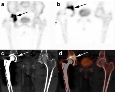
With the increasing clinical availability of SPECT/CT devices, SPECT/CT imaging covering the prosthesis can easily be added with an extra imaging time of approximately 15 min. The resulting anatomical correlation of increased uptake and the supplementary morphological information from the CT component often improves the resulting report and has an impact on therapeutic decision making (Fig.2). The value of SPECT/CT in the evaluation of hip prostheses is striking in daily routine but not well supported by the very limited avail-able literature. Several reviews are currently availavail-able, but prospective focused studies are lacking [27].
The many types of hip prosthesis available and range of different materials and surface conditions have a sub-stantial influence on the interpretation of SPECT/CT images. Increased tracer uptake reflects the biomechanical stress reac-tion of the bone. Thus, knowledge and understanding of the different mechanisms of anchorage of the implants is very helpful. Alignment and orientation of total hip implant components influence bone SPECT/CT uptake distribution. A close collaboration among nuclear medicine physicians, orthopaedic surgeons and radiologists is crucial to find the reason for painful hip prostheses and the best therapeutic approach.
The increased sensitivity of SPECT/CT compared to planar scintigraphic images should not lead to overinterpretation. Generally, all available metabolic and morphological infor-mation given by the SPECT/CT study should be incorporated into the interpretation: location, intensity and pattern of scin-tigraphic activity in all phases often pinpoint the causative lesion. Early-phase uptake can indicate soft-tissue inflamma-tion, prosthesis infection or synovitis. Linear late-phase take can be related to periprosthetic fractures. Increased up-take around the femoral and/or the acetabular component often reflects loosening. In the presence of nonspecific uptake on planar or SPECT images, the dedicated interpretation of the CT part of the study often helps identify corresponding osteolysis, fracture or soft-tissue abnormalities such as calci-fications, ossicalci-fications, joint effusion, ganglion cysts or tu-mours. Malpositioning of the femoral or acetabular compo-nent may be detected and measured on CT images. Especially in older patients without any pathology in the region of the implant, additional lumbar spine or whole-body images can help identify pathologies responsible for radiating pain.
Prosthetic joint infection occurs in 1–2 % of primary and 3–5 % of revision implants [28]. Adding SPECT/CT in patients with suspected prosthetic joint infections, substan-tially improves imaging with99mTc-labelled antigranulocyte antibodies, as shown by Graute et al. [29] in 31 patients. Sensitivity and specificity increased from 66 %/60 % with planar images and 89 %/45 % with SPECT alone to 89 %/ 73 % with SPECT/CT. Filippi and Schillaci reported a signifi-cant clinical contribution of additional SPECT/CT to planar 99m
patients (36 %), including two patients with a knee prosthesis and five patients with a hip prosthesis [30].
In addition to prosthesis imaging, a number of other SPECT/CT applications are promising. SPECT/CT provides
important additional information in patients with suspicion of osteonecrosis or impaired vitality of the femoral head due to trauma (or other reasons) [31]. SPECT/CT is especially help-ful if MR imaging is impaired by metal implants. With new
Fig. 2 A 79-year-old woman with pain in the right hip 6 years after total hip arthroplasty. MIP image (a) and coronal99mTc-DPD
SPECT image (b) show increased tracer uptake in the right acetabulum (arrows) with osteolysis on the CT image (c, arrow) and SPECT/CT image (d, arrow) around the acetabular component. Additionally, material wear was observed at the acetabular cup inlay. No increased uptake is seen around the femoral component. Loosening of the acetabular component was verified intraoperatively
Fig. 3 A 27-year-old woman with pain in the left hip. MIP image (a) and coronal99mTc-DPD SPECT image (b) show focally increased tracer uptake (arrows) in the subtrochanteric region of the femur corresponding to an osteolytic lesion with central nidus on the CT image (c, arrow) and fused SPECT/CT image (d, arrow), characteristic of osteoid osteoma
metal artifact-reduction sequences MR imaging of hip pros-theses has made promising progress and comparative studies between SPECT/CT and MRI are needed to delineate the advantages and disadvantages of the different modalities [32]. Stress fractures and osteoid osteomas (Fig.3) are other entities encountered in the hip that can be imaged adequately with SPECT/CT [33].
Knee
Evaluation of osteoarthritis, osteochondral defects, painful knee prostheses and bone tumours represent the majority of 99m
Tc-DPD SPECT/CT knee examinations performed in daily routine. Conventional radiography is still the first-line imaging modality for the evaluation of the painful knee joint. Often an additional MRI scan is necessary for the evaluation of menisci, ligaments, cartilage and osseous structures. Although MRI covers the majority of pathologies encountered in the knee, SPECT/CT may serve as an important complementary modality, not only in patients with contraindications to MRI. Buck et al. found that bone scan is more sensitive for mechanical bone overload compared to MRI in patients with chronic medial knee pain [34]. By assessing the uptake in the three knee joint compartments (femoropatellar, medial femorotibial, lateral femorotibial) with combined morphological and metabolic information, SPECT/CT provides important information for therapy planning, e.g. for the decision between partial or
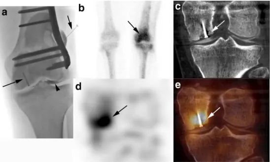
total joint arthroplasty. Recently we introduced SPECT/CT arthrography of the knee to increase the value of SPECT/CT alone. This technique enables the visualization of the articular cartilage, menisci, synovial structures, and loose bodies after intraarticular administration of contrast agent [35]. In our experience this technique can be performed easily and is especially promising in the evaluation of the stability and activity of osteochondral defects (Fig.4).
Painful knee implants are a common clinical problem. Traditionally, conventional three-phase bone scintigraphy is used when conventional radiographs are not sufficient to explain the clinical situation. Taking into consideration that physiological periprosthetic bone turnover leads to increased scintigraphic uptake for at least 1 year after joint replacement, bone scintigraphy has shown good sensitivity (88– 92 %) and specificity (76 – 100 %) in postoperative knee replacement complications [36,37]. The combined SPECT/CT examina-tion of knee implants offers a lot of addiexamina-tional informaexamina-tion about the position of the prosthesis, joint effusion, osteolyses and fractures. Hirschmann et al. implemented a standardized approach for the evaluation of uptake in knee implants [38]. They claimed a high inter- and intraobserver agreement using this approach. They also provided evidence for a diagnostic impact of SPECT/CT in 83 % of 23 consecutive patients with a painful knee implant [39]. Femoropatellar osteoarthritis (11 patients), and loosening of the tibial component (3 patients) and femoral component (2 patients) were the major SPECT/CT diagnoses. A similar approach has been reported by the same
Fig. 4 A 25-year-old man after osteotomy of the left femur and surgical refixation of an osteochondral lesion of the medial femoral condyle 1 year previously. The radiograph shows the needle (short arrow) positioning for injection of intraarticular contrast agent (arrowhead), as well as the small screw fixing the osteochondral fragment (long arrow). Planar bone scan (b) shows increased uptake in the left femur distally (arrow). On the
coronal99mTc-DPD SPECT image (d), increased tracer uptake (arrow) is seen in the medial femoral condyle corresponding to the well-fixed osteochondral fragment on the CT image (c, arrow) and the SPECT/ CT image (e, arrow). The articular cartilage surface is nicely visible and well preserved on SPECT/CT arthrography
group for the evaluation of anterior cruciate ligament grafts, trying to correlate the location of increased uptake with biome-chanical appreciation and clinical symptoms [40,41]. A novel four-dimensional SPECT/CT approach correlates tracer up-take and joint replacement component positioning in patients after total knee arthroplasty, showing excellent inter- and intraobserver reliabilities, and might therefore be the evaluation standard of the future [42,43].
Although SPECT/CT is increasingly used in daily routine with convincing results, well-founded studies addressing the additional value of SPECT/CT are still rare and the level of evidence is limited [44]. Close collaboration among orthopae-dic surgeons and nuclear meorthopae-dicine physicians seems to be essential to integrate all important clinical information and imaging findings into a therapeutic decision.
In cases of benign and malignant bone tumours in the knee joint, such as osteoid osteoma, enchondroma, osteoblastoma, giant cell tumours, sarcomas and others, SPECT/CT provides
important information on the extent and activity of disease and should always be combined with whole body images to ex-clude multifocal bone disease.
Foot
Due to the close proximity of multiple small and complex joints, the clinical evaluation of the painful foot is often challenging. Combining SPECT and high-resolution CT enables the exact anatomical correlation of increased uptake in traumatic, inflam-matory, neoplastic and degenerative disease (including congen-ital variants), e.g. accessory sesamoid bones, tarsal coalition and osteochondrosis dissecans [45]. The assessment of the extent of osteoarthritis is a common clinical request in the foot that can be addressed precisely by SPECT/CT, thus facilitating therapy planning, e.g. corticosteroid injection or arthrodesis. Pagenstert et al. found that the interobserver reliability of
Fig. 5 A 45-year-old patient after pilon fracture of the tibia. Severe activated posttraumatic osteoarthritis and cartilage loss is seen in the anterior part of the ankle joint on the CT image (a), sagittal99mTc-DPD
SPECT image (b) and SPECT/CT arthrography image (c) (arrows). The subtalar joint is well preserved
Fig. 6 An 83-year-old woman with pain in the ankle without a history of trauma. Radiographs (not shown) and initial CT image (a) are normal. Planar
scintigraphy image (b) and sagittal99mTc-DPD SPECT
image (c) obtained 5 days later demonstrate band-like increased uptake (arrows) in the distal tibial metaphysis corresponding to a sclerotic line on the CT image (d, arrow) and fused SPECT/CT (e, arrow) due to an insufficiency fracture
SPECT/CT in the localization of osteoarthritic changes in the foot was excellent (k =0.86), and significantly higher than that of CT and bone scan [46]. Singh et al. investigated 50 patients with SPECT/CT of the foot [47]. In 39 patients (78 %) the SPECT/CT findings led to a modification of the treatment plan. The accuracy, sensitivity, specificity, and posi-tive and negaposi-tive predicposi-tive values of SPECT/CT in this series was 94 %, 95 %, 83 %, 98 % and 71 %, respectively. Chicklore et al. found that SPECT/CT was useful for the evaluation of impingement syndromes and soft-tissue pathologies [48]. SPECT/CT can be used for the postoperative evaluation of the foot after arthrodesis to assess nonunion or the development of osteoarthritis in adjacent joints due to mechanical overload. SPECT/CT can also be used to evaluate bone healing in calca-neal fractures after osteosynthesis, and the development of subtalar joint osteoarthritis. In rare cases of osteoid osteoma in the foot SPECT/CT is the ideal imaging modality to show the nidus and increased uptake, while MRI might even be misleading due to extensive bone marrow oedema rendering the detection of the nidus difficult [49].
As in the wrist and knee joint, SPECT/CT arthrography increases the diagnostic performance of SPECT/CT alone by showing the articular cartilage, synovial changes and loose intraarticular bodies (Fig.5). Contrast agent injection into the ankle joint under fluoroscopic guidance can be performed easily. Our preliminary results in eight ankle joints (unpublished data) support the feasibility of this new technique. Especially in osteoarthritis of the ankle joint and osteochondral lesions of the talus, SPECT/CT arthrography provides important information on bone metabolism, articular cartilage and stability of the osteochondral lesion with an impact on therapy. Achilles tendonitis and plantar fasciitis are other pathologies that may be diagnosed with SPECT/CT. As clinical examination, ultrasonography and MRI are very powerful tools for assessing tendon pathologies, we believe that the major advantage of bone scan is in “staging” the whole skeleton for foci of enthesiopathy in patients with rheumatological disorders suffering from spondyloarthropathy.
Stress fractures to the foot are not rare and are often visualized by the combination of fracture lines or hyperdense areas on CT images and corresponding increased activity on SPECT images (Fig.6). Painful accessory bone syndromes, such as those caused by an os trigonum or accessory navicular bone, show increased uptake on SPECT/CT images and can be treated with injections of local anaesthetic. Tarsal coalition is a deformity resulting from abnormal fibrous, cartilaginous or osseous bridging between two or more tarsal bones. Calcaneonavicular and talocalcaneal coalitions can be missed on conventional radiographs, an CT and even MRI images [50]. Combined metabolic and morphological evaluation of coalitions by SPECT/CT and comparison with the contralateral side—as bilateral involvement occurs in about half of cases— are very helpful in symptomatic patients.
Several studies assessing foot osteomyelitis with SPECT/CT have been published, most often in patients with ,diabetes and/or peripheral arterial disease. Heiba et al. proposed a dual-isotope SPECT/CT protocol using sequential99mTc bone scan and111In leucocyte scanning to evaluate the diabetic foot [5]. This approach was highly accurate and improved the detection and discrimination of soft-tissue infection and osteomyelitis considerably. In another study in 17 patients with 19 clini-cally suspected sites of infection, 99mTc-HMPAO-labelled leucocyte SPECT/CT imaging resulted in a change to the interpretation compared to SPECT and planar images alone in ten sites (53 %), and thus contributed significantly to the correct evaluation [51].
Conclusion
In conclusion, SPECT/CT of the extremities is a very promising field in clinical imaging. To utilize the whole power of metabolic and morphological information in SPECT/CT, profound knowledge of musculoskeletal CT imaging is essen-tial to enable integrated reporting. Although increasingly used in daily routine with convincing results, well-founded litera-ture is still rare.
Acknowledgments We express our gratitude to Geoffrey Warnock, PhD, for his valuable help preparing the manuscript.
Conflicts interest Klaus Strobel received an ISS grant from Philips Healthcare.
Author’s contributions K.S. and M.H. participated in the design and coordination of the review and drafted the manuscript. All authors read and approved the final manuscript.
References
1. Hegenscheid K, Puls R, Rosenberg C. Imaging strategies for knee injuries. Radiologe. 2012;52(11):980–6.
2. Kaur J, Chopra R. Three dimensional CT reconstruction for the evaluation and surgical planning of mid face fractures: a 100 case study. J Maxillofac Oral Surg. 2010;9(4):323–8.
3. Chen KN, Wang G, Cao LG, Zhang MC. Differences of percutaneous retrograde screw fixation of anterior column acetabular fractures between male and female: a study of 164 virtual three-dimensional models. Injury. 2009;40(10):1067–72.
4. Brown GA, Firoozbakhsh K, Gehlert RJ. Three-dimensional CT modeling versus traditional radiology techniques in treatment of acetabular fractures. Iowa Orthop J. 2001;21:20–4.
5. Heiba SI, Kolker D, Mocherla B, Kapoor K, Jiang M, Son H, et al. The optimized evaluation of diabetic foot infection by dual iso-tope SPECT/CT imaging protocol. J Foot Ankle Surg. 2010;49 (6):529–36
6. Yang DC, Ratani RS, Mittal PK, Chua RS, Pate SM. Radionuclide three-phase whole-body bone imaging. Clin Nucl Med. 2002;27 (6):419–26.
7. Pankaj A, Kotwal PP, Mittal R, Deepak KK, Bal CS. Diagnosis of post-traumatic complex regional pain syndrome of the hand: current role of sympathetic skin response and three-phase bone scintigraphy. J Orthop Surg (Hong Kong). 2006;14(3):284–90.
8. Huellner MW, Burkert A, Schleich FS, Schurch M, Hug U, von Wartburg U, et al. SPECT/CT versus MRI in patients with nonspecific pain of the hand and wrist– a pilot study. Eur J Nucl Med Mol Imaging. 2012;39(5):750–9.
9. Linke R, Kuwert T, Uder M, Forst R, Wuest W. Skeletal SPECT/CT of the peripheral extremities. AJR Am J Roentgenol. 2010;194(4): W329–35.
10. Ito S, Yamamoto Y, Tanii T, Aga F, Nishiyama Y. SPECT/CT imaging in ulnocarpal impaction syndrome. Clin Nucl Med. 2013. doi:10.1097/ RLU.0b013e31828da39d
11. Allainmat L, Aubault M, Noel V, Baulieu F, Laulan J, Eder V. Use of hybrid SPECT/CT for diagnosis of radiographic occult fractures of the wrist. Clin Nucl Med. 2013;38(6):e246–51.
12. Schleich FS, Schurch M, Huellner MW, Hug U, von Wartburg U, Strobel K, et al. Diagnostic and therapeutic impact of SPECT/CT in patients with unspecific pain of the hand and wrist. EJNMMI Res. 2012;2(1):53.
13. Tetsumura A, Honda E, Sasaki T, Kino K. Metallic residues as a source of artifacts in magnetic resonance imaging of the temporo-mandibular joint. Dentomaxillofac Radiol. 1999;28(3):186–90. 14. Bagheri MH, Ahmadloo N, Rezaian S. Artifacts in magnetic
reso-nance imaging after surgical resection of brain tumors. Magn Reson Imaging. 2013;31(5):700–2.
15. Henderson RA, Lachiewicz PF. Groin pain after replacement of the hip: aetiology, evaluation and treatment. J Bone Joint Surg Br. 2012;94(2):145–51.
16. Classen T, Zaps D, Landgraeber S, Li X, Jager M. Assessment and management of chronic pain in patients with stable total hip arthroplasty. Int Orthop. 2013;37(1):1–7.
17. Chang EY, McAnally JL, Van Horne JR, Statum S, Wolfson T, Gamst A, et al. Metal-on-metal total hip arthroplasty: do symptoms correlate with MR imaging findings? Radiology. 2012;265(3):848–57. 18. Kruger T, Hug U, Hullner MW, Schleich F, Veit-Haibach P, von
Wartburg U, et al. SPECT/CT arthrography of the wrist in ulnocarpal impaction syndrome. Eur J Nucl Med Mol Imaging. 2011;38(4):792. 19. Hirschmann MT, Schmid R, Dhawan R, Skarvan J, Rasch H, Friederich NF, et al. Combined single photon emission computerized tomography and conventional computerized tomography: clinical value for the shoulder surgeons? Int J Should Surg. 2011;5(3):72–6. 20. Degreef I, Verduyckt J, Debeer P, De Smet L. An unusual cause of shoulder pain: osteoid osteoma of the acromion– a case report. J Shoulder Elbow Surg. 2005;14(6):643–4.
21. Berquist TH. Imaging of joint replacement procedures. Radiol Clin North Am. 2006;44(3):419–37.
22. Temmerman OP, Raijmakers PG, Berkhof J, Hoekstra OS, Teule GJ, Heyligers IC. Accuracy of diagnostic imaging techniques in the diagnosis of aseptic loosening of the femoral component of a hip prosthesis: a meta-analysis. J Bone Joint Surg Br. 2005;87(6):781–5. 23. Temmerman OP, Raijmakers PG, David EF, Pijpers R, Molenaar MA, Hoekstra OS, et al. A comparison of radiographic and scintigraphic techniques to assess aseptic loosening of the acetabular component in a total hip replacement. J Bone Joint Surg Am. 2004;86-A(11): 2456–63.
24. Aliabadi P, Tumeh SS, Weissman BN, McNeil BJ. Cemented total hip prosthesis: radiographic and scintigraphic evaluation. Radiology. 1989;173(1):203–6.
25. Callaghan JJ, Van Nostrand D, Dysart SH, Savory CG, Hopkins WJ. Prospective serial technetium diphosphonate and indium-111 white blood cell labeled imaging in primary uncemented total hip arthroplasty. Iowa Orthop J. 1996;16:104–12.
26. Venesmaa P, Vanninen E, Miettinen H, Kroger H. Periprosthetic bone turnover after primary total hip arthroplasty measured by
single-photon emission computed tomography. Scand J Surg. 2012;101 (4):241–8.
27. Strobel K, Steurer-Dober I, Huellner MW, Veit-Haibach P, Allgayer B. Importance of SPECT/CT for knee and hip joint prostheses. Radiologe. 2012;52(7):629–35.
28. Love C, Marwin SE, Palestro CJ. Nuclear medicine and the infected joint replacement. Semin Nucl Med. 2009;39(1):66–78.
29. Graute V, Feist M, Lehner S, Haug A, Muller PE, Bartenstein P, et al. Detection of low-grade prosthetic joint infections using 99mTc-antigranulocyte SPECT/CT: initial clinical results. Eur J Nucl Med Mol Imaging. 2010;37(9):1751–9.
30. Filippi L, Schillaci O. Usefulness of hybrid SPECT/CT in 99mTc-HMPAO-labeled leukocyte scintigraphy for bone and joint infec-tions. J Nucl Med. 2006;47(12):1908–13.
31. Luk WH, Au-Yeung AW, Yang MK. Diagnostic value of SPECT versus SPECT/CT in femoral avascular necrosis: preliminary results. Nucl Med Commun. 2010;31(11):958–61.
32. Sutter R, Ulbrich EJ, Jellus V, Nittka M, Pfirrmann CW. Reduction of metal artifacts in patients with total hip arthroplasty with slice-encoding metal artifact correction and view-angle tilting MR imag-ing. Radiology. 2012;265(1):204–14.
33. Bryant LR, Song WS, Banks KP, Bui-Mansfield LT, Bradley YC. Comparison of planar scintigraphy alone and with SPECT for the initial evaluation of femoral neck stress fracture. AJR Am J Roentgenol. 2008;191(4):1010–5.
34. Buck FM, Hoffmann A, Hofer B, Pfirrmann CW, Allgayer B. Chronic medial knee pain without history of prior trauma: correlation of pain at rest and during exercise using bone scintigraphy and MR imaging. Skeletal Radiol. 2009;38(4):339–47.
35. Strobel K, Wiesmann R, Tornquist K, Steurer-Dober I, Muller U. SPECT/CT arthrography of the knee. Eur J Nucl Med Mol Imaging. 2012;39(12):1975–6.
36. Sacchetti GM, Ghisellini F, Brambilla M, De Consoli A, Fornara P, Rizzo E, et al. Quantitative scintigraphic evaluation of total knee arthroplasties: a feasibility study. Clin Orthop Relat Res. 1996;325: 181–9.
37. Smith SL, Wastie ML, Forster I. Radionuclide bone scintigraphy in the detection of significant complications after total knee joint replacement. Clin Radiol. 2001;56(3):221–4.
38. Hirschmann MT, Iranpour F, Konala P, Kerner A, Rasch H, Cobb JP, et al. A novel standardized algorithm for evaluating patients with painful total knee arthroplasty using combined single photon emis-sion tomography and conventional computerized tomography. Knee Surg Sports Traumatol Arthrosc. 2010;18(7):939–44.
39. Hirschmann MT, Konala P, Iranpour F, Kerner A, Rasch H, Friederich NF. Clinical value of SPECT/CT for evaluation of patients with painful knees after total knee arthroplasty– a new dimension of diagnostics? BMC Musculoskelet Disord. 2011;12:36.
40. Hirschmann MT, Mathis D, Afifi FK, Rasch H, Henckel J, Amsler F, et al. Single photon emission computerized tomography and conven-tional computerized tomography (SPECT/CT) for evaluation of patients after anterior cruciate ligament reconstruction: a novel standardized algorithm combining mechanical and metabolic information. Knee Surg Sports Traumatol Arthrosc. 2013;21(4):965–74.
41. Hirschmann MT, Mathis D, Rasch H, Amsler F, Friederich NF, Arnold MP. SPECT/CT tracer uptake is influenced by tunnel orien-tation and position of the femoral and tibial ACL graft insertion site. Int Orthop. 2013;37(2):301–9.
42. Rasch H, Falkowski AL, Forrer F, Henckel J, Hirschmann MT. 4D-SPECT/CT in orthopaedics: a new method of combined quantitative volumetric 3D analysis of SPECT/CT tracer uptake and component position measurements in patients after total knee arthroplasty. Skeletal Radiol. 2013;42(9):1215–23.
43. Testa EA, Rasch H, Forrer F, Hirschmann MT. Clinical value of 4D-SPECT/CT in patients with painful total knee arthroplasty. Br J Sports Med. 2013;47(10):e3.
44. Hirschmann MT, Henckel J, Rasch H. SPECT/CT in patients with painful knee arthroplasty– what is the evidence? Skeletal Radiol. 2013;42(9):1201–7.
45. Biersack HJ, Wingenfeld C, Hinterthaner B, Frank D, Sabet A. SPECT-CT of the foot. Nuklearmedizin. 2012;51(1):26–31. 46. Pagenstert GI, Barg A, Leumann AG, Rasch H, Muller-Brand J,
Hintermann B, et al. SPECT-CT imaging in degenerative joint disease of the foot and ankle. J Bone Joint Surg Br. 2009;91(9):1191–6. 47. Singh VK, Javed S, Parthipun A, Sott AH. The diagnostic value of
single photon-emission computed tomography bone scans combined with CT (SPECT-CT) in diseases of the foot and ankle. Foot Ankle Surg. 2013;19(2):80–3.
48. Chicklore S, Gnanasegaran G, Vijayanathan S, Fogelman I. Potential role of multislice SPECT/CT in impingement syndrome and soft-tissue pathology of the ankle and foot. Nucl Med Commun. 2013;34(2):130–9. 49. Davies M, Cassar-Pullicino VN, Davies AM, McCall IW, Tyrrell PN. The diagnostic accuracy of MR imaging in osteoid osteoma. Skeletal Radiol. 2002;31(10):559–69.
50. Newman JS, Newberg AH. Congenital tarsal coalition: multimodality evaluation with emphasis on CT and MR imaging. Radiographics. 2000;20(2):321–32; quiz 526–7, 532.
51. Filippi L, Uccioli L, Giurato L, Schillaci O. Diabetic foot infection: usefulness of SPECT/CT for 99mTc-HMPAO-labeled leukocyte imaging. J Nucl Med. 2009;50(7):1042–6.