DOI 10.1007/s00418-013-1146-1 ORIGINAL PAPER
Reduction in Golgi apparatus dimension in the absence
of a residential protein, N‑acetylglucosaminyltransferase V
Zhizhong Dong · Christian Zuber · Michael Pierce · Pamela Stanley · Jürgen Roth
Accepted: 4 September 2013 / Published online: 28 September 2013 © Springer-Verlag Berlin Heidelberg 2013
cross-sectioned cisternal stacks between Lec4 and con-trol CHO cells, but significantly reduced Golgi stack hits were observed in cross-sectioned Lec4 cells. Therefore, the Golgi apparatus dimensional change in Lec4 and Lec4A cells may be due to a compaction of the organelle.
Keywords Golgi apparatus ·
N-acetylglucosaminyltransferase V ·
N-acetylglucosaminyltransferase I · CHO Lec4 cells · CHO Lec1 cells · β1,6-branched N-glycans · Golgi volume density · Golgi α-mannosidase II
Abbreviations
GlcNAcT-I N-acetylglucosaminyltransferase I GlcNAcT-V, MGAT5 N-acetylglucosaminyltransferase V CHO Chinese hamster ovary cells GlcNAc N-acetylglucosamine
Man Mannose
L-PHA Leukoagglutinating Phaseolus
vulgaris lectin
Dig Digoxigenin
Introduction
In higher eukaryotic cells, the Golgi apparatus forms a continuous branching and anastomosing ribbon consisting of stacks of cisternae bridged by non-compact, fenestrated regions, and tubular networks at its cis and trans face (Day et al. 2013; Klumperman 2011; Martínez-Alonso et al.
2013). Depending on cell-type, the number of cisternae making up the stack and the area comprising the cis and
trans Golgi networks may vary (Han et al. 2013; Rambourg and Clermont 1997; Sengupta and Linstedt 2011; Uemura and Nakano 2013). While structurally complex, the Golgi
Abstract Various proteins are involved in the generation
and maintenance of the membrane complex known as the Golgi apparatus. We have used mutant Chinese hamster ovary (CHO) cell lines Lec4 and Lec4A lacking N-acetyl-glucosaminyltransferase V (GlcNAcT-V, MGAT5) activity and protein in the Golgi apparatus to study the effects of the absence of a single glycosyltransferase on the Golgi appa-ratus dimension. Quantification of immunofluorescence in serial confocal sections for Golgi α-mannosidase II and electron microscopic morphometry revealed a reduction in Golgi volume density up to 49 % in CHO Lec4 and CHO Lec4A cells compared to parental CHO cells. This reduc-tion in Golgi volume density could be reversed by stable transfection of Lec4 cells with a cDNA encoding Mgat5. Inhibition of the synthesis of β1,6-branched N-glycans by swainsonine had no effect on Golgi volume density. In addition, no effect on Golgi volume density was observed in CHO Lec1 cells that contain enzymatically active Glc-NAcT-V, but cannot synthesize β1,6-branched glycans due to an inactive GlcNAcT-I in their Golgi apparatus. These results indicate that it may be the absence of the GlcNAcT-V protein that is the determining factor in reducing Golgi volume density. No dimensional differences existed in
Z. Dong · C. Zuber · J. Roth (*)
Division of Cell and Molecular Pathology, Department of Pathology, University of Zürich, 8091 Zürich, Switzerland e-mail: [email protected]
M. Pierce
Complex Carbohydrate Research Center, Department
of Biochemistry and Molecular Biology, University of Georgia, Athens, GA 30602, USA
P. Stanley
Department of Cell Biology, Albert Einstein College of Medicine, New York, NY 10461, USA
apparatus is a highly dynamic organelle (Boncompain and Perez 2013; Colanzi et al. 1997; Cole et al. 1996; Polish-chuk and Lutsenko 2013; Presley et al. 1998, 2002; Sciaky et al. 1997; Tillmann et al. 2013; Willett et al. 2013) and most important for cellular traffic (Boncompain and Perez
2013; Chia et al. 2013; Farquhar and Hauri 1997; Macha-mer 2013; Polishchuk and Lutsenko 2013; Sandvig et al.
2013; Tillmann et al. 2013; Warren 2013; Willett et al.
2013). Not surprisingly therefore, variations in the size of the Golgi apparatus or parts thereof have been reported to depend on the functional state of the cell and to become altered under disease conditions (Clermont et al. 1995; Fujita et al. 2002; Griffiths et al. 1989; Maeda et al. 2008; Noske et al. 2008; Rambourg et al. 1993; Sengupta and Linstedt 2011; Stieber et al. 1998).
There is much interest in understanding the molecular mechanisms responsible for generating and maintaining the integrity of the Golgi apparatus, and various types of pro-teins involved in this process have been identified. While microtubules and associated proteins are important for positioning the Golgi apparatus (Kreis et al. 1997; Presley et al. 1997; Zhu and Kaverina 2013), microtubule disas-sembly results in Golgi apparatus vesiculation (Thyberg and Moskalewski 1999). Cytoplasmic dynein and probably other motor proteins as well as actin filaments seem to be addi-tionally involved in the formation and maintenance of Golgi apparatus structure (Allan 1996; Burkhardt 1998; Dippold et al. 2009; Egea et al. 2006, 2013; Harada et al. 1998; Yadav et al. 2012). Much information about the Golgi stack reassembly has been obtained through studies on the Golgi apparatus during mitosis (Acharya and Malhotra 1996; Barr and Warren 1996; Rabouille and Kondylis 2007; Shorter and Warren 2002). Golgi reassembly stacking proteins (Barr et al. 1997; Feinstein and Linstedt 2008; Puthenveedu et al.
2006; Sengupta et al. 2009; Shorter et al. 1999; Xiang and Wang 2010), a cis Golgi matrix protein, GM130 (Lowe et al. 1998; Marra et al. 2007; Nakamura et al. 1995, 1997), an NSF-like ATPase, p97, and NSF together with SNAPs and p115, a vesicle docking protein (Nelson et al. 1998; Rabouille et al. 1995b, 1998), seem to be important for the formation of the cisternal stack. In interphase cells, proteins cycling between the endoplasmic reticulum and the Golgi apparatus, such as Rab1b (Haas et al. 2007; Monetta et al.
2007; Romero et al. 2013; Tomas et al. 2012; Wilson et al.
1994), Arf1 (Boal et al. 2010; Lin et al. 2011; Manolea et al.
2008; Zhang et al. 1994) and TAP/p115 (Nelson et al. 1998; Puthenveedu and Linstedt 2001; Radulescu et al. 2011), are involved in maintaining Golgi apparatus morphology. Fur-thermore, the spectrin membrane skeleton (Nelson et al.
1998) is required for Golgi apparatus architecture.
Glycosyltransferases are Golgi residential proteins (Dunphy and Rothman 1983; Goldberg and Kornfeld 1983; Roth and Berger 1982; Roth et al. 1985), and the activity
of a particular subset results in the synthesis of complex
N-glycans (Kornfeld and Kornfeld 1985; Zuber and Roth
2009). Together with other proteins, glycosyltransferases seem to be involved in the formation and maintenance of the Golgi apparatus architecture. An illustrative example for this function was provided by studies on the parasitic protozoan Giardia lamblia (Lujan et al. 1995). During dif-ferentiation from throphozoites to cysts, the developmen-tal induction of Golgi enzyme activities correlated with the appearance of a morphologically identifiable Golgi appara-tus, which was absent in non-encysting cells. There are also data that N-acetylglucosaminyltransferase I (GlcNAcT-I) is involved in maintaining the structure of the cisternal stack (Nilsson et al. 1996). Replacement of part or the entire membrane-spanning domain of this type II membrane pro-tein (Kumar et al. 1990) with leucine residues transformed the part of the Golgi stack housing the mutated GlcNAcT-I from flat cisternae into tubulo-vesicular membranes.
A number of Chinese hamster ovary (CHO) cells defec-tive in glycosylation, due to mutated glycosyltransferases, have been isolated and characterized (Stanley and Ioffe
1995). In the CHO Lec4 and Lec4A cell mutants, N-acetyl-glucosaminyltransferase V (GlcNAcT-V) activity is affected (Chaney et al. 1989; Weinstein et al. 1996). Glc NAcT-V adds a β1,6 N-acetylglucosamine (GlcNAc) branch to the core α1,6-mannose (Man) in complex N-linked glycans attached to proteins. Lec4 cells lack GlcNAcT-V activity, due to a base insertion at nucleotide 822 of the Magt5 gene that shifts the open reading frame. A 155 amino acid trun-cated GlcNAcT-V (instead of a full length 740 amino acid enzyme) may be synthesized, which consists of the cyto-solic and transmembrane domains and a short piece of the stem region. The fate of this truncated GlcNAcT-V is not known, but recognition by the protein quality control and subsequent proteolysis by ER-associated protein degrada-tion may occur, as shown for other proteins (Roth et al.
2010). By contrast, CHO Lec4A cells have GlcNAcT-V activity equivalent to that of parental CHO cells in cellular homogenates. However, a single-point mutation, changing leucine to arginine at position 188, causes GlcNAcT-V to mislocalize to the endoplasmic reticulum and consequently to be functionally inactive in vivo. In both CHO Lec4 and Lec4A cells, complex N-glycans lack a β1,6 GlcNAc branch. In addition, the Golgi apparatus in Lec4A, and probably also in Lec4 mutant cells, lacks the residential protein GlcNAcT-V.
The structure of the Golgi apparatus in cells stably trans-fected and therefore overexpressing different glycosyltrans-ferases appears normal (Lee et al. 1989; Nilsson et al. 1993; Rabouille et al. 1995a). However, more recent quantitative studies (Guo and Linstedt 2006) provided evidence that overexpression of the Golgi glycosyltransferase GlcNAcT-II results in a 1.3-fold increase in the size of the Golgi apparatus
and that this change is sustained depending on interaction of GlcNAcT-II with COPII-coated transport carriers.
Currently, there is no information on the effects on Golgi apparatus structure and size of eliminating a Golgi residen-tial protein such as a glycosyltransferase. Here, we dem-onstrate that the absence of the Golgi apparatus residential protein GlcNAcT-V causes a reduction in the dimensional size of the Golgi apparatus without affecting its general architecture.
Materials and methods
Cell lines
Chinese hamster ovary mutant cell lines Pro−Lec4.7b,
Pro−Lec4A.12.2, Pro−5, and Pro–5 Lec1.3c were
previ-ously characterized (Chaney et al. 1989; Puthalakath et al.
1996; Stanley et al. 1982; Weinstein et al. 1996). Cells were maintained in α-MEM supplemented with 10 % fetal calf serum, glutamine and DNA, and RNA precursors. Transfection of Lec4 cells with Mgat5 cDNA
Rat Mgat5 cDNA (Shoreibah et al. 1993) was subcloned into the EcoRI site of the pcDNA3 expression vector (Inv-itrogen, San Diego, CA). Lec4 cells were plated on 35 mm cell culture dishes and grown in α-MEM medium supple-mented with 10 % fetal calf serum. Plasmid DNA (1 μg per 35 mm culture dish) was transfected into Lec4 cells, using Fugene™ 6 transfection reagent (Boehringer Man-nheim, Germany) according to the manufacturer’s protocol. Following transfection, clonal cell lines were established by selection for G418 resistance and tested for cell sur-face expression of β1,6 GlcNAc-branched N-glycans using digoxigenin-conjugated leukoagglutinating Phaseolus
vul-garis lectin (dig L-PHA; Boehringer Mannheim, Germany)
as described below. The positive clonal cell lines were des-ignated Lec4 GnTV-N5, Lec4 GnTV-N10, and Lec4GnTV-N30. Lec4 cells were mock-transfected with the pcDNA3 expression vector as described above, and clonal cell lines were established and designated Lec4 pcDNA3.
GlcNAcT-V assay
Cells were grown to confluence, harvested in 50 mM PBS, and concentrated to a pellet by centrifugation in microfuge tubes. Cell pellets were frozen on dry ice and shipped to Michael Pierce (Athens, GA, USA). For the assay, an approximately equal volume of ice-cold buffer (0.1 M MES, pH 6.5) was added to each pellet, followed by rapid thawing and sonication as described (Palcic et al. 1990). Each assay tube contained 106 cpm of UDP-[3H]-GlcNAc (25 cpm/
pmol) and 10 nmol of synthetic trisaccharide acceptor (octyl
6–O–[2–O–(2-acetamido-2-deoxy-β-d-glucosyl-pyranosyl)-α-d-mannopyranosyl]-β-d-glucopyranoside) that were dried
under vacuum in a 1.5-ml tube. The dried contents of each tube were resuspended in 0.05 ml of assay buffer (0.05 mM MES, pH 6.5, 2.0 % Triton X-100). Next, 5 μl of cell lysate was added to the tube, and the contents gently mixed by pipetting. Tubes were incubated for 6 h at 37 °C. Reactions were quenched by adding 0.5 ml water. Radiolabeled prod-uct was isolated using a C18 Sep-Pak (Waters) cartridge, eluted with 2 ml of methanol, and subjected to scintillation counting. Assay values for each extract were conducted in duplicate or triplicate and averaged. Aliquots of cell lysates were removed prior to enzymatic assay for protein determi-nation in triplicate using the Bio-Rad protein assay for cal-culation of GlcNAcT-V specific activities.
Detection of β1,6-branched N-glycans with L-PHA For lectin blots (Zuber et al. 1998), cells grown to 50– 80 % confluence were removed by EDTA treatment (0.1 % EDTA-PBS at 37 °C for 10 min), pelleted by centrifu-gation (1,500×g, 5 min at 4 °C) and lysed in three vol-umes of 1 % Triton X-100 containing 1 mM AEBSF and 1 % aprotinin on ice for 30–40 min. After centrifugation (8,000×g, 10 min at 4 °C), the supernatant was denatured by boiling in Laemmli buffer. Samples were electropho-retically resolved in 3–10 % gradient polyacrylamide gels (120 μg protein per lane) and transferred to nitrocellu-lose. After blocking in 0.05 % Tween 20 and 1 % BSA in PBS, nitrocellulose strips were incubated with dig L-PHA (2 μg/ml for 2 h), rinsed, and incubated in alkaline phos-phatase-conjugated polyclonal sheep anti-dig (Fab)2 frag-ments (5,000-fold diluted, Boehringer Mannheim, Ger-many) for 1 h. Alkaline phosphatase activity was revealed by the NBT-BCIP color reaction according to the manu-facturer’s instructions.
For light microscopic cell surface staining, cells grown on coverslips were fixed in 2 % paraformaldehyde in Ear-le’s balanced salt solution (EBSS) (pH 7.2–7.4) for 5 min at 37 °C, and then at ambient temperature for 25 min, and the fixation was stopped by immersing the cover slips in 50 mM NH4Cl in PBS for 30 min (Roth et al. 1989). The coverslips were blocked in 2 % BSA in PBS for 30 min, incubated with digoxigenated (dig) L-PHA (2 μg/ml) for 2 h, followed by three rinses in PBS containing 0.1 % BSA (5 min each) and rhodamine-conjugated sheep anti-digox-igenin (Fab)2 fragments (200-fold diluted in 2 % BSA in PBS) for 1 h. After rinses, coverslips were embedded in Mowiol and mounted on glass slides. Cells were observed with a Leica TCS4D confocal laser scanning microscope.
For scanning electron microscopy, cells grown on glass coverslips were fixed in 2 % paraformaldehyde–0.1 %
glutaraldehyde in EBSS as described above. Cells were incubated with directly 8 nm gold particle-labeled L-PHA (absorbance at 525 nm = 0.12) for 1 h. Following rinses and critical point drying, samples were coated and observed in a scanning electron microscope under standard and back-scattering mode (Hermann et al. 1996).
Golgi α-mannosidase II immunostaining and morphometric analysis
Cells grown on coverslips were fixed as described above and permeabilized with PBS containing 0.15 % saponin and 0.1 % BSA for 15 min. After rinses with buffer, the cover-slips were incubated with rabbit anti-Golgi α-mannosidase
ΙΙ (1,000-fold diluted in PBS containing 0.5 % saponin and
0.1 % BSA; antibody kindly provided by Dr. Kelley More-men, University of Georgia, Athens, GA) for 2 h, rinsed in PBS containing 0.1 % BSA, and incubated in rhodamine-conjugated affinity-purified goat anti-rabbit IgG (1,000-fold diluted in 0.1 % BSA-PBS; Jackson ImmunoResearch Laboratories Inc., West Grove, PA) for 1 h, rinsed with 0.1 % BSA in PBS and distilled water and embedding in Mowiol. After storage overnight at 4 °C, the cells were observed under a Leica TCS4D confocal laser scanning microscope. Cells grown in the presence of swainsonine (see below) were processed in the same way.
We estimated Golgi apparatus volume density by confocal laser scanning microscopy based on differential voxel count-ing of consecutive serial optical sections. As per each experi-mental set, three different views containing 30–40 cells were recorded randomly with a 40x objective and consecutive serial optical sections (0.4-μm optical resolution and optical section distance) were taken from the top to the bottom of each cell. The digitized images were analyzed using Photo-shop®. Since Golgi α-mannosidase II has a broad Golgi
dis-tribution in CHO cells (Velasco et al. 1993), it was used to delineate the Golgi apparatus by immunofluorescence. The entire cells could be discerned by the presence of a faint cytoplasmic fluorescence. Using a histogram program, the sum of pixels for Golgi α-mannosidase II immunofluores-cence and for the cytoplasmic fluoresimmunofluores-cence was obtained separately. From the ratio of the sum of pixels for Golgi
α-mannosidase II immunofluorescence and the sum of pixels
for the remaining cytoplasm, the volume density of the Golgi apparatus was calculated using the following equation:
In this equation, v/V (G/C) represents the volume density of the Golgi apparatus; m and n represent the initial gray level for the Golgi apparatus (G) and cytoplasm (C), respectively; and gi represents the number of pixels at each gray level i.
v/V (G/C) =
i=255 i=m gi
i=255 i=n gi
Transmission electron microscopy and morphometric analysis
Cells grown on Thermanox coverslips (Nalge Nunc. Inter-national, Roskilde, Denmark) were fixed in 2 % paraform-aldehyde–1 % glutaraldehyde in EBSS (pH 7.2–7.4) for 5 min at 37 °C and then at ambient temperature for 25 min. After rinses with buffer and immersion in 50 mM NH4Cl in PBS for 30 min (Roth et al. 1989), cells were postfixed in 1 % reduced osmium tetroxide (Karnovsky 1971) for 30 min, rinsed in distilled water and stained with 2 % aque-ous uranyl acetate for 15 min. The cells were embedded in Epon according to standard protocol. Ultrathin sections were contrasted with uranyl acetate and lead citrate. From each cell line, photographs were taken randomly from 30 to 40 cross-sectioned cells. In addition, photographs at the original magnification of 25,000 were taken from each cell containing the Golgi apparatus in the cross section. The number of cisternae per Golgi stack, the length and thick-ness of cisternal stacks, as well as the area occupied by the Golgi stacks and the size of the cell profile in the cross sec-tions were measured on digitized photographic negatives. The volume density of the Golgi apparatus was calculated by the ratio of the surface area of the Golgi stacks, and the surface size of the cell profile in the cross sections (Weibel
1979).
Swainsonine treatment of cell lines and lectin spot plots Swainsonine (1 μg/ml; Sigma Chemical Co, Ltd., St. Louis, MO) was added to the culture medium of cells growing at 30–40 % confluence, and the cells were ana-lyzed 12 and 24 h after addition of the drug. For L-PHA spot blot analysis, cells were harvested and lysed as described above. About 1 μl aliquots of serially diluted cell lysates were pipetted onto nitrocellulose strips and air-dried, and β1,6-branched N-glycans detected with dig L-PHA as described above. The quantitative analysis was performed with Wincam® software (Camtek Oy, Vantaa,
Finland). Using the gray levels of the serial dilutions of the zero time-point homogenates as reference, the gray levels (relative percentage) of the spots from the homogenates of the cells treated with swainsonine for 12 and 24 h were estimated.
Results
Expression of β1,6-branched N-glycans in various CHO cell lines
Parent and mutant CHO cell lines were studied by confo-cal laser scanning microscopy and lectin-gold scanning
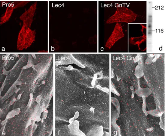
electron microscopy as well as by lectin blotting using the leukoagglutinating Phaseolus vulgaris lectin (L-PHA) to detect β1,6-branched complex N-glycans (Cummings and Kornfeld 1982), the product of GlcNAcT-V. While wild-type CHO Pro−5 cells were reactive with L-PHA,
CHO Lec4 and Lec4A mutant cells lacking GlcNAcT-V and mock-transfected Lec4pcDNA3 cells were unreactive (Fig. 1a, b, e, f). The three clonal cell lines stably express-ing GlcNAcT-V exhibited cell surface L-PHA stainexpress-ing undistinguishable from that observed for CHO Pro−5 cells
(Fig. 1c, g). In L-PHA blots, two major bands at 140 and 85 kDa and several minor bands were evident in parent CHO and Lec4 GlcNAcT-V cells lines (Fig. 1d), but CHO Lec4, CHO Lec4A, and mock-transfected CHO Lec4 cell glycoproteins did not bind L-PHA (data not shown). Dif-ferent GlcNAcT-V activity levels were found in the trans-fected cell lines (data not shown). The Lec4 GnTV-N5 transfectants had enzyme activity similar to CHO Pro−5
cells and, based on densitometric evaluation of spot blots, synthesized similar amounts of β1,6-branched N-glycans. Therefore, CHO Lec4 GnTV-N5 cells were used in most comparative analyses.
Golgi apparatus dimension is reduced in the absence of GlcNAcT-V
To measure the dimension of the Golgi apparatus at the light microscopic level, immunofluorescence for Golgi
α-mannosidase II, a cis/medial Golgi resident protein in
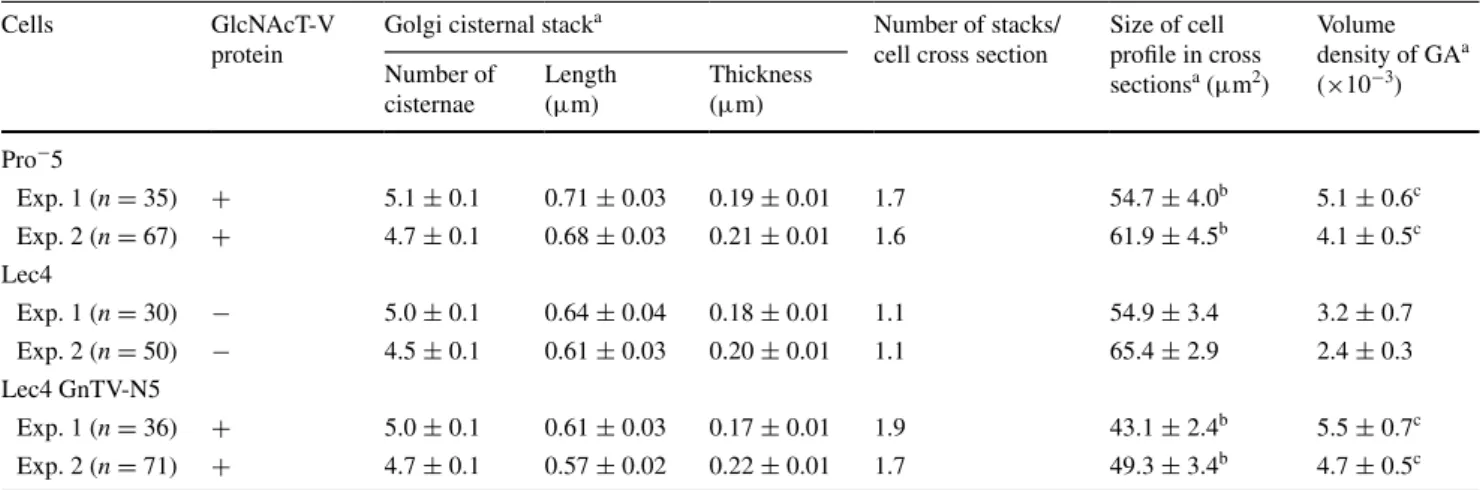
CHO cells (Velasco et al. 1993), was quantified by confocal laser scanning microscopy (Fig. 2). As detailed in Table 1, the volume density of the Golgi apparatus of CHO Lec4 and CHO Lec4A cells, as well as of CHO Lec4 pcDNA3 cells, was significantly smaller to that of CHO parental cells and of the three clonal CHO Lec4 GnTV cell lines. By electron microscopy, no differences in the structure of the Golgi apparatus were apparent between CHO Pro−5
(Fig. 3a), CHO Lec4 (Fig. 3b), and CHO Lec4 GnTV (Fig. 3c). The results of the electron microscopic morpho-metric analyses are presented in Table 2 and confirm the subjective impression. The mean number of Golgi stack hits per cross-sectioned CHO Lec4 cells was reduced by 31–42 % as compared to CHO Pro−5 and CHO Lec4
GnTV cells. Therefore, the volume density of the Golgi apparatus was significantly smaller in CHO Lec4 cells
Fig. 1 Demonstration of β1,6-branched N-glycans on the CHO cell surface using dig L-PHA and confocal laser scanning microscopy (a–c) and lectin-gold scanning electron microscopy (e–g). The CHO Pro−5 cells (a, e) and CHO Lec4 GnTV-N5 cells (c, g) showed
posi-tive cell surface staining with microvilli being strongly posiposi-tive. In
contrast, CHO Lec4 cells (b, f) showed no specific cell surface labe-ling. ×1,100 (a–c), ×9,000 (e–g). Dig L-PHA—blot of homogenates from CHO Lec4 GnTV cells revealed two major bands at 140 and 85 kDa and several minor bands (d)
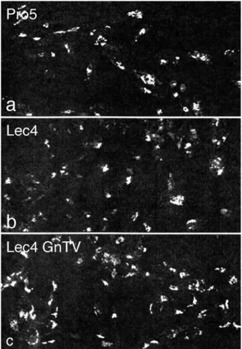
Fig. 2 Confocal laser scanning immunofluorescence for Golgi
α-mannosidase II. CHO Pro−5 cells (a), CHO Lec4 cells (b), and
CHO Lec4 GnTV-N5 cells (c) exhibit a typical perinuclear Golgi staining pattern in a single confocal optical section (×560)
Table 1 Morphometric analysis of the Golgi mannosidase II immunofluorescence
a Volume density of Golgi apparatus, data are expressed as mean ± SD. Three different views were evaluated in each experiment b Reduction rate of the volume density of the Golgi apparatus in Lec4 cells as compared to other cell lines
c Indicates significant difference as compared with Lec4, Lec4 PcDNA3, or Lec4A cells (p < 0.01) d Indicates significant difference as compared with Lec4, Lec4 PcDNA3, or Lec4A cells (p < 0.05) e GlcNAcT-V protein mislocated in the endoplasmic reticulum
Cells GlcNAcT-V
protein
Exp. 1 Exp. 2 Exp. 3
v/Va R (%)b v/Va R (%)b v/Va R (%)b Pro−5 + 30 ± 1c 43 27 ± 4c 44 28 ± 1c 36 Lec4 − 17 ± 1 – 15 ± 1 – 18 ± 1 – Lec4 GnT-N5 + 25 ± 1c 32 26 ± 1c 42 27 ± 3c 33 Lec4 GnT V-N10 + 25 ± 5d 32 – – Lec4 GnT V-N30 + 30 ± 5c 43 – – Lec4 PcDNA3 − 17 ± 4 – – – Lec4A +e 18 ± 1 6 – –
Fig. 3 Transmission electron micrographs showing the morphology of the Golgi apparatus (GA) in CHO Pro−5 cells (a), CHO Lec4 cells
(b), and CHO Lec4 GnTV-N5 cells (c). Note the structural similar-ity in appearance of the Golgi apparatus in the different cells lines. N part of the nucleus (×41,300)
(reduced by 37–49 %) as compared to CHO Pro−5 and
CHO Lec4 GnTV cells. Interestingly, the mean number of cisternae per stack, as well as the horizontal and vertical dimension of the Golgi cisternal stacks, was similar in all cell lines studied (Table 2). Therefore, the most important contribution for the reduced volume density of the Golgi apparatus in CHO Lec4 cells came from the reduced Golgi stack hits in cross-sectioned cells. In order to obtain more insight into this phenomenon, we analyzed the distribu-tion pattern of several other parameters. We found that the number of cisternae per stack, as well as the horizontal and vertical dimension of the Golgi stacks, had a Poisson bution for each cell line (not shown). However, the distri-bution of the Golgi apparatus volume density was consid-erably different between CHO Lec4 cells and CHO Pro−5
as well as CHO Lec4 GnTV cells (Fig. 4). The number
of cross-sectioned cells not exhibiting Golgi stacks was much higher in Lec4 cells than in Pro−5 and Lec4 GnTV.
N5 cells. This parameter was the main contributor to differ-ences in the mean value of Golgi apparatus volume density (Table 3).
Golgi apparatus dimension is not influenced by the absence of GlcNAcT-V glycosylation product
At this point of the study, the question arose: Is the physi-cal absence of the GlcNAcT-V protein in the Golgi appa-ratus, and/or the lack of its enzymatic activity and gly-cosylation products, the basis for the changes in Golgi apparatus dimension in Lec4 cells? Unfortunately, no spe-cific GlcNAcT-V inhibitor was available for in vivo studies with intact cells. Thus, to address this question indirectly, we investigated CHO Lec1 cells, which contain an enzy-matically inactive GlcNAcT-I protein in the Golgi appa-ratus (Kumar et al. 1990; Puthalakath et al. 1996; Stanley et al. 1975). As a consequence, no acceptor substrate for GlcNAcT-V is synthesized in Lec1 cells. Thus, although
Table 2 Morphometric analysis of the Golgi apparatus by electron microscopy
a Data are expressed as mean ± SEM
b Indicates significant difference as compared with Lec4 cells (p < 0.01, Wilcoxon test) within the same experiment c Indicates significant difference as compared with Lec4 cells (p < 0.05, Wilcoxon test) within the same experiment
Cells GlcNAcT-V
protein
Golgi cisternal stacka Number of stacks/ cell cross section
Size of cell profile in cross sectionsa (μm2) Volume density of GAa (×10−3) Number of cisternae Length (μm) Thickness (μm) Pro−5 Exp. 1 (n = 35) + 5.1 ± 0.1 0.71 ± 0.03 0.19 ± 0.01 1.7 54.7 ± 4.0b 5.1 ± 0.6c Exp. 2 (n = 67) + 4.7 ± 0.1 0.68 ± 0.03 0.21 ± 0.01 1.6 61.9 ± 4.5b 4.1 ± 0.5c Lec4 Exp. 1 (n = 30) − 5.0 ± 0.1 0.64 ± 0.04 0.18 ± 0.01 1.1 54.9 ± 3.4 3.2 ± 0.7 Exp. 2 (n = 50) − 4.5 ± 0.1 0.61 ± 0.03 0.20 ± 0.01 1.1 65.4 ± 2.9 2.4 ± 0.3 Lec4 GnTV-N5 Exp. 1 (n = 36) + 5.0 ± 0.1 0.61 ± 0.03 0.17 ± 0.01 1.9 43.1 ± 2.4b 5.5 ± 0.7c Exp. 2 (n = 71) + 4.7 ± 0.1 0.57 ± 0.02 0.22 ± 0.01 1.7 49.3 ± 3.4b 4.7 ± 0.5c
Fig. 4 The distribution pattern of volume density of the Golgi appa-ratus in the various CHO cell lines. The number of cross-sectioned cells containing no cisternal stacks was much higher in CHO Lec4 cells than in CHO Pro−5 as well as CHO Lec4 GnTV-N5 cells
Table 3 Mean values of Golgi apparatus volume density with or without Golgi stack hits in cross-sectioned cells
a Calculations were performed by considering only cross sections containing Golgi apparatus
b Calculations were performed by including cross sections with and without Golgi apparatus hits
Cells Mean volume density of
Golgi apparatusa × 10−3 Mean volume density of Golgi apparatusb × 10−3
Pro−5 5.1 6.1
Lec4 3.2 5.6
they contain enzymatically active GlcNAcT-V in their Golgi apparatus, β1,6-branched N-glycans cannot be syn-thesized. We found that the volume density of the Golgi apparatus in CHO Lec1 cells, based on the evaluation of Golgi α-mannosidase II immunofluorescence, was not dif-ferent from that of Pro−5 or Lec4 GnTV cells (Table 4).
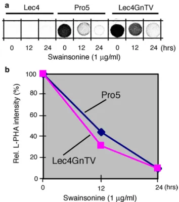
Second, we used swainsonine, an inhibitor of Golgi
α-mannosidase II activity, to block the synthesis of
accep-tor substrate for GlcNAcT-V. Both CHO Pro−5 cells and
CHO Lec4 GnTV cells treated for 24 h with 1 μg/ml of swainsonine contained less than 10 % L-PHA reactivity, as compared to control or swainsonine-treated CHO Lec4 cells (Fig. 5). However, the swainsonine treatment had no influence on the dimension of the Golgi apparatus in CHO Pro−5 and CHO Lec4 GnTV cells, or in CHO Lec4 cells
(Table 4).
Discussion
The present studies were undertaken to investigate whether the absence of a single glycosyltransferase protein, and/or the product of its transferase activity, has an influence on the morphology of the Golgi apparatus. For this, we have made use of CHO cells lines with mutations in glycosyl-transferase genes (Patnaik and Stanley 2006) and have focused on GlcNAcT-V. GlcNAcT-V with a molecular mass of 95 kDa is a comparatively large glycosyltrans-ferase of the Golgi apparatus (Shoreibah et al. 1993). It initiates the synthesis of tri- and tetra-antennary complex
N-glycans, whose expression is associated with malignancy and the metastatic potential of tumor cells (Dennis 1999; Dennis et al. 1987; Granovsky et al. 2000; Partridge et al.
2004; Seelentag et al. 1998). The absence of tri- and tetra-antennary, β1,6-branched N-glycans has no influence on the viability of CHO Lec4 and CHO Lec4A cells although
they differ in gross morphology from the parental cells (Stanley and Sudo 1981). The present study demonstrates that the defect in CHO Lec4 and CHO Lec4A cells is asso-ciated with a significant decrease in the volume density of their respective Golgi apparatus. This defect could be fully compensated by stable transfection of a cDNA encoding GlcNAcT-V into CHO Lec4 cells. The analysis of Golgi apparatus dimension in cell lines treated with swainso-nine to block the synthesis of β1,6-branched N-glycans, or in CHO Lec1 cells, which cannot synthesize the acceptor substrate for GlcNAcT-V, provides strong evidence that the observed change of volume density in CHO Lec4 and CHO Lec4A cells does not simply result from the synthe-sis of truncated N-glycans. Rather it seems that the absence of the GlcNAcT-V protein in these cell lines leads to the change in Golgi apparatus volume density. This assumption is also supported by our analysis of CHO Lec1 cells, which contain enzymatically inactive Golgi apparatus located Glc-NAcT-I (Puthalakath et al. 1996) and which have the same Golgi apparatus dimension as the parental CHO Pro−5 cell
line. In this context, it is interesting to note that the Golgi apparatus in wild-type yeast Saccharomyces cerevisiae lacks clearly defined stacks of cisternae and appears in the form of single cisternae or individual networks of tubules
Table 4 The volume density of the Golgi apparatus in various CHO cell lines following swainsonine treatment
a Data are expressed as mean ± SD and based on three different eval-uations each comprising three different randomly taken views con-taining 30–40 cells
b Cells were treated for 24 h with 1 μg/ml swainsonine
c Indicates significant difference as compared with Lec4 or Lec4A cells (p < 0.01, x2 test, t test)
Cells GlcNAcT-V
protein Volume density (×10
−3)a Control Swainsonine treatmentb Pro−5 + 27 ± 4c 26 ± 4c Lec4 − 15 ± 3 16 ± 3 Lec4 GnTV-N5 + 27 ± 3c 26 ± 3c Lec1 + 26 ± 3c –
Fig. 5 Reduction in the β1,6-branched N-glycans in CHO cells after swainsonine treatment. Swainsonine (1 μg/ml) was added to the cell culture medium, and cells were collected after 0, 12, and 24 h of treatment. Homogenates were processed for L-PHA spot blots, and the results densitometrically analyzed. The CHO Lec4 cells served as control
(MorinGanet et al. 1998; Preuss et al. 1992; Rambourg et al. 1995). It is tempting to speculate that the structurally simple Golgi apparatus of yeast cells may be related to the low number of Golgi glycosyltransferases, essentially five mannosyltransferases (Herscovics and Orlean 1993; Lehle and Tanner 1995), as compared to the large families of gly-cosyltransferases existing in the Golgi apparatus of higher eukaryotic cells.
The reduction in volume density of the Golgi apparatus by up to 49 % in the absence of GlcNAcT-V is surpris-ing even if one considers its molecular mass of 95 kDa (Shoreibah et al. 1993). We noticed from electron micro-scopic analyses that the number of cisternae per Golgi stacks, as well as the length and the thickness of the Golgi cisternal stacks, was the same in parental and the CHO Lec1 cell mutant. Thus, in contrast to the replace-ment of part or the entire membrane-spanning domain of GlcNAcT-I by leucine, which disrupted part of cisternal structure (Nilsson et al. 1996), the absence of GlcNAcT-V protein was compatible with normal cisternal struc-ture. However, our quantitative analysis revealed that the mean number of Golgi stack hits per cell cross section was clearly reduced in CHO Lec4 cells as compared to CHO Pro−5 cells and CHO Lec4 cells stably expressing
GlcNAcT-V.
How could these data be related to the measured reduc-tion in the volume density of the Golgi apparatus in CHO Lec4 cells? All current evidence indicates that the inter-phase Golgi apparatus in higher eukaryotic cells can be regarded as a single organelle composed of numerous dynamic and interrelated units (Hermo and Smith 1998; Rambourg and Clermont 1997). Non-compact regions composed of tubules link these units, which correspond to compact cisternal regions, to each other laterally, and there-fore forming a twisting ribbon that bifurcates and rejoins. Although there can be variability in the compact cisternal regions, the mean number of cisternae in one stack, and the size of the units, can be considered to be cell-type specific and relatively constant under steady-state conditions. One possible explanation for the present results could be that the unit size of the Golgi apparatus is reduced in the absence of GlcNAcT-V. Although a reduction in the number of Golgi apparatus units cannot be ruled out, this seems rather unlikely. A more likely explanation, which would account for the reduction in Golgi apparatus volume density, could be a change in the overall three-dimensional shape of the organelle, resulting in a more compact structure in CHO Lec4 and CHO Lec4A cells. The absence of GlcNAcT-V may result in secondary effects on, for example, interac-tions with intercisternal matrix proteins and other protein– protein interactions, which have been proposed to be of importance for Golgi apparatus structure (Sengupta and Linstedt 2011).
In summary, our results indicate that the absence of the Golgi residential protein GlcNAcT-V results in the adaptive reduction in volume density of the Golgi apparatus without altering its overall architecture. Although the mechanism leading to this phenomenon remains to be clarified, this finding adds a novel aspect to the structural flexibility of this dynamic steady-state organelle.
Acknowledgments We are grateful to Kelley Moremen for the Golgi α-mannosidase II antibody. This work was supported by the Swiss National Science Foundation (to J. R.) and the National Cancer Institute RO1 36434 (to P. S.) and partially supported by the Albert Einstein Cancer Center NCI PO1 13330 (to P. S.). P. S. thanks Subha Sundaram for technical assistance.
References
Acharya U, Malhotra V (1996) Reconstitution of Golgi stacks from vesiculated Golgi membranes in permeabilized cells. Semin Cell Dev Biol 7:511–516
Allan V (1996) Role of motor proteins in organizing the endoplasmic reticulum and Golgi apparatus. Semin Cell Dev Biol 7:335–342 Barr FA, Warren G (1996) Disassembly and reassembly of the Golgi
apparatus. Semin Cell Dev Biol 7:505–510
Barr FA, Puype M, Vandekerckhove J, Warren G (1997) GRASP65, a protein involved in the stacking of Golgi cisternae. Cell 91:253–262
Boal F, Guetzoyan L, Sessions RB, Zeghouf M, Spooner RA, Lord JM, Cherfils J, Clarkson GJ, Roberts LM, Stephens DJ (2010) LG186: an inhibitor of GBF1 function that causes Golgi disas-sembly in human and canine cells. Traffic 11:1537–1551 Boncompain G, Perez F (2013) The many routes of Golgi-dependent
trafficking. Histochem Cell Biol 140:251–260
Burkhardt JK (1998) The role of microtubule-based motor proteins in maintaining the structure and function of the Golgi complex. Biochim Biophys Acta Mol Cell Res 1404:113–126
Chaney W, Sundaram S, Friedman N, Stanley P (1989) The Lec4A CHO glycosylation mutant arises from miscompartmentaliza-tion of a Golgi glycosyltransferase. J Cell Biol 109:2089–2096 Chia P, Gunn P, Gleeson P (2013) Cargo trafficking between
endosomes and the trans-Golgi network. Histochem Cell Biol 140:307–315
Clermont Y, Rambourg A, Hermo L (1995) Trans-Golgi network (TGN) of different cell types: three-dimensional structural char-acteristics and variability. Anat Rec 242:289–301
Colanzi A, Mironov A, Weigert R, Limina C, Flati S, Cericola C, Di Tullio G, Di Girolamo M, Corda D, De Matteis MA, Luini A (1997) Brefeldin A-induced ADP-ribosylation in the struc-ture and function of the Golgi complex. Adv Exp Med Biol 419:331–335
Cole NB, Smith CL, Sciaky N, Terasaki M, Edidin M, Lippincott-Schwartz J (1996) Diffusional mobility of Golgi proteins in membranes of living cells. Science 273:797–801
Cummings R, Kornfeld S (1982) Characterization of the structural determinants required for the high affinity interaction of aspar-agine-linked oligosaccharides with immobilized Phaseolus vul-garis leukoagglutinating and erythroagglutinating lectins. J Biol Chem 257:11230–11234
Day KJ, Staehelin LA, Glick BS (2013) A three-stage model of Golgi structure and function. Histochem Cell Biol 140:239–249 Dennis J (1999) Protein glycosylation in development and disease.
Dennis JW, Laferte S, Waghorne C, Breitman ML, Kerbel RS (1987) Beta 1-6 branching of Asn-linked oligosaccharides is directly associated with metastasis. Science 236:582–585
Dippold HC, Ng MM, Farber-Katz SE, Lee SK, Kerr ML, Peter-man MC, Sim R, Wiharto PA, Galbraith KA, Madhavarapu S, Fuchs GJ, Meerloo T, Farquhar MG, Zhou H, Field SJ (2009) GOLPH3 bridges phosphatidylinositol-4- phosphate and acto-myosin to stretch and shape the Golgi to promote budding. Cell 139:337–351
Dunphy WG, Rothman JE (1983) Compartmentation of asparagine-linked oligosaccharide processing in the Golgi apparatus. J Cell Biol 97:270–275
Egea G, Lazaro-Dieguez F, Vilella M (2006) Actin dynamics at the Golgi complex in mammalian cells. Curr Opin Cell Biol 18:168–178 Egea G, Serra-Peinado C, Salcedo-Sicilia L, Gutiérrez-Martínez E (2013)
Actin acting at the Golgi. Histochem Cell Biol 140:347–360 Farquhar M, Hauri H-P (1997) Protein sorting and vesicular traffic in
the Golgi apparatus. In: Berger E, Roth J (eds) The Golgi appa-ratus. Birkhäuser Verlag, Basel, pp 63–129
Feinstein TN, Linstedt AD (2008) GRASP55 regulates Golgi ribbon formation. Mol Biol Cell 19:2696–2707
Fujita Y, Okamoto K, Sakurai A, Kusaka H, Aizawa H, Mihara B, Gonatas NK (2002) The Golgi apparatus is fragmented in spinal cord motor neurons of amyotrophic lateral sclerosis with baso-philic inclusions. Acta Neuropathol 103:243–247
Goldberg DE, Kornfeld S (1983) Evidence for extensive subcellular organization of asparagine-linked oligosaccharide processing and lysosomal enzyme phosphorylation. J Biol Chem 258:3159–3165 Granovsky M, Fata J, Pawling J, Muller WJ, Khokha R, Dennis JW
(2000) Suppression of tumor growth and metastasis in Mgat5-deficient mice. Nat Med 6:306–312
Griffiths G, Fuller S, Back R, Hollinshead M, Pfeiffer S, Simons K (1989) The dynamic nature of the Golgi complex. J Cell Biol 108:277–297
Guo Y, Linstedt AD (2006) COPII-Golgi protein interactions regulate COPII coat assembly and Golgi size. J Cell Biol 174:53–63 Haas AK, Yoshimura S, Stephens DJ, Preisinger C, Fuchs E, Barr
FA (2007) Analysis of GTPase-activating proteins: Rab1 and Rab43 are key Rabs required to maintain a functional Golgi complex in human cells. J Cell Sci 120:2997–3010
Han H-M, Bouchet-Marquis C, Huebinger J, Grabenbauer M (2013) Golgi apparatus analyzed by cryo-electron microscopy. Histo-chem Cell Biol. doi:10.1007/s00418-013-1136-3
Harada A, Takei Y, Kanai Y, Tanaka Y, Nonaka S, Hirokawa N (1998) Golgi vesiculation and lysosome dispersion in cells lacking cytoplasmic dynein. J Cell Biol 141:51–59
Hermann R, Walther P, Muller M (1996) Immunogold labeling in scanning electron microscopy. Histochem Cell Biol 106:31–39 Hermo L, Smith C (1998) The structure of the Golgi apparatus: a
sperm’s eye view in principal epithelial cells of rat epididymis. Histochem Cell Biol 109:431–447
Herscovics A, Orlean P (1993) Glycoprotein biosynthesis in yeast. FASEB J 7:540–550
Karnovsky M (1971) Use of ferrocyanide-reduced osmium tetroxide in electron microscopy. Abstracts of Fourteenth Annual Meet-ing American Society of Cell Biology, p 114
Klumperman J (2011) Architecture of the Mammalian Golgi. Cold Spring Harb Perspect Biol 3:a005181
Kornfeld R, Kornfeld S (1985) Assembly of asparagine-linked oligo-saccharides. Ann Rev Biochem 54:631–664
Kreis T, Goodson H, Perez F, Rönnholm R (1997) Golgi apparatus-cytoskeleton interactions. In: Berger E, Roth J (eds) The Golgi apparatus. Birkhäuser Verlag, Basel, pp 179–193
Kumar R, Yang J, Larsen R, Stanley P (1990) Cloning and expres-sion of N-acetylglucosaminyltransferase I, the medial Golgi
transferase that initiates complex N-linked carbohydrate forma-tion. Proc Natl Acad Sci USA 87:9948–9952
Lee EU, Roth J, Paulson JC (1989) Alteration of terminal glycosyla-tion sequences on N-linked oligosaccharides of Chinese ham-ster ovary cells by expression of β-galactoside α2,6 sialyltrans-ferase. J Biol Chem 264:13848–13855
Lehle L, Tanner W (1995) Protein glycosylation in yeast. In: Mon-treuil J, Vliegenthart JFG, Schachter H (eds) Glycoproteins, vol 29a. Elsevier, Amsterdam, pp 475–509
Lin YC, Chiang TC, Liu YT, Tsai YT, Jang LT, Lee FJ (2011) ARL4A acts with GCC185 to modulate Golgi complex organization. J Cell Sci 124:4014–4026
Lowe M, Rabouille C, Nakamura N, Watson R, Jackman M, Jamsa E, Rahman D, Pappin DJC, Warren G (1998) Cdc2 kinase directly phosphorylates the cis-Golgi matrix protein GM130 and is required for Golgi fragmentation in mitosis. Cell 94:783–793 Lujan HD, Marotta A, Mowatt MR, Sciaky N, Lippincott-Schwartz
J, Nash TE (1995) Developmental induction of Golgi structure and function in the primitive eukaryote Giardia lamblia. J Biol Chem 270:4612–4618
Machamer C (2013) Accommodation of large cargo within Golgi cis-ternae. Histochem Cell Biol 140:261–269
Maeda Y, Ide T, Koike M, Uchiyama Y, Kinoshita T (2008) GPHR is a novel anion channel critical for acidification and functions of the Golgi apparatus. Nat Cell Biol 10:1135–1145
Manolea F, Claude A, Chun J, Rosas J, Melancon P (2008) Distinct functions for Arf guanine nucleotide exchange factors at the Golgi complex: GBF1 and BIGs are required for assembly and maintenance of the Golgi stack and trans-Golgi network, respectively. Mol Biol Cell 19:523–535
Marra P, Salvatore L, Mironov A Jr, Di Campli A, Di Tullio G, Trucco A, Beznoussenko G, Mironov A, De Matteis MA (2007) The biogenesis of the Golgi ribbon: the roles of membrane input from the ER and of GM130. Mol Biol Cell 18:1595–1608 Martínez-Alonso E, Tomás M, Martínez-Menárguez J (2013) Golgi
tubules: their structure, formation and role in intra-Golgi trans-port. Histochem Cell Biol 140:327–339
Monetta P, Slavin I, Romero N, Alvarez C (2007) Rab1b interacts with GBF1 and modulates both ARF1 dynamics and COPI association. Mol Biol Cell 18:2400–2410
MorinGanet MN, Rambourg A, Clermont Y, Kepes F (1998) Role of endoplasmic reticulum-derived vesicles in the formation of Golgi elements in sec23 and sec18 Saccharomyces cerevisiae mutants. Anat Rec 251:256–264
Nakamura N, Rabouille C, Watson R, Nilsson T, Hui N, Slusarewicz P, Kreis TE, Warren G (1995) Characterization of a cis-Golgi matrix protein, GM130. J Cell Biol 131:1715–1726
Nakamura N, Lowe M, Levine TP, Rabouille C, Warren G (1997) The vesicle docking protein p115 binds GM130, a cis-Golgi matrix protein, in a mitotically regulated manner. Cell 89:445–455 Nelson D, Alvarez C, Gao Y, Garcia-Mata R, Fialkowski E, Sztul
E (1998) The membrane transport factor TAP/p115 cycles between the Golgi and earlier secretory compartments and con-tains distinct domains required for its localization and function. J Cell Biol 143:319–331
Nilsson T, Pypaert M, Hoe MH, Slusarewicz P, Berger EG, Warren G (1993) Overlapping distribution of two glycosyltransferases in the Golgi apparatus of HeLa cells. J Cell Biol 120:5–13 Nilsson T, Rabouille C, Hui N, Watson R, Warren G (1996) The
role of the membrane-spanning domain and stalk region of N-acetylglucosaminyltransferase I in retention, kin recogni-tion and structural maintenance of the Golgi apparatus in HeLa cells. J Cell Sci 109:1975–1989
Noske AB, Costin AJ, Morgan GP, Marsh BJ (2008) Expedited approaches to whole cell electron tomography and organelle
mark-up in situ in high-pressure frozen pancreatic islets. J Struct Biol 161:298–313
Palcic MM, Ripka J, Kaur KJ, Shoreibah M, Hindsgaul O, Pierce M (1990) Regulation of N-acetylglucosaminyltransferase V activ-ity. Kinetic comparisons of parental, Rous sarcoma virus-trans-formed BHK, and l-phytohemagglutinin-resistant BHK cells using synthetic substrates and an inhibitory substrate analog. J Biol Chem 265:6759–6769
Partridge EA, LeRoy C, DiGuglielmo GM, Pawling J, Cheung P, Granovsky M, Nabi IR, Wrana JL, Dennis JW (2004) Regula-tion of cytokine receptors by Golgi N-glycan processing and endocytosis. Science 306:120–124
Patnaik SK, Stanley P (2006) Lectin-resistant CHO glycosylation mutants. Meth Enzymol 416:159–182
Polishchuk R, Lutsenko S (2013) Golgi in copper homeostasis: a view from the membrane trafficking field. Histochem Cell Biol 140:285–295
Presley JF, Cole NB, Schroer TA, Hirschberg K, Zaal KJM, Lippin-cott-Schwartz J (1997) ER-to-Golgi transport visualized in liv-ing cells. Nature 389:81–85
Presley JF, Smith C, Hirschberg K, Miller C, Cole NB, Zaal KJM, Lippincott-Schwartz J (1998) Golgi membrane dynamics. Mol Biol Cell 9:1617–1626
Presley JF, Ward TH, Pfeifer AC, Siggia ED, Phair RD, Lippincott-Schwartz J (2002) Dissection of COPI and Arf1 dynamics in vivo and role in Golgi membrane transport. Nature 417:187–193 Preuss D, Mulholland J, Franzusoff A, Segev N, Botstein D (1992)
Characterization of the Sacharomyces cerevisiae Golgi complex through the cell cycle by immunoelectron microscopy. Mol Biol Cell 3:789–803
Puthalakath H, Burke J, Gleeson PA (1996) Glycosylation defect in Lec1 Chinese hamster ovary mutant is due to a point muta-tion in N-acetylglucosaminyltransferase I gene. J Biol Chem 271:27818–27822
Puthenveedu MA, Linstedt AD (2001) Evidence that Golgi structure depends on a p115 activity that is independent of the vesicle tether components giantin and GM130. J Cell Biol 155:227–237 Puthenveedu MA, Bachert C, Puri S, Lanni F, Linstedt AD (2006)
GM130 and GRASP65-dependent lateral cisternal fusion allows uniform Golgi-enzyme distribution. Nat Cell Biol 8:238–248 Rabouille C, Kondylis V (2007) Golgi ribbon unlinking: an
organelle-based G2/M checkpoint. Cell Cycle 6:2723–2729
Rabouille C, Hui N, Hunte F, Kieckbusch R, Berger EG, Warren G, Nilsson T (1995a) Mapping the distribution of Golgi enzymes involved in the construction of complex oligosaccharides. J Cell Sci 108:1617–1627
Rabouille C, Levine TP, Peters JM, Warren G (1995b) An NSF-like ATPase, p97, and NSF mediate cisternal regrowth from mitotic Golgi fragments. Cell 82:905–914
Rabouille C, Kondo H, Newman R, Hui N, Freemont P, Warren G (1998) Syntaxin 5 is a common component of the NSF- and p97-mediated reassembly pathways of Golgi cisternae from mitotic Golgi fragments in vitro. Cell 92:603–610
Radulescu AE, Mukherjee S, Shields D (2011) The Golgi protein p115 associates with gamma-tubulin and plays a role in Golgi struc-ture and mitosis progression. J Biol Chem 286:21915–21926 Rambourg A, Clermont Y (1997) Three-dimensional structure of the
Golgi apparatus in mammalian cells. In: Berger E, Roth J (eds) The Golgi apparatus. Birkhäuser Verlag, Basel, pp 37–61 Rambourg A, Clermont Y, Chretien M, Olivier L (1993) Modulation
of the Golgi apparatus in stimulated and nonstimulated prolac-tin cells of female rats. Anat Rec 235:353–362
Rambourg A, Clermont Y, Ovtracht L, Kepes F (1995) Three-dimen-sional structure of tubular networks, presumably Golgi in nature, in various yeast strains: a comparative study. Anat Rec 243:283–293
Romero N, Dumur CI, Martinez H, García IA, Monetta P, Slavin I, Sampieri L, Koritschoner N, Mironov AA, De Matteis MA, Alvarez C (2013) Rab1b overexpression modifies Golgi size and gene expression in HeLa cells and modulates the thyrotrophin response in thyroid cells in culture. Mol Biol Cell 24:617–632 Roth J, Berger EG (1982) Immunocytochemical localization of
galactosyltransferase in HeLa cells: codistribution with thia-mine pyrophosphatase in trans—Golgi cisternae. J Cell Biol 93:223–229
Roth J, Taatjes DJ, Lucocq JM, Weinstein J, Paulson JC (1985) Dem-onstration of an extensive trans-tubular network continuous with the Golgi apparatus stack that may function in glycosyla-tion. Cell 43:287–295
Roth J, Taatjes DJ, Warhol MJ (1989) Prevention of non-specific interactions of gold-labeled reagents on tissue sections. Histo-chemistry 92:47–56
Roth J, Zuber C, Park S, Jang I, Lee Y, Kysela KG, Le Fourn V, Santi-maria R, Guhl B, Cho JW (2010) Protein N-glycosylation, pro-tein folding, and propro-tein quality control. Mol Cells 30:497–506 Sandvig K, Skotland T, van Deurs B, Klokk TI (2013) Retrograde
transport of protein toxins through the Golgi apparatus. Histo-chem Cell Biol 140:317–326
Sciaky N, Presley J, Smith C, Zaal KJM, Cole N, Moreira JE, Tera-saki M, Siggia E, Lippincott-Schwartz J (1997) Golgi tubule traffic and the effects of Brefeldin A visualized in living cells. J Cell Biol 139:1137–1155
Seelentag WK, Li WP, Schmitz SF, Metzger U, Aeberhard P, Heitz PU, Roth J (1998) Prognostic value of β1,6-branched oli-gosaccharides in human colorectal carcinoma. Cancer Res 58:5559–5564
Sengupta D, Linstedt AD (2011) Control of organelle size: the Golgi complex. Ann Rev Cell Dev Biol 27:57–77
Sengupta D, Truschel S, Bachert C, Linstedt AD (2009) Organelle tethering by a homotypic PDZ interaction underlies formation of the Golgi membrane network. J Cell Biol 186:41–55 Shoreibah M, Perng GS, Adler B, Weinstein J, Basu R, Cupples R,
Wen DZ, Browne JK, Buckhaults P, Fregien N, Pierce M (1993) Isolation, characterization, and expression of a cDNA encoding N-acetylglucosaminyltransferase V. J Biol Chem 268:15381–15385 Shorter J, Warren G (2002) Golgi architecture and inheritance. Ann
Rev Cell Dev Biol 18:379–420
Shorter J, Watson R, Giannakou ME, Clarke M, Warren G, Barr FA (1999) GRASP55, a second mammalian GRASP protein involved in the stacking of Golgi cisternae in a cell-free system. EMBO J 18:4949–4960
Stanley P, Ioffe E (1995) Glycosyltransferase mutants: key to new insights in glycobiology. FASEB J 9:1436–1444
Stanley P, Sudo T (1981) Microheterogeneity among carbohydrate structure at the cell surface may be important in recognition phenomena. Cell 23:763–769
Stanley P, Caillibot V, Siminovitch L (1975) Selection and characteri-zation of eight phenotypically distinct lines of lectin-resistant Chinese hamster ovary cell. Cell 6:121–128
Stanley P, Vivona G, Atkinson P (1982) Carbohydrate heterogeneity of vesicular stomatitis virus G glycoprotein allows localization of the defect in a glycosylation mutant of CHO cells. Arch Bio-chem Biophys 219:128–139
Stieber A, Chen YJ, Wei S, Mourelatos Z, Gonatas J, Okamoto K, Gonatas NK (1998) The fragmented neuronal Golgi apparatus in amyotrophic lateral sclerosis includes the trans-Golgi-net-work: functional implications. Acta Neuropathol 95:245–253 Thyberg J, Moskalewski S (1999) Role of microtubules in the
organi-zation of the Golgi complex. Exp Cell Res 246:263–279 Tillmann K, Millarte V, Farhan H (2013) Regulation of traffic and
organelle architecture of the ER-Golgi interface by signal trans-duction. Histochem Cell Biol 140:297–306
Tomas M, Marin MP, Martinez-Alonso E, Esteban-Pretel G, Diaz-Ruiz A, Vazquez-Martinez R, Malagon MM, Renau-Piqueras J, Martinez-Menarguez JA (2012) Alcohol induces Golgi fragmentation in differentiated PC12 cells by deregulating Rab1-dependent ER-to-Golgi transport. Histochem Cell Biol 138:489–501
Uemura T, Nakano A (2013) Plant TGNs: dynamics and physiological functions. Histochem Cell Biol 140:341–345
Velasco A, Hendricks L, Moremen KW, Tulsiani DRP, Touster O, Far-quhar MG (1993) Cell type-dependent variations in the subcel-lular distribution of alpha-mannosidase-I and alpha-mannosi-dase-II. J Cell Biol 122:39–51
Warren G (2013) Transport through the Golgi in Trypanosoma brucei. Histochem Cell Biol 140:235–238
Weibel E (1979) Stereological methods. 1. Practical methods for bio-logical morphometry. Academic Press, New York
Weinstein J, Sundaram S, Wang XH, Delgado D, Basu R, Stanley P (1996) A point mutation causes mistargeting of Golgi GlcNAc-TV in the Lec4A Chinese hamster ovary glycosylation mutant. J Biol Chem 271:27462–27469
Willett R, Ungar D, Lupashin V (2013) The Golgi puppet master: COG complex at center stage of membrane trafficking interac-tions. Histochem Cell Biol 140:271–283
Wilson BS, Nuoffer C, Meinkoth JL, Mccaffery M, Feramisco JR, Balch WE, Farquhar MG (1994) A rab1 mutant affecting gua-nine nucleotide exchange promotes disassembly of the Golgi apparatus. J Cell Biol 125:557–571
Xiang Y, Wang Y (2010) GRASP55 and GRASP65 play complemen-tary and essential roles in Golgi cisternal stacking. J Cell Biol 188:237–251
Yadav S, Puthenveedu MA, Linstedt AD (2012) Golgin160 recruits the dynein motor to position the Golgi apparatus. Dev Cell 23:153–165
Zhang C, Rosenwald A, Willingham M, Skuntz S, Clark J, Kahn R (1994) Expression of a dominant allele of human ARF1 inhibits membrane traffic in vivo. J Cell Biol 124:289–300
Zhu X, Kaverina I (2013) Golgi as an MTOC: making microtubules for its own good. Histochem Cell Biol 140:361–367
Zuber C, Roth J (2009) N-Glycosylation. In: Gabius H (ed) The sugar code. Wiley-VCH, Weinheim, pp 87–110
Zuber C, Li W-P, Roth J (1998) Blot analysis with lectins for the eval-uation of glycoproteins in cultured cells and tissues. In: Rho-des JM, Milton JD (eds) Methods in molecular biology series. Humana Press, Totowa, pp 159–166