Publisher’s version / Version de l'éditeur:
IEEE Transactions on Biomedical Engineering, 58, 3, pp. 750-754, 2011-03
READ THESE TERMS AND CONDITIONS CAREFULLY BEFORE USING THIS WEBSITE. https://nrc-publications.canada.ca/eng/copyright
Vous avez des questions? Nous pouvons vous aider. Pour communiquer directement avec un auteur, consultez la
première page de la revue dans laquelle son article a été publié afin de trouver ses coordonnées. Si vous n’arrivez pas à les repérer, communiquez avec nous à [email protected].
Questions? Contact the NRC Publications Archive team at
[email protected]. If you wish to email the authors directly, please see the first page of the publication for their contact information.
Archives des publications du CNRC
This publication could be one of several versions: author’s original, accepted manuscript or the publisher’s version. / La version de cette publication peut être l’une des suivantes : la version prépublication de l’auteur, la version acceptée du manuscrit ou la version de l’éditeur.
For the publisher’s version, please access the DOI link below./ Pour consulter la version de l’éditeur, utilisez le lien DOI ci-dessous.
https://doi.org/10.1109/TBME.2010.2090880
Access and use of this website and the material on it are subject to the Terms and Conditions set forth at
Towards brain first-aid : a diagnostic device for conscious awareness
D'Arcy, Ryan C. N.; Ghosh Hajra, Sujoy; Liu, Careesa; Sculthorpe, Lauren
D.; Weaver, Donald F.
https://publications-cnrc.canada.ca/fra/droits
L’accès à ce site Web et l’utilisation de son contenu sont assujettis aux conditions présentées dans le site LISEZ CES CONDITIONS ATTENTIVEMENT AVANT D’UTILISER CE SITE WEB.
NRC Publications Record / Notice d'Archives des publications de CNRC:
https://nrc-publications.canada.ca/eng/view/object/?id=bc4bcbb8-3446-404c-a1a7-4952222e33b2 https://publications-cnrc.canada.ca/fra/voir/objet/?id=bc4bcbb8-3446-404c-a1a7-4952222e33b2Towards Brain First-Aid: A Diagnostic Device
for Conscious Awareness
Ryan C. N. D’Arcy*, Sujoy Ghosh Hajra, Member, IEEE, Careesa Liu, Member, IEEE,
Lauren D. Sculthorpe, and Donald F. Weaver
Abstract—When the brain is damaged, evaluating an individ-ual’s level of awareness can be a major diagnostic challenge (Is he or she in there?). Existing tests typically rely on behavioral indicators, which are incorrect in as many as one out of every two cases. The current paper presents a diagnostic device that addresses this problem. The technology circumvents behavioral limitations through noninvasive brain wave measurements (elec-troencephalography, or EEG). Unlike traditional EEG, the device is designed for point-of-care use by incorporating a portable, user-friendly, and stable design. It uses a novel software algorithm that automates subject stimulation, data acquisition/analysis, and the reporting of results. The test provides indicators for five identifiable levels of neural processing: sensation, perception, attention, mem-ory, and language. The results are provided as rapidly obtained diagnostic, reliability, validity, and prognostic scores. The device can be applied to a wide variety of patients across a host of different environments. The technology is designed to be wireless-enabled for remote monitoring and assessment capabilities. In essence, the device is developed to scan for conscious awareness in order to optimize subsequent patient care.
Index Terms—Health monitoring, neuroscience, point-of-care diagnostics, sensor technologies utilization, wireless reporting and assessment.
I. INTRODUCTION
W
HILE diagnostic imaging methods, such as computer-ized tomography (CT) and magnetic resonance imag-ing (MRI), have long been employed to determine the extent of structural insults to the brain, clinical testing for the functional integrity of the brain has traditionally been left to the domain of tests that rely on behavioral responses (verbal or motor). These tests are typically designed for awake, alert patients who areca-Manuscript received July 30, 2010; revised October 5, 2010; accepted October 26, 2010. Date of publication November 11, 2010; date of current version February 18, 2011. This work was supported by the National Research Council Canada. Asterisk indicates corresponding author.
*R. C. N. D’Arcy is with the Institute for Biodiagnostics (Atlantic), National Research Council Canada, Halifax, NS B3H 3A7, Canada, and also with the Departments of Radiology and Neuroscience, Dalhousie University, Halifax, NS B3H 4R2, Canada (e-mail: [email protected]).
S. G. Hajra, C. Liu, and L. D. Sculthorpe are with the Institute for Biodiag-nostics (Atlantic), National Research Council Canada, Halifax, NS B3H 3A7, Canada.
D. F. Weaver is with the School of Biomedical Engineering, Department of Medicine (Neurology), and the Department of Chemistry, Dalhousie University, Halifax, NS B3H 4R2, Canada, and also with the Queen Elizabeth II Health Center, Halifax, NS B3H 3A7, Canada.
Color versions of one or more of the figures in this paper are available online at http://ieeexplore.ieee.org.
Digital Object Identifier 10.1109/TBME.2010.2090880
pable of providing such responses. Brain injury, however, com-monly induces altered states of consciousness, unconsciousness, and/or paralysis—making standard testing impossible. This is all too often the case for a host of brain disorders and dis-eases (e.g., traumatic brain injury, stroke, Alzheimer’s disease, Parkinson’s disease, autism, etc.).
For many years, the “gold standard” for testing conscious awareness has been the Glasgow Coma Scale (GCS). First re-ported more than 35 years ago [1], this and other long-standing clinical tools rely on the capacity for voluntary movement. Re-liance on behavioral responses fundamentally limits these di-agnostic tests [2], and estimates of misdiagnosis using such methods are as high as 43% [3]. The prevalence of such misdiag-noses leaves basic questions about a patient’s level of function-ing unanswered. Cases like Terri Schiavo and Rom Houben— in which level of conscious awareness was hotly debated— represent real-world examples of the problem. Unfortunately, the problem exists across levels of conscious awareness, from locked-in (fully alert, but cannot respond), minimally con-scious, vegetative, coma, to brain death [2]. The human, fam-ily, medical, ethical, and legal implications of misdiagnosis are significant.
Advances in functional brain imaging technologies are pro-viding a more objective, physiological solution [4]–[7]. Func-tional MRI (fMRI) is one popular neuroimaging method for such work, but requires large and expensive instrumentation. As such, fMRI cannot realistically provide portable, point-of-care diagnosis. Tests like the GCS are pervasive in medicine due to three key features—they are practical, easy to implement, and the results can be rapidly communicated [1].
It has been suggested that electroencephalography (EEG) may be the neuroimaging method that is best suited to point-of-care consciousness assessment [2]. Event-related potentials (ERPs), derivatives of the EEG obtained by signal averaging, can even be used to obtain indices of specific stages of sen-sory and cognitive information processing. Indeed, more than a decade of comprehensive research in this [8]–[11] and other labs [12]–[16] has demonstrated the diagnostic power of sev-eral ERPs in assessing cognitive function in behaviorally unre-sponsive patients. The current letter presents a novel brain wave technology that executes this kind of ERP-based test rapidly and provides a diagnostic measure that blends the clinical features of the GCS with the physiological measurement of conscious awareness. An overview of the scientific and technical features is presented next. Additional detail can be found in the US pro-visional patent application for the Halifax Consciousness Scan (HCS).
II. METHODS
EEG measures the volume-conducted electrical currents pro-duced by neural activity. The neural activity occurs in real time, on the order of milliseconds, therefore providing an online record of brain activity. From these data, it is possible to extract neural indicators of cognitive function (e.g., sensation, percep-tion, attenpercep-tion, memory, and language). One of the most com-mon methods relies on signal averaging to isolate ERPs. Clinical ERP applications have been shown to replace behavioral tests and successfully evaluate functional status in patients [8]–[11]. These studies demonstrated the general concept of decoupling the diagnosis of intact function from the limitations of behavior in brain-damaged patients.
The prior work did not, however, address the practical chal-lenges in developing a point-of-care diagnostic device. At least five such challenges exist. First, the test should run on a portable, stable, and noise-resistant EEG device, which is easily inte-grated into a wide variety of environments (small and robust).
Second, the test cannot be reliant on advanced expertise/training, but rather should be easy to administer, with no prior knowl-edge/training (similar to a home blood pressure monitor). Third, a spectrum of EEG-based cortical responses is needed (rather than assessing one specific brain function in isolation). These responses must be integrated into a rapid, meaningful clinical test. Fourth, the analysis software should incorporate a norma-tive database so as to provide standardized test results. It should be noted that building and utilizing a normative database for comparison purposes requires that the procedure used for data acquisition be standardized (i.e., the stimulus sequence, EEG hardware, and analysis software must be held constant). And,
fifth, the test should produce a range of results covering di-agnosis, reliability, validity, and prognosis. The results should be easily interpreted and readily communicated using current IT capabilities (hand-held computer, wireless communications, etc.). Such a device, if made portable, could not only be used in hospitals and clinics, but also in a range of other settings (ambulances, arenas, nursing homes, home care, etc.). It could be easily integrated into the critical care cascade—the contin-uum of care from prehospital assistance to ICU discharge and rehabilitation [12].
A. HCS Device Design
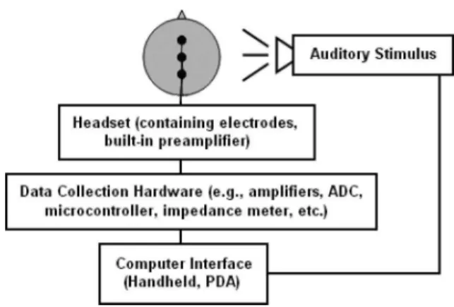
Fig. 1 provides a schematic overview of the HCS. The EEG system is comprised of recording electrodes, a headset with ear-phones, an electrode interface, EEG data collection hardware (amplifiers and supporting electronics including ADC, micro-controllers, etc.), an impedance monitor, and a computer in-terface. The electrodes are configured in the headset to cover the anterior–posterior axis (three channels). Ground, reference, and ocular monitoring electrodes are also integrated into the device (four additional channels). Earphones provide auditory stimulation. The electrodes and earphones are embedded in the headset. They are designed for easy application yet high qual-ity contact with the scalp. This is accomplished with the use of preamplifiers near the sensors, which present high effective input impedance allowing for the rejection of interference from
Fig. 1. Schematic overview of the HCS. The headset contains recording elec-trodes and built-in preamplifier. The data collection hardware amplifies and processes the signals before sending them to the computer. Auditory stimuli are presented via earphones and are also controlled by the computer.
60 Hz mains and other sources of noise. The headset, electrodes, and earphones are designed to be both reusable and disposable. The headset plugs into a permanent electrode interface that con-nects to the data collection hardware. The hardware employs multistage amplification using instrumentation amplifiers. The design of the electrode lead cabling includes shielding and in-corporates guarding circuits for noise attenuation. The amplifier uses driven right leg circuitry to minimize common mode volt-ages, which can result in erroneous conclusions of biological activity [18]. Postamplification, the EEG signals are processed using hardware bandpass filters and then sampled using a 16-bit ADC before being transmitted to the host computer. The am-plifiers are designed as a low cost, stable, and differential sys-tem, with active noise cancellation electronics. The hardware is preset to a specific impedance and noise verification test, analogue-to-digital sampling rate (256 Hz), and bandpass filter (0.1–100 Hz).
The computer is small, portable, and integrated within the device. It is wireless-enabled and communicates test results to other devices. The computer is comprised of a display screen, keyboard input, and system “test” and “run” buttons. It has headphone, USB, and battery charging interfaces. The computer interfaces with the EEG data collection hardware with the help of bluetooth communication protocol. The amplifier receives and logs external triggers coded using TTL pulses from the computer. The triggers are used as event markers for signal averaging to derive ERPs. Stimulation, acquisition, and analysis are coded as scripted routines.
B. HCS Algorithm Development
An overview of the HCS method is provided in Fig. 2, with preprocessing (digital filtering, signal averaging, artifact correc-tion, etc.), peak identificacorrec-tion, and score generation representing the three main stages. The algorithm converts the recorded EEG data into a numerical score of conscious awareness. The re-sults provide quantitative data for the evaluation of five key indicators: 1) sensory processing; 2) perceptual processing; 3) attentional processing; 4) memory processing; and 5) lan-guage processing. A quantitative scale is the main test result
Fig. 2. Overview of HCS method.
and is designed to be fast and easy to interpret. Scale bars depict the results for each of the five indices ranked against their respec-tive normarespec-tive data. The score is designed such that alterations in consciousness will lead to a drop in ERP peak amplitude, and subsequently the total possible score.
Computer software has been developed to preprocess the EEG data and derive the signal-averaged responses. This software in-cludes (but is not limited to) down sampling, digital filtering (bandpass 1–20 Hz and 60 Hz notch filter), segmentation (−100 to 800 ms), ocular correction [19], baseline correction, and con-ditional averaging. Stimulus presentation is temporally varied (e.g., jittered) to reduce the influence of ongoing EEG rhythms that obscure the ERP (e.g., alpha waves). Both transient and steady state signal-averaged data are analyzed. S/N optimiza-tion is done through ocular correcoptimiza-tion (rather than rejecoptimiza-tion), trial averaging, and specific denoising routines (such as pattern recognition for artifacts). Stimulation uses both auditory tones and speech stimuli to elicit the five indicators for conscious awareness. The test duration is approximately 5 min.
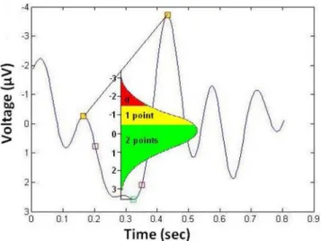
The HCS scores are derived from the signal-averaged wave-form data by quantifying ERP component amplitudes. This re-quires, first, identification of the relevant peak, and second, measurement of its amplitude. Peaks are identified based on the time latency and polarity that are known for the ERP component in question (e.g., P1 is a positive deflection that peaks between 50 and 150 ms poststimulus). Traditionally, ERP amplitudes are measured against a prestimulus baseline voltage. At rapid rates
of stimulation, however, there is no true prestimulus baseline. The HCS, therefore, measures amplitudes against a component-specific baseline, calculated as the average voltage of ERP onset and offset points. In general, these points are defined as the peaks of opposite polarity that surround the identified ERP component. The HCS score reflects a comparison of the obtained ERP amplitude to normative data. The normative data include the average amplitude values (as measured against either baseline or a comparison condition) as well as a measure of the variance. The normative data can be tailored to the specific patient (e.g., taking factors like age into account). Scores are quantified based on the individual patient’s component amplitude relative to the lower bound of the normative data variance measure. In addition, the normative comparison is converted to a standard reference framework, which allows for the patient specific amplitude data to be plotted against the norm using a numeric index (e.g., 0.5 ± SD). Scores are based on established ERP peaks that have been used to test for sensory and cognitive functions in behaviorally unresponsive patients [e.g., P1, N1, P2, mismatch negativity (MMN), P300, N400, and P600]. These are elicited by a number of different means, including the patient’s own name [20], nonverbal emotional exclamations [21], number sequences [22], and standardized neuropsychological tests [11].
III. RESULTS ANDDISCUSSION
Typical waveforms forall five indicators are presented in Fig. 3. Peaks (open green boxes), inflections (open red boxes), peaks before and after (red boxes with yellow fill) have been identified by the algorithm. Sensation, perception, attention, memory, and language are properly indexed by the P1, MMN, P300 (tones), P300 (speech), and N400. Following peak detec-tion, amplitude is measured and compared to the normative val-ues. Fig. 4 shows score derivation for the P300 (positive-going peak at 300 ms), as an attentional index of increased stimulus intensity.
An overview of the results display is provided in Fig. 5. The results are divided into four different categories: diagnostic score (Dx), reliability score (Rx), validity score (Vx), and prog-nostic score (Px). The total Dx is out of ten, with two possible points for each of the five indicators (0 = abnormal, 1 = bor-derline, and 2 = normal). The Rx and Vx are allotted based on the repeatability and features of the waveform characteristics, respectively. The Px is calculated using a classification approach to assess fit to prior patient data divided on the basis of recovery versus no recovery for a specific indicator. The scores (Dx, Rx,
Vx, Px) are communicated via graphical output to the screen of the portable device, as well as outputs that are integrated with current communications technologies. The device generates re-ports that include “On Device Display” (Fig. 5), “Short Rere-ports” in e-mail format (Fig. 6) and “Long Reports” via internet access to a secure domain. The test results can be sent via telecommu-nication networks for rapid dissemination and easy integration into the critical care cascade.
In summary, a prototype device and algorithm were devel-oped for EEG-based testing of conscious awareness (the HCS). The HCS is portable, stable, and noise-resistant (Challenge 1).
Fig. 3. Typical waveforms for all five indicators plotted in voltage (microvolts) versus time (seconds). Open green boxes delineate ERP component peaks, open red boxes show the inflection points, and red boxes with yellow fill show the peaks before and after.
Fig. 4. Sample score derivation methodology.
The hard coded settings and programmed algorithm elimi-nate the requirement for advanced expertise/training and prior knowledge/training (Challenge 2). A spectrum of EEG-based responses were successfully elicited, identified, and integrated into a rapid clinical test (Challenge 3). The analysis software uses a normative database for comparison purposes, thereby pro-viding standardized results (Challenge 4). The test results are
Fig. 5. HCS score as displayed on the device. Note: this is a theoretical result as testing on controls yields perfect scores.
Fig. 6. E-mail sample for the “Short Report”. Note: this is a theoretical result as testing on controls yields perfect scores.
presented in a clinically friendly format, which can be readily communicated (Challenge 5).
IV. FUTUREDIRECTIONS
A clinical study of the device is underway. Continued hard-ware development will incorporate improved noise cancella-tion methods, other stimulacancella-tion modalities (e.g., visual), and expanded clinical applications (e.g., anesthetic monitoring). Wireless capabilities will also be utilized to allow for multi-device data collection and monitoring (i.e., scalability). Ad-ditional development will focus on more advanced neurocog-nitive testing using other brain imaging technologies (e.g., magnetoencephalography).
V. CONCLUSION
Approximately one in three people are affected by brain dis-orders and diseases at some point in their lives. Front-end di-agnostics have the greatest potential to impact treatment. The market for such devices is estimated to be as high as US$8 billion by 2015. Given the intricate relationship between brain and behavior, diagnostics for one cannot be dependent on the other. Instead, novel solutions must be found at the interface between biomedical engineering and neuroscience. The HCS device attempts to address this challenge.
REFERENCES
[1] G. Teasdale and B. Jennett, “Assessment of coma and impaired conscious-ness: A practical scale,” Lancet, vol. 2, no. 7872, pp. 81–84, Jul. 1974. [2] J. R. Gawryluk, R. C. D’Arcy, J. F. Connolly, and D. F. Weaver, “Improving
the clinical assessment of consciousness with advances in electrophysio-logical and neuroimaging techniques,” BMC Neurol., vol. 10, no. 1, p. 11, Jan. 2010.
[3] C. Schnakers, A. Vanhaudenhuyse, J. Giacino, M. Ventura, M. Boly, S. Majerus, G. Moonen, and S. Laureys, “Diagnostic accuracy of the vegetative and minimally conscious state: Clinical consensus versus stan-dardized neurobehavioral assessment,” BMC Neurol., vol. 9, no. 1, p. 35, Jul. 2009.
[4] M. Boly, M.-E. Faymonville, P. Peigneux, B. Lambermont, P. Damas, G. Del Fiore, C. Degueldre, G. Franck, A. Luxen, M. Lamy, G. Moonen, P. Maquet, and S. Laureys, “Auditory processing in severely brain injured patients: Differences between the minimally conscious state and the per-sistent vegetative state,” Arch. Neurol., vol. 61, no. 2, pp. 233–238, Feb. 2004.
[5] W. Staffen, M. Kronbichler, M. Aichhorn, A. Mair, and G. Ladurner, “Selective brain activity in response to one’s own name in the persis-tent vegetative state,” J. Neurol. Neurosurg. Psychiatry, vol. 77, no. 12, pp. 1383–1384, Dec. 2006.
[6] H. B. Di, S. M. Yu, X. C. Weng, S. Laureys, D. Yu, J. Q. Li, P. M. Qin, Y. H. Zhu, S. Z. Zhang, and Y. Z. Chen, “Cerebral response to patient’s own name in the vegetative and minimally conscious states,” Neurology, vol. 68, no. 12, pp. 895–899, Mar. 2007.
[7] A. M. Owen, M. R. Coleman, M. Boly, M. H. Davis, S. Laureys, and J. D. Pickard, “Detecting awareness in the vegetative state,” Science, vol. 313, no. 5792, p. 1402, Sep. 2006.
[8] J. F. Connolly, J. M. Byrne, and C. A. Dywan, “Assessing adult receptive vocabulary with event-related potentials: An investigation of cross-modal and cross-form priming,” J. Clin. Exp. Neuropsychol., vol. 17, no. 4, pp. 548–565, Aug. 1995.
[9] J. F. Connolly and R. C. N. D’Arcy, “Innovations in neuropsychological assessment using event-related brain potentials,” Int. J. Psychophysiol., vol. 37, no. 1, pp. 31–47, Jul. 2000.
[10] J. F. Connolly, R. C. N. D’Arcy, N. R. Lynn, and R. Kemps, “The ap-plication of cognitive event-related brain potentials (ERPs) in language-impaired individuals: Review and case studies,” Int. J. Psychophysiol., vol. 38, no. 1, pp. 55–70, Oct. 2000.
[11] R. C. N. D’Arcy, Y. Marchand, G. Eskes, E. R. Harrison, S. J. Phillips, A. Major, and J. F. Connolly, “Electrophysiological assessment of lan-guage function following stroke,” Clin. Neurophysiol., vol. 114, no. 4, pp. 662–672, Apr. 2003.
[12] D. Morlet, P. Bouchet, and C. Fischer, “Mismatch negativity and N100 monitoring: Potential clinical value and methodological advances,”
Au-diol. Neurootol., vol. 5, no. 3/4, pp. 198–206, May–Aug. 2000. [13] B. Kotchoubey, S. Lang, G. Mezger, D. Schmalohr, M. Schneck,
A. Semmler, V. Bostanov, and N. Birbaumer, “Information processing in severe disorders of consciousness: Vegetative state and minimally con-scious state,” Clin. Neurophysiol., vol. 116, no. 10, pp. 2441–2453, Oct. 2005.
[14] V. J. M. Wijnen, G. J. M. van Boxtel, H. J. Eilander, and B. de Gelder, “Mismatch negativity predicts recovery from the vegetative state,” Clin.
Neurophysiol., vol. 118, no. 3, pp. 597–605, Mar. 2007.
[15] A. Vanhaudenhuyse, S. Laureys, and F. Perrin, “Cognitive event-related potentials in comatose and post-comatose states,” Neurocrit. Care, vol. 8, no. 2, pp. 262–270, Oct. 2008.
[16] J. Daltrozzo, N. Wioland, V. Mutschler, P. Lutun, B. Calon, A. Meyer, T. Pottecher, S. Lang, A. Jaeger, and B. Kotchoubey, “Cortical information processing in coma,” Cogn. Behav. Neurol., vol. 22, no. 1, pp. 53–62, Mar. 2009.
[17] A. Capone-Neto and S. B. Rizoli, “Linking the chain of survival: Trauma as a traditional role model for multisystem trauma and brain injury,” Curr.
Opin. Crit. Care, vol. 15, no. 4, pp. 290–294, Aug. 2009.
[18] A. C. Metting van Rijin, A. Peper, and C. A. Grimbergen, “High qual-ity recording of bioelectric events,” Med. Biol. Eng. Comput., vol. 28, pp. 389–397, Sep. 1990.
[19] G. Gratton, M. G. Coles, and E. Donchin, “A new method for off-line removal of ocular artifact,” Electroencephalogr. Clin. Neurophysiol., vol. 55, no. 4, pp. 468–484, Apr. 1983.
[20] I. Berland and H. Pratt, “P300 in response to the subject’s own name,”
Electroencephalogr. Clin. Neurophysiol., vol. 96, no. 5, pp. 472–474, Sep. 1995.
[21] V. Bostanov and B. Kotchoubey, “Recognition of affective prosody: Con-tinuous wavelet measures of event-related brain potentials to emotional exclamations,” Psychophysiology, vol. 41, no. 2, pp. 259–268, Mar. 2004.
[22] S. Lang and B. Kotchoubey, “Brain responses to number sequences with and without active task requirement,” Clin. Neurophysiol., vol. 113, no. 11, pp. 1734–1741, Nov. 2002.