environment, from the genus to the population level
The MIT Faculty has made this article openly available. Please share
how this access benefits you. Your story matters.
Citation
Takemura, Alison F., Diana M. Chien, and Martin F. Polz.
“Associations and Dynamics of Vibrionaceae in the Environment,
from the Genus to the Population Level.” Frontiers in Microbiology 5
(2014).
As Published
http://dx.doi.org/10.3389/fmicb.2014.00038
Publisher
Frontiers Research Foundation
Version
Final published version
Citable link
http://hdl.handle.net/1721.1/87578
Terms of Use
Article is made available in accordance with the publisher's
policy and may be subject to US copyright law. Please refer to the
publisher's site for terms of use.
REVIEW ARTICLE
published: 11 February 2014 doi: 10.3389/fmicb.2014.00038
Associations and dynamics of Vibrionaceae in the
environment, from the genus to the population level
Alison F. Takemura†, Diana M. Chien†and Martin F. Polz *Parsons Lab for Environmental Science and Engineering, Department of Civil and Environmental Engineering, Massachusetts Institute of Technology, Cambridge, MA, USA
Edited by:
Daniela Ceccarelli, University of Maryland, USA
Reviewed by:
Anwar Huq, University of Maryland, USA
Darrell J. Grimes, The University of Southern Mississippi, USA
*Correspondence:
Martin F. Polz, Parsons Lab for Environmental Science and Engineering, Department of Civil and Environmental Engineering, Massachusetts Institute of Technology, Building 48, Room 417, 15 Vassar Street, Cambridge, MA 02139, USA
e-mail: mpolz@mit.edu †These authors have contributed equally to this work.
the causative agent of cholera, and V. vulnificus, the deadliest seafood-borne pathogen, are a well-studied family of marine bacteria that thrive in diverse habitats. To elucidate the environmental conditions under which vibrios proliferate, numerous studies have examined correlations with bulk environmental variables—e.g., temperature, salinity, nitrogen, and phosphate—and association with potential host organisms. However, how meaningful these environmental associations are remains unclear because data are fragmented across studies with variable sampling and analysis methods. Here, we synthesize findings about Vibrio correlations and physical associations using a framework of increasingly fine environmental and taxonomic scales, to better understand their dynamics in the wild. We first conduct a meta-analysis to determine trends with respect to bulk water environmental variables, and find that while temperature and salinity are generally strongly predictive correlates, other parameters are inconsistent and overall patterns depend on taxonomic resolution. Based on the hypothesis that dynamics may better correlate with more narrowly defined niches, we review evidence for specific association with plants, algae, zooplankton, and animals. We find that Vibrio are attached to many organisms, though evidence for enrichment compared to the water column is often lacking. Additionally, contrary to the notion that they flourish predominantly while attached, Vibrio can have, at least temporarily, a free-living lifestyle and even engage in massive blooms. Fine-scale sampling from the water column has enabled identification of such lifestyle preferences for ecologically cohesive populations, and future efforts will benefit from similar analysis at fine genetic and environmental sampling scales to describe the conditions, habitats, and resources shaping Vibrio dynamics.
Keywords: Vibrio, population, environmental correlation, ecology, niche, attachment, planktonic
INTRODUCTION
The family Vibrionaceae (or vibrios for short) comprises a genet-ically and metabolgenet-ically diverse group of heterotrophic bacteria that are routinely found in all ocean environments, ranging from coastal to open and surface to deep water (Thompson et al., 2004; Thompson and Polz, 2006). Moreover, a few Vibrio species have extended their range beyond the marine environment, occurring predominantly in brackish and even freshwater environments (Thompson et al., 2004). The study of the environmental distri-bution and dynamics of vibrios has a long history, largely because many species contain potential human and animal pathogens (Thompson et al., 2004, 2005). Hence there is considerable public health and economic interest in determining factors correlated to increased abundance of vibrios (Stewart et al., 2008). Moreover, vibrios are easily cultured on standard and selective media and thus were highly visible in the pre-molecular era of microbial ecology. In recent years, environmental dynamics have also been studied with culture-independent methods allowing for a more fine-scale assessment of environmental drivers of occurrence, and the vibrios have become a model for bacterial population biol-ogy and genomics. In fact, presently, the vibrios represent one of
the best-studied models for the ecology and evolution of bacterial populations in the wild.
The early discovery that some fish species harbor high numbers of vibrios (e.g.,Liston, 1954, 1957; Aiso et al., 1968; Sera et al., 1972) has led to the widespread notion that these bacteria are only transient members of microbial assemblages of the water column. Instead, vibrios were regarded as specifically associated with ani-mals, and occurrence in water samples was thought to be primarily due to their excretion with fecal matter. This picture was enforced by the discovery that several luminescent Vibrio (Allivibrio) and related Photobacterium species form intimate symbioses with ani-mals (e.g., fish, squid) (Ruby and Nealson, 1976; Stabb, 2006). More recent work has, however, revealed that the notion of vibrios being “enterics of the sea” (Liston, 1954) represents an oversimpli-fication. Many Vibrio species grow actively in ocean water either in the free-living phase or associated with various types of organic particles, many of which are of non-animal origin (Lyons et al., 2007; Froelich et al., 2012). Thus although association with animals can be an important part of the life cycle of many Vibrio species, there are others that only loosely associate with animals or not at all, an aspect we explore in detail in this review.
Another widely held belief about vibrios is that they play a relatively minor role in chemical transformations in the ocean, despite the wide range of metabolisms [e.g., chitin degradation (Hunt et al., 2008a; Grimes et al., 2009)] of which they are capa-ble. This belief is largely based on low to medium average relative abundance of Vibrionaceae in ocean water. Yet three considera-tions suggest that the role of vibrios has been underestimated. First, it has been pointed out that although vibrios’ abundances are generally only around 103to 104 cells per ml seawater (i.e., on the order of few percent of total bacteria), they have very high biomass (Yooseph et al., 2010). For example, an actively grow-ing Vibrio can have 100× the biomass of Pelagibacter, which, at∼105cells per ml, is typically the most abundant heterotrophic
member of bacterial assemblages in the ocean (Yooseph et al., 2010). Second, new time-series analysis shows that vibrios are capable of blooms in the water column during which they can even become the predominant members of the total bacterial assemblage (Gilbert et al., 2012). These blooms had been missed previously because they are of relatively short duration, yet they confirm that vibrios, which are capable of very rapid growth in laboratory media, can reach high doubling rates in the envi-ronment. Finally, vibrios might be disproportionately subject to predation by protozoa and viruses (Worden et al., 2006; Suttle, 2007), likely due to their comparatively large size. For example, cells were found in one study to measure more than three times the community average in volume, and, along with other similarly large genera, suffered especially high grazing mortality (Beardsley et al., 2003). Taken together, these considerations suggest that vibrios should be re-evaluated for their role in biogeochemical processes in the ocean since they have disproportionately high biomass that is subject to high turnover by rapid growth in concert with high predation.
The purpose of this review is to provide an overview of known environmental factors and ecological associations affecting Vibrio abundance and dynamics. We note that although we look at the dynamics of potentially pathogenic species, we purposefully exclude data on pathogenesis itself since this is outside the scope of this review. We first focus on total Vibrio (i.e., the assessment of occurrence of members of the genus or family), which have often been measured as a proxy for potential pathogen occur-rence, asking whether they can be treated as an environmentally cohesive unit. To what extent do total vibrios correlate to specific environmental variables, and do these measures have predictive power for individual species? To address this question, we present meta-analyses of the dynamics of V. cholerae, V. parahaemolyticus, and V. vulnificus, three species harboring genotypes potentially pathogenic to humans. The limitation to these three is necessary since public health interests have driven much of the research so that the literature is highly biased toward human pathogens. In this context, a further important question is to what extent easily measurable bulk parameters, such as temperature, salinity, nutri-ents, dissolved oxygen and/or chlorophyll a are good correlates for total vibrios or specific species, allowing easy and cost-effective risk assessment.
However, because our meta-analysis suggests poor or incon-sistent performance of most bulk parameters, we researched alternative, frequently finer-scale environmental variables. These
include associations with different animals, plants and algae, as well as organic polymers, which may occur as suspended par-ticulate matter in the water column and provide resources for attached bacteria. Although such attached lifestyles are common for vibrios, recent research also suggests that many species can occur free-living at least part of the time and be engaged in relative short-lived blooms.
Finally, we summarize recent research aimed at defining habi-tat characteristics and phylogenetic bounds of ecologically cohe-sive populations among co-existing vibrios, using the water col-umn and macroinvertebrates as examples of adaptive landscapes. This research demonstrates that such populations, which may or may not correspond to named (taxonomic) species, represent eco-evolutionary units that allow testing of hypotheses of how populations are structured by environmental selection and gene flow.
ENVIRONMENTAL CORRELATES OF Vibrio PRESENCE AND ABUNDANCE
To better understand under what conditions vibrios occur and proliferate, most studies have investigated environmental vari-ables that can be measured from bulk seawater such as tem-perature, salinity, dissolved oxygen, nitrogen, phosphorus, and chlorophyll a concentrations. These are attractive since they are easily measured and many are observable remotely by buoy or satellite (e.g.,Lobitz et al., 2000) so that potential for presence of pathogenic vibrios might be easily assessed. In addition, several studies have extended measurements to more complex physico-chemical and biotic variables, including dissolved organic carbon (DOC) and zoo- and phyto-plankton taxa.
In the following, we first ask how informative these variables are by conducting a meta-analysis to compare correlations across studies, for both total Vibrio as well as the potential pathogens
V. cholerae, V. parahaemolyticus, and V. vulnificus, and, second,
determine if the genus and species levels exhibit similar patterns. To determine the potential impact of environmental variables, we looked at how strong their correlations are by comparing coef-ficient of determination values, R2, reported in the literature. A goodness of fit parameter, R2 varies from 0 (no explanation of variance in the dependent variable) to 1 (perfect explanation), giving us a means of assessing, for example, whether tempera-ture better predicts abundance of total Vibrio, than salinity does. Studies included have regression analyses with associated R2 -values, or Spearman or Pearson correlations, whose rho values were squared to obtain R2. Additionally, we compare how their abundances trend along gradients in two particularly well-studied variables, salinity and temperature.
TOTAL Vibrio
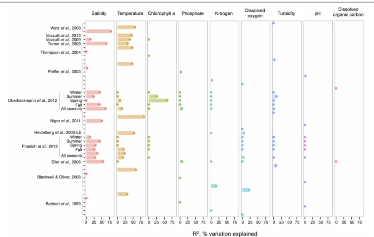
When correlations across studies are compared, we see that the strongest environmental correlates to total Vibrio are temperature and salinity. These two variables most often explain the great-est amount of variance in total Vibrio abundance in the water column (Figure 1), whereas consideration of additional variables often makes only marginal improvements (e.g., in Heidelberg et al., 2002a,b; Oberbeckmann et al., 2012; Froelich et al., 2013). However, a minority of analyses has found temperature and
Takemura et al. Vibrio ecology
FIGURE 1 | An overview of regression analyses indicate that temperature and salinity explain most variation in bulk-water total
Vibrio abundance. The R2, or pseudo-R2, values associated with regression
analyses are shown for selected environmental variables that are well-represented across studies. An individual study may perform multiple analyses because variables are considered for correlation independently (for
ex.Wetz et al., 2008); because datasets are split (e.g., between seasons in Oberbeckmann et al., 2012); or because different sets of variables are considered sequentially (e.g., two variables versus six variables in the two All Seasons models fromFroelich et al., 2013). Dots indicate bar heights, and where a dot occurs without a bar, R2was non-significant (i.e., R2= 0).
Variables may have been log or exponentially transformed in references.
salinity to be non-significant toward explaining Vibrio abun-dance. This inconsistency might be a result of the ranges con-sidered; for instance, temperature may be found non-significant due to a narrow range observed, such that Vibrio abundance varies little. In fact, evidence supports this hypothesis; the cor-relation strength of temperature to vibrios varies by season (Oberbeckmann et al., 2012; Froelich et al., 2013), suggesting the magnitude of the correlation may depend on the tempera-ture range examined. For instance,Oberbeckmann et al. (2012)
andFroelich et al. (2013)both observed the highest correlation of temperature and Vibrio during the seasons with the broadest tem-perature ranges, spring, and fall, respectively. Additionally, it is possible that at lower temperatures vibrios exhibit less variation in abundance; two studies assessing total vibrios in the cooler waters of the Baltic Sea and North Sea found non-significant correlations (Eiler et al., 2006; Oberbeckmann et al., 2012).
Compared to salinity and temperature, other environmental measures usually explain less variance in total Vibrio. Dissolved oxygen has had little explanatory power; for instance, in Figure 1, its largest R2 was less than half that of temperature in the same analysis (Blackwell and Oliver, 2008). The same is true
for nitrogen, whose highest R2 was still less than temperature’s
(Blackwell and Oliver, 2008). In the environments examined, phosphate, pH, and turbidity explain little variance, and DOC explains none at all, albeit the number of studies used for DOC in this meta-analysis is limited. Of interest, though not depicted, potential host organisms, copepods, decapods, and cyanobac-teria, have been found to explain relatively little variance in total vibrios when considered in a model that already incor-porates temperature (Turner et al., 2009; Vezzulli et al., 2009), and similarly for dinoflagellates when salinity is first consid-ered (Eiler et al., 2006). Turner et al. (2009) did observe that diatoms explained more variance than temperature. While this might imply a physical association, the correlation was negative, suggesting that total Vibrio, at least as a whole, do not associate with diatoms.
Chlorophyll a, on the other hand, has had noted impor-tance in two datasets: the spring and summer of the study by
Oberbeckmann et al. (2012), with R2-values of 60 and 26%, respectively. These were in fact higher than correlations to tem-perature or salinity in these seasons. Perhaps during this period, as temperature warms, growth conditions favor phytoplankton
blooms that impact Vibrio abundance (Oberbeckmann et al., 2012). However,Froelich et al. (2013)did not make these same observations in their seasonal datasets. This inconsistency may be a product of the fact that different Vibrio species likely affiliate with or feed on exudates of specific algal taxa only, rather than algae in general, a subject further discussed in the section The Evidence for a Planktonic, Free-Living Lifestyle.
Given the frequent strength of temperature and salinity as cor-relates, we asked, how do total vibrios distribute with respect to these variables when their combined effect is considered? A few studies have modeled the bivariate relationship, finding that total
Vibrio abundance increases as temperature and salinity increase
(Hsieh et al., 2008; Turner et al., 2009; Froelich et al., 2013). The ranges investigated were also broad, lending confidence that these results are general; for example,Hsieh et al. (2008)modeled from 2.5 to 32.5◦C and 0 to 27 ppt, respectively.
V. cholerae, V. parahaemolyticus, AND V. vulnificus
We compare environmental correlates and trends noted in total
Vibrio to three species that have been well-sampled across locales: V. cholerae, V. parahaemolyticus, and V. vulnificus. While it
would also be interesting to consider species beyond potential pathogens, their environmental data is much more limited.
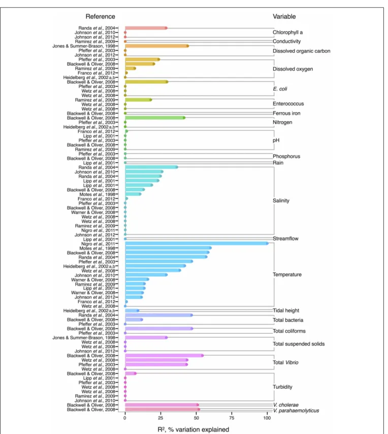
In V. cholerae, we see an interesting shift from total Vibrio in the strength of correlating environmental variables: some biotic variables are as strong or, in fact, stronger than temperature or salinity (Figure 2). Total Vibrio, congenerics V. vulnificus and V.
parahaemolyticus, as well as a dinoflagellate genus (Prorocentrum)
and cladoceran species (Diaphanosoma mongolianum) have all significantly correlated to V. cholerae abundance (Eiler et al., 2006; Blackwell and Oliver, 2008; Kirschner et al., 2011; Prasanthan et al., 2011). Moreover, V. parahaemolyticus abundance has explained more V. cholerae abundance variance than nitrogen, temperature, or salinity in (Prasanthan et al., 2011), and dinoflag-ellate abundance has explained more variance than phosphorus, salinity, or temperature (Eiler et al., 2006). While correlations to plankton may represent direct associations, such high correlation of vibrios to each other is likely not indicative of causal inter-actions, but rather stems from overlap in environmental ranges and/or habitats (Blackwell and Oliver, 2008). E. coli and total coliforms have also correlated to V. cholerae abundance, though both groups may simply be responding to anthropogenic nutrient influxes favoring growth of heterotrophs (Blackwell and Oliver, 2008).
Long thought to be a reservoir of toxigenic V. cholerae, zoo-plankton, and particularly copepods, are hypothesized to cor-relate to V. cholerae abundance. Surprisingly, however, whende Magny et al. (2011)examined several zooplankton genera and species, including copepods Cyclops and Diaptomus, they did not find significant correlations to any zooplankter except the rotifer Brachionus angularis (not depicted in Figure 2, because Monte Carlo analysis did not yield R2-values). While the asso-ciation between V. cholerae O1/O139 and the copepod Acartia
tonsa has also been studied (Huq et al., 2005; Lizárraga-Partida et al., 2009), quantitatively significant correlation in the environ-ment has remained elusive. For instance,Lizárraga-Partida et al. (2009)demonstrated only a qualitative link between V. cholerae
O1 presence coincident with an increase in A. tonsa, even though laboratory studies have shown ready attachment (e.g.,Huq et al., 1984; Rawlings et al., 2007).
V. cholerae has also been hypothesized to correlate with
chloro-phyll a, a potential proxy of algal and zooplankton growth, and/or a eutrophic environment conducive to heterotroph growth, but chlorophyll a’s general predictive value is unclear. While sig-nificant in Eiler et al. (2006), other studies have observed no correlation of chlorophyll a to V. cholerae abundance (Jiang and Fu, 2001; Kirschner et al., 2008; Mishra et al., 2012). Yet V.
cholerae growth has been observed experimentally to depend on
DOC, which could relate to phytoplankton abundance and thus chlorophyll a (Eiler et al., 2007). In microcosm experiments,
Eiler et al. (2007)demonstrated that adding 2.1 mg carbon L−1 of cyanobacterial-derived dissolved organic matter influenced bacterial growth more than a 12–25◦C change in temperature. The inconsistency of chlorophyll a, and, incidentally, bulk DOC (which showed no significant correlation) (Eiler et al., 2006; Blackwell and Oliver, 2008; Kirschner et al., 2008; Neogi et al., 2012) as correlates might be due to the quality of exudates; its composition of refractory humic substances (Kirschner et al., 2008) or derivation from different algal species, differentially stimulating V. cholerae growth [(Worden et al., 2006), see also section The Evidence for a Planktonic, Free-Living Lifestyle]. Interestingly, the lack of clear support for chlorophyll a’s influ-ence on V. cholerae environmental abundance is in contrast to the fact that chlorophyll a can correlate with cholera disease incidence (de Magny et al., 2008), and has been used in predictive mod-els for cholera in Bangladesh (Bertuzzo et al., 2012; Jutla et al., 2013).
Like V. cholerae, V. parahaemolyticus abundance in water sam-ples is also strongly correlated to temperature, and was found significant in all but one analysis reviewed here (DePaola et al., 1990; Zimmerman et al., 2007; Blackwell and Oliver, 2008; Caburlotto et al., 2010; Deter et al., 2010; Johnson et al., 2010, 2012; Böer et al., 2013), with maximal R2= 50.6% (Deter et al., 2010) (Figure 3).Blackwell and Oliver (2008)found that V.
para-haemolyticus correlates both to total Vibrio and congenerics, as
well as coliforms and E. coli. These variables were only considered in a single study, however, so it is not known if the relationships hold across different sampling locations. The significance of salin-ity is variable for V. parahaemolyticus with only three of seven studies having non-zero R2-values (Figure 3) (Zimmerman et al., 2007; Caburlotto et al., 2010; Johnson et al., 2010), but this may be due to V. parahaemolyticus colonizing a large salinity range, as detailed below (Figure 6).
Correlation to environmental variables has also frequently been studied for V. parahaemolyticus occurring in sediment and shellfish, though trends remain unclear. In sediment, considered a potential reservoir (Vezzulli et al., 2009), individual regressions of
V. parahaemolyticus abundance to temperature, salinity, and total
organic carbon have yielded moderate R2-values, at times above 30% (Blackwell and Oliver, 2008; Deter et al., 2010; Johnson et al., 2012; Böer et al., 2013). However, some studies have found salin-ity or temperature to be a non-significant explanatory variable (Blackwell and Oliver, 2008; Deter et al., 2010; Johnson et al., 2010).
Takemura et al. Vibrio ecology
FIGURE 2 | Variation in V. cholerae abundance or percent positive samples is best explained by temperature, other organisms, and salinity. R2, or pseudo-R2, values from analyses across studies are depicted grouped
by variable, and then in rank order, with their associated reference. A
reference may conduct multiple analyses for a given variable (e.g., on subsets of data or considering different variables combinations for data regression). Dots indicate bar heights, and where a dot occurs without a bar, R2was
non-significant (i.e., R2= 0).
In shellfish, a common vehicle of virulent vibrios to humans, the incidence of temperature and salinity as correlates to V.
parahaemolyticus is also inconsistent. Salinity has been found
explanatory in some studies, with R2 as high as 42% (DePaola et al., 2003; Johnson et al., 2010, 2012) and non-significant in others (Deepanjali et al., 2005; Deter et al., 2010; Sobrinho et al., 2010). Temperature can explain moderate amounts of vari-ance in V. parahaemolyticus abundvari-ance (DePaola et al., 1990, 2003; Cook et al., 2002; Johnson et al., 2010, 2012; Sobrinho
et al., 2010), with significant R2 as high as 44% (Cook et al., 2002), though other studies have found little or no correla-tion (Deepanjali et al., 2005; Duan and Su, 2005; Deter et al., 2010). The absence of correlation is surprising, given that tem-perature’s effect is amplified by influencing shellfish’s ability to concentrate V. parahaemolyticus from surrounding water. Oysters can enrich V. parahaemolyticus over 100-fold (DePaola et al., 1990; Shen et al., 2009), and the magnitude of con-centration is temperature-dependent, with effects greatest at
FIGURE 3 | Variation in V. parahaemolyticus abundance or percent positive samples is best explained by temperature and other organisms. R2, or pseudo-R2, values from analyses across studies are depicted grouped
by variable, and then in rank order, with their associated reference. A
reference may conduct multiple analyses for a given variable (e.g., on subsets of data or considering different variables combinations for data regression). Dots indicate bar heights, and where a dot occurs without a bar, R2was
non-significant (i.e., R2= 0).
32◦C and less, but still evident, in cooler waters (Shen et al., 2009).
For V. vulnificus isolated from the water column, temperature is the strongest correlate among measured environmental vari-ables, and often explains more variance in V. vulnificus than for other species or total Vibrio; several analyses found temperature explained over 50% of the variance in V. vulnificus sampled from water (Motes et al., 1998; Randa et al., 2004; Blackwell and Oliver, 2008; Nigro et al., 2011) (Figure 4). Moreover, temperature has been a stronger correlate than chlorophyll a (Randa et al., 2004; Johnson et al., 2010, 2012), dissolved oxygen (Pfeffer et al., 2003; Blackwell and Oliver, 2008; Ramirez et al., 2009), and nitrogen (Pfeffer et al., 2003; Blackwell and Oliver, 2008). While DOC is an inconsistent correlate, it has been more explanatory than tem-perature in at least one study (Jones and Summer-Brason, 1998). The variable pH, however, is not a significant correlate (Lipp et al., 2001; Pfeffer et al., 2003; Blackwell and Oliver, 2008; Ramirez et al., 2009; Franco et al., 2012), nor is phosphorus (Pfeffer et al.,
2003; Blackwell and Oliver, 2008). Turbidity has been found non-significant in several studies (Lipp et al., 2001; Pfeffer et al., 2003; Wetz et al., 2008; Ramirez et al., 2009), or not as explanatory as temperature (Blackwell and Oliver, 2008). While salinity, when significant, has generally been less informative than temperature (Motes et al., 1998; Randa et al., 2004; Warner and Oliver, 2008; Johnson et al., 2010), it has, in one analysis, been more (Lipp et al., 2001).
Biotic correlates have also been identified for V. vulnificus. Total bacteria (Pfeffer et al., 2003; Randa et al., 2004; Blackwell and Oliver, 2008), enteroccous (Wetz et al., 2008; Ramirez et al., 2009), coliforms (Pfeffer et al., 2003; Blackwell and Oliver, 2008) and E. coli (Pfeffer et al., 2003; Blackwell and Oliver, 2008; Wetz et al., 2008) have been studied only sporadically, but their correlation strength to V. vulnificus has usually been less than tem-perature’s; one exception, however, is enterococcus in (Ramirez et al., 2009), potentially indicative of a surge in nutrients over-taking temperature’s effect on growth. Interestingly, total Vibrio
Takemura et al. Vibrio ecology
FIGURE 4 | Variation in V. vulnificus abundance or percent positive samples is best explained by temperature, and other organisms, including Vibrio. R2, or pseudo-R2, values from analyses across studies are
depicted grouped by variable, and then in rank order, with their associated
reference. A reference may conduct multiple analyses for a given variable (e.g., on subsets of data or considering different variables combinations for data regression). Dots indicate bar heights, and where a dot occurs without a bar, R2was non-significant (i.e., R2= 0).
have explained substantial variance (R2= 43–54%) in V.
vulnifi-cus in more instances than for other Vibrio species (Pfeffer et al., 2003; Blackwell and Oliver, 2008; Wetz et al., 2008), suggest-ing they are respondsuggest-ing similarly to their environments under the conditions studied. However, instances do occur where total
Vibrio and V. vulnificus do not correlate (Høi et al., 1998; Wetz et al., 2008), underscoring that a species is not a constant com-ponent of a genus, and may respond to environmental conditions independently.
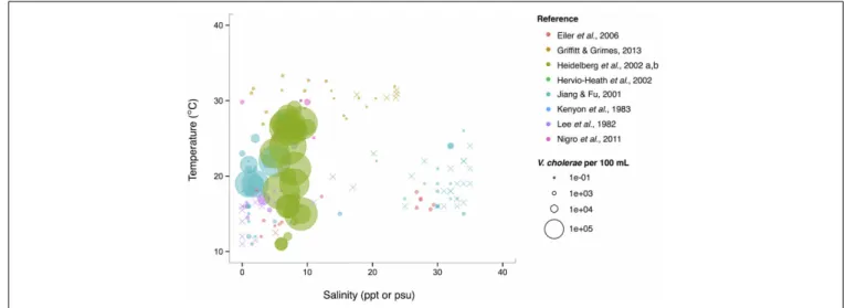
Isolations of the three potentially pathogenic species across salinity and temperature gradients were also looked at, and found to exhibit different patterns. V. cholerae has a wide temperature range (∼10–30◦C) in brackish water (1–10 ppt), and generally decreases with increasing salinity over the entire range examined (0–40 ppt) (Figure 5). Observed V. cholerae abundance is greatest around 20◦C and 0–10 ppt, on the order of 103 cells per mL. At less-favorable, higher salin-ities, V. cholerae has been found around this temperature, though in much lower abundances (on the order of 1 cell per mL). Interestingly, V. cholerae’s realized niche is much smaller than its fundamental one, as it has maximal tempera-ture and salinity tolerances around 38◦C and 75 ppt (Materna et al., 2012), suggesting other controls on its abundance in the environment.
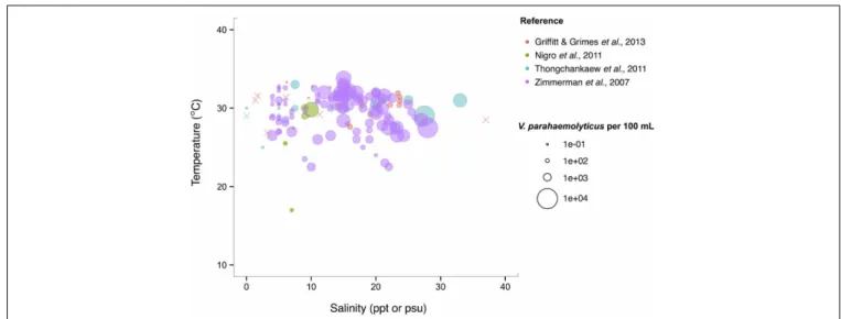
V. parahaemolyticus contrasts V. cholerae by having a more
constant abundance that is broadly spread out over salinities of 3–35 ppt in a narrow, much warmer temperature range, centered roughly around 29◦C. (Figure 6). Consistent with this finding, it has been noted that this species prefers warmer waters (>20◦C) (Martinez-Urtaza et al., 2012), and has been observed to grow best at 25◦C in vitro (Nishina et al., 2004). However, isolations from shellfish can exhibit different trends from those observed in the water column; Martinez-Urtaza et al. (2008) detected
V. parahaemolyticus in mussels gathered in much cooler, 15◦C
water, consistent with the potential for shellfish to concentrate V.
parahaemolyticus.
A previous literature-based analysis showed V. vulnificus to have a more complicated relationship to temperature and salin-ity than either V. cholerae or V. parahaemolyticus. It has a narrow temperature range at higher salinities (>10 ppt) while at low salinities (between 5 and 10 ppt) its temperature range more than doubles—from 22–30◦C to 10–32◦C (Randa et al., 2004). This suggests that, in temperate climates, this species is found year-round in estuarine, low salinity environments but can expand into full strength seawater during warmer months. In the tropics, this species should be endemic to the ocean.
CONCLUSIONS FROM META-ANALYSIS
From this meta-analysis, we find, first, that temperature and salinity often explain more variance than any other bulk water parameter, like phosphate, nitrogen, pH, or DOC. Yet some of the difficulty in making general statements regarding the rela-tionship of vibrios to individual environmental variables likely stems from the fact that their strength can depend on the ranges examined, e.g., as for temperature, or in quality of the variable, such as DOC, which will encompass carbon derived from differ-ent sources that may impact Vibrio growth differdiffer-entially. Second, we observe that trends that apply to the whole genus Vibrio do not necessarily reflect those of individual species. Total vibrios and the well-studied potential pathogens V. cholerae, V.
para-haemolyticus, and V. vulnificus correlate with shared and distinct
environmental variables. For V. parahaemolyticus and V.
vulnifi-cus, temperature often explains more variance than does salinity
in the same analysis, and for V. cholerae, diverse biotic variables, including specific phyto- and zooplankton taxa, can be stronger correlates than abiotic variables. Unfortunately, biotic variables, particularly individual plankton taxa, have rarely been studied in more than one instance, making these observations difficult
FIGURE 5 | V. cholerae favors lower salinity and occupies a broad temperature range. V. cholerae concentrations, i.e., MPN-estimated CFU or molecular marker gene copies per 100 mL, reported in different studies are plotted against the temperature (◦C) and salinity values (ppt or psu) at which they were found. All studies report V. cholerae,
including O1/O139 and non-O1/non-O139, except for Heidelberg et al. (2002a,b); DeLoney-Marino et al. (2003), whose genetic marker detected V. cholerae/V. mimicus. Circle (◦) sizes correspond to concentrations, but note the breaks are scaled for clearer visualization, and not linearly. (×) indicates no V. cholerae found in that sample.
Takemura et al. Vibrio ecology
FIGURE 6 | V. parahaemolyticus favors high temperatures but is relatively unconstrained by salinity. Concentrations, i.e., MPN-estimated CFU or molecular marker gene copies per 100 mL, reported in different studies are plotted against the temperature (◦C) and salinity values (ppt
or psu) at which they were found in bulk water samples. Circle (◦) correspond to concentrations, but note the breaks are scaled for clearer visualization, and not linearly. (×) indicates no V. parahaemolyticus found in that sample.
to generalize. But the correlations reviewed above hint that there may be ecological relationships between Vibrio and plankton that merit deeper investigation.
Across salinity and temperature gradients, the pattern also differs between total Vibrio and individual species, and species’ patterns differ from each other. Indeed, differences may occur even within taxonomic species; V. parahaemolyticus pathogenic genotypes have been observed to be a variable fraction of total
V. parahaemolyticus (Zimmerman et al., 2007). For example, at their Alabama site, total V. parahaemolyticus—detected via ther-molabile hemolysin marker (tlh)—remained at a more constant concentration of between 1 and 10 cells per mL, while toxigenic genotypes—thermolabile hemolysin+ and thermostable direct hemolysin+ cells—fluctuated in a much wider range: between 0.0001 and 10 cells per mL. This result argues against using the total species to infer the potential pathogens. Taken together with the results from the meta-analysis, these findings suggest that finer-scale sampling—of both the environmental parameters and the Vibrio population of interest—is necessary to link ecological parameters to cellular abundances.
ASSOCIATIONS WITH COMPLEX AND PARTICULATE MARINE GROWTH SUBSTRATES
The previous sections demonstrate that, with the exception of temperature and salinity, parameters measured in bulk seawa-ter have shown limited power in explaining the environmental dynamics of Vibrio species. This may, in part, be due to the narrow focus on only a few (potentially) pathogenic species, and frequently limited comparability of measured parameters across studies. It is also likely, however, that bulk measurements, such as dissolved oxygen, nitrogen and phosphate concentration in seawater, only poorly capture the ecological parameters that
Vibrio populations are associated with or respond to. Vibrios
are often presumed to primarily attach to biological surfaces,
yet may also subsist on dissolved resources of biological origin while free-living. Taking these resource associations into account, their environmental dynamics may be somewhat decoupled from parameters measurable in bulk seawater, and may depend more on the concentration and properties of relevant solid or dis-solved resources. We review in the following sections the ample evidence for surface-associated niches, as well as more recent evidence for environmental dynamics including free-living states and formation of blooms.
From the perspective of bacteria attaching to surfaces, these are either metabolically inert or can be degraded as a source of growth substrates. Vibrios have the ability to attach to and degrade a considerable number of polymeric substrates (Johnson, 2013), suggesting that specific association with surfaces is an important growth strategy. For example, nearly all vibrios can metabolize the abundant biopolymer chitin (present in both crustacean and diatom shells in the marine environment) (Hunt et al., 2008a; Grimes et al., 2009), and various representatives can metabolize an array of plant/algal polysaccharides: agar, alginate, fucoidan, mannan, cellulose, pectin, and laminarin (Goecke et al., 2010). In addition, vibrios may metabolize plastic wastes, as suggested by a recent study documenting that vibrios make up the majority of bacteria attached to plastic wastes floating in the ocean, with elec-tron microscopy showing individual cells residing at the bottom of pits (Zettler et al., 2013). Although this suggests that these plas-tics, which had been thought to be largely biologically inert, could be degraded by vibrios, such activity remains to be confirmed.
Evidence is also accumulating that vibrios may play a role in oil spill degradation: Vibrio representatives can metabolize oil-derived compounds (West et al., 1984; Moxley and Schmidt, 2010), and have been found to comprise a sizable fraction of oil-associated microbial communities from the Deepwater Horizon spill, both from sea-surface samples (>31% in the molecu-lar study ofHamdan and Fulmer, 2011) and salt-marsh plants
contaminated with oil mousse (57% in the study ofLiu and Liu, 2013). While a clear positive effect of crude oil on Vibrio growth has yet to be demonstrated in vitro, it appears that many vib-rios can at least persist in the presence of oil (Stephens et al., 2013). Vibrio representatives furthermore show resistance to inhi-bition by the oil dispersant Corexit (Hamdan and Fulmer, 2011), which was widely used following the Deepwater Horizon spill; this resistance may additionally support an ability to persist after oil spills.
Most associations with specific surfaces have, however, been described for plants, algae, and animals, and the following section explores these organisms as potential biological niches for vibrios.
BIOLOGICAL NICHES FOR Vibrio
Vibrio have been detected on a plethora of aquatic biological
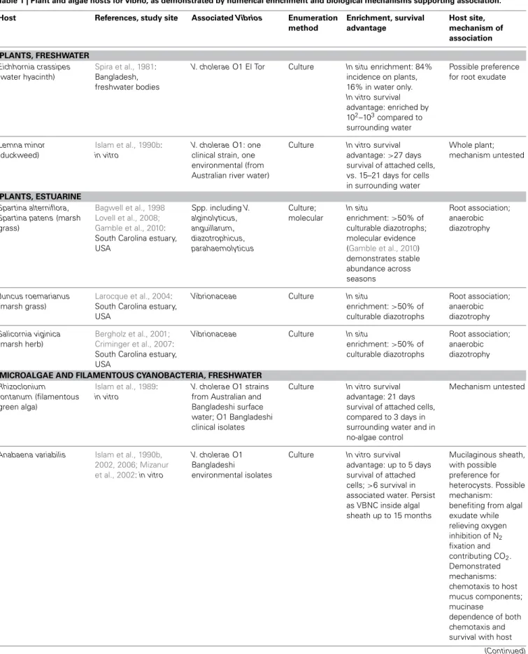
surfaces, but which of these associations represent more than transient, incidental attachments? In the following sections we consider which aquatic plants (Table 1) and animals (Table 2) may represent sustained Vibrio niches, on the basis of (1) numer-ical enrichment compared to the surrounding medium, and (2) knowledge of biological mechanisms, e.g., availability of nutrition and shelter, potentially supporting an association. In doing so, we also draw attention to the need for more quantitative and mech-anistic approaches to understanding the ecological associations that allow vibrios to flourish—approaches that could underpin more powerful predictions of Vibrio dynamics arising from these diverse associations. We note also that many of the following observations are limited to V. cholerae because of its prominence as a pathogen, but the same niches may be available to other vibrios with similar biological activities.
ASSOCIATIONS WITH PLANTS
Vibrio survival is enhanced in association with certain freshwater and estuarine plants (Table 1). Plant hosts can provide nutri-tion (Andrews and Harris, 2000) and the opportunity to form predation-resistant biofilms (Matz et al., 2005), and have been postulated to modulate unfavorably cold temperatures as well (Criminger et al., 2007). Two freshwater aquatic plants have been observed to support both in situ enrichment (in freshwater bod-ies of Bangladesh) and in vitro survival advantage for V. cholerae: duckweed, Lemna minor (Islam et al., 1990b), and water hyacinth,
Eichhornia crassipes (Spira et al., 1981), with preference for roots of the latter. Concentration on E. crassipes roots may indicate that root exudate is a particularly rich nutritional source, but may also be an artifact of the fact that the roots represent the greatest area exposed to water, and hence to inoculation by planktonic Vibrio. By contrast, duckweed’s minimal structure, lacking stem or devel-oped leaves, means that almost the entire plant is in contact with the water and thus available for inoculation.
Among estuarine plants, nitrogen-fixing representatives of several Vibrio taxa—including V. diazotrophicus, V. natriegens,
V. cininnatiensis (Urdaci et al., 1988), and V. parahaemolyticus (Criminger et al., 2007)—appear to be noteworthy members of the rhizosphere, given that they represent more than half of the culturable diazotrophs associated with the dominant marsh grasses Spartina sp. and Juncus roemerianus (Bagwell et al., 1998; Larocque et al., 2004), and the herb Salicornia virginica (Bergholz
et al., 2001; Criminger et al., 2007). While this numerical dom-inance may reflect culturing bias, later molecular studies of the
S. alterniflora rhizosphere confirmed that vibrios (not
taxonomi-cally resolved below the level of the family) are stable constituents of the community (Lovell et al., 2008), with little seasonal fluc-tuation (Gamble et al., 2010). Nitrogen fixation thus appears to be an effective strategy supporting Vibrio survival in the anaero-bic rhizosphere, demonstrating the ecological breadth granted by vibrios’ facultatively anaerobic metabolism.
ASSOCIATIONS WITH MICROALGAE AND FILAMENTOUS CYANOBACTERIA
While early culture-based studies have demonstrated numeri-cal dominance of vibrios on phytoplankton surfaces compared to surrounding water, e.g.,Simidu et al. (1971), little is known about direct, physical associations with specific phytoplankton. Algal cells represent a nutritional opportunity in that they often excrete a high proportion of their photosynthetically fixed car-bon, thereby creating a diffusive sphere (the phycosphere) around them, with elevated organic carbon concentration compared to the bulk (Paerl and Pinckney, 1996). However, in vitro survival advantage and persistence have been thus far been demonstrated only for V. cholerae in physical association with two microal-gae: with the filamentous freshwater green alga Rhizoclonium
fontanum (Islam et al., 1989), and inside the mucilaginous sheath of Anabaena sp. cyanobacteria under both freshwater (Islam et al., 1990a, 1999) and saline conditions (Ferdous, 2009) (Table 1).
Recent work has illuminated mechanistic details of the V.
cholerae association with Anabaena, which may follow the
canon-ical model of symbioses between heterotrophic bacteria and nitrogen-fixing freshwater cyanobacteria. In such associations, heterotrophs locate their hosts via chemotaxis and benefit from rich cyanobacterial exudate (Paerl and Gallucci, 1985). In return, their oxidative metabolism both relieves oxygen inhibition of nitrogen fixation (which would otherwise limit rapid algal growth), and generates carbon dioxide for photosynthetic assim-ilation (Paerl and Gallucci, 1985). For V. cholerae, chemotactic preference for components of the Anabaena mucilaginous sheath has been demonstrated (Mizanur et al., 2002). Furthermore, investigators have shown that both chemotaxis to and survival on Anabaena depend on V. cholerae’s expression of mucinase (Islam et al., 2002, 2006). The exact role of mucinase has yet to be defined, but activity of secreted mucinase might liberate from mucus the relevant chemotactic attractants, aid colonizing Vibrio in physical penetration of the mucilage, and/or convert mucilage to nutritive compounds supplementary to the cyanobacterial exudate.
ASSOCIATIONS WITH MACROALGAE
Numerous studies have shown that vibrios are one of the most abundant culturable constituents of macroalgal communi-ties (Table 1): a recent meta-analysis of 161, predominantly culture-dependent macroalgal-bacterial studies determined that vibrios on average comprised 10% of these communities (Hollants et al., 2013), with 28, 28, and 44% of them found on brown, green, and red macroalgae, respectively. While no molecular studies have yet quantified Vibrio within macroalgal
Takemura et al. Vibrio ecology
Table 1 | Plant and algae hosts for vibrio, as demonstrated by numerical enrichment and biological mechanisms supporting association. Host References, study site Associated Vibrios Enumeration
method Enrichment, survival advantage Host site, mechanism of association PLANTS, FRESHWATER Eichhornia crassipes (water hyacinth) Spira et al., 1981: Bangladesh, freshwater bodies
V. cholerae O1 El Tor Culture In situ enrichment: 84%
incidence on plants, 16% in water only. In vitro survival advantage: enriched by 102–103compared to surrounding water Possible preference for root exudate
Lemna minor
(duckweed)
Islam et al., 1990b:
in vitro
V. cholerae O1: one
clinical strain, one environmental (from Australian river water)
Culture In vitro survival
advantage:>27 days
survival of attached cells, vs. 15–21 days for cells in surrounding water
Whole plant; mechanism untested
PLANTS, ESTUARINE
Spartina alterniflora, Spartina patens (marsh
grass)
Bagwell et al., 1998 Lovell et al., 2008; Gamble et al., 2010: South Carolina estuary, USA Spp. including V. alginolyticus, anguillarum, diazotrophicus, parahaemolyticus Culture; molecular In situ enrichment:>50% of culturable diazotrophs; molecular evidence (Gamble et al., 2010) demonstrates stable abundance across seasons Root association; anaerobic diazotrophy Juncus roemarianus (marsh grass) Larocque et al., 2004: South Carolina estuary, USA
Vibrionaceae Culture In situ
enrichment:>50% of culturable diazotrophs Root association; anaerobic diazotrophy Salicornia viginica (marsh herb) Bergholz et al., 2001; Criminger et al., 2007: South Carolina estuary, USA
Vibrionaceae Culture In situ
enrichment:>50% of culturable diazotrophs
Root association; anaerobic diazotrophy
MICROALGAE AND FILAMENTOUS CYANOBACTERIA, FRESHWATER
Rhizoclonium fontanum (filamentous green alga) Islam et al., 1989: in vitro V. cholerae O1 strains
from Australian and Bangladeshi surface water; O1 Bangladeshi clinical isolates
Culture In vitro survival
advantage: 21 days survival of attached cells, compared to 3 days in surrounding water and in no-algae control
Mechanism untested
Anabaena variabilis Islam et al., 1990b, 2002, 2006; Mizanur et al., 2002: in vitro
V. cholerae O1
Bangladeshi
environmental isolates
Culture In vitro survival
advantage: up to 5 days survival of attached cells;>6 survival in associated water. Persist as VBNC inside algal sheath up to 15 months Mucilaginous sheath, with possible preference for heterocysts. Possible mechanism: benefiting from algal exudate while relieving oxygen inhibition of N2 fixation and contributing CO2. Demonstrated mechanisms: chemotaxis to host mucus components; mucinase dependence of both chemotaxis and survival with host
Table 1 | Continued
Host References, study site Associated Vibrios Enumeration method Enrichment, survival advantage Host site, mechanism of association MACROALGAE, MARINE Brown algae
Ascophyllum nodosum Chan and McManus, 1969: Canada
Vibrio spp. Culture In situ enrichment:
Dominant culturable bacteria; enriched by 102–104compared to water column Algal polysaccharide metabolism
Laminaria spp. Laycock, 1974: Nova Scotia, Canada;Wang et al., 2009
Spp. incl. V.
tasmaniensis
Culture In situ enrichment:
Dominant culturable bacteria Algal polysaccharide metabolism; laminaranolytic activity in particular demonstrated Red algae Hypnea spp. Lakshmanaperumalsamy and Purushothaman, 1982: tropical estuary, Africa
Vibrio spp. Culture In situ enrichment:
Dominant culturable bacteria
Algal polysaccharide metabolism
Polysiphonia lanosa Chan and McManus, 1969: Canada.Islam et al., 1988: in vitro.; Wang et al., 2009 Vibrio spp., incl. V. tasmaniensis, splendidus; in vitro experiments with V. cholerae O1
Culture In situ enrichment:
Dominant culturable bacteria; enriched by 102–104compared to
water column. In vitro survival advantage demonstrated
Algal polysaccharide metabolism
Porphyra yezoensis Duan et al., 1995: China Vibrio spp. Culture, scanning electron microscopy In situ enrichment: Dominant microscopically identifiable and culturable bacteria Algal polysaccharide metabolism Green algae Chaetomorpha spp. Lakshmanaperumalsamy and Purushothaman, 1982: tropical estuary, Africa
Vibrio spp. Culture In situ enrichment:
Dominant culturable bacteria Algal polysaccharide metabolism Enteromorpha intestinalis, linza Lakshmanaperumalsamy and Purushothaman, 1982: tropical estuary, AfricaIslam et al., 1988: in vitro
Vibrio spp.; in vitro
experiments with V.
cholerae O1
Culture In situ enrichment:
Dominant culturable bacteria. In vitro survival advantage demonstrated
Algal polysaccharide metabolism
Ulva lactuca, pertusa Islam et al., 1988:
in vitro;Duan et al., 1995: China;Nakanishi et al., 1996; Patel et al., 2003; Tait et al., 2005 Vibrio spp.; in vitro experiments with V. cholerae O1 Culture, scanning electron microscopy In situ enrichment: Dominant microscopically identifiable and culturable bacteria. In
vitro survival advantage
demonstrated Algal polysaccharide metabolism; modulation of host processes: developmental morphogenic effects, spore germination stimulation
communities, numerical enrichment of culturable vibrios has been demonstrated for the brown algae Ascophyllum nodosum (Chan and McManus, 1969), and Laminaria longicruris (Laycock, 1974); the red algae Hypnea sp. (Lakshmanaperumalsamy
and Purushothaman, 1982), Polysiphonia lanosa (Chan and McManus, 1969), and Porphyra yezoensis (Duan et al., 1995); and the green algae Chaetomorpha sp. (Lakshmanaperumalsamy and Purushothaman, 1982), Enteromorpha sp.
Takemura et al. Vibrio ecology
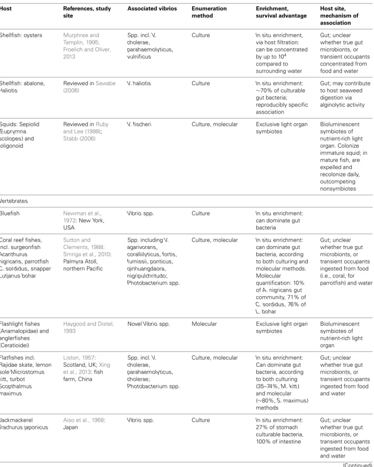
Table 2 | Animal hosts for vibrio, as demonstrated by numerical enrichment and biological mechanisms supporting association.
Host References, study
site
Associated vibrios Enumeration method Enrichment, survival advantage Host site, mechanism of association INVERTEBRATES Freshwater Acanthamoeba protozoa Abd et al., 2005, 2007, 2010; Sandström et al., 2010: in vitro V. cholerae O1, O139; V. mimicus
Culture, microscopy In vitro survival
advantage: replicate intracellularly>14 days Cytoplasm, cysts; protected from antibiotics and predation Chironomid midge egg masses
Broza and Halpern, 2001; Halpern et al., 2003, 2008: in vitro
V. cholerae isolates
from Israeli rivers and
waste-stabilization ponds
Culture In vitro survival
advantage: 103
greater cell counts compared to growth in medium alone
Gelatinous egg matrix; can use gelatinous material as sole carbon source, degrading via secreted hemag-glutinin/protease Zooplankton: cladoceran Diaphanosoma mongolianum, from alkaline lake, Germany Kirschner et al., 2011: in vitro V. cholerae non-O1/non-O139 isolate from alkaline lake, Germany
Fluorescence in situ hybridization
In vitro survival
advantage, but not enrichment: up to 6-fold increase in growth rate of cells in surrounding medium; 105–107cells attached compared to 106–107cells in surrounding medium
Probable use of host exudates
Estuarine and marine
Zooplankton: Estuarine copepods, espp. Acartia and
Eurytemora
Simidu et al., 1971: Japan;Sochard et al., 1979: Gulf of Mexico;Huq et al., 1983, 1984: in vitro; Colwell, 1996: in vitro;Mueller et al., 2007: in vitro; Preheim et al., 2011a: Massachusetts estuary, USA Vibrio spp., espp. V. cholerae
Culture In situ and in vitro
enrichment shown in some cases, with up to 105cells per
host. Can dominate culturable surface-and gut-attached communities
Possible preference for oral region and egg sac, due to proximity to host exudates; preference for live versus dead hosts unclear Corals, incl. Acropora hyacinthus, Oculina patagonica, Mussimilia hispida, Stylophora pistillata Koren and Rosenberg, 2006: Israel;Kvennefors et al., 2010: Great Barrier Reef; Chimetto et al., 2008; Sharon and Rosenberg, 2008; Koenig et al., 2011; Krediet et al., 2013 Spp. incl. V. alginolyticus, harveyi, splendidus
Culture, molecular In situ enrichment:
can dominate mucus community, according to both culturing and molecular methods; can dominate culturable diazotrophs (found for Mussimilia hispida) Mucus. Metabolize mucus; diazotrophs likely contribute nitrogen to hosts; may adapt to host antimicrobials via antibiotic-resistance gene acquisition; can inhibit pathogen colonization
Shellfish: blue crabs,
Callinectes sapidus
Davis and Sizemore, 1982: Texas, USA
Spp. incl. V.
cholerae, vulnificus, parahaemolyticus
Culture In situ enrichment:
Dominant culturable bacteria in hemolymph Hemolymph; mechanism untested (Continued)
Table 2 | Continued
Host References, study
site
Associated vibrios Enumeration method Enrichment, survival advantage Host site, mechanism of association
Shellfish: oysters Murphree and Tamplin, 1995; Froelich and Oliver, 2013
Spp. incl. V.
cholerae, parahaemolyticus, vulnificus
Culture In situ enrichment,
via host filtration: can be concentrated by up to 104
compared to surrounding water
Gut; unclear whether true gut microbionts, or transient occupants concentrated from food and water
Shellfish: abalone,
Haliotis
Reviewed inSawabe (2006)
V. haliotis Culture In situ enrichment:
∼70% of culturable gut bacteria; reproducibly specific association
Gut; may contribute to host seaweed digestion via alginolytic activity Squids: Sepiolid (Euprymna scolopes) and loligonoid Reviewed inRuby and Lee (1998); Stabb (2006)
V. fischeri Culture, molecular Exclusive light organ symbiotes Bioluminescent symbiotes of nutrient-rich light organ. Colonize immature squid; in mature fish, are expelled and recolonize daily, outcompeting nonsymbiotes
Vertebrates
Bluefish Newman et al., 1972: New York, USA
Vibrio spp. Culture In situ enrichment:
can dominate gut bacteria
Coral reef fishes, incl. surgeonfish Acanthurus nigricans, parrotfish C. sordidus, snapper Lutjanus bohar Sutton and Clements, 1988; Smriga et al., 2010: Palmyra Atoll, northern Pacific Spp. including V. agarivorans, coralliilyticus, fortis, furnissii, ponticus, qinhuangdaora, nigripulchritudo; Photobacterium spp.
Culture, molecular In situ enrichment:
can dominate gut bacteria, according to both culturing and molecular methods. Molecular quantification: 10% of A. nigricans gut community, 71% of C. sordidus, 76% of L. bohar Gut; unclear whether true gut microbionts, or transient occupants ingested from food (i.e., coral, for parrotfish) and water
Flashlight fishes (Anamalopidae) and anglerfishes (Ceratioidei)
Haygood and Distel, 1993
Novel Vibrio spp. Molecular Exclusive light organ symbiotes Bioluminescent symbiotes of nutrient-rich light organ Flatfishes incl. Rajidae skate, lemon sole Microstomus
kitt, turbot Scopthalmus maximus
Liston, 1957: Scotland, UK;Xing et al., 2013: fish farm, China Spp. incl. V. cholerae, parahaemolyticus, cholerae; Photobacterium spp.
Culture, molecular In situ enrichment:
Can dominate gut bacteria, according to both culturing (35–74%, M. kitt) and molecular (∼80%, S. maximus) methods Gut; unclear whether true gut microbionts, or transient occupants ingested from food and water
Jackmackerel
Trachurus japonicus
Aiso et al., 1968: Japan
Vibrio spp. Culture In situ enrichment:
27% of stomach culturable bacteria, 100% of intestine
Gut; unclear whether true gut microbionts, or transient occupants ingested from food and water
Takemura et al. Vibrio ecology
Table 2 | Continued
Host References, study
site
Associated vibrios Enumeration method Enrichment, survival advantage Host site, mechanism of association Salmonidae, incl. pink salmon Onchorhynchus gorbuscha, chum salmon O. keta, sockeye salmon O. nerka, Chinook salmon O. tshawytscha Yoshimizu and Kimura, 1976: Japanese coast, East Bering Sea
Vibrio spp. Culture In situ enrichment:
dominate gut bacteria of saltwater-dwelling (but not freshwater) salmonids; on average represent 69% of saltwater gut community
Gut; unclear whether true gut microbionts, or transient occupants ingested from food and water
Sea bream Pagrus
major, Acanthopagrus schlegeli
Muroga et al., 1987: Japan
Vibrio spp. Culture In situ enrichment:
∼45% of culturable gut bacteria
Gut; unclear whether true gut microbionts, or transient occupants ingested from food and water
(Lakshmanaperumalsamy and Purushothaman, 1982), and
Ulva pertusa (Duan et al., 1995). For V. cholerae, in vitro survival advantage has been shown on the green algae Ulva lactuca and
Enteromorpha intestinalis and the red alga Polysiphonia lanosa
(Islam et al., 1988).
As mentioned above, vibrios can metabolize many algal polysaccharides; they have furthermore been implicated in several other biological activities facilitating symbiosis with macroalgal hosts. These include antagonism directed toward potential bacterial or algal competitors for host surface area (Dobretsov and Qian, 2002; Kanagasabhapathy et al., 2008), developmental morphogenic effects on Ulva pertusa (Nakanishi et al., 1996), and stimulation of spore germination for Ulva sp. (Patel et al., 2003; Tait et al., 2005). Hence multi-ple lines of evidence point to significant Vibrio association with Ulva sp. (enrichment, survival, morphogenesis and spore modulation) and Polysiphonia sp. (enrichment, survival) in particular.
ASSOCIATIONS WITH ANIMALS
Vibrio interactions with animals include both specific, stable
bioses, and less well-defined associations (Table 2). Stable sym-bioses have been described for luminescent V. fischeri (Aliivibrio) with sepiolid squids (Euprymna scolopes) and loligonoid squids (Ruby and Lee, 1998), and for various luminescent Vibrio with flashlight fishes (Anamalopidae) and anglerfishes (Ceratioidei) (Haygood and Distel, 1993). The dynamics of the V.
fischeri-Euprymna symbiosis have been particularly well-explicated: V. fischeri from surrounding waters colonize the developing squid
light organ, successfully outcompeting non-symbionts in this process, which triggers a developmental program in the host. Once established, the symbionts undergo daily cycles of expul-sion and regrowth (Ruby and Lee, 1998; Stabb, 2006). Thus the symbiosis regularly seeds the water column, such that lumi-nous V. fischeri are enriched in the water surrounding E. scolopes (Ruby and Lee, 1998). This expedites continual recoloniza-tion of immature squid, which is likely further facilitated by
V. fischeri chemotaxis toward squid mucus (DeLoney-Marino et al., 2003).
Some Vibrio have also been deemed facultative intracellu-lar symbionts of Acanthamoeba protozoa: Vibrio cholerae O1 and O139, and Vibrio mimicus (Abd et al., 2005, 2007, 2010; Sandström et al., 2010). These vibrios can replicate intra-cellularly for at least 14 days without affecting host health, at least in nutrient-replete artificial medium, and have been observed in both cytoplasm and cysts of the protozoa. Like sev-eral other microbial taxa, then, most famously the pathogen
Legionella (Rowbotham, 1980), vibrios appear capable of evad-ing Acanthamoeba endocytosis to shelter intracellularly. Thus they gain protection from antibiotics (Abd et al., 2005, 2007, 2010), predation, and perhaps other adverse conditions, e.g., cold tem-peratures. Still to be investigated are the questions of why some
Acanthamoeba cells encyst their Vibrio inhabitants while others
do not; why the Vibrio do not appear to be detrimental to host survival; and how often Vibrio might be released following host lysis, or even actively ejected, thus returning to the water column. Moreover, all studies of the Vibrio-Acanthamoeba relationship have been experimental: in situ surveys are necessary to estab-lish the environmental relevance of this potential symbiosis, and assess any effects on Vibrio population dynamics.
Vibrios may be neutral or benign inhabitants of coral hosts: they have been shown to comprise a significant portion of the mucus-dwelling bacterial community of healthy corals (e.g.,
Koren and Rosenberg, 2006; Kvennefors et al., 2010), being able to subsist on coral mucus as their sole carbon and nitrogen source (Sharon and Rosenberg, 2008). V. splendidus, for example, consti-tuted 50–68% of clone libraries derived from Oculina patagonica coral mucus, but was scarce in the coral tissue itself (Koren and Rosenberg, 2006). Moreover, nitrogen-fixing Vibrio representa-tives, primarily V. harveyi and V. alginolyticus, have been found to dominate the culturable diazotrophs of the coral Mussimilia
hisp-ida (Chimetto et al., 2008), and likely share fixed nitrogen with either or both coral and zooxanthellae. Evidence also suggests immune interaction between Vibrio and coral hosts: adaptation
of Vibrio commensals to coral antimicrobials has been suggested by significant antibiotic-resistance gene cassette content of their integrons (Koenig et al., 2011), while one V. harveyi coral isolate has been found to help defend its host by inhibiting colonization by a pathogen (Krediet et al., 2013).
In freshwater habitats, V. cholerae have been found to prolifer-ate on egg masses of the abundant, widely distributed chironomid midges (Broza and Halpern, 2001; Halpern et al., 2008). These egg masses are embedded in thick, gelatinous material, which
V. cholerae can use as a sole carbon source (Broza and Halpern, 2001); their degradation of the gelatinous matrix via secreted hemagglutinin/protease appears to be the primary cause of egg mass disintegration (Halpern et al., 2003). Accordingly,Halpern et al. (2006)were able to show correlations of chironomid egg mass with the abundance of attached V. cholerae, although they have not yet investigated any correlation of V. cholerae dynamics in the surrounding aquatic environment.
Zooplankton, primarily estuarine copepods such as Acartia and Eurytemora, have been investigated as a major reservoir of
V. cholerae in particular, but while attachment has been
demon-strated, it remains unclear whether the association is specific, and whether attached vibrios are consistently enriched compared to surrounding waters. Individual copepods have been shown to be able to host up to 105 V. cholerae cells (Colwell, 1996;
Mueller et al., 2007), with preference often shown for attach-ment to the oral region and egg sac (next to the anal pore)—that is, regions offering close access to host exudates (Huq et al., 1983, 1984). Culture-based studies have detected enriched Vibrio occurrence on copepods compared to the surrounding water col-umn (e.g., Simidu et al., 1971; Sochard et al., 1979), and one culture-based study showed Vibrio dominance of wild copepods’ surface- and gut-attached bacterial communities (Sochard et al., 1979). However, other studies, both in vitro and in situ, have observed V. cholerae remaining predominantly free-living in the presence of copepods (Worden et al., 2006; Neogi et al., 2012) or attaching with greater preference to phytoplankton (Tamplin et al., 1990). Additionally, one culture-independent environmen-tal study detected greater concentrations of Vibrio, including V.
cholerae, in water compared to zooplankton (Heidelberg et al., 2002a,b). Perhaps such variability of association with copepods helps explain the difficulty in detecting correlated Vibrio-copepod dynamics, as mentioned above in the section Environmental Correlates of Vibrio Presence and Abundance.
Other uncertainties regarding Vibrio association with cope-pods exist. There is a lack of quantitative evidence demonstrat-ing long-term proliferation of copepod-attached Vibrio: existdemonstrat-ing studies assessing survival advantage of Vibrio cultured with cope-pods have only demonstrated increased abundance of Vibrio in surrounding water, without monitoring attached abundance (Huq et al., 1983, 1984). Finally, it is not clear whether vibrios prefer colonizing live or dead copepods. While several in vitro studies have noted V. cholerae attachment preference for dead or detrital copepods (Huq et al., 1990; Tamplin et al., 1990; Mueller et al., 2007), one study instead observed survival advantage only upon association with live copepods, and found little attachment to dead copepods (Huq et al., 1983). Perhaps this question could be resolved by investigating from which part(s) exactly of the
copepod vibrios derive nutrition: from oral/anal exudates or gut contents of actively feeding copepods, from degradation of the chitinaceous exoskeleton which for live copepods is protected by a waxy epicuticle that resists attachment (Tarsi and Pruzzo, 1999), or from degradation of other copepod detritus. In addition, vari-able host traits such as immune defenses, age, and time since molting or death (which likely affect epicuticle condition) should be taken into account. As of yet, evidence of association with live copepods as an ecological specialization has been demonstrated for only one Vibrio sp. nov. (F10) (Preheim et al., 2011a).
In addition, zooplankton other than copepods may represent potential Vibrio hosts as well.Kirschner et al. (2011)found clado-ceran Diaphanosoma mongolianum to enhance growth more than the copepod Arctodiaptomus spinosus in microcosm experiments; when cladocerans were added, they enhanced the growth of V.
cholerae strains in the surrounding medium relative to controls
where cladocerans were excluded, while copepods did not. In addition, the number of cells attached to cladocerans per indi-vidual was on average 100 times higher than on copepods. When a back-of-the-envelope calculation is done to consider whether V.
cholerae is enriched on zooplankton, however, we find that they
are not, even on cladocerans; from six microcosms, 105–107cells were estimated attached and 106–107cells not attached, a result suggesting that cladocerans might enhance overall growth with frequent dispersal, rather than supporting exclusively attached growth.
For other animals in which Vibrio have been found to be abundant—fish, and shellfish—it has not yet been determined whether vibrios form specific, lasting associations as gut micro-biota, or are merely transient occupants, temporarily proliferating on favorable nutrients until excreted or otherwise detached. In marine fish, numerous studies, both culturedependent and -independent, have demonstrated that Vibrio are major gut inhab-itants, often dominating the community, and hence are substan-tially enriched compared to surrounding seawater. Surveyed fish include flatfish (Liston, 1957; Xing et al., 2013), jackmackerel (Aiso et al., 1968), bluefish (Newman et al., 1972), salmonids (Yoshimizu and Kimura, 1976), sea bream (Muroga et al., 1987), and various coral reef fishes (Sutton and Clements, 1988; Smriga et al., 2010). Notably, Vibrio abundances often appear compara-ble between culture-based and -independent studies: e.g., 35–74 and 83.4%, respectively, of flatfish inhabitants (Liston, 1957; Xing et al., 2013). The ability of Vibrio representatives to resist low pH and bile supports their survival within the fish gut (Yoshimizu and Kimura, 1976). Whether food or water intake is the greater source of inoculation is an open question: some studies have found a strong effect of food source on gut Vibrio composition (e.g.,Grisez et al., 1997), whereas others found a stronger influ-ence of Vibrio representation in the water column (e.g.,Blanch et al., 2009). Conversely, Vibrio content of the fish gut has also been shown to be responsible for increasing Vibrio abundance in surrounding water when fish were introduced into a tank that did not otherwise support Vibrio growth, demonstrating significant excretion of viable cells from the fish gut (Sugita et al., 1985). Hence, regardless of length of association, the fish gut appears to represent a favorable refuge where Vibrio can rapidly prolif-erate, prior to being released again to the water column. Indeed,