ANALYSIS OF THE ROLE OF THE RETINOBLASTOMA SUSCEPTIBILITY GENE IN MURINE DEVELOPMENT AND TUMORIGENESIS
by Bart O. Williams
B. S., Biology and Chemistry, Carroll College, 1991
Submitted to the Department of Biology in Partial Fulfillment of the Requirements for the Degree of
DOCTOR OF PHILOSOPHY
at the
Massachusetts Institute of Technology May 1996
@ Bart O. Williams 1996
All rights reserved
The author hereby grants to M. I. T. permission to reproduce and to distribute publicly copies of this thesis document in whole or in part.
Signature of Author
Department of Biology May 15, 1996
Certified by
Professor Tyler Jacks Thesis Supervisor
Accepted by ,-,.. , ,
A t Professor Frank Solomon
Chairman of the Biology Graduate Committee
1iASSACHUSETTS INSTITUTE
OF TECHNOLOGY
JUL 0
81996
ARCHIVES
ABSTRACT
Studies on the retinoblastoma susceptibility gene (RB) have played a central role in the conceptual development of the tumor suppressor gene field. Patients inheriting a mutation in the RB gene develop the rare pediatric eye cancer retinoblastoma with 90% penetrance. In addition, somatic mutations in the RB gene have been found in many sporadically occurring human tumors. A great deal of insight has also been gained from studies of retinoblastoma protein (pRB) in tissue culture. These studies have shown that the protein product plays an important role in cell cycle
coordination, perhaps by modulating some aspect of the restriction point during the G 1 phase of the cell cycle. Despite this vast amount of information, until recently very little was known about the normal function of RB in development. Techniques developed in the last decade have allowed mice with targeted mutations to be created. Several groups have exploited these techniques to create mice with germline inactivating mutations in the murine version of the retinoblastoma susceptibility gene (Rb). Initial analysis of these mice indicated that heterozygotes did not develop retinoblastoma, instead they develop tumors in the intermediate lobe of the pituitary and in the thyroid. Mice homozygous for the mutant allele die between 13.5 and 15.5 days of gestation with associated defects in hematopoiesis and neurogenesis.
The focus of this work has been on expanding the use of these mice to better understand RB function. One surprising observation is the relatively limited tumor spectrum observed in both mice and humans which are constitutionally heterozygous for an RB mutation. A potential
explanation for this is that other genetic alterations must occur in combination with Rb mutation to cause tumorigenesis in most tissues. To test this model, Rb-deficient mice were crossed with a
p53 mutant strain. Animals carrying mutations in both genes are susceptible to a wider spectrum
of tumors. A potential mechanism for this cooperativity is presented which shows that at least in one system (the developing murine lens) Rb-deficiency leads to p53-dependent apoptotic cell death. This apoptotic induction could serve to eliminate potentially transformed cell from the organism.
One limitation to the utility of the Rb-deficient strain was the attendant embryonic lethality. To bypass this lethality and assess the effects of Rb-deficiency during later stages of development and in adult mice, ES cells were created which carried targeted mutations in both copies of the Rb gene and used them to create chimeric animals. Analysis of tissue homogenenates from these chimeras showed that Rb-deficient cells could contribute to most tissues of the adult mouse without
pathological consequences. To further this analysis, Rb-deficient ES cells have been created which carry a transgene directing nearly ubiquitous expression of B-galactosidase. Staining of chimeric tissues derived from these ES cells for B-galactosidase activity has enabled us to identify and analyze Rb-deficient cells on a single cell level within these chimeras.
Thesis Advisor: Tyler Jacks
Title: Assistant Investigator, Howard Hughes Medical Institute and Assistant Professor of Biology
BIOGRAPHICAL NOTE BART O. WILLIAMS Date of Birth: Place of Birth: August 5, 1969 Madison, Wisconsin Education
Wild Rose High School, Wild Rose, WI 1987
GPA: 3.96
Carroll College, Waukesha, WI B.S. 1991 Biology and Chemistry
GPA: 3.95 Class Rank: 1 of 369
Massachusetts Institute of Technology, Cambridge, MA
Ph.D. (May 1996) Biology Awards and Honors
1987-1991 Presidential Scholarship Award, Carroll College
1988 American Association of University Professors Award for Sophomores
1989 Undergraduate Research Grant Award from the National Science Foundation (through the Center for Great Lakes Studies,
Milwaukee, WI)
1990 Delta Sigma Nu Scholastic Honor Society 1990-1991 USA Today's All-USA Academic Third Team 1991 Outstanding Senior Biology Student, Carroll College 1996 Young Investigator Travel Grant Award, AACR Meeting,
Lausanne, Switzerland (January 17-20, 1996)
1996 Postdoctoral Fellowship Award from the Damon Runyon-Walter Winchell Cancer Research Fund
Research Experience
1989-1990 Undergraduate Research Student, Carroll College, Department of Biology, Waukesha, WI (Dr. Leslie D. Zettergren, Advisor)
1990-1991 Undergraduate Research Student, Medical College of Wisconsin, Department of Biochemistry, Milwaukee, WI (Dr. Arthur L. Haas, Advisor)
1991-1996
1996-Graduate Student, Department of Biology and Center for Cancer Research, MIT, Cambridge, MA (Dr. Tyler Jacks, Advisor)
Damon Runyon-Walter Winchell Cancer Research Fund Postdoctoral Fellow, Varmus Laboratory, National Cancer Institute, Bethesda, MD (Dr. Harold E. Varmus, Sponsor) Teaching Experience
1989 1989-1991 1990 1990 1992 1994
Teaching Assistant -Introductory Biology Laboratory, Carroll College (Dr. Ted Michaud, Instructor)
Tutor for Introductory Biology Lectures, Carroll College (Dr. Ted Michaud, Dr. John Clausz, and Dr. John Mellen, Instructors) Teaching Assistant -Microbiology Laboratory, Carroll College (Dr. John Clausz, Instructor)
Teaching Assistant -Comparative Anatomy Laboratory, Carroll College (Dr. Ted Michaud, Instructor)
Teaching Assistant -Experimental Molecular Biology Laboratory, Massachusetts Institute of Technology (Dr. Phillips Robbins,
Instructor)
Teaching Assistant -Undergraduate Cell Biology, Massachusetts Institute of Technology (Dr. Richard Young and Dr. Tyler Jacks, Instructors)
Publications
Zettergren, L.D., Cutlan, R., and Williams. B.O. (1991) Embryonic dorsal region compartments in B lymphopoiesis: Rana pipiens, Gallus gallus, and Mus musculus, Developmental and Comparative Immunology, 15:105S
Chen, J.C., Gorman, J.R., Stewart, V., Williams, B., Jacks, T., and Alt, F.W. (1993). Generation of normal lymphocyte populations from Rb-deficient ES cells. Current Biology 3(7); 405-413. Jacks, T., Remington, L., Williams, B.O., Schmitt, E.M., Halachmi, S., Bronson, R.T., and Weinberg, R.A. (1994) Tumor spectrum analysis of p53-mutant mice. Current Biology 4(1): 1-7.
Slebos, R.J.C., Lee, M.H., Plunkett, B.S., Kessis, T.D., Williams, B.O., Jacks, T., Hedrick, L., Kastan, M.B., and Cho, K.R. (1994) p53-dependent G1 arrest involves pRB related proteins and is disrupted by the human papillomavirus 16 E7 oncoprotein. Proc Natl Acad Sci USA 91:
5320-5324.
Williams, B.O., Remington, L., Albert, D.M., Mukai, S., Bronson, R.T., and Jacks, T. (1994) Cooperative tumorigenic effects of germline mutations in Rb and p53. Nature Genetics 7:
480-484.
Williams, B.O., Schmitt, E.M., Remington, L., Bronson, R.T., Albert, D.M., Weinberg, R.A., and Jacks, T. (1994) Extensive contribution of Rb-deficient cells to adult chimeric mice with limited histopathological consequences. EMBO J. 13: 4521-4529.
Morgenbesser, S.D., Williams. B.O., Jacks, T., and DePinho, R.A. (1994) p53-dependent apoptosis produced by Rb-deficiency in the developing mouse lens. Nature 371: 72-74.
Williams, B.O., Morgenbesser, S.D., DePinho, R.A., and Jacks, T. (1995) Developmental and tumorigenic consequences of Rb and p53 mutations. Cold Spring Harbor Symp Quant Biol, 59:449-457.
Haas, A.L., Baboshina, O., Williams, B.O., and Schwartz, L.M., (1995) Exponential increases in ubiquitin conjugate pools accompany the developmentally programmed cell death of insect skeletal muscle, Journal Biological Chemistry 270(16):9407-9412..
Sah, V.P., Attardi, L.D., Mulligan, G.M., Williams, B.O., Bronson, R.T., and Jacks, T. (1995) A subset of p53-deficient female embryos develop exencephaly. Nature Genetics 10:175-180. Tischler, A.S., Shih, T.S., Williams. B.O., and Jacks, T. (1995) Characterization of
pheochromocytomas in a mouse strain with a targeted disruptive mutation of the Neurofibromatosis gene Nfl. Endocrine Pathology 6(4): 323-335.
Williams, B.O. and Jacks, T. (1996) Mechanisms of carcinogenesis and the mutant mouse. Current Opinion in Genetics and Development 6(1): 65-69.
Herrera, R.H., Sah, V.P., Williams, B.O., Makela, T., Weinberg, R.A., and Jacks, T. (1996) Altered cell cycle kinetics, gene expression, and G1 restriction point regulation in Rb-deficient fibroblasts. Molecular and Cellular Biology 16(5): 2402-2406.
Lee, M.H., Williams, B.O., Mulligan, G.J., Mukai, S., Bronson, R.T., Dyson, N., Harlow, E., and Jacks, T. (1996) Targeted disruption of pl07: evidence for functional overlap with Rb. Genes and Development (in press).
ACKNOWLEDGMENTS
The last 5 years of my life have been exciting and rewarding and I owe a multitude of people thanks for that.
I have had the great pleasure of working for Tyler Jacks over the past four years. In that time, I have witnessed a tremendous change in the size of our group (from three of us working with Tyler when he was a Weinberg post-doc to a present day group size of 19) and a well deserved increase in his scientific stature. I can honestly say that Tyler has remained the same supportive advisor he was in the beginning. He has had a great positive influence on every aspect of my development as a scientist.
I would also like to express my gratitude to Bob Weinberg, who allowed me to work in his lab with Tyler for a few months. In addition, I would like to thank Bob for his suggestions over the years and for chairing my thesis committee. I would also like to thank Jackie Lees, David Baltimore, and Nick Dyson for serving on my committee as well.
In terms of people who have worked with me in lab, I have shared a room (and for most of the time) a bay with Shane Shih. Shane has become a great friend and always made lab interesting
and enjoyable. He has also raised my awareness about important issues such as heat conservation. Earlene Schmitt and Lee Remington were also there at the beginning and provided a great deal of camaraderie and technical expertise.
For the first two years of my lab work, I was fortunate enough to have Valerie Tan (now Sah) work with me as a UROP. As I told her when she left for graduate school at UCSD, I will probably end up working for her some day.
The other members of the lab in the Rb group, George Mulligan and Kay (Fergie) Macleod, have been instrumental in advancing much of this work through suggestions, technical help, and interesting conversations. In particular my friendship with Kay has allowed me to meet and discuss science with some prominent individuals (it must be the accent?).
I would also like to thank the other members of the lab for making it such a fun and interesting place to work.
Andi McClatchey for advice and suggestions Leisa Johnson for the same
Reuben Shaw for good conversation and for making 7.06 TAing go so smoothly Jennie Sue Smith, Jimmy Brugarolas, Zemer Gitai, Mariana Nacht, Laura Attardi, Doron Greenbaum, Yvonne Chan, Ichiko Saotome, and Elizabeth Farrell for suggestions and conversation.
I would also like to especially thank Kim Mercer whose hard work and technical expertise have allowed me and others to do much of the work that we do. John Mkandawire has also helped further some of these projects with technical assistance.
I have learned almost as much from Rod Bronson as I have from anyone else during my time here. I thank him for many hours of interesting and informative conversation and instruction. Shiz Mukai has also been a great source of information in my time at MIT.
In addition, I would like to thank all my other collaborators, especially Hong Yang, Ralph Herrera, and Myung-Ho Lee, Nick Dyson, and Ed Harlow.
Also, I want to acknowledge the professors and advisors I had during my undergraduate days at Carroll College. Ted Michaud was my biology advisor and Richard Watkins was my chemistry advisor. Les Zettergren first exposed me to the excitement of doing research and Art Haas (at the Medical College of Wisconsin in Milwaukee) was also an important influence.
Finally I would like to thank the people who have been the most important.
My parents always encouraged me to pursue a course which would make me happy and supported me (along with all four of my grandparents) in everything I did. Whether it was a basketball game, musical, or anything else my parents, grandparents (and even my uncles, aunts, cousins, and great aunts and uncles); my family was always there to support me. This would not have been possible without them.
Last but not least, I would like to thank my wife Wendy and daughter Alissa for their love and support. Wendy worked in the early morning and weekends just so I could do something I enjoyed. In addition, she selflessly moved here with me at the beginning of graduate school (where we didn't know a single person) just so I could go to what I thought (and now know) was one of the best graduate schools in the world.
TABLE OF CONTENTS TITLE PAGE ABSTRACT BIOGRAPHICAL NOTE ACKNOWLEDGMENTS TABLE OF CONTENTS
LIST OF FIGURES AND TABLES
ABBREVIATIONS USED
NOMENCLATURE
CHAPTER 1
CHAPTER 2
Tumor suppressor genes Literature Cited
The retinoblastoma susceptibility gene
Identification and cloning of the retinoblastoma susceptibility gene
In vitro evidence that the RB gene product can directly
suppress cell growth
Analysis of the RB gene product
The RB gene product interacts with several DNA tumor virus oncoproteins
The RB gene product binds to E2F, a cellular transcription factor
pRB can interact with a number of cellular proteins
RB is a member of a multigene family
Regulation of pRB phosphorylation
Methods for creating mice carrying targeted gene inactivations in the germline
Creation of mouse strains with mutations in tumor suppressor genes
Initial analysis of mice carrying an inactivating
8
CHAPTER 3
CHAPTER 4
mutation in the Rb gene
Fetal liver hematopoiesis is defective in Rb-deficient embryos
Rb-deficient embryos display defects in neurogenesis Apoptosis
Rb-deficiency leads to apoptosis in a variety of contexts Overall goals of this work
Literature Cited
The p53 tumor suppressor gene
Identification of the p53 protein and gene
p53 was originally characterized as an oncogene but
later recognized to be a tumor suppressor gene
Inheritance of a p53 mutation in humans is associated with Li-Fraumeni syndrome
p53 is targeted by other DNA tumor virus oncoproteins Cellular functions ofp53
Creation of mouse strains with a targeted disruption in p53
The molecular mechanisms of p53 function Literature Cited
Cooperative tumorigenesis between Rb and p53 mutations
Introduction
Williams BO, Remington L, Albert DM, Mukai S, Bronson RT, and Jacks T. (1994) Cooperative
tumorigenic effects of germline mutations in Rb and p53. Nature Genetics 7: 480-484.
Rational for examining lens development in Rb-deficient embryos
Morgenbesser SD, Williams BO, Jacks T,
and DePinho RA. (1994) p53-dependent apoptosis
51 53 53 55 55 57 74 75 77 79 80 80 83 83 88 95 96 98 104 106
produced by Rb-deficiency in the developing mouse lens. Nature 371: 72-74. Discussion Literature Cited CHAPTER 5 CHAPTER 6 CHAPTER 7
Use of chimeric mice to assess effects of Rb-deficiency in adults
Introduction to chimeric analysis
Williams BO, Schmitt EM, Remington L, Bronson RT, Albert DM, Weinberg RA, and
Jacks T. (1994) Extensive contribution of Rb-deficient cells to adult chimeric mice with limited
histopathological consequences. EMBO J. 13: 4521-4529. Limitations and implications Literature Cited
Derivation and analysis of chimeras created from Rb-deficient; ROSA-26 positive ES cells
Introduction
Materials and Methods Results
Discussion Literature cited
Creation of ES cells deficient for both Rb and p53 Introduction
Materials and methods Results
Discussion Literature cited
CHAPTER 8 Summary and Implications
109 111 114 115 119 128 130 131 132 135 140 154 157 158 159 160 161 163 165 166
Literature Cited APPENDIX A
APPENDIX B
APPENDIX C
Analysis of the effects of p53-deficiency on murine tumorigenesis and development
Notes of attribution Summary
Jacks T, Remington L, Williams BO, Schmitt EM, Halachmi S, Bronson RT, and Weinberg RA. (1994) Tumor spectrum analysis of p53-mutant mice. Current Biology 4(1): 1-7.
Sah VP, Attardi LD, Mulligan GM, Williams BO, Bronson RT, and Jacks T. (1995) A subset of p53
-deficient female embryos develop exencephaly. Nature Genetics 10:175-180.
Literature cited
Characterization of Rb-deficient fibroblast growth Notes of attribution
Introduction
Materials and methods Results
Discussion
Herrera RE, Sah VP, Williams BO, Makela TP, Weinberg RA, and Jacks T. (1996) Altered cell cycle kinetics, gene expression, and G1 phase restriction point regulation in Rb-deficient fibroblasts
Literature Cited
Phenotypes of mice carrying targeted mutations in both Rb and p107 Notes of attribution Summary 174 177 178 179 180 187 193 194 195 196 196 198 202 203 209 210 211 212
APPENDIX D
Lee MH, Williams BO, Mulligan GJ, Mukai S,
Bronson RT, Dyson N, Harlow E, and Jacks T. (1996) Targeted disruption of pl07: evidence for functional overlap with Rb. Genes and Development (in press). Future Directions
Literature Cited
Analysis of phenotypes associated with a hypomorphic allele of Rb in the mouse (RbI3Neo)
Introduction
Materials and methods Results
Discussion Literature Cited
APPENDIX E Generation of ES cells deficient for multiple members of the Rb gene family
Introduction
Materials and methods Results Discussion Literature Cited 213 253 254 255 256 257 265 275 277 278 279 279 280 284 285
LISTS OF FIGURES AND TABLES
CHAPTER 1
Table 1-1. Functions of human tumor suppressor genes 23 and tumors associated with their inactivation
CHAPTER 2
Figure 2-1 Characteristics of pRB 32
Figure 2-2 Structural homology between members of the 39 RB-gene family
Figure 2-3 Regulation of pRB phosphorylation 45
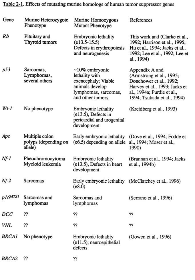
Table 2-1 Effects of mutating murine homologs of human 49 tumor suppressor genes
CHAPTER 3
Figure 3-1 Characteristics of p53 protein 76
Figure 3-2 p53 pathway 86
CHAPTER 4
Figures within:
Williams BO, Remington L, Albert DM, Mukai S, Bronson RT, and Jacks T. (1994) Cooperative tumorigenic effects of germline mutations in Rb and p53. Nature Genetics 7: 480-484.
Figure 1 Survival of mice with mutations in p53 and Rb 100 Table 1 Incidence of pathological lesions in mice with 100
different combinations of Rb and p53 mutations
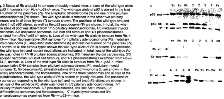
Figure 2 Status of Rb and p53 in tumors of doubly 101 mutant mice
Figure 3 Novel pathology present in Rb+/-;p53-/- animals 102 Figure 4 Models for cooperativity between Rb and p53 102
mutations Figures within:
Morgenbesser SD, Williams BO, Jacks T, and DePinho RA. (1994)
p53-dependent apoptosis produced by Rb-deficiency in the developing
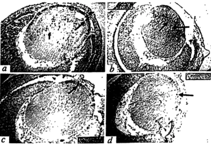
Morphological development of lenses in E13.5 day wild type and mutant embryos
Lens cell proliferation in E13.5 wild-type and mutant embryos
Cellular differentiation in lenses derived from E14.5 wild-type and mutant embryos
Apoptotic cell death in lenses from E13.5 wild-type and mutant embyros
CHAPTER 5
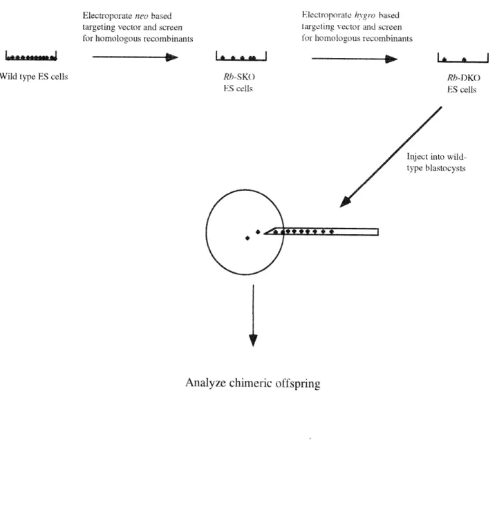
Figure 5-1 Rb-DKO chimera generation 116
Figures within:
Williams BO, Schmitt EM, Remington L, Bronson RT, Albert DM, Weinberg RA, and Jacks T. (1994) Extensive contribution of Rb-deficient cells to adult chimeric mice with limited histopathological consequences. EMBO J. 13: 4521-4529.
Figure 1 Figure 2 Figure 3 Figure 4 Figure 5 Figure 6
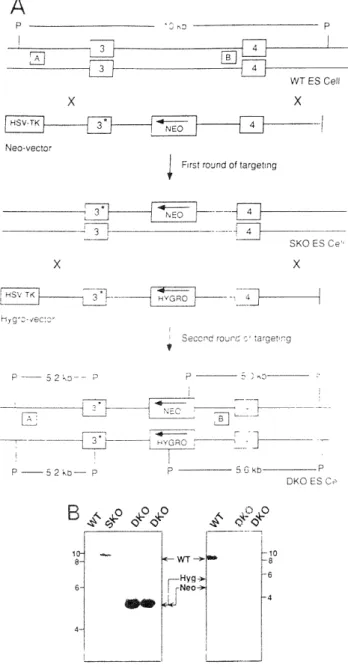
Construction of Rb-deficient cells GPI Isoenzyme Assay
Normal erythropoiesis in Rb-DKO chimeras Histopathology
Immunohistochemistry of pituitary tumors Viability of Rb-DKO chimeras
120 121 121 122 124 124 CHAPTER 6 Figure Figure Figure 6-1 6-2 6-3 Figure 6-4 Figure 6-5
Generation of ROSA-26 Double Knockout Chimerasl34
Analysis of 13.5 day fetal liver 142
Analysis of Rb-DKO fetal chimera 144
central nervous system
Adrenal X-gal staining in adult Rb-DKO chimeras 146 Representative X-gal staining of tissues from 150 chimeras derived from ES2 and ES4 injected
blastocysts Figure 1 Figure 2 Figure 3 Figure 4 107 107 107 108
Figure 6-6 Representative X-gal staining of tissues from 152 chimeras derived from ES2 and ES4 injected
blastocysts
CHAPTER 7
Table 7-1 Selection of Rb-/-;p53+/- clones in increased 162
concentrations of hygromycin APPENDIX A
Figures within:
Jacks T, Remington L, Williams BO, Schmitt EM, Halachmi S, Bronson RT, and Weinberg R A. (1994) Tumor spectrum analysis of p53-mutant mice. Current Biology 4(1): 1-7.
Figure 1 p53 gene targeting 181
Figure 2 Immunoprecipitation of p53 protein 181
Figure 3 Effects of p53 mutation on survival 182
Figure 4 Tumor distribution in heterozygous and 182
homozygous mutant animals
Figure 5 Histopathology of representative tumors 183
Figure 6 Loss of heterozygosity 184
Figures and tables within:
Sah VP, Attardi LD, Mulligan GM, Williams BO, Bronson RT, and Jacks T. (1995) A subset of p53-deficient female embryos develop exencephaly. Nature Genetics 10:175-180.
Table 1 p53 genotype of adult and embryonic mice 188
Figure 1 Whole mount view of p53-/- exencephalic mice 188 and littermate controls
Figure 2 Histological analysis of p53-/- exencephalic 189 embryos
Figure 3 Sex determination of exencephalic p53-/- embryos 190 Figure 4 Whole mount TUNEL analysis of p53-/- embryos 190
APPENDIX B
Figure B-1 Relative size of Rb +/+ and Rb -/- MEFs 199
Table B-1 Summary of cell cycle kinetics experiments 200
Figures and tables within:
Herrera RE, Sah VP, Williams BO, Makela TP, Weinberg RA, and Jacks T. (1996) Altered cell cycle kinetics, gene expression, and G1 phase restriction point regulation in Rb-deficient fibroblasts
Figure 1 Altered size and cell cycle kinetics in Rb-/- fibroblasts 204 Figure 2 Analysis of the R point of late G1 in Rb-/- and Rb+/+ 205
primary fibroblasts
Figure 3 Immunoblot analysis of protein lysates prepared after 206 the readdition of serum to starved cells
Figure 4 Northern blot analysis of RNA prepared after the 207 readdition of serum to starved cells
APPENDIX C
Figures and tables within:
Lee MH, Williams BO, Mulligan GJ, Mukai S, Bronson RT,
Dyson N, Harlow E, and Jacks T. (1996) Targeted disruption of pl07: evidence for functional overlap with Rb. Genes and Development (in press).
Table 1 Offspring viability of Rb+/-;pl107-/- mice at 237 3-4 weeks of age
Figure 1 Targeted disruption of mouse p107 gene 238
Figure 2 Western blot and northern blot analysis of pl07 241 mutant mice and cells
Figure 3 Phenotype of Rb+/-;p107-/- mice 244
Figure 4 Retinal lesions in Rb+/-;pl07-/- mice 247
Figure 5 Viability of Rb-/-;pl07-/- mice 249
APPENDIX D APPENDIX E Table E-1I Figure E- 1 Generation of ES cells p107/p130 chimera histology
Figure D-1 Schematic of Rbl3Neo allele generation
Figure D-2 Schematic of genomic structure and expected restriction digest pattern using Pstl digested genomic DNA Figure D-3 Southern blot tissues and tumors from RbX3t and
Rbl3Neo heterozygous animals
Figure D-4 Survival of wild-type mice (50 mice) relative to
Rbl3Neo (25 mice) and RbX3t heterozygotes
Table D-1 Presence of normal Rbl3Neo homozygotes during development
Table D-2 Animals surviving to 3 weeks of age derived from
RbI3Neo heterozygote crosses
Table D-3 Animals surviving to 3 weeks of age derived from
RbI3Neo -/- by Rbl3Neo +/- crosses
Table D-4 Results of crosses between RbI3Neo heterozygotes and
RbX3t heterozygotes
Figure D-5 Western blot analysis of pRb in intron mutant embryos Figure D-6 RT-PCR analysis of splicing in Rbl3Neo allele
Figure D-7 Schematic diagram of the normal splicing event and the abnormal event taking place at some frequency from the
Rbl3Neo allele
Figure D-8 Pathology associated with the RbI3Neo mutation
280 282 258 261 262 266 268 268 269 269 271 273 274 276
ABBREVIATIONS USED B-gal BrdU DNA ES cells FACS FdU G418 GPI hygroR kd Lac Z LOH MEFs mg ml neoR pRb RB Rb Rb-DKO Chimeras Rb-DKO ES cells ROSA-26 X-gal Beta galactosidase Bromo-deoxyuridine Deoxyribonucleic acid Embryonic stem cells
Fluorescent activated cell sorter Fluoro-deoxyuridine
A synthetic analog of neomycin Glucose phosphate isomerase
Gene conferring resistance to hygromycin Kilodalton
Beta galactosidase gene Loss of heterozygosity
Murine embryonic fibroblasts milligram
milliliter
Gene conferring resistance to neomycin or G418 Retinoblastoma susceptibility protein
Retinoblastoma susceptibility gene (human) Retinoblastoma susceptibility gene (mouse)
Chimera derived from injection of Rb-deficient ES cells into blastocysts (Rb Double Knock Out) ES cells with both copies of the RB gene mutated Reverse orientation splice acceptor (Lac Z
transgenic mice)
NOMENCLATURE
I have used the accepted nomenclature in referring to genes and proteins throughout this
manuscript. Gene names are italicized while the same abbreviation referring to the protein product of the gene is not. In addition, when the gene referred to is a mouse gene (and where it is
applicable) the gene abbreviation is written as the first letter being capitalized and all subsequent letters being lowercase (Rb and Apc). When the human counterpart is referred to all the letters in the abbreviation are capitalized (RB and APC).
CHAPTER 1
The discovery and characterization of oncogenes in the late 1970s and early 1980s revolutionized the study of cancer (Bishop, 1995). Cancer could now be considered as a genetic disease in which mutations in molecularly definable genes could be linked to tumor development. Most oncogenes were originally isolated either because their coding sequences had been coopted by retroviruses to give them transforming potential or because their addition to normal cells caused normal growth control mechanisms to be compromised leading to transformation (Bishop, 1985; Bishop, 1995; Land et al., 1983; Ruley, 1983; Shih et al., 1981; Shih and Weinberg, 1982; Stehelin et al., 1976).
Because the carcinogenic effects of tumor suppressor gene mutations were usually the result of loss of function mutations in both cellular copies of the gene (Lee et al., 1987; Levine, 1993), they proved more challenging to isolate and characterize. The existence of tumor suppressor genes was originally postulated on the basis of somatic cell fusion experiments in which some fusions
between transformed and normal cells lead to an inhibition of the transformed cell phenotype (Harris et al., 1969). This observation was later expanded to show that the introduction of small regions of individual chromosomes from normal cells could suppress the transformed properties of some tumor cells (reviewed in Stanbridge, 1990). These data, along with the seminal work of Knudson (described in Chapter 2) helped establish the concept that cancer could result from the loss of negative growth control in addition to gain of function mutations in growth-promoting oncogenes. The development of positional cloning methods in the 1980s has facilitated the identification of many tumor suppressor genes to date.
Almost all of the tumor suppressor genes identified to date have been cloned on the basis of inherited mutations in these genes segregating with an obvious familial cancer predisposition. The prototypic example of the identification of the retinoblastoma susceptibility gene (RB) will be described in detail. At least ten other tumor suppressor gene mutations identified by their
association with familial cancer syndromes have been described. Like the products of oncogenes, the protein products of these genes participate in a wide spectrum of cellular functions. The
functions of these genes and the inherited cancer syndromes associated with them are summarized in Table 1-1. Besides the RB gene, this work has focused heavily on the activities of the p53 gene. Because of this, the identification and activities of the p53 gene will also be reviewed in detail.
Table 1-1. Functions of human tumor suppressor genes and tumors associated with their inactivation
(Adapted from Williams and Jacks, 1996)
Protein Features
Nuclear phosphoprotein, Binds transcription factors
Nuclear phosphoprotein, Transcription factor Zinc fingers, Transcription factor Coiled-coil motif, Binds B-catenin ras-GAP activity Membrane cytoskeletal attachment
pl6MTSI Cyclin/cdk inhibitor
Inherited Syndromes (Human)# Familial retinoblastoma Li-Fraumeni syndrome Wilms tumor Familial adenomatous polyposis coli, Turcot's syndrome Neurofibromatosis Type I Neurofibromatosi s Type II Familial melanoma N-CAM homology Associated Cancers (Human)# Retinoblastoma; osteo-sarcoma; breast, lung, and bladder carcinoma Several (50% of all cancer)
Nephroblastoma
Colon and Brain
Neurofibromatosis, Colon carcinoma, Astrocytoma Vestibular Schwannoma, Meningioma, Ependymoma Several Colon VHL Inhibits transcriptional elongation
BRCA1 Zinc finger, secreted
granin ?, nuclear protein ?
BRCA2 granin ? von-Hippel-Lindau syndrome Familial breast/ovarian cancer Familial breast/ ovarian cancer
Renal cell carcinoma, pheochromocytoma
Breast and ovarian
Breast and ovarian
References or reviews for the information summarized in Table 1 are as follows: RB
(Hollingsworth et al., 1993; Hollingsworth, 1993; Riley et al., 1994; Weinberg, 1992; Weinberg, 1995) and below, p53 (Greenblatt et al., 1994; Levine, 1992; Levine et al., 1991) and below, WT-1 (Hastie, 1994), APC (Miyaki et al., 1995; Polakis, 1995), NF-i (McCormick, 1995; Vishochil et Gene RB p53 Wt-I APC NF-1 NF-2 DCC
al., 1993), NF-2 (Gusella et al., 1996), pi6MTSI (Harper and Elledge, 1996), DCC (Cho and
Fearon, 1995), VHL (Duan et al., 1995a; Duan et al., 1995b; Kibel et al., 1995), BRCA1 (Holt et al., 1996; Jensen et al., 1996; Miki et al., 1994), and BRCA2 (Jensen et al., 1996; Tavtigian et al.,
Literature Cited
Bishop JM. (1985). Trends in oncogenes. Trends Genet. 1: 245-249.
Bishop JM. (1995). Cancer: the rise of the genetic paradigm. Genes Dev. 9: 1309-1315.
Cho KR and Fearon ER. (1995). DCC: linking tumor suppressor genes and altered cell surface interactions in cancer? Curr Opin Genet Dev. 5: 72-78.
Duan DR, Humphrey JS, Chen DY, Weng Y, Sukegawa J, Lee S, Gnarra,JR, Linehan WM and Klausner RD. (1995a). Characterization of the VHL tumor suppressor gene product: localization, complex formation, and the effect of natural inactivating mutations. Proc Natl Acad Sci USA. 92: 6459-6463.
Duan DR, Pause A, Burgess WH, Aso T, Chen DYT, Garrett KP, Conaway RC, Conaway JW, Linehan WM and Klausner RD. (1995b). Inhibition of transcriptional elongation by the VHL tumor suppressor protein. Science. 269: 1402-1406.
Greenblatt MS, Bennett WP, Hollstein M and Harris CC. (1994). Mutations in the p53 tumor suppressor gene: clues to cancer etiology and molecular pathogenesis. Cancer Res. 54: 4855-4878. Gusella J, Ramesh V, MacCollin M, and Jacoby LB. (1996). Neurofibromatosis 2: loss of
merlin's protective spell. Curr Opin Genet Dev. 6: 87-92.
Harper JW and Elledge SJ. (1996). Cdk inhibitors in development and cancer. Curr Opin Genet Dev. 6: 56-64.
Harris H, Miller OJ, Klein G, Worst P and Tachibana T. (1969). Suppression of malignancy by cell fusion. Nature. 223: 363-368.
Hastie, ND. (1994). The genetics of Wilms tumor-a case of disrupted development. Annu Rev Genet. 28: 523-558.
Hollingsworth J, R. E., Chen P-L and Lee W-H. (1993). Integration of cell cycle control with transcriptional regulation by the retinoblastoma protein. Curr Opin Cell Biol. 5: 194-200.
Hollingsworth RE, Hensey CE, and Lee W-H. (1993). Retinoblastoma protein and the cell cycle. Curr Opin Genet Dev. 3: 55-62.
Holt JT, Thompson ME, Szabo C, Robinson-Benion C, Arteaga CL, King M-C and Jensen RA. (1996). Growth retardation and tumor inhibition by BRCAl. Nat Genet. 12: 298-302.
Jensen RA, Thompson ME, Jetton TJ, Szabo CI, van der Meer R, Helou B, Tronick SR, Page DL, King M-C and Holt JT. (1996). BRCA1 is secreted and exhibits properties of a granin. Nat Genet. 6: 303-308.
Kibel A, Iliopaoulos 0, DeCaprio JA and Kaelin WG. (1995). Binding of the von-Hippel-Lindau tumor suppressor protein to elongin B and C. Science. 269: 1444-1446.
Land H, Parada LF and Weinberg RA. (1983). Tumorigenic conversion of primary embryo fibroblasts requires at least two cooperating oncogenes. Nature. 304: 596-602.
Lee WJ, Bookstein R, Hong F, Yong LJ, Shew J-Y and Lee EY-HP. (1987). Human
retinoblastoma susceptibility gene: cloning identification and sequence. Science. 235: 1394-1399. Levine AJ. (1992). The p53 tumour suppressor gene and product. Cancer Surv. 12: 59-79. Levine AJ. (1993). The tumor suppressor genes. Annu Rev Biochem. 62: 623-651.
Levine AJ, Momand J and Finlay CA. (1991). The p53 tumour suppressor gene. Nature. 351: 453-456.
McCormick F. (1995). Ras signaling and NF1. Curr Opin Genet Dev. 5: 72-78.
Miki Y, Swensen J, Shattuck-Eidens D, Futreal PA, Harshman K, Tavtigian S, Liu Q, Cochran C, Bennett LM, Ding W et al. (1994). A Strong Candidate for the Breast and Ovarian Cancer
Susceptibility Gene BRCAl. Science. 266: 66-71.
Miyaki M, Tanaka K, Kikuchi-Yanoshita R, Muraoka,M and Konishi M. (1995). Familial polyposis: recent advances. Crit Rev Oncol/Hematol. 19: 1-31.
Polakis P. (1995). Mutations in the APC gene and their implications for protein structure and function. Curr Opin Genet Dev. 5: 66-71.
Riley DJ, Lee EY-HP and Lee W-H. (1994). The retinoblastoma protein: more than a tumor suppressor. Annu Rev Cell Biol. 10: 1-29.
Ruley HE. (1983). Adenovirus early region 1A enables viral and cellular transforming genes to transform primary cells in culture. Nature. 304: 602-606.
Shih C, Padhy LC, Murray M and Weinberg RA. (1981). Transforming genes of carcinomas and neuroblastomas introduced into mouse fibroblasts. Nature. 290: 261-264.
Shih C and Weinberg RA. (1982). Isolation of a transforming sequence from a human bladder carcinoma cell line. Cell. 29: 161-169.
Stanbridge EJ. (1990). Human tumor suppressor genes. Annu Rev Genet. 24: 615-658. Stehelin D, Varmus HE, Bishop JM and Vogt PK. (1976). DNA related to the transforming gene(s) of avian sarcoma viruses is present in normal avian DNA. Nature. 260: 170-173. Tavtigian SV, Simard J, Rommens J, Couch F, Shattuck-Eidens D, Neuhausen S, Merajver S, Thorlacius S, Offit K, Stoppa-Lyonnet D, Belanger C, et al. (1996). The complete BRCA2 gene and mutations in chromosomal 13q-linked kindreds. Nat Genet. 12: 333-337.
Vishochil D, White R and Cawthon R. (1993). The neurofibromatosis type 1 gene. Annu Rev Neurosci. 16: 183-205.
Weinberg RA. (1995). The retinoblastoma protein and cell cycle control. Cell. 81: 323-330. Williams BO and Jacks T. (1996). Mechanisms of carcinogenesis and the mutant mouse. Curr
Opin Genet Dev. 6: 65-70.
Wooster R, Bignell G, Lancaster J, Swift S, Seal S, Mangion J, Collins N, Gregory S, Gumbs C, Micklem G et al. (1995). Identification of the breast cancer susceptibility gene BRCA2. Nature. 378: 789-792.
CHAPTER 2
Identification and cloning of the retinoblastoma susceptibility gene
Retinoblastoma is a rare tumor of the retina which develops in approximately 1 in 20,000 children in the United States each year (Dryja, 1989; Dunphy, 1964; Weinberg, 1992). It was first reported in the early nineteenth century, and in those relatively primitive medical times was almost always fatal (Dryja, 1989; Dunphy, 1964; Weinberg, 1992). However, better methods of treatment allowed survival of affected individuals, and around the turn of the century vertical transmission of the disease was reported (DeGouvea, 1886; Dryja, 1989; Dunphy, 1964; Ridley, 1905; Weinberg,
1992). As more such cases were examined it became evident that retinoblastomas occurring in familial clusters differed from those occurring sporadically. The inherited cases were much more likely to occur earlier in life and present as multifocal and bilateral. In 1971, Alfred Knudson published an analysis of the two different forms of the disease which came to several conclusions that would lay the conceptual framework for the discovery and analysis of tumor suppressor genes (Knudson, 1971). Knudson suggested the difference between the two types of retinoblastoma was that in the sporadic cases two relatively rare events needed to occur to initiate tumor development, while in the inherited cases only one event was necessary. Knudson later extended this "two-hit" hypothesis to explain the differences between the familially associated and sporadic cases of Wilms tumor, a childhood cancer of the kidney (Knudson and Strong, 1972).
The concept that inactivating mutations in both alleles of a single gene could explain the "two-hits" proposed by Knudson was first advanced by Comings (Comings, 1973). Experimental support for this idea began to emerge in 1978 when Yunis and Ramsey described the deletion of an area of chromosome 13 (band q14) in some DNA samples derived from retinoblastomas (Yunis and Ramsey, 1978). Five years later, several groups reported that retinoblastoma patients who were constitutionally heterozygous for DNA markers in the 13q14 region underwent reduction to homozygosity in this region in their tumor DNA (Bendedict et al., 1983; Cavenee et al., 1983; Dryja et al., 1984; Godbout et al., 1984; Sparkes et al., 1983). Shortly thereafter, the cloning of the
gene underlying retinoblastoma development was reported by Friend et al. (Friend et al., 1986) and later confirmed by other groups (Fung et al., 1987; Lee et al., 1987b). The gene was found to span approximately 200 kilobases of DNA in the 13q14 region (Hong et al., 1989; McGee et al., 1989). Further analysis revealed that the gene contained 27 exons ranging in size from 31 to 1889
nucleotides (Hong et al., 1989; McGee et al., 1989). In addition, detailed examination of the promoter region of the gene showed it to be GC rich and contain binding sites for the transcription factors ATF, Sp-1, and E2F (reviewed in Riley et al., 1994). While the initial isolation of the gene was dependent on the analysis of individual tumors from patients who had inherited large deletions in the gene, it was subsequently found that a large number of retinoblastomas were actually
associated with point mutations in the RB gene (Canning and Dryja, 1989; Dunn et al., 1988; Fung et al., 1987; Gallie et al., 1990; Horowitz et al., 1989; Kaye et al., 1990; Lee et al., 1987b; Yandell et al., 1989). The end result of all the point mutations was to create functionally inactive pRB. The
significance of the promoter binding sequences is highlighted by the fact that several examples of familially-associated retinoblastoma have been associated with deletions or point mutations within
these binding sites (Bookstein et al., 1990a; Sakai et al., 1992).
After the gene was cloned, several groups screened DNA from a variety of different tumor types to examine whether RB function was lost via deletion or point mutation. Surprisingly, even though patients who inherit a defective RB allele are not significantly predisposed to any tumors besides retinoblastoma (Friend et al., 1986; Fung et al., 1987; Lee et al., 1987b), osteosarcoma (Friend et al., 1986), and perhaps pinealoblastoma (Jakobiec et al., 1977; Pesin and Shields, 1989; Stannard et al., 1985), somatic mutations in the RB gene were shown to occur in several other tumor types. These included some types of sarcomas (Friend et al., 1987; Shew et al., 1989; Stratton et al., 1990) as well as carcinomas of the cervix (Scheffner et al., 1991), prostate (Bookstein et al., 1990b), bladder (Cairns et al., 1991; Ishikawa et al., 1991; Takahashi et al., 1991), breast (Lee et al., 1988; T'Ang et al., 1988; Varley et al., 1989), and lung (Hensel et al., 1990; Ookawa et al., 1993; Rygaard et al., 1990; Yokota et al., 1988). In addition, mutations affecting other genes involved in the regulation of RB protein (pRB) activity have been estimated
to occur in a still larger spectrum of human tumors (discussed below). The data implied that pRB played a potentially important role in growth control in many cell types besides retinoblasts.
In vitro evidence that the RB gene can directly suppress cell proliferation
The first direct evidence that RB could suppress transformation and tumorigenesis came from studies in which RB was reintroduced into cell lines which were deficient for RB as a result of mutation (reviewed in Riley et al., 1994). In several cases such reintroduction was shown to have effects including the inhibition of cell proliferation in vitro, inducing changes in cellular
morphology, reducing the growth potential of cells in soft agar, and inhibiting the growth of tumor cell lines upon injection into nude mice. Examples of tumor cell lines which are affected by the reintroduction of RB include those derived from retinoblastomas (Huang et al., 1988; Medraperla et al., 1991; Xu et al., 1991), osteosarcomas (Huang et al., 1988), and carcinomas of the breast (Wang et al., 1993b), bladder (Goodrich et al., 1992; Takahashi et al., 1991), and prostate (Bookstein et al., 1990b). Assays based on these systems, especially the ability of RB to induce cell cycle arrest and morphological changes in the osteosarcoma cell line SAOS-2 (Hinds et al.,
1992; Qian et al., 1992), have been heavily utilized to assess aspects of RB function (see below).
Analysis of the RB gene product
The cDNA derived from the RB gene is 4757 nucleotides long and contains an open reading frame predicted to encode a protein containing 928 amino acids (reviewed in (Riley et al., 1994;
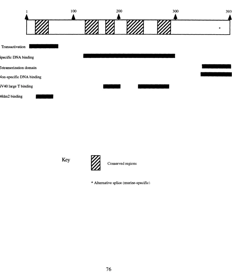
Weinberg, 1992). Northern and subsequent Western blot analysis revealed that the gene was expressed in an almost ubiquitous fashion (Bernards et al., 1989; Lee et al., 1987a). The predicted molecular mass of the protein was 105 kD. However, initial analysis of pRB sequence provided few clues to its potential function. A schematic representation of the domains discussed below is
Figure 2-1. Characteristics of pRB
Amino
Acid # 1 250 500 750 928
Pocket (Oncoprotein/E2F binding)
-c-abl binding
* Denotes a serine or threonine within located in a cdk consensus recognition site. Residues are 249, 252, 356, 373, 608, 612, 788, 795, 807, 811, and 821. Residue 249 is not a perfect consensus site
but has been shown to be phosphorylated in vitro by cdc2 (Lees et al., 1991).
The protein was first identified by Wen-Hwa Lee and colleagues in 1987 with antibodies raised against the RB protein derived from expression in E. coli (Lee et al., 1987a). Initial analysis revealed that the protein was present at all phases of the cell cycle suggesting that its activity was regulated post-translationally (Chen et al., 1989; Lee et al., 1987a). Western blot analysis of cell extracts with this antibody revealed that it recognized a number of proteins migrating between 110 and 116 kD (Ludlow et al., 1990; Shew et al., 1989). Subsequent analysis showed that these phosphopeptides were phosphorylated exclusively on serine and threonine residues (Chen et al., 1989; DeCaprio et al., 1988; DeCaprio et al., 1989; Furukawa et al., 1990; Lees et al., 1991; Lin et al., 1991; Ludlow et al., 1990; Shew et al., 1989). It was later shown that many of these residues were phosphorylated by the activity of cdk family members based on the observations that purified cdc2 could phosphorylate pRB in vitro on many of the same sites that they were phosphorylated in
vivo (Lees et al., 1991; Lin et al., 1991; Taya et al., 1989). Furthermore, the first five of the in vitro
phosphorylated sites mapped were found to contain a consensus cdc2 recognition motif (Lees et al., 1991). Much in vivo and in vitro evidence now supports the model that cyclin dependent kinases are the major, if not only, kinases involved in pRB phosphorylation (described below).
The pattern of pRB phosphorylation oscillates during different stages of the cell cycle (Buchkovich et al., 1989; Chen et al., 1989; DeCaprio et al., 1988; DeCaprio et al., 1989; Furukawa et al., 1990; Mihara et al., 1989). During GO and early to mid Gl, hypophosphorylated forms of the protein predominate. As the cell progresses through the restriction point late in G 1, the protein becomes
highly phosphorylated probably due to cdklcyclin kinase activity (discussed below). These highly (or hyper-) phosphorylated forms of pRB remain through S, G2, and most of M phase. The protein then begins to become dephosphorylated again during anaphase. This process then is completed during early G1. Relatively little is known about the mechanisms underlying the dephosphorylation of pRB. However, the facts that pRB was shown to interact with the catalytic subunit of phosphoprotein phosphatase type 1 (PP-1) (Durfee et al., 1993) and that inhibitors specific for PP-1 block pRB dephosphorylation (Hollingsworth, 1993) suggest that PP-1 may control it.
The RB aene product interacts with several DNA tumor virus oncoproteins
All DNA tumor viruses need to replicate their own viral genome to propagate and survive. Large DNA tumor viruses (such as herpes simplex virus) typically encode most of the enzymes
necessary for this process and need only minor components of the cellular environment for its completion (Knipe, 1989) On the other hand, some of the smallest DNA tumor viruses, such as parvoviruses, have evolved the opposite replication strategy. These viruses are heavily dependent on cellular factors for all aspects of viral replication and thus only replicate their DNA when the cellular DNA replicates (reviewed in Dyson and Harlow, 1992).
Several DNA tumor viruses (most notably adenoviruses, papillomaviruses, and polyomaviruses) have evolved strategies which lie between these two extremes (Dyson and Harlow, 1992). These viruses are still heavily dependent on cellular factors, but instead of merely waiting for DNA replication to occur, their genomes encode proteins which help stimulate infected cells to enter S phase. These proteins include the E1A protein produced by adenovirus, the E7 protein encoded for by the papillomavirus genome, and the large T antigens of polyomaviruses.
The E1A protein are the first polypeptides expressed from the viral genome following adenovirus infection and have been shown to have a number of different functions (Lewis and Mathews, 1980; Nevins, 1981). In order to gain further insight into the mechanisms underlying E1A function, two groups used antibodies derived against the E1A protein to identify cellular proteins which interacted with E1A (Harlow et al., 1986; Yee and Branton, 1985). Remarkably, one protein which coimmunoprecipitated with E1A was pRB (Whyte et al., 1988). Further analysis showed that was a direct interaction. Stimulated by this observation, DeCaprio et al. showed that a polyomavirus protein, the simian virus 40 (SV40) large T antigen, could also form a complex with the RB protein (DeCaprio et al., 1988). Subsequently it was shown that the E7 oncoproteins of
human papillomaviruses and large T antigens of other polyomaviruses could also interact with pRB (DeCaprio et al., 1988; Dyson et al., 1990; Dyson et al., 1989; Munger et al., 1989).
Since E1A (Dyson and Harlow, 1992; Houweling et al., 1980; Land et al., 1983; Ruley, 1983) (as well as SV40 large T (Livingston, 1992) and E7 (Munger et al., 1992)) could act as oncogenes and loss of RB function was associated with the development of human tumors, a simple hypothesis was forwarded that the binding of these proteins to pRB resulted in pRB functional inactivation (Dyson and Harlow, 1992; Livingston, 1992; Munger et al., 1992). This would create a situation equivalent to that seen in cells which had lost RB function via mutation. Importantly, the
hypophosphorylated forms of pRB were the only ones targeted by the viral oncoproteins suggesting that they were the functionally active forms (Dyson and Harlow, 1992; Livingston, 1992; Munger et al., 1992). Such functional inactivation of the growth suppressing RB protein would help drive the infected cell into S phase and facilitate viral replication.
This model of viral oncoprotein and pRB function has been supported by several lines of
experimental work which themselves have revealed important insights into RB function. First, the regions of the viral oncoproteins which mediate binding to pRB are necessary for full viral activity (Dyson and Harlow, 1992; Livingston, 1992; Munger et al., 1992). In addition, the regions of pRB which bind to the viral oncoproteins (referred to as the "pocket" region) have been shown to be favored sites for point mutations associated with human tumors (Hu et al., 1990; Huang et al., 1990; Kaelin et al., 1990). This implied that the viral oncoproteins were targeting a region of pRB which normally acted in a growth inhibitory manner.
The RB gene product binds to E2F. a cellular transcription factor
Work during the mid-1980s had shown that the E1A could dissociate complexes containing the cellular transcription factor E2F resulting in the release of free E2F (Bagchi et al., 1990; Nevins,
complex (Nevins, 1992) and has subsequently been shown to have the ability to stimulate cellular proliferation and transactivate promoters of many genes which are thought to play a role in stimulating cell cycle progression. A partial list of genes which contain putative E2F target sites in their promoters include dihydrofolate reductase (Blake and Azizkhan, 1989; Hiebert et al.,
1991), thymidine kinase (Kim and Lee, 1991), DNA polymerase ax (Pearson et al., 1991), c-myc (Hiebert et al., 1989; Thalmeier et al., 1989), N-myc (DePinho et al., 1986; Mudryj et al., 1990),
cdc2 (Dalton, 1992), B-myb(Lam and Watson, 1993), PCNA (Lee et al., 1995a), and cyclin E
(Ohtani et al., 1995).
Since E1A both bound pRB and caused the release of free E2F from higher order complexes, several groups tested whether binding of E1A released pRB from an interaction with E2F which allowed E2F to perform its cellular functions. In 1991, four groups reported that this indeed was the case (Bagchi et al., 1991; Bandara and La Thangue, 1991; Chellappan et al., 1991; Chittenden et al., 1991). E2F was shown to bind to hypophosphorylated form of pRB and be released upon E1A binding to pRB. The binding site for E2F within the pRB protein was found to map partially to the E1A-binding site (the pocket) and also to another area C-terminal to the pocket (see Figure 2-1).
The simple model that emerged from these studies was that pRB normally inhibits cell cycle progression by binding to and negatively regulating the activity of E2F complexes (Nevins, 1992). While in general this model has proven to be accurate, it has become necessarily more complex due to several observations in recent years. A great deal of accumulating evidence suggests that besides simply negatively regulating E2F activity, pRB may actually repress transcriptional activity from certain promoters via its association with promoter sites through its E2F interaction (since E2F complexes have specific DNA binding activity) (Weintraub et al., 1995; Weintraub et al.,
1992). To add another level of complexity to the equation, E2F has been shown to be a dimeric
complex between one E2F and one DP subunit (reviewed in (La Thangue, 1994). The first E2F subunit, E2F- 1 was cloned in 1992 . Subsequently, the identification of four other E2Fs (E2F
2-5) has been reported (Beijersbergen et al., 1994; Ginsberg et al., 1994; Hijmans et al., 1995; Ivey-Hoyle et al., 1993; Lees et al., 1993; Sardet et al., 1995). In addition, multiple genes encoding for DP subunits have also been identified, although relatively little has been reported about their functions (Girling et al., 1994; Girling et al., 1993).
Consistent with the model that pRB inhibits cell cycle progression, overexpression of E2F-1 has been shown to be sufficient to drive arrested cells into S phase (Johnson et al., 1993).
Furthermore, SAOS-2 cells overexpressing E2F-1 are able to overcome the block in cell cycle progression induced by pRB introduction (Qin et al., 1995).
As discussed below, the different E2F subunits appear to confer specificity of each complex to associate differentially with different pRB-related proteins. Whether the different E2F subunits (or even the DP subunits) direct E2F complexes to specific promoters remains to be determined.
pRB can interact with a number of cellular proteins
Besides its binding to E2F complexes, pRB has been reported to interact, at least in vitro, with several other cellular proteins. In general, these proteins appear to interact preferentially with the hypophosphorylated form of pRB. A partial list of these proteins includes the transcription factors c-myc (Rustgi et al., 1991), c-abl (Welch and Wang, 1993), elf-i (Wang et al., 1993a), Id-2 (Iavarone et al., 1994), myoD (Gu et al., 1993), myogenin (Gu et al., 1993), PU.1 (Hagemeier et al., 1993), ATF-2 (Kim et al., 1992), UBF (Cavanaugh et al., 1995), and BRG-1 (Dunaief et al.,
1994) as well as D-type cyclins (Dowdy et al., 1993; Ewen et al., 1993; Kato et al., 1993), the protein phosphatase PP1-cx2 (Durfee et al., 1993), mdm2 (Xiao et al., 1995), and the human homolog of a yeast ras regulatory protein MSI1 (Qian et al., 1993). The importance of these individual interactions or their physiological relevance in modulating pRB function is not clear in many cases.
Extending the concept of the pRB:E2F interaction, a model that has been suggested based on the ability of pRB to interact with this plethora of cellular transcription factors is that pRB acts as a gatekeeper for the initiation of cell cycle progression (Hollingsworth et al., 1993; Hollingsworth,
1993; Riley et al., 1994; Weinberg, 1995). In the hypophosphorylated state it binds to and inhibits a number of these transcription factors. Upon stimulation of cell cycle progression by external signals, the machinery to phosphorylate pRB is activated resulting in hyperphosphorylated pRB and the coordinated release and activation of these transcription factors. The fact that pRB becomes hyperphosphorylated coincident with the restriction point (Weinberg, 1995), the point after which mammalian cells are committed to going through the cell cycle to the next G 1 phase (Pardee, 1989), further supports this model.
RB is a member of a multigene family
Work on DNA tumor virus oncoproteins also impacted the retinoblastoma field in another significant way in helping establish that RB is actually one of a three member gene family which share significant functional and structural homology. The work which lead to this understanding was based again on studies of proteins which could be shown to interact with DNA tumor virus oncoproteins by coimmunoprecipitation. Some of the same mutant versions of E1A (as well as large T antigen) which had lost the ability to interact with a number of the other
coimmunoprecipitating proteins (Dyson and Harlow, 1992). Among these were proteins referred to by their estimated molecular weight, p107 and p130. The genes encoding for p107 (Ewen et al., 1991) and p130 (Hannon et al., 1993; Li et al., 1993; Mayol et al., 1993) have now been cloned. The three family members share considerable homology (see Figure 2-2 and reviewed in (Cobrinik, 1996).
Figure
2-2.
Structural Homology
Between
Members
Of The
Rb-gene
Family
RBE[Z
n130
I
p107
L
U
Homology whose functional relevance is unknown
Homology in the E1A/large T/E7/E2F binding site
Homology between the spacer regions (cdk/cyclin binding site) of p107 and p130
(Adapted from Li et al.. 1993)
M
r4 -r///A : 7
r// I1I 1~
1Itff
f
P
I
Further analysis of the protein products of these genes has confirmed that they do share functional characteristics with pRB. Besides the shared ability to bind to DNA tumor virus oncoproteins which was the basis for their identification, the proteins also bind to members of the E2F family of transcription factors with pRB having been shown to bind to E2F complexes containing E2F-1, -2, -3, or -4 (Bagchi et al., 1991; Chellappan et al., 1991; Fagan et al., 1994; Helin et al., 1993; Helin et al., 1992; Hiebert et al., 1992; Horio et al., 1993 ; Kaelin et al., 1992; Krek et al., 1993; Lees et al., 1993; Moberg et al., 1996) and p107 and p130 binding to complexes containing E2F-4 or -5 in vivo (Beijersbergen et al., 1994; Hijmans et al., 1995; Moberg et al., 1996; Sardet et al., 1995; Vairo et al., 1995). The distribution of these complexes has been shown to change during progression through the cell cycle (Chittenden et al., 1993; Cobrinik et al., 1993; Moberg et al., 1996; Schwarz et al., 1993; Shirodkar et al., 1992). pl30:E2F complexes make up the majority of E2F complexes during GO and early G1. During mid to late G1, pRB becomes the major E2F binding partner, while during late G1 and S p107 becomes the predominant binding partner.
Further support for grouping these gene products into a gene family comes from analysis of the cellular effects of reintroduction or ectopic expression of these genes. As briefly mentioned above, the introduction of the retinoblastoma gene into several cell types suppressed aspects of the
transformed phenotype. Introduction of pl07 and p130 into tumor cell lines has shown that they also have the ability to suppress aspects of the transformed phenotype when overexpressed. However, the mechanism underlying this suppression appears to differ among the RB-family
members. For example, while the ability of RB to suppress colony formation when introduced into SAOS-2 cells can be overcome by overexpression of E2F-1, while the ability of pl07 to suppress colony formation is unaffected (Zhu et al., 1993). In addition, some cell lines are differentially sensitive to suppression by pRB-family members. For example, colony formation and proliferation in the cervical carcinoma cell line C33-A is not inhibited by the introduction of
RB, but it inhibited by p107 (Zhu et al., 1993). Another example is the glioblastoma cell line
T98G in which colony formation is inhibited by p130 but not by p107 or RB (Claudio et al., 1994).
The suggestion that pl07 and p130 play roles in suppressing aspects of cellular transformation is difficult to reconcile with other observations. The most important of which is that despite great efforts to identify human tumors carrying mutations in p107 and/or p130, no evidence for any such mutations has been reported. It should be noted, however, that it has been suggested that
p130 may be a mutational target during the development of nasopharyngeal tumors in Chinese
garment workers (Claudio et al., 1994; Mascola et al., 1992). Whether p130 will be the important target of these mutational events remains to be seen. In addition, the creation of mice carrying targeted mutations in the p107 and p130 genes (see Appendices C and E) has revealed that mice deficient for either one of these genes fail to show any tumor predisposition (see Appendix C and
(Cobrinik et al., 1996)). Extending this even further, mice heterozygous for a mutation in one of the genes and completely deficient for the other (eitherpl07+/-;pl30-/- orpl]07-/-,pl30+/-) still do not display any predispositions to tumor development (Cobrinik et al., 1996).
Determining why p107 and p130 can suppress aspects of transformation in vitro but inactivating mutations in these genes have not been associated with human or murine cancer remains a key question in the RB field. Perhaps one explanation for this apparent paradox could be that the in
vitro assays are based on overexpressing the proteins. Such overexpression could lead to these
proteins simply acting as a surrogate for pRB by binding to E2F complexes which normally only bind to pRB. This could result in the inhibition of these complexes and lead to growth arrest. While this scenario remains possible, the fact that coexpression of E2F- I1 can overcome suppression by RB but not by p107 is strong evidence against this (Zhu et al., 1993).
Undoubtedly the availability of reagents derived from p107, p130, and Rb-deficient mice will allow a better understanding of the functions of these proteins in the future.
Regulation of pRB phosphorylation
The cellular machinery involved in regulating the phosphorylation status of pRB (as well as p107 and p130) has become well understood in the last several years (see Figure 2-3). Cell cycle progression is controlled by the regulated activity of cyclin-dependent kinases (cdks) (Morgan,
1995). Cyclin dependent kinases were originally identified in yeast and amphibians and
subsequently found to comprise a multigene family in mammals (Meyerson et al., 1992; Morgan, 1995). Three of these (cdk2, cdk4, and cdk6) have been recognized to play important roles in progression through the GI phase of the cell cycle (Sherr, 1994). The catalytic activity of these serine-threonine kinases is positively regulated by their association with cyclin proteins, a family of which at least 7 different subfamilies have been recognized in mammals based on structural
homology and function (Hunter and Pines, 1994; Sherr, 1993; Sherr, 1994). In terms of progression from G1 to S phase, D type cyclins (via their association with cdk4 and cdk6) and
Cyclin E (through its association with cdk2) play important roles (Draetta, 1994; Sherr, 1993; Sherr, 1994). Both of these cyclin/cdk complexes phosphorylate and presumably inactivate RB function (Draetta, 1994; Hatakeyama et al., 1994; Hunter and Pines, 1994; Sherr, 1993; Sherr,
1994) and overexpression of both Cyclins D and E can abrogate the block in cell cycle progression induced by RB (Hinds et al., 1992). Cyclins A and B are thought to play important roles in the progression of cells through S and G2/M (Pines, 1995). Cyclin H is a component of the cdk-activating kinase complex (Fisher and Morgan, 1994; Makala et al., 1994; Matsuoka et al., 1994), while the functions of Cyclins C and G have not been determined (Pines, 1995).
Recent data suggests that the only role of D type cyclins (of which three have been identified) in cell cycle control may be in regulating the phosphorylation status of pRB. An elegant set of experiments by Jiri Bartek and colleagues has shown that microinjection of antibodies which inactivate D type cyclins inhibits cell cycle progression only in cells which contain functional retinoblastoma protein (Lukas et al., 1995).
The importance of these regulatory proteins (as well as the pRB pathway in general) is emphasized by the fact that alterations of cdk and/or cyclin expression by chromosomal translocation, gene amplification, or mutation has been observed in many human tumors (Adelaide et al., 1995; Arnold et al., 1992; Motokura and Arnold, 1993; Motokura et al., 1991; Rosenberg et al., 1991; Wolfel et al., 1995; Zukerberg et al., 1995). In addition, analysis of mice deficient for specific D type cyclins are beginning to offer new insights into RB function. For example, mice deficient in cyclin D1 exhibit reduced perinatal size and viability, retinal abnormalities associated with
decreased retinal size, and a failure to induce proliferation in the breast epithelium in response to pregnancy (Sicinsky et al., 1995). It is tempting to speculate that a failure to inhibit the activity of pRb in these cell types depends specifically on the presence of cyclin D1 (but not D2 or D3). Further evidence suggesting a role for D type cyclins in development and tumorigenesis come from observations of transgenic mice overexpressing Cyclin D1 in the breast (Wang et al., 1994). These mice are predisposed to the development of mammary adenocarcinomas. In addition, coexpression of cyclin D1 and myc in lymphoid tissue can induce lymphomagenesis (Bodrug et al., 1994; Lovec et al., 1994).
The kinase activity of these cyclin/cdk complexes is itself a target of complex regulation. Besides their required association with cyclins for kinase activity, cdks also require the phosphorylation of an internal threonine residue (Thrl60 in CDK2) for activity (Solomon, 1994). The
phosphorylation of these residues in these cdks is controlled the cyclin/cdk activating kinase (CAK) (Solomon, 1994). CAK was found to be a heterodimer of yet another cyclin/cdk pair (cdk7 and Cyclin H) (Fisher and Morgan, 1994; Makala et al., 1994; Matsuoka et al., 1994). Furthermore, the activity of the CAK is partially controlled by the activity of a yet unidentified
kinase. In addition to this level of regulation, cdk activation also requires the removal of inhibitory phosphates from Thrl4 and Tyrl5 by cdc25 phosphatases (Hoffman and Karsenti, 1994).
The activities of cyclin/cdk complexes are also controlled by their association with so-called cyclin-dependent kinase inhibitors (CDI). The WAF1/CIP/ISDI1/p21 is a CDI encoding gene (El-Deiry