HAL Id: tel-02555205
https://tel.archives-ouvertes.fr/tel-02555205
Submitted on 27 Apr 2020HAL is a multi-disciplinary open access archive for the deposit and dissemination of sci-entific research documents, whether they are pub-lished or not. The documents may come from teaching and research institutions in France or abroad, or from public or private research centers.
L’archive ouverte pluridisciplinaire HAL, est destinée au dépôt et à la diffusion de documents scientifiques de niveau recherche, publiés ou non, émanant des établissements d’enseignement et de recherche français ou étrangers, des laboratoires publics ou privés.
Molecular interactions between the kelp saccharina
latissima and algal endophytes
Miriam Bernard
To cite this version:
Miriam Bernard. Molecular interactions between the kelp saccharina latissima and algal endophytes. Symbiosis. Sorbonne Université, 2018. English. �NNT : 2018SORUS105�. �tel-02555205�
Sorbonne Université
Ecole doctorale Sciences de la Nature et de l’Homme (ED 227)
Laboratoire de Biologie Intégrative des Modèles Marins UMR 8227Equipe Biologie des algues et interactions avec l’environnement
Molecular interactions between the kelp Saccharina
latissima and algal endophytes
Par Miriam Bernard
Thèse de doctorat de Biologie Marine
Dirigée par Catherine Leblanc et Akira F. Peters
Présentée et soutenue publiquement le 07/09/2018
Devant un jury composé de :
Dr. Florian Weinberger Chercheur GEOMAR Kiel Rapporteur
Dr. Sigrid Neuhauser Chercheur Univ. Innsbruck Rapportrice
Pr. Soizic Prado Professeur MNHN Examinatrice
Pr. Christophe Destombe Professeur Sorbonne Université Représentant UPMC Dr. Catherine Leblanc Directrice de Recherche Directrice de thèse
Acknowledgements
First of all, I would like to thank my supervisors Catherine Leblanc and Akira Peters. You were the two most important persons to contribute to the success of this thesis. Thank you for giving me the opportunity to work on this project and for your guidance and advice over the last three years. It has really been a pleasure to work with you.
I would also like to thank everyone else who has been involved in this work, in particular Sylvie Rousvoal, who not only developed several protocols with me, but also helped to improve my French. Whenever I had problems in the lab you had the ideas to make things work.
Qikun Xing has provided incredible help with the analysis of the RNAseq data. Thank you so much, I would not have been able to finish this project on time without you! Here I would also like to acknowledge the advice and support from Erwan Corre during the transcriptomic analysis.
Thanks also to Laurence Dartevelle for showing me how to handle the kelp cultures and for helping out with the kelp stocks and to Sandrine Boulben for the continuous detective work on the luminometer. I hope it will work forever from now on.
I would also like to thank my co-authors Martina Strittmatter, Pedro Murúa, Svenja Heesch, Ga Youn Cho, Bertrand Jacquemin, Sylvie Rousvoal, Marion Ballenghien, Nadia Collet, Tristan Le Goff and Philippe Potin for their valuable attributions and the friendly collaborations.
Furthermore, I am very grateful for valuable discussions of my work with the members of my thesis committee (thanks also for being spontaneous enough to move the committee meeting to my living room) and with the ABIE team.
A big thank you goes to the ALFFies. You are the best! I really enjoyed all the meetings and also the time we spent together outside of the ALFF meetings, such as a crazy karaoke night in Prague and Sri’s wedding in India. Thanks also in particular to Claire Gachon who brought ALFF to life and hosted me twice during my secondments at SAMS. Here I also want to thank everyone else at SAMS for the help during my secondments in Oban.
I would also like to thank Job Schipper at Hortimare and Olavur Gregersen, Urd Grandorf Bak and Nicoline Korsvold at Ocean Rainforest for giving me hands-on experience in kelp aquaculture and for being so welcoming and supportive.
A very special thanks goes to Hetty KleinJan: back then when we met for the first time in Bonn, we didn’t know yet what we would get involved in. I am so happy that I had you by my side during these 3 years, not only as a colleague, but as a really good friend.
Of course, I would also like to thank all my other colleagues at the station. In particular Margot Tragin, Zujaila Qui Minet, Svenja Heesch and Martina Strittmatter for being such good companions. Virginie Glippa, I guess that we sometimes might have been a bit too loud when joking around in the lab in three different languages (or the corridor…!), but you made the labwork so much better. Also, I am sure that no other lab in the station is decorated as nicely as ours. Thanks also to the “office girls”: Amandine Simeon, Maria Matard, Laure Mignerot and Eugenie Grigorian.
Looking back into the past, this thesis would not have happened without Inka Bartsch and Katharina Zacher: You really aroused my love for kelps and I am incredibly thankful that I had your continuous support even during my PhD.
Zum Schluss möchte ich mich bei meiner Familie und bei Valentin bedanken. Danke, dass ihr immer für mich da seid. Ihr seid meine Helden!
Table of contents
General Introduction ... 1
1. Evolutionary context of studying seaweed interactions ... 1
2. Kelps ... 2
2.1 Life cycle and ecological relevance of kelps ... 2
2.2. Global seaweed aquaculture and ecological relevance of kelps ... 4
3. Pigmented algal endophytes ... 6
3.1 Defining algal host-endophyte interactions ... 6
3.2 Endophytic red algae ... 8
3.3 Endophytic green algae ... 9
3.4 Endophytic brown algae ... 10
4. Algal defence reactions against biotic stresses ... 13
4.1 Recognition of the attacker ... 13
4.2 Inducible defence responses ... 15
4.2.1 Oxidative burst ... 15
4.2.2 Free fatty acids and oxylipins release ... 16
4.2.3 Halogenation ... 16
4.2.4 Transcriptomic regulation ... 17
4.3 Systemic responses and distance signalling ... 20
5. Thesis project and outline ... 20
Chapter I. Diversity, biogeography and host specificity of kelp endophytes with a focus on the genera Laminariocolax and Laminarionema ... 23
1. Introduction ... 26
2. Material and Methods ... 28
2.1 Sampling and isolation of endophytes... 28
2.2 DNA extraction, barcode markers, amplification and sequencing ... 28
3. Results ... 30
3.1 Molecular systematics ... 30
3.2 Hosts and geographic origin of the isolated strains ... 34
3.3 Laminariocolax atlanticus M. Bernard, Strittmatter & A.F. Peters sp. nov. ... 35
4. Discussion... 37
4.1 Molecular phylogeny of kelp endophytes ... 37
4.2 Species delimitation in Laminariocolax ... 39
4.3 Variability of host specificity ... 41
5. Supplementary Material ... 42
Chapter II. qPCR-based relative quantification of the brown algal endophyte Laminarionema elsbetiae in Saccharina latissima: variation and dynamics of host-endophyte interactions ... 51
1. Introduction ... 54
2. Material and Methods ... 55
2.1 In situ algal sampling ... 55
2.2 Monospecific algal cultures ... 56
2.3 DNA extraction ... 57
2.4 qPCR and evaluation of the assay ... 58
2.5 Data analysis ... 59
3. Results ... 59
3.1 Set-up and validation of the qPCR assay ... 59
3.2 Distribution of endophyte filaments along the thallus of S. latissima ... 60
3.3 Infection rates in young kelps ... 61
3.4 Natural infection of laboratory-grown samples in a seaweed farm ... 62
3.5 Seasonal variation of relative infection rates ... 63
3.6 Geographic variation of relative infection rates ... 64
3.7 Host specificity ... 65
4.1 A specific and reliable qPCR approach for epidemiological studies ... 66
4.2 Early occurrence of the infection in nature ... 67
4.3 Variation of infection rates ... 68
4.4 Host specificity of L. elsbetiae ... 69
5. Supplementary Material ... 71
Chapter III. A highly prevalent filamentous algal endophyte in natural populations of the sugar kelp Saccharina latissima is not detected during cultivation in Northern Brittany ... 75
1. Introduction ... 78
2. Material and methods ... 79
2.1 Algal Material ... 79
2.2 Spore release and seeding procedure ... 79
2.3 Offshore cultivation ... 80
2.4 Sampling ... 80
2.5 DNA extraction ... 81
2.6 qPCR ... 82
3. Results and discussion ... 82
Chapter IV. Physiological and molecular responses of kelps to an infection by Laminarionema elsbetiae, a filamentous brown algal endophyte ... 87
1. Introduction ... 90
2. Material & Methods ... 92
2.1 Biological material ... 92
2.2 Co-cultivation bioassay ... 93
2.3 Extension of the co-cultivation bioassay: preliminary experiments ... 95
2.3.1 GG pre-treatment ... 95
2.3.2 Addition of fungal extracts ... 96
2.5 Transcriptomic analysis ... 97
2.5.1 Experimental set-up for transcriptomic analysis ... 97
2.5.2 RNA extraction, quality assessment and sequencing ... 98
2.5.3 De novo assembly and annotation of the transcriptome ... 98
2.5.4 Identification of differentially expressed (DE) genes ... 99
2.5.5 Laminarionema elsbetiae read analysis ... 99
3. Results ... 99
3.1 The effect of co-cultivation with algal endophytes on kelp growth ... 99
3.2 Preliminary experiments to modify kelp responses towards algal endophytes ... 101
3.2.1 GG pre-treatment ... 101
3.2.2 Addition of fungal extracts ... 102
3.3 Oxidative burst measurement ... 104
3.4 Transcriptomics ... 105
3.4.1 General overview of the transcriptomes ... 105
3.4.2 Differentially expressed genes ... 107
3.4.3 Functional annotation of differentially expressed genes ... 109
3.4.4 Laminarionema elsbetiae reads ... 110
4. Discussion... 113
4.1 The two kelp species show a different physiological response during the co-cultivation with the endophyte ... 113
4.2 Defense elicitation can modify the physiological response of L. digitata towards the algal endophyte ... 115
4.3 Gene regulation during the first contact with the endophytes differs in the two hosts ... 115
4.4 A complex cross-talk between kelp and endophyte could be involved in host specificity .. 118
4.5 Conclusion: first insights into host specificity in kelp-endophyte interactions ... 120
Conclusions and perspectives ... 129
1. Different molecular and chemical responses of kelps to algal endophytes could be the basis of natural infection patterns ... 129
3. Variation of kelp-endophyte relationships: a complex interaction depending on different abiotic
and biotic factors ... 132
4. Future directions for studying kelp-endophyte interactions ... 133
References ... 135
List of abbreviations ... 165
Appendix I: Oral presentations ... 167
Appendix II: Presented posters ... 169
Appendix III: Microspongium alariae in Alaria esculenta: a widely-distributed non-parasitic brown algal endophyte that shows cell modifications within its host (Murúa et al. 2018) ... 173
Abstract ... 189
General introduction
1
General Introduction
1. Evolutionary context of studying seaweed interactions
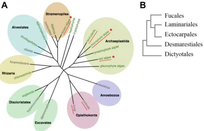
Marine macroalgae – commonly known as seaweeds – are of vital importance for the functioning of coastal ecosystems (Bold & Wynne 1985). Seaweeds are an evolutionary diverse, polyphyletic group with representatives in all three major algal lineages - green (Chlorophyta), red (Rhodophyta) and brown algae (Phaeophyceae, see asterisks Fig. 1A).
Fig 1: A. Eukaryotic tree of life (edited from Cock & Coelho 2011). Lineages showing complex multicellularity are underlined in red. Lineages containing seaweeds are marked with red asterisks. [Printed with the permission of Oxford University Press] B. Relationship of selected brown algal orders (based on Silberfeld et al. 2010). Kelps belong to the Laminariales, the endophytes Laminariocolax and Laminarionema belong to the Ectocarpales.
Most rocky shore habitats in temperate and northern polar seas are dominated by brown seaweeds of the orders Fucales and Laminariales (Fig. 1B, Dayton 1985). Brown algae are part of the Stramenopiles, a lineage that originated from a secondary endosymbiosis event between an ancestral non-photosynthetic protist and a red alga approximately 1 billion years ago (Baldauf 2003). Since multicellularity has evolved independently from the other multicellular groups in the Phaeophyceae, they provide an ideal basis for comparative studies of evolutionary
General introduction
2
processes. For instance, research on the brown algal model Ectocarpus siliculosus has contributed to a better understanding of key cellular processes, such as carbon storage and cell wall biosynthesis (Michel et al. 2010a; reviewed by Cock & Coelho 2011). The anatomy of brown algae ranges from crusts over filamentous thalli to more complex differentiated tissues (Lobban & Harrison 1994). The largest and morphologically most complex brown seaweeds are found within the order of Laminariales (Fig. 1B) which are commonly known as kelps. Due to their important role in coastal habitats, kelps are involved in various biotic interactions with associated micro- and macroorganisms (reviewed by Leblanc et al. 2011 and Potin 2012).
Despite their phylogenetic distance, brown algae have been shown to share certain basic defence mechanisms against biotic stress with the other multicellular eukaryotic lineages (reviewed by Cosse et al. 2007). However, while the molecular and physiological bases of biotic interactions are very well studied in animals and terrestrial plants, brown algae remain poorly understood to a large extent in this regard (reviewed by Brodie et al. 2017). An experimental investigation of biotic interactions in this lineage could provide a better understanding of the underlying biological processes from an evolutionary point of view.
2. Kelps
2.1 Life cycle and ecological relevance of kelps
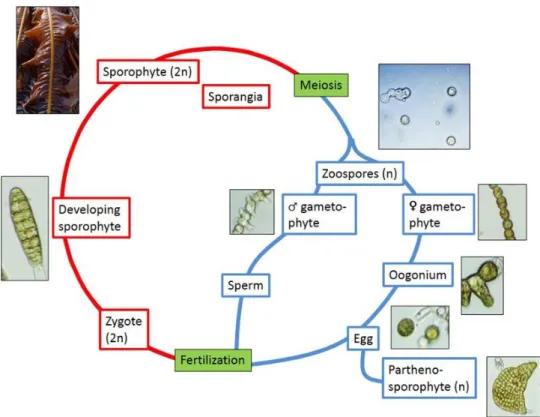
Kelps are characterized by a complex and strongly heteromorphic, haploid-diploid life cycle consisting of microscopic haploid gametophytes and diploid sporophytes of up to several meters length (Fig. 2). Sporangia develop in areas on the blades of the diploid sporophytes referred to as sori (Bold & Wynne 1985). Within these sporangia, haploid zoospores of 4-8 μm size are formed, which are released under environmentally-controlled mechanisms (Amsler & Neushul 1989a) and dispersed by currents (Dayton 1985). Germinated spores grow into male and female gametophytes and produce motile spermatozoids from antheridia and egg cells from oogonia, respectively. After fertilization, the diploid zygote develops into a macroscopic sporophyte, whereas unfertilized egg cells can grow to haploid parthenosporophytes (Dayton 1985). Due to the large size of the sporophytes, the kelp life cycle is usually completed only partially in laboratory cultures. Cultures can be started from gametophyte stocks or freshly released spores which develop into young sporophytes that can be used for experimentation.
General introduction
3
Fig. 2: Life cycle of Laminariales. Red: diploid phase, Blue: haploid phase (Bernard 2014).
Kelps are the major component of rocky intertidal and subtidal habitats (Bold & Wynne 1985). They form vast underwater forests which are among the most diverse and productive ecosystems in the world (Mann 1973). Kelp forests support complex food webs and provide habitats and breeding areas for a variety of animals, such as fish, molluscs, crustaceans and mammals (reviewed by Bartsch et al. 2008). They also play an important role in carbon sequestration (Chung et al. 2013) and significantly affect currents and water flows (Jackson 1983).
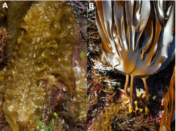
The sugar kelp Saccharina latissima (Fig. 3A) is a short-lived perennial species with a circumpolar distribution in the Northern hemisphere (Bolton et al. 1983). In Europe, it can be found in cold waters of the high Arctic to temperate regions in northwest Spain and northern Portugal (Ardré 1970; Gulliksen et al. 1999; Cires Rodriguez & Moliner 2010). Its undivided, characteristically dimpled and wrinkled blade reaches lengths of up to four meters. The common habitats of S. latissima are sheltered intertidal pools and the shallow subtidal where it grows with its rhizoid attached to rocks, boulders or large stones (Hanelt 1998).
Laminaria digitata (Fig. 3B), commonly known as oarweed or finger kelp, is a perennial North
General introduction
4
Spain along American and European coasts, respectively (Miranda 1931; Schneider et al. 1979; Gulliksen et al. 1999). The blade is split into finger-like segments of up to 2.5 m length with the number of digits varying depending on wave exposure (Lobban & Wynne 1981). L. digitata usually forms dense forests in the lower intertidal and shallow subtidal, but occasionally reaches depths of up to 25 m at its northern distribution range (Birkett et al. 1998; Cabioc’h et al. 2006).
Fig. 3: A. Sporophytes of Saccharina latissima showing the characteristic undivided, wrinkled blade (© Mike Guiry). B. Sporophytes of Laminaria digitata with blades split into finger-like segments (© Mike Guiry).
2.2. Global seaweed aquaculture and ecological relevance of kelps
The use of seaweed by humans has a long history. The earliest written record of seaweed used as food in China dates back more than 2500 years (reviewed by Anis et al. 2017) whereas archaeological evidence of algae being collected and used by humans exists even from the Palaeolithic age (Dillehay et al. 2008). Today, the global seaweed aquaculture is rapidly expanding (Buschmann et al. 2017) and the production as well as the associated value have increased exponentially over the last decades (Fig. 4, FAO Food and Agriculture Organization of the United Nations).
General introduction
5
Approximately 80 % of the produced biomass is used for human diet. Other applications include the use as fertilizers, animal feed and cosmetic or medical products (McHugh 2003; Loureiro et al. 2015). Furthermore, seaweeds have a high potential for the sustainable production of bioethanol and biogas (Adams et al. 2009; Mazarrasa et al. 2014; reviewed by Chen et al. 2015).
Fig. 4: Seaweed aquaculture biomass (histogram) and value (red curve) over the period from 1950-2015 (data obtained from FAO Food and Agriculture Organization of the United Nations).
S. latissima is the closest European relative to the Asian S. japonica, a dominant species in the
Asian seaweed industry. It is one of the fastest-growing European kelp species and has a high carbohydrate content (Skjermo et al. 2014). Traditionally, S. latissima was collected as a fertilizer in agriculture and as animal feed. Today, the off-shore cultivation of this species in Europe is increasing (Mesnildrey et al. 2012; Skjermo et al. 2014) with additional applications in human diet, abalone feeding and as an extract for the cosmetic industry.
Laminaria digitata, on the other hand, is one of the most strongly harvested species in France
with 40.000-60.000 tons harvested per year and an annual turn-over of 1.7 to 2.7 million Euro (Mesnildrey et al. 2012). While it has traditionally been harvested as a fertilizer and animal feed, it is now mainly used for alginate production (Chapman & Chapman 1980; Mesnildrey et al. 2012).
General introduction
6
3. Pigmented algal endophytes
Seaweeds do not only serve as food source or habitats for animals, they also provide a substratum for smaller organisms growing on (epiphytes) or inside of (endophytes) their thalli, such as fungi, oomycetes or filamentous algae of all three macroalgal lineages (Dayton 1985; reviewed by Bartsch et al. 2008 and Gachon et al. 2010). Algal epiphytes penetrate into the outermost cell layers of the host tissue mainly for mechanical support (Setchell 1918). Algal endophytes, on the other hand, may grow entirely within a host and only reproductive structures are formed at the host surface (Peters 1991). A clear distinction of epi- and endophytes is not always possible because certain species may represent a continuum between an epiphytic and endophytic lifestyle (Peters 2003; Gauna & Parodi 2008). Furthermore, pigmented algal endophytes are usually photosynthetically independent from their hosts (Potin 2012) and life stages of such species can also be found outside of their hosts (Küpper et al. 2016). For simplicity, the term endophyte is used in this thesis to describe algae that possess the ability to grow inside of an algal host and penetrate deeper than the cortex.
Endophytic algae have attracted the interest of phycologist mainly due to the fact they occasionally coincide with morphological changes or disease symptoms in their hosts (Apt 1988a; Correa et al. 1988), which can also have a direct impact on the economic value of kelps (Yoshida & Akiyama 1979). Despite an increasing interest in this topic due to the economic importance of seaweed aquaculture (Chen 2004), still little is known about the identity, phylogeny and life cycles of pigmented algal endophytes.
3.1 Defining algal host-endophyte interactions
The term endophyte describes an "organism living within a host plant" (greek: éndon = inside; phytón = plant; Womersley 1987) and thus defines the spatial relationship of this interaction. It does, however, not give a further assessment of it as being detrimental, neutral, or beneficial for each partner. The following definitions can be used instead to describe the nature of algal interactions more precisely.
A symbiosis characterizes a close interaction between two different organisms, regardless of the effect they have on each other (Table 1, De Bary 1879; Correa 1994; Begon 2006). Symbioses can be obligatory, if one or both partners depend on each other, or facultative. As pigmented algal endophytes are usually independent from their host in regard to their nutrition (Peters 1991; Correa 1994; Gauna & Parodi 2008), they can be referred to as facultative
General introduction
7
endosymbionts. Within a symbiosis, the effects of host and symbiont on each other may be either beneficial, innocuous or harmful (Table 1, Correa 1994).
Mutualism describes a relationship between organisms of different species that results in a mutual benefit for each partner (Table 1, Begon 2006). It usually involves the direct exchange of either nutrients or services, such as shelter or transport (Begon 2006).
Commensalism, on the other hand, describes a relationship between two organisms where one partner benefits and the other one is neither significantly harmed nor helped (Table 1, Begon 2006). The commensal may obtain nutrients, shelter, support or locomotion from a host that is unaffected by the former.
Parasitism is a non-mutual relationship between two organisms that is beneficial for one member (the parasite) and harmful for the other (the host, Table 1, Correa 1994; Begon 2006). Parasites develop on or in their host and derive at least a part of their nutrition from the host (Begon 2006).
Table 1: Overview on terms used to describe associations between different organisms. n.d. = not defined. + = positive effect. 0 = neutral, no effect. - = negative effect.
Term Host Endophyte
Symbiosis n.d n.d.
Mutualism + +
Commensalism 0 +
Parasitism - +
If the presence of a symbiont has a negative effect on its host, it can be referred to as a pathogen. Pathogens are organisms that cause a disease in their hosts, i. e. an abnormal physiological or developmental condition (Correa 1994).
The following postulates have been formulated by Koch (1876) as a reference in evaluating causal relationships between diseases and infectious agents (see also Evans 1976 for a revision of the Koch’s postulates):
1. The putative pathogen must be present in all stages of the disease.
2. The putative pathogen must be isolated from the diseased host and be grown in pure culture.
General introduction
8
3. When healthy hosts are infected with the putative pathogen from the pure culture, the specific symptoms of the disease must re-occur.
4. The organism must be re-isolated from the diseased host and correspond to the original putative pathogen.
Although the Koch postulates can be useful to describe pathogenicity of certain organisms, a major constraint is the fact that some pathogens cannot be grown in isolated cultures. These rules therefore have to be adapted according to the studied organisms (Evans 1976).
Natural associations, such as kelp-endophyte interactions, cannot always be clearly labelled with the terms described above as it is often difficult to obtain solid data on the effect of the interaction on either partner. Particular endophyte species may be referred to as pathogens, in cases where evidence proofs a harmful effect on the vital functions of the host, like retarded growth, loss of regeneration capacity or severe cellular damage (Yoshida & Akiyama 1979; Apt 1988a; Correa & McLachlan 1992; Correa et al. 1993). A general classification of endophytes as pathogens, however, cannot be made. Instead, host-endophyte pairs have to be studied individually to assess the effects – beneficial or harmful – on each partner.
3.2 Endophytic red algae
Extensive literature exists on parasitic red algae that either possess highly reduced photosynthetic pigments (Kugrens & West 1973), or have lost their coloration entirely (Evans et al. 1973, Callow et al. 1979). Pigmented red algal endophytes, on the other hand, which are commonly members of the family Achrochaetiaceae, have received less research attention (Tam et al. 1987). Although red algal endophytes are most often associated with red algal hosts (Fig. 5A), they have also been found infecting brown algae, such as Desmarestia aculeata (Fig. 5B, Selivanova & Zhigadlova 2013).
Little is known about the epidemiology of these organisms and macroscopically detectable disease symptoms in infected hosts have only been described for few species. The filamentous endophyte Rhododrewia porphyrae, for instance, causes red spots in the economically important red alga Porphyra, whereas infections of other hosts, such as the red alga
Pterosiphonia bipinnata, are usually not associated with macroscopic disease symptoms (Tam
General introduction
9
Fig. 5: A. Endophytic filaments of Colaconema endophyticum in Membranoptera dimorpha (source: Selivanova & Zhigadlova 2013). B. Endophytic filaments of Colaconema desmarestiae in Desmarestia aculeata (source: Selivanova & Zhigadlova 2013).
3.3 Endophytic green algae
The green algal genus Ulvella (formerly Acrochaete) contains several well-studied endophytic filamentous green algae. Ulvella operculata and Ulvella heteroclada, for instance, are considered as primary pathogens of the sporophytes of Chondrus crispus, an economically important rhodophyte (Correa & McLachlan 1994). They do, however, not penetrate beyond the outer cell layers of the gametophyte of C. crispus (Correa &McLachlan 1991). Green algal endophytes can have a negative impact on the growth, reproductive output, carrageenan yield, wound healing and regeneration of their host (Correa & McLachlan 1992; Faugeron et al. 2000) and facilitate secondary infections by pathogenic bacteria (Correa & McLachlan 1994).
Fig. 6: A. The thallus of Hymenena falklandica showing green spots (arrows) as a symptom of infection with Epicladia heterotricha (source: Gauna & Parodi 2008). [Printed with the permission of John Wiley and Sons] B. Thallus of E. heterotricha surrounding a cell of the host H. falklandica (source: Gauna & Parodi 2008). [Printed with the permission of John Wiley and Sons]
General introduction
10
Endophytic green algae can reach a very high prevalence in their host populations. Epicladia
heterotricha, for instance, has been found infecting 100% of the individuals within a population
of its host, the red alga Hymenena falklandica, in Argentina (Gauna & Parodi 2008). It grows between the hosts’ cells (Fig. 6A) and forms macroscopically visible green spots on its host (Fig. 6B, Gauna & Parodi 2008).
3.4 Endophytic brown algae
Endophytic brown algae are most commonly found in kelps (Andrews 1977; Lein et al. 1991; Peters and Schaffelke 1996; Ellertsdóttir and Peters 1997). They are microscopic, with filamentous thalli, diffuse growth, and usually possess plastids with pyrenoids (Burkhardt & Peters 1998). Most endophytic brown algae are included in the Ectocarpales sensu lato due to their morphologically reduced nature and the presence of pedunculated pyrenoids but the phylogenetic relationships are not fully explored and classifications undergo continuous changes (Fig. 1B, Burkhardt & Peters 1998). Limited sampling due to the difficult isolation of these algae from infected hosts has so far prevented a comprehensive revision of the taxonomy of endophytic brown algae.
The most commonly reported genera of kelp endophytes are Laminariocolax (Russel 1964; Ellertsdóttir & Peters 1997; Thomas et al. 2009) and Laminarionema (Kawai & Tokuyama 1995; Peters & Ellertsdóttir 1996; Ellertsdóttir & Peters 1997).
Laminarionema
The genus Laminarionema consists currently of only one species, i.e. Laminarionema
elsbetiae. It has been first described in 1995 infecting Saccharina japonica in Japan, but none
of the other kelp species in the direct vicinity, such as Costaria costata or Undaria pinnatifida (Kawai & Tokuyama 1995).
Furthermore, it was found on Helgoland infecting S. latissima and – in lower amounts –
Laminaria digitata (Ellertsdóttir & Peters 1997). In Argentina, L. elsbetiae was found not in kelps but in the red alga Rhodymenia pseudopalmata (Gauna et al. 2009a).
General introduction
11
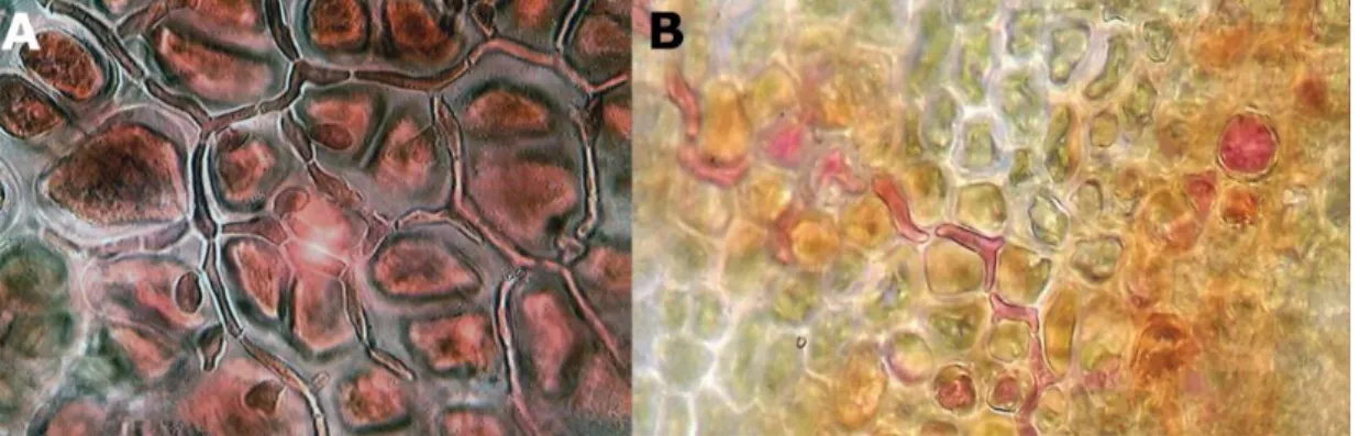
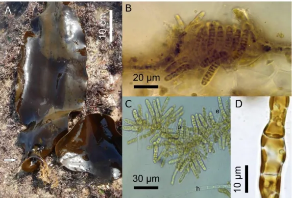
Fig. 7: A. Cross section of the blade of S. latissima: Vegetative Laminarionema elsbetiae filaments (arrow) growing between the host cells. B. Released macrospore of L. elsbetiae (source: Kawai & Tokuyama 1995). [Printed with the permission of John Wiley and Sons] C. L. elsbetiae isolate from S. latissima in unialgal culture.
Laminarionema elsbetiae is characterised by a strictly endophytic thallus (Fig. 7A), with only
phaeophycean hairs emerging from the host. Its large macrosporangia form a single very large macrospore of 23 – 30 µm length, one of the largest flagellated cells in brown algae (Fig. 7B, Kawai & Tokuyama 1995; Peters & Ellertsdóttir 1996).
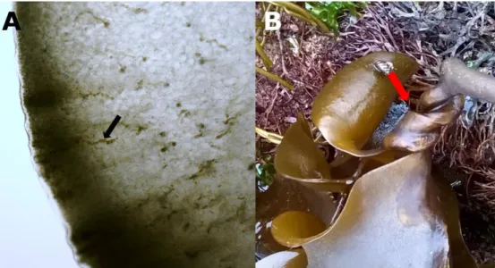
Fig. 8: A. Twisted stipe (arrow) of S. latissima. B. Distorted blade and warts on S. latissima, infected with L. elsbetiae.
Like other pigmented algal endophytes, it can be isolated from the host tissue and be grown in unialgal culture (Fig. 7C). Laminarionema elsbetiae has been associated with the following
General introduction
12
disease symptoms: dark spots, twisted stipes (Fig. 8A), wart-like protrusions and degenerated phylloids (Fig. 8B, Peters & Ellertsdóttir 1996; Ellertsdóttir & Peters 1997). However, while 93% of individuals in a natural Saccharina latissima population on Helgoland were infected with L. elsbetiae, only half of the infected kelps showed morphological alterations visible by eye (Peters & Ellertsdóttir 1996; Ellertsdóttir & Peters 1997). Thus, the presence of L. elsbetiae alone is not causing disease symptoms and it can therefore not generally be described as a pathogen. Other factors, for instance endophyte density or the distribution in the host, may be crucial for the occurrence of disease symptoms (Gauna et al. 2009a).
Laminariocolax
Two endophytic species of the genus Laminariocolax have been described to date:
Laminariocolax aecidioides and Laminariocolax tomentosoides.
Fig. 9: A. Cross section of the stipe of L. digitata: Vegetative Laminariocolax tomentosoides filaments (arrow) growing between the host cells. B. Distorted blade of L. digitata (arrow), infected with L. tomentosoides.
L. aecidioides was originally described from Greenland (Rosenvinge 1893) and includes the taxa L. eckloniae and L. macrocystis, which have formerly been described as distinct species (Peters et al. 2015). It is found in temperate to polar regions worldwide and known to infect a broad range of kelps, including Ecklonia maxima (Burkhardt & Peters 1998), Macrocystis
pyrifera (Peters 1991), Saccharina sessilis (Setchell & Gardner 1922) and Undaria pinnatifida (Yoshida & Akiyama 1979), but also other brown algal hosts, such as Fucus vesiculosus
General introduction
13
(Fucales, Nielsen & Gunnarson 2001), Saccorhiza polyschides (Tilopteridales, Dixon 1961) and Himantothallus grandifolius (Desmarestiales, Peters 2003). L. aecidioides has been associated with various disease symptoms in kelps, in particular dark spots (Peters & Schaffelke 1996; Gauna et al. 2009b) wart-like protrusions, galls (Lein et al. 1991), crippled thallus (Peters & Schaffelke 1996) and tumours (Thomas et al. 2009). However, similarly to
Laminarionema elsbetiae, not all infected hosts show disease symptoms (Gauna et al. 2009b).
Laminariocolax aecidioides can reach a very high prevalence in host populations, infecting up
to 100% of the host individuals, as reported for a S. latissima population in Kiel (Peters & Schaffelke 1996) and for a population of Laminaria hyperborea on the southwestern coast of Norway (Lein et al. 1991).
Laminariocolax tomentosoides is the type species of the genus Laminariocolax. It was first
described as Ectocarpus tomentosoides by Farlow (1889) infecting Laminaria species in Massachusetts (United States). It has been isolated from seaweeds along the North Pacific (Lee 1980; Klochkova et al. 2009; Lindstrom 2006; Liu 2008) and North Atlantic coasts (Russel 1964). In Europe, Laminariocolax tomentosoides is most commonly found in Laminaria
digitata (Fig. 9A, Russel 1964; Kornmann & Sahling 1977), but it also infects other brown and red seaweeds, like Palmaria palmata (Russel 1964) and Grateloupia turuturu (Villalard-Bohnsack & Harlin 2001). Disease symptoms associated with Laminariocolax tomentosoides include twisted stipes and fronds (Fig. 9B, Peters 2003). Similar to what has been reported for other endophyte species, the prevalence of L. tomentosoides within a host population can be very high (up to 87%, Ellertsdóttir & Peters 1997).
4. Algal defence reactions against biotic stresses 4.1 Recognition of the attacker
In biotic interactions, the key for an effective defence is the early recognition of an attacker in order to stop it before irreversible damage is done (Weinberger 2007). A common feature of innate immunity in eukaryotes is the recognition of exogenous microbe- or pathogens-associated molecular patterns (MAMPs or PAMPs, Nürnberger et al. 2004). MAMPs are highly conserved patterns in the cell envelope or cell wall, which are found only on the attacker, but not on the host itself (Küpper et al. 2006; Weinberger 2007).
In addition to MAMPs, algae can also recognize endogenous elicitors that induce defence responses, such as oligosaccharides derived from the degradation of their own cell wall
General introduction
14
following a biotic attack (Küpper et al. 2001, 2002). Alginate – the main component of the brown algal cell wall - is a linear polymer composed of two different monomers: β-D-mannuronate (M) and α-L-guluronate (G, Fig. 10). They are linked either in homopolymeric guluronate blocks (GG), homopolymeric mannuronate blocks (MM) or alternating mannuronate and guluronate blocks (MG, Fig. 10, Paredes Juárez et al. 2014). Only the guluronate-containing blocks are recognized as endogenous elicitors by Laminaria digitata, with GG blocks inducing a much stronger oxidative burst (description see below) than MG blocks. MM blocks and alginate polymers are not recognized by the kelp and thus cannot elicit measurable defence reactions (Küpper et al. 2001).
Fig. 10: Chemical structure of alginate. Linear block polymers of β-D-mannuronate (M) and α-L-guluronate (G) with a variation in composition and sequential arrangements (source: Paredes Juárez et al. 2014).
The endophytic green alga Ulvella operculata expresses carrageenolytic activity to degrade and penetrate into the cell wall of its host, the red alga Chondrus crispus (Bouarab et al. 1999). Similarly, Heesch & Peters (1999) suggested that the spores of the brown algal endophytes
Laminarionema elsbetiae and Laminariocolax aecidioides penetrate the surface of S. latissima
by locally dissolving the cell wall using alginolytic enzymes. GG blocks are likely to be released during the interaction with alginolytic organisms and could therefore act as endogenous elicitors during kelp-endophyte interactions.
General introduction
15
4.2 Inducible defence responses 4.2.1 Oxidative burst
One of the defence reactions following the perception of exogenous or endogenous elicitors is the oxidative burst, i.e. the massive production of reactive oxygen species (ROS), such as superoxide ions, hydrogen peroxide or hydroxyl radicals, through the activation of plasma membrane-associated NADPH oxidases (Bouarab et al. 1999; Küpper et al. 2001; Weinberger and Friedlander 2000). The oxidative burst is a component of innate immunity conserved among eukaryotes, from animals to terrestrial plants and marine macroalgae (Halliwell & Gutteridge 2007). It is rapid – in Laminaria digitata it was measured 2 to 3 minutes after the addition of GG – and transient, lasting no longer than 30 minutes (Küpper et al. 2001).
In kelp sporophytes, an oxidative burst can be induced either by exogenous or endogenous elicitors: lipopolysaccharides from the cell wall of gram-negative pathogenic bacteria (Küpper et al. 2006) as well as GG derived from its own cell wall (Küpper et al. 2001) induced a strong oxidative burst in L. digitata. GG equally induced an oxidative burst in sporophytes of other kelp species, such as S. latissima, L. hyperborea, Laminaria ochroleuca and Laminaria pallida, whereas kelp gametophytes generally did not respond to these elicitors (Küpper et al. 2002).
Interestingly, the response to GG seems to be restricted to the sporophytes of Laminariales and Desmarestiales. Other brown algae, like members of the Fucales and Ectocarpales, do not respond to the addition of endogenous elicitors although their cell walls also contain alginate (Küpper et al. 2002).
The released ROS have direct cytotoxic effects that can help to control and supress the growth of pathogenic bacteria (Weinberger & Friedlander 2000; Küpper et al. 2001; Küpper et al. 2002). Furthermore, they serve as a signal to induce and mediate the activation of defence genes (Hancock et al. 2001; Neill et al. 2002).
In concordance with reports about the importance of H2O2 in the systemic acquired resistance
of terrestrial plants (Torres et al. 2006), ROS also seem to play an essential role in the resistance of seaweeds against algal endophytes. Sporophytes of the red alga C. crispus, which are susceptible to an infection by the green algal endophyte U. operculata, released only low amounts of H2O2 when challenged with extracts of the endophyte (Bouarab et al. 1999). In
contrast, the gametophytes of C. crispus – the naturally resistant generation – responded with a strong oxidative burst. In the kelp L. digitata, an oxidative burst elicited by GG treatment around 1 week prior to the infection increased the resistance of L. digitata against the algal
General introduction
16
endophyte Laminariocolax tomentosoides (Küpper et al. 2002). The authors hypothesized that ROS induced secondary long-term defence mechanisms in Laminaria digitata, including the mediation of cell-wall modifications in order to provide a barrier against penetration by the pathogen (Küpper et al. 2002).
4.2.2 Free fatty acids and oxylipins release
Another common response of eukaryotes following the perception of an attacker is the production of free fatty acids and oxygenated derivatives known as oxylipins (Weinberger 2007). Many inducible defence genes in terrestrial plants are regulated by signalling pathways involving oxylipins, such as jasmonic acid (Dave & Graham 2012). In marine algae, oxylipins are produced from C20 and C18 fatty acids (Gerwick et al. 1999) and are involved in the responses to abiotic and biotic stresses. The red alga Gracilaria chilensis releases oxylipins as part of its defence against epiphytes (Lion et al. 2006). Furthermore, oxylipins are essential in the natural resistance of the C. crispus gametophyte against the endophyte U. operculata, as an inhibition of the oxylipin pathways increased the susceptibility of C. crispus gametophytes to the endophyte significantly (Bouarab et al. 2004). Free fatty acids and oxylipins also seem to play an important role in the interactions between kelps and endophytes: Küpper et al. (2009) showed that a pre-incubation of L. digitata with methyl jasmonate, a volatile derivative of jasmonic acid, induced resistance of the kelp against the endophyte Laminariocolax
tomentosoides.
4.2.3 Halogenation
The emission of iodinated, brominated or chlorinated low-molecular-weight carbon skeletons (volatile halogenated organic compounds, VHOCs) is a rapid, phylum-specific defence response of marine macroalgae (reviewed by Leblanc et al. 2006; Cosse et al. 2009). It is well known that marine algae, and kelps in particular, are concentrating halides from the environment. The dry weight of young L. digitata sporophytes consists of up to 4.7% of iodine dependent on the tissue, the season and the age of the plant (Ar Gall et al. 2004), whereas red algae are important accumulators of bromine (Saenko et al. 1978). A particular class of peroxidases – vanadium-dependent haloperoxidases (vHPO) – plays a key role in the halogen metabolism of marine algae. They catalyse the oxidation of halides in the presence of H2O2. A
General introduction
17
form VHOCs (reviewed by Leblanc et al. 2006). Iodine is mainly accumulated as iodide in kelps, which is considered an important scavenger of H2O2 and other ROS that are formed
during the oxidative responses (Küpper et al. 2008). VHOCs, on the other hand, are likely to play a direct role in the defence of marine algae against biotic and abiotic stresses (Leblanc et al. 2006; La Barre et al. 2010). The production of VHOCs is increased under abiotic stresses, such as high light, UV exposure or temperature stress (Mtolera et al. 1996; Abrahamsson et al. 2003; Laturnus et al. 2004). Furthermore, an upregulation of VHOCs production has been observed as a response of the red algae Gracilaria sp. and C. crispus to oligosaccharide defence elicitors (Cosse et al. 2007; Weinberger et al. 2007a). In L. digitata, GG elicitation was followed by the emission of iodine-containing halocarbons and molecular iodine I2 (Palmer et
al. 2005; Ball et al. 2010; Leigh et al. 2010). Cosse et al. (2009) furthermore proposed a putative role of vHPOs in oxidative cross‐linking of alginates and polyphenols, which leads to cell wall strengthening and mechanical protection against herbivores and pathogens, such as endophytic algae.
4.2.4 Transcriptomic regulation
Regulating the gene expression is a key response of eukaryotes to biotic and abiotic stresses (reviewed by Shinozaki & Yamaguchi-Shinozaki 2007 and de Nadal et al. 2011). Insights into an organism’s transcriptome - the complete set of transcripts and their quantity - can help to reveal and identify genes that are differentially regulated during specific interactions, such as host-pathogen interactions (reviewed by Westermann et al. 2012). While various technologies have been used over time to measure the gene expression of different organisms, the contemporary two main techniques are the hybridisation of transcripts to an array of probes (microarray technology) and the more recent RNA-sequencing (RNAseq, see the review by Lowe et al. 2017 for the development of transcriptomics technologies). In 1995, microarrays were used for the first time to study the gene expression of Arabidopsis (Schena et al. 1995). The development of RNAseq followed in 2006 (Bainbridge et al. 2006) and new high-throughput sequencing technologies have since led to a rapid increase in the amount of RNAseq experiments in plant and animal research (reviewed by Lowe et al. 2017).
In seaweeds, on the other hand, there has been a significant delay in the publication of transcriptomic data. Collén et al. (2007) were the first to perform a microarray-based transcriptomic study on defence mechanisms of C. crispus, showing that seaweeds respond to
General introduction
18
abiotic stresses with multiple transcriptomic changes. Since then, the amount of publications in this field has increased significantly within the last decade using both microarrays and RNAseq technologies. The biggest part of available literature focusses on transcriptomic responses to abiotic factors (Deng et al. 2012; Liu et al. 2014; Heinrich et al. 2015; Sun et al. 2015; Lee et al. 2017) and only few studies have investigated the transcriptomic regulation associated to biotic stresses so far. Recently, a transcriptomic analysis of the brown seaweed
Fucus vesiculosus showed that only a small amount of genes was up- or downregulated in
response to grazing by Littorina obtusata after 3 and 12 days (Flöthe et al. 2014). Similarly, Ritter et al. (2017) found only 0.8% of the totally identified genes of the kelps Laminaria
digitata and Lessonia spicata to be differentially expressed during grazing. Although they
presented a set of candidate genes that were specifically induced by grazing, the biological role of these genes remains unclear due to few homologies with known gene functions (Ritter et al. 2017).
When C. crispus gametophytes were challenged with cell-free extracts of U. operculata, Bouarab et al. (2004) observed an upregulation of phenylalanine ammonia lyase, an enzyme involved in the biosynthesis of aromatic compounds, which was correlated with an increased resistance against the endophyte. Besides this study, the gene expression of seaweeds upon an infection with algal endophytes has never been studied until now.
However, Cosse et al. (2009) demonstrated a rapid regulation of the transcriptome of L. digitata after GG elicitation, with the maximal numbers of upregulated genes after 6 hours. As GG blocks are likely to be released by kelps during an infection with algal endophytes, gene expression might be similar during these interactions. Certain general transcriptomic responses towards stress were shown to be conserved among eukaryotes, such as antioxidant mechanisms, signalling or the production of antimicrobial secondary compounds, whereas other mechanisms, like the involvement of iodine metabolism in defence responses, appear to be a novel trait among marine algae (Cosse et al. 2009). GG elicitation also induced a number of C5 epimerases which convert MM-rich alginates into GG-rich polysaccharides, thereby potentially strengthening the cell wall as mechanical protection against pathogens (Cosse et al. 2009). Although the transcriptomic responses of kelps towards endogenous elicitors have been described partially, genome-wide transcriptomic response patterns during biotic interactions remain poorly understood.
A schematic overview of the different hypothetically induced defence responses of kelps during interactions with algal endophytes is presented in Fig. 11.
19 Gen er al in tr o d u ctio n
Fig. 11: Scheme of hypothetical pathways induced in kelp –endophyte interactions (based on Cosse et al. 2007). Blue: oxidative burst, green: oxylipin pathway, brown: halogen pathway. SOD = superoxide dismutase, vHPO = vanadium-dependent haloperoxidase, VHCOs = volatile halogenated organic compounds.
General introduction
20
4.3 Systemic responses and distance signalling
In animals and plants, in order to restrict the spread of an attacker, defence mechanisms may not only be induced locally, but also in distant tissue that is not challenged directly (reviewed by Boehm 2012 and Dempsey & Klessig 2012). These so-called systemic responses have rarely been studied in seaweeds. Potin et al. (2002) showed that resistance of C. crispus against the endophyte U. operculata triggered by oligosaccharide elicitation occurred only at the part of the thallus that had been challenged with the oligosaccharides, but not at distant parts. The authors therefore concluded that C. crispus is not able to transfer signals internally in order to restrict potential infections (Potin et al. 2002).
On the contrary, when the kelp L. digitata was challenged with GG, oxidative responses were not only triggered locally at the site of elicitation, but also at distant parts of the kelp’s thallus (Thomas et al. 2014). Using a pharmacological approach the authors showed that – unlike in terrestrial plants – ROS were not involved in the long distance signalling in L. digitata. Instead, their results suggested that free fatty acids and their derivatives are translocated through the sieve elements of kelps to distant parts of the thallus where they activate ROS production or are further metabolized to oxylipins (Thomas et al. 2014). The role of systemic responses and distance signalling during kelp-endophyte interactions remains to be studied.
5. Thesis project and outline
This thesis project is part of ALFF (Algal Microbiome: Friends and Foes), a Marie Skłodowska-Curie Initial Training Network funded by the European Union. In the course of the global increase of algal aquaculture, research in applied phycology mainly focuses on yield improvement and engineering bottlenecks or the discovery of new metabolites. A big challenge that has so far been largely understudied, however, is the understanding of the role of microorganisms (the so-called algal microbiome) on algal growth and development which may be beneficial, neutral or harmful. In this context, my thesis focuses on the interactions of kelps with filamentous algal endophytes.
The challenges that remain to be tackled regarding this topic are very diverse. Due to the difficult isolation of the endophytes from their hosts, extensive sampling campaigns are rare. Therefore, not only the phylogeny of these organisms is undergoing continuous changes (Burkhardt & Peters 1998, Peters et al. 2015), but also little is known about the biogeographic distributions and host ranges of different endophyte species (see Eggert et al. 2010 for a
General introduction
21
discussion of these aspects). In addition, important parts of the biology of filamentous endophytic algae have only been studied partially. For instance, while the life cycle of most endophyte species has been described under laboratory conditions (Kawai & Tokuyama 1995; Peters & Ellertsdóttir 1996; Gauna et al. 2009b), it has rarely been followed in nature (Peters 1991). The endophytes spread via zoospores that are released from plurilocular sporangia on infected host plants (Peters & Ellertsdóttir 1996; Heesch & Peters 1999), but spore release by the endophyte has never been followed over the course of a year and it is unclear which mechanisms are causing the spore release in nature. Furthermore, former studies imply that specific relationships between endophytes and kelps exist (Russel 1964; Kawai & Tokuyama 1995; Ellertsdóttir & Peters 1997), but the molecular bases of host specificity are hardly understood. A local dissolution of the kelp surface by enzymes has been suggested as the mechanisms for invasion by the endophyte spores (Heesch & Peters 1999) – similar to what has been described for the green algal endophyte U. operculata (Correa & McLachlan 1994) – but further biochemical and molecular studies are necessary to confirm this hypothesis.
Another important point that remains to be investigated is whether the disease symptoms that are usually co-occurring with the presence of endophytic algae are actually caused by the endophytes, as no experimental proof based on the Koch postulates (Koch 1876) exists. It is also unclear if these disease symptoms could decrease the economic value of cultivated kelps. Unlike a lot of other biotic stressors, such as epiphytes, bryozoans, amphipods or gastropods (Forbord et al. 2012; Handå et al. 2013; Peteiro & Freire 2013; Lüning & Mortensen 2015), the impact of endophytic infections on kelp aquaculture has rarely been investigated (Yoshida & Akiyama 1979),
The lack of a reliable method to quantify endophytic infections makes epidemiological studies and experiments on the variation and dynamics of endophytic infections very difficult. Former epidemiological studies have mainly been based on visual assessments of microscopic sections and the subsequent isolation of endophytic filaments in order to identify them by morphological or molecular characters. However, this approach is not only time-consuming, but also less adapted for an actual quantification.
It is now established that kelps feature innate immunity as other eukaryotic multicellular lineages and that they activate defense responses during biotic attacks (Küpper et al. 2001; Küpper et al. 2002; Cosse et al. 2009; Flöthe et al. 2014; Ritter et al. 2017). However, an overall picture of how kelps respond towards endophytic infections on a molecular level is missing.
General introduction
22
To obtain a better understanding of kelp-endophyte interactions on a physiological and molecular level, I combined different methodological approaches in my thesis:
In chapter I, I investigated the diversity of endophyte strains isolated from different kelp species in Europe, Chile, Korea and New Zealand by sequencing two unlinked molecular markers. The data allowed not only a revision of the molecular phylogeny of kelp endophytes and a comparison of their biogeographic distribution ranges, but also inferences on specificity of certain kelp-endophyte relationships.
In chapters II+III, I describe a qPCR-based quantitative method to follow spatio-temporal dynamics of endophytic infection patterns of Laminarionema elsbetiae in S. latissima, using a long-term approach in natural kelp populations (chapter II) and short-term approach in seaweed aquaculture (chapter III). The results also provided new insights into the life cycle of
Laminarionema elsbetiae.
Chapter IV compares the physiological and molecular responses of two different kelp species to an infection by the endophyte Laminarionema elsbetiae. While the endophyte is very common in natural populations of S. latissima, it is only occasionally found in Laminaria
digitata, suggesting that the two kelp species react differently towards the infection. To test
this hypothesis, I developed a co-cultivation bioassay to measure the impact of L. elsbetiae on the growth of both hosts. Furthermore, large-scale RNA sequencing was used to compare the regulation of the gene expression of both kelp species during the first contact with the endophyte.
Chapter I
23
Chapter I. Diversity, biogeography and host specificity of kelp endophytes with a focus on the genera Laminariocolax and Laminarionema
The first important step towards a better understanding of kelp-endophyte interactions is the investigation of the diversity of algal endophytes, not only in order to obtain an overview of the common species, but also to find out whether consistent patterns in the specificity of endophytes towards certain hosts exist. Due to their morphologically reduced nature, filamentous brown algae – which are often found as epi- and endophytes in kelps - are commonly included in the Ectocarpales sensu lato. However, their phylogenetic relationships are not fully explored and their classifications underwent continuous changes since the first description of Laminariocolax as a kelp endophyte in the late 19th century (Farlow 1889). As species identification based exclusively on morphological characters has turned out to be insufficient, a combination of descriptive data with DNA barcoding has emerged as a well-suited tool to catalogue the diversity and unravel the phylogeny of filamentous brown algal endophytes (Thomas et al. 2009; Peters et al. 2015). In order to obtain DNA of endophytic algae, they have first to be isolated and cultivated in unialgal cultures in a difficult and time-consuming process which has so far prevented a comprehensive revision of the endophyte taxonomy.
The study presented in this chapter included 56 endophyte strains which were isolated from seven different kelp species in Europe, Korea, Chile and New Zealand. They were grown in unialgal cultures until enough material for DNA extraction was available to investigate their molecular diversity by sequencing two independent molecular markers, the mitochondrial 5’-COI and the nuclear ITS1. The new molecular data were combined with published sequences as well as records based on morphological descriptions in order to revise the phylogeny of the identified species. A new species of the genus Laminariocolax was described here as
Laminariocolax atlanticus sp. nov. Using the data, it was also possible to define the global
biogeographic distribution ranges of four different endophyte species and to obtain first insights into the specificity of host-endophyte relationships.
Chapter I
24
Article
Diversity, biogeography and host specificity of kelp endophytes with a focus on the genera Laminarionema and Laminariocolax (Ectocarpales, Phaeophyceae)
Authors: Miriam Bernard1*, Martina Strittmatter2, Pedro Murúa2,3, Svenja Heesch1, Ga Youn Cho4, Catherine Leblanc1, Akira F. Peters5*
1 Sorbonne Université, CNRS, UMR 8227, Integrative Biology of Marine Models, Station
Biologique de Roscoff, Roscoff, France
2 The Scottish Association for Marine Science, Scottish Marine Institute, Culture Collection
for Algae and Protozoa, Oban, Argyll, PA37 1QA, Scotland, United Kingdom.
3 Aberdeen Oomycete Laboratory, International Centre for Aquaculture Research and
Development, University of Aberdeen, Foresterhill, Aberdeen, AB25 2ZD, Scotland, United Kingdom.
4 National Institute of Biological Resources, Incheon 22689, Korea
5 Bezhin Rosko, 29250 Santec, France
* Corresponding authors
Chapter I
25
Abstract
Endophytic filamentous brown algae are known to invade stipes and fronds of kelps with potentially negative effects for the hosts. They have simple filamentous thalli and are difficult to identify based on morphology. We investigated the molecular diversity of 56 endophytes isolated from seven different kelp species from Europe, Chile, Korea and New Zealand by sequencing two unlinked molecular markers (5’COI and ITS1). A majority of 49 of the isolated endophytes (88%) belonged to the genera Laminarionema and Laminariocolax. The endophyte
Laminarionema elsbetiae was isolated from Saccharina latissima and S. japonica tissues in
Europe and Korea, respectively, showing highly similar sequences in both regions. In contrast, three different species of the genus Laminariocolax were identified, the most common of which being L. aecidioides, an endophyte with a worldwide distribution and a broad host range. The other two species - L. tomentosoides and a species described here as Laminariocolax atlanticus sp. nov.- were associated with different kelp species in the Northern hemisphere and the North Atlantic, respectively. Our results suggest that specific host-endophyte patterns could exist locally, as found in kelps in Brittany, where all endophytes isolated from S. latissima in Brittany were L. elsbetiae, all endophytes isolated from Laminaria digitata were
Laminariocolax tomentosoides, and the two species L. atlanticus and L. aecidioides both
isolated from Laminaria hyperborea. However, this pattern was not consistent with the results from other places, like Western Scotland and Helgoland, where the same kelp species are present.
Keywords
Chapter I
26
1. Introduction
Kelps are essential elements of lower eulittoral and sublittoral zones of rocky shore coastal ecosystems in temperate and northern polar seas (Bartsch et al. 2008). While they serve as food source or habitats for animals, they also provide a substratum for smaller algae growing on (epiphytes) and inside (endophytes) their thalli (Dayton 1985; Bartsch et al. 2008). Epiphytes penetrate into the outermost cell layers of the host tissue mainly for mechanical support (Setchell 1918). Endophytes, on the other hand, may grow entirely within a host and only reproductive structures are formed at the host surface (Peters 1991). A clear distinction of epi- and endophytes is not always possible because certain species may represent a continuum between an epiphytic and endophytic lifestyle (Peters 2003; Gauna & Parodi 2008). Furthermore, most of these associations are facultative and life stages of such endophytic species may also be found outside of their hosts (Peters et al. 2015; Küpper et al. 2016). In this study, we use the term endophyte to describe organisms that possess the ability to penetrate deeper than the cortex and grow inside of an algal host. Infections by filamentous endophytic brown algae have been reported from kelp species worldwide (e.g., Peters 1991; Kawai & Tokuyama 1995; Ellertsdóttir & Peters 1997; Amsler et al. 2009; Gauna et al. 2009a+b), with a prevalence of up to 100% of infected individuals within a population (Lein et al. 1991). The presence of endophytes in kelps often coincides with disease symptoms, such as dark spots on fronds, warts or twists of fronds and stipes (Yoshida & Akiyama 1979; Apt 1988a; Peters & Schaffelke 1996; Ellertsdóttir & Peters 1997; Thomas et al. 2009). However, not all infected hosts show morphologic changes (Gauna et al. 2009b; Bernard et al. 2017), and until now the basic underlying molecular mechanisms of this interaction and the profits or disadvantages for either partner are still unclear.
Endophytes of kelps are in most cases microscopic brown algae, with filamentous thalli, diffuse growth, and plastids with pyrenoids (Burkhardt & Peters 1998). Due to their morphologically reduced nature they are included in the Ectocarpales sensu lato, but their phylogenetic relationships are not fully explored, and classifications undergo continuous changes. The species Laminariocolax aecidioides (Rosenvinge) A.F.Peters, for instance, was originally classified in the genus Ectocarpus, as E. aecidioides Rosenvinge (1893). Later it was assigned to the genera Phycocelis, Myrionema, Entonema, Gononema and Streblonema, based on different aspects of the endophyte’s morphology (Burkhardt & Peters 1998). A molecular systematic study finally classified it in the genus Laminariocolax within the Chordariaceae (Burkhardt & Peters 1998). As the description of filamentous endophytic brown algae based
Chapter I
27
exclusively on morphological characters has turned out to be insufficient, a combination of descriptive data with DNA barcoding emerged as a well-suited method to catalogue the diversity and unravel the phylogeny of this group of organisms (Thomas et al. 2009; Peters et al. 2015). However, limited sampling due to the difficult isolation of these algae from infected hosts has so far prevented a comprehensive revision of the endophyte taxonomy. Furthermore, little is known about their biogeographic distributions and host ranges (see Eggert et al. 2010 for a discussion of these aspects).
In this study, we isolated 56 endophyte strains from seven different kelp species in Europe, Korea, Chile and New Zealand and investigated their molecular diversity using two independent molecular markers, 5’COI and ITS1. The mitochondrial cytochrome oxidase I locus (5’COI) was proposed as a universal marker for DNA barcoding of animals by Herbert et al. (2003). It is suitable for species delimitation of various organisms, such as insects (Herbert et al. 2004), zooplankton (Bucklin et al. 2010), but also red algae (Saunders 2005; Le Gall & Saunders 2010) and several brown algal groups, such as Fucus (Kucera & Saunders 2008), Laminariaceae (McDevit & Saunders 2010), Sargassum (Mattio & Payri 2010),
Desmarestia (Yang et al. 2014) and Ectocarpales (Peters et al. 2015; Montecinos et al. 2017). The internal transcribed spacer 1 (ITS1) is a nuclear marker, separating the 18S and 5.8S subunits of the rDNA. While the 18S subunit is commonly used as a nuclear marker to roughly classify microbial eukaryotes (e.g. Tragin et al. 2016), it is not sufficiently variable to distinguish between different species of brown algae (Saunders & Kraft 1995). The ITS1 region, evolving much faster than the adjacent subunit regions of the rDNA (Baldwin 1992; Goff et al. 1994), has therefore been established as a common nuclear marker to distinguish closely related species in the Phaeophyceae (Burkhardt & Peters 1998; Kucera & Saunders 2008; Kogame et al. 2015; Montecinos et al. 2017).
The aims of this paper were to study the molecular phylogeny of the isolated endophytes and to compare their biogeographic distribution ranges based on published and new molecular data as well as on morphological records. Our data also allowed inferences on specific host-endophyte relationships.
Chapter I
28
2. Material and Methods
2.1 Sampling and isolation of endophytes
Endophytes were isolated from kelp tissue as described by Peters & Ellertsdóttir (1996), with most strains deriving from tissue showing obvious morphological alterations, like dark spots, warts or twists on the kelp fronds and stipes (Eggert et al. 2010). The kelps were usually collected in situ during low tide and one endophyte strain was isolated per host individual. In total, 56 clonal endophyte strains were included in the study (Table S1 in the supplementary material). They were isolated from seven different kelp species: Laminaria digitata (Hudson) J.V.Lamouroux, L. hyperborea (Gunnerus) Foslie (Brittany, Helgoland), Saccharina latissima (L.) C.E.Lane et al. (Brittany, German Baltic and North Sea, Scotland and England),
Saccharina japonica (Areschoug) C.E.Lane et al. (Korea), Saccharina nigripes (J.Agardh)
Lontin & G.W.Saunders (Svalbard), Lessonia berteroana Montagne (Chile) and Macrocystis
pyrifera (L.) C.Agardh (New Zealand). Furthermore, a filamentous brown alga (BI-041)
isolated from incubated substratum from Baffin Island in the Canadian Arctic (Küpper et al. 2016) has been added to the present study. The endophytes from temperate regions were cultivated at 14°C, Arctic isolates at 4°C, with monthly changes of the culture medium (half-strength Provasoli enrichment, Coelho et al. 2012). Light irradiance was 5 μmol photons s-1 m -2 at 12 h light/day.
2.2 DNA extraction, barcode markers, amplification and sequencing
Algal material from actively growing cultures was freeze-dried and ground in a mechanical bead grinder (Tissuelyser II, Qiagen, Germany) twice for 2 min at 30 Hz. DNA was extracted using the Nucleospin Plant II kit (Macherey-Nagel, Germany). The mitochondrial marker (5’COI, primers GazF2 and GazR2, Lane et al. 2007) was PCR-amplified in all samples. Additionally, the nuclear ribosomal marker (ITS1, primers AFP4L and 5.8S1R, Peters & Burkhardt 1998) was amplified in representative isolates (at least one isolate from each locality). The total PCR reaction volume consisted of 20 μL, containing 3 mM MgCl2., 5x
Green GoTaq Flexi buffer (Promega, US), 1 μL template DNA, primers at 400 nM, 0.2mM dNTP each and 1 unit of GoTaq Flexi Polymerase (Promega, US). An initial 4-min denaturation step at 95°C was followed by 35 cycles of 30 s at 95°C, 30 s at 55°C and 1 min at 72°C and a final extension at 72ºC for 10 min. PCR products were commercially Sanger