Bioreactor technologies to support liver function in vitro
The MIT Faculty has made this article openly available.
Please share
how this access benefits you. Your story matters.
Citation
Ebrahimkhani, Mohammad R., Jaclyn A. Shepard Neiman, Micha
Sam B. Raredon, David J. Hughes, and Linda G. Griffith. “Bioreactor
Technologies to Support Liver Function in Vitro.” Advanced Drug
Delivery Reviews 69–70 (April 2014): 132–157.
As Published
http://dx.doi.org/10.1016/j.addr.2014.02.011
Publisher
Elsevier
Version
Author's final manuscript
Citable link
http://hdl.handle.net/1721.1/99380
Terms of Use
Creative Commons Attribution
Bioreactor Technologies to Support Liver Function In Vitro
Mohammad R Ebrahimkhani1, Jaclyn A Shepard Neiman1,2, Micah Sam B Raredon1,3,David J Hughes4, and Linda G Griffith1,5
1Department of Biological Engineering, Massachusetts Institute of Technology, Cambridge, MA
02139
2Department of Chemical Engineering, Massachusetts Institute of Technology, Cambridge, MA
02139
3Department of Materials Science and Engineering, Massachusetts Institute of Technology,
Cambridge, MA 02139
4CN Bio Innovations Ltd, Oxford, United Kingdom
5Center for Gynepathology Research, Massachusetts Institute of Technology, Cambridge, MA
02139
Abstract
Liver is a central nexus integrating metabolic and immunologic homeostasis in the human body, and the direct or indirect target of most molecular therapeutics. A wide spectrum of therapeutic and technological needs drive efforts to capture liver physiology and pathophysiology in vitro, ranging from prediction of metabolism and toxicity of small molecule drugs, to understanding off-target effects of proteins, nucleic acid therapies, and off-targeted therapeutics, to serving as disease models for drug development. Here we provide perspective on the evolving landscape of bioreactor-based models to meet old and new challenges in drug discovery and development, emphasizing design challenges in maintaining long-term liver-specific function and how emerging technologies in biomaterials and microdevices are providing new experimental models.
Keywords
Tissue engineering; bioreactor; microfluidic; organ on chip; hepatocytes; liver non-parenchymal cells; drug development; drug toxicity
© 2014 Elsevier B.V. All rights reserved.
Corresponding author information: Linda G Griffith, MIT Biological Engineering, 77 Massachusetts Ave., Rm. 16-429, Cambridge, MA 02139, Tel: 001-617-253-0013; Fax: 001-617-253-2400; griff@mit.edu.
Publisher's Disclaimer: This is a PDF file of an unedited manuscript that has been accepted for publication. As a service to our customers we are providing this early version of the manuscript. The manuscript will undergo copyediting, typesetting, and review of
NIH Public Access
Author Manuscript
Adv Drug Deliv Rev. Author manuscript; available in PMC 2015 April 01. Published in final edited form as:
Adv Drug Deliv Rev. 2014 April ; 0: 132–157. doi:10.1016/j.addr.2014.02.011.
NIH-PA Author Manuscript
NIH-PA Author Manuscript
1. Introduction
The human liver is the largest solid organ in the body and carries out a complex array of functions, ranging from metabolic homeostasis of nutrients and many hormones in the systemic circulation, to synthesis of virtually all plasma proteins, and serving as a nexus for myriad immune functions that balance innate and adaptive immunity and thus control tolerance and inflammation. The liver also metabolizes drugs, xenobiotics, and endogenous hormones and waste products, and produces bile to aid digestion and as route of excretion of liver waste. Acute and chronic ailments of the liver arising from infection, pharmacological drug consumption, poor diet and lifestyle, alcoholism, cancer metastasis, and other factors remain significant sources of morbidity and death around the world. Over 360 million people have chronic liver infections, which can result in end-stage liver disease and death. While some of these ailments can be prevented or treated with known approaches, many lack suitable therapeutic options due to incomplete mechanistic understanding. Hence, a wide spectrum of therapeutic and technological needs drive efforts to capture liver physiology and pathophysiology in vitro.
Given the diversity of liver functions, especially its role connecting metabolism and
immunologic regulation, it is not surprising that a multitude of different in vitro models have arisen to capture both individual facets and more integrated functions of the liver. It is arguably unlikely that a single in vitro model of liver will ever serve all needs in the drug development or basic research communities, as any model is a balance between what information is needed and the cost and complexity of setting up, validating, and maintaining the model. Here we provide perspective on the evolving landscape of models to meet old and new challenges in drug discovery and development. An analysis of liver models directed toward commercial, pre-clinical drug development appears elsewhere in this issue (A. Roth et al, this issue) and in other recent reviews [1–3]. Hence, we focus here on design and application of bioreactor formats designed to support complex liver-specific functions in culture for a range of applications, noting that only a few of these formats have been commercialized and that much of the potential for bioreactor technologies in drug development is yet to be realized.
We consider “bioreactors” to include approaches that involves a designed or programmed fluid flow as an integral part of the culture format, where flow is used to enhance molecular transport, provide mechanical stimulation, control addition of drugs or biological regulators on a diurnal or other temporal basis, or to otherwise influence cell function and/or assay performance in ways that are not readily accessible in static culture formats. The evolution of bioreactor formats over the past few decades has taken place in parallel with tremendous advances in synthesis and fabrication of functional biomaterials scaffolds for driving 2D and 3D organotypic tissue organization and function, as well as mechanistic understanding of how cells integrate biophysical and chemical cues in their environments [4]. After providing a brief overview of liver physiology, we examine features of various bioreactor formats in the context of application needs, describe engineering parameters and their influence on in vitro hepatic function, and identify emerging technologies aimed at modeling more complex tissue functions and integrating systems biology approaches for analysis of complex system behavior. In light of the growing interest in models based on human cells, we especially
NIH-PA Author Manuscript
NIH-PA Author Manuscript
highlight the recent advances in modeling human liver behaviors with bioreactor systems. We note that many organotypic systems are operated in static rather than bioreactor format, and refer the reader to other reviews for additional perspective on these culture formats [1,2,5].
1.1.The liver feeds and protects us
The basic energy needs of organisms must be fulfilled continuously despite dramatic fluctuations in nutrient availability during normal diurnal feeding cycles and in times of starvation or excess. These demanding metabolic forces resulted in evolution of the liver, a metabolic integrator that, at 2–4% of body weight, is the largest solid organ in the body. The liver is located at a unique hemodynamic confluence where, unlike other organs, it receives blood from arterial and venous supplies. The hepatic portal vein brings in nutrient-rich blood rich from the intestine while the hepatic artery brings oxygen-rich blood from the heart. Of the total hepatic blood flow (100–130 mL/min per 100 g of liver, 25% of cardiac output), one-fifth to one-third is supplied by the hepatic artery [6]. With the exception of some fats that are adsorbed into intestinal lymph, compounds absorbed by the intestine first pass through the liver, which senses and regulates the concentrations of glucose, lipids, amino acids that circulate systemically to brain, muscle, and other organs. Metabolic functions extend beyond homeostasis of nutrient levels to a wide spectrum of metabolic
transformations of xenobiotics encountered in the portal (or systemic) blood. In parallel with these metabolic transformations, the liver regulates diverse body functions through the production of most serum proteins. The liver controls the availability of lipophilic hormones (through production of binding proteins ranging from albumin to transcortin), guides clotting processes (through production of fibrinogen and many enzymes involved in clotting), and regulates inflammation (through production of proteases and protease inhibitors).
In addition to the vascular network, the liver is permeated by a biliary network that originates as small canaliculi between adjacent hepatocytes, draining hepatocyte-produced bile into biliary ductules that coalesce and empty into a common duct leading to the gall bladder, which stores bile for secretion into the intestine during digestion. Bile salts, along with many other small molecules secreted into bile, are in large part re-absorbed by the ileum, and returned to the liver by way of the portal vein, thus completing a portal
enterohepatic circulation [7]. Maintenance of the enterohepatic circulation is vital for several liver and gastroeintestinal functions including bile flow, solubilization and excretion of cholesterol, clearance of toxic molecules, intestinal absorption of lipophilic nutrients, and metabolic and antimicrobial processes [8].
The liver is also the first line of defense against microbes that breach the gut barrier, and against a number of antigens arising from food and from gut microflora. Microbial products such as flagellin, lipopolysaccharide (LPS; endotoxin), and peptidoglycans permeate into portal blood in healthy individuals (e.g. LPS is present at ∼1.0 ng/mL in portal venous blood [9]) where they provide constant stimulation to the extensive immune system in the liver [10] and influence immune tolerance in both the liver and the entire body [11].
NIH-PA Author Manuscript
NIH-PA Author Manuscript
1.2. Liver structure and function
Hepatocytes are relatively large, mitochondrial and Golgi-rich cells organized into epithelial plates and are the primary cell type responsible for metabolic functions in the liver such as synthesis of serum proteins, intermediary metabolism of amino acids, lipids, and
carbohydrates, and detoxification of xenobiotic compounds. Hepatocytes comprise over 80% of liver volume but 50–60% of total liver cell number. Hepatocytes are traditionally described as “parenchymal” cells and together with cholangiocytes, the cells that form the biliary tree, they comprise the epithelial cells within the liver (Figure 1). The primary functional unit of the liver is the hepatic lobule. It consists of a roughly hexagonal
arrangement of rows of hepatocytes (epithelial plates) radiating outward from a central vein. Located along the lobule perimeter, the portal triad consists of a small portal vein, hepatic artery, and bile duct. Blood enters the lobules through branches of the portal vein and hepatic artery, and it flows through liver sinusoids toward the central vein. On their basolateral side, hepatocytes are surrounded by the sub-endothelial space of Disse where lymph is collected for delivery to lymphatic capillaries [12] (Figure 1). Quiescent hepatic stellate cells comprise 5–8% of total liver cells, are present within the space of Disse at a ratio of approximately 1 stellate cell for 6 hepatocytes, store 80% of body vitamin A, and participate in hepatic wound healing, regulation of sinusoidal blood flow, angiogenesis and hepatocyte growth [13]. Sinusoidal endothelial cells comprise 15–20% total liver cells and form a fenestrated endothelium that is unique among capillary beds. Most fenestrations (<200 nm) are clustered together in groups of 10–100 called liver sieve plates, which occupy 2–20% of the liver endothelial surface [14,15]. These fenestrations allow direct cell-to-cell contact and free diffusion of molecular and macromolecular complexes like LDL (25–27 nm in diameter) and HDL particles (8–11 nm in diameter).
Kupffer cells are resident liver macrophages that comprise 8–12% of total liver cells and reside within the hepatic sinusoids where they act as one of the first lines of defense against antigens passing through the gastrointestinal barrier. Although the liver sinusoids lack a true basement membrane, the space of Disse also contains basement-membrane-like matrix, which is essential for the differentiated and normal functions of all the hepatic cellular compartments [16]. Hepatic stellate cells, sinusoidal endothelial cells, and Kupffer cells, together with additional cells from the immune system (e.g. multiple subsets of intrahepatic lymphocytes such as Natural Killer (NK) cells, Natural Killer T (NKT) cells or hepatic dendritic cells) comprise the “non-parenchymal” hepatic cells (NPCs), which account for approximately 40% of total liver cells [11,17,18].
The apical faces of adjacent hepatocytes form bile canaliculi. Bile is secreted by hepatocytes into the bile canaliculus and then drains toward bile ducts, which are lined by duct epithelial cells named cholangiocytes. The link between hepatocyte canaliculi and the biliary tree is named the Canal of Hering, which is also believed to be a niche for liver progenitor cells [19]. While hepatocytes initially secrete bile into the bile canaliculus, cholangiocytes modify bile of canalicular origin by a series of coordinated spontaneous and hormone or peptide regulated secretion/reabsorption of water and electrolytes before it reaches the small intestine [20]. Although parenchymal liver cells are critical in delivering key functional attributes of liver such as metabolic function, the non-parenchymal cells are equally critical
NIH-PA Author Manuscript
NIH-PA Author Manuscript
to define liver microstructure and function, can sense and decode many environmental cues such as the presence of LPS, viral particles, dead cells or a toxin, and can communicate with hepatocytes to generate appropriate phenotypic response. This response includes, but is not limited to, cell death, cell proliferation, and in some cases altering metabolic pathways. Recreation of liver physiology in culture requires isolation and reassembly of at least some subset of these heterogeneous cell types, preferably from human sources. Although
elutriation and other methods have been used by specialized laboratories to isolate relatively pure populations of individual cell types, widespread access to culture the non-parenchymal cells requires methods for preserving and shipping them from the point of isolation. Currently, cryopreserved hepatocytes and cryopreserved Kupffer cells are available commercially, enabling at least one important part of the immune system (liver
macrophages) to be included in routine cultures. Whether iPS-derived cells will emerge to fill the gaps in availability of liver NPCs is an open question, as the utility of iPS-derived hepatocytes is only slowly being defined on an application-by-application basis.
1.3.The liver as an immunological organ
The liver is a central integrator of immunological modulators. It contains the single largest population of macrophages in the body [21], but also the highest density of NK cells and NKT cells, and the largest reticulo-endothelial cell network [11,22]. Adding further to the complexity of the immunological networks in liver, hepatic parenchymal cells exhibit some immunological properties such as expression of Toll-like receptors (TLRs) and secretion of IL-6 and other cytokines [23,24]. Remarkably, the healthy liver routinely takes up immune activators from the portal blood, and LPS can drop 100-fold in concentration between portal and peripheral venous blood without exhibiting an overt innate immune response [9]. At the same time, however, failure to clear pathogens can result in systemic infections, often leading to death. These facts suggest that there are unique hepatic immunoregulatory mechanisms that maintain a balance between tolerance and immunity. This tolerogenic attribute of liver creates a window of vulnerability for well-adapted pathogens such as hepatitis C virus, and is illustrated by the persistence of microbial infections and tumor metastases in the liver.
This tolerogenic state inside the liver comes about through cooperation between both innate and adoptive immune systems. Unusual cell types that act as antigen-presenting cells (APCs) exist in the liver and include resident hepatic cells such as sinusoidal endothelial cells, Kupffer cells, and stellate cells [25]. Such antigen presentation usually results in tolerance characterized by CD8+ T cells with low effector functions, such as IFN-gamma production [25,26]. Additionally, the threshold of innate immune response is higher within the hepatic microenvironment. Thus, low-level constitutive exposure to portal LPS has been shown to induce regulatory mediators such as IL-10, causing cell-autonomous hypo-reactivity to subsequent LPS stimuli [27–29]. It can be stated that liver’s immune system is maintained in a steady state of active tolerance that can be reversed by sufficiently strong pathogen-specific signal.
Due to the high threshold for the initiation of an adaptive T cell response in the liver, innate immune mechanisms assume greater significance. To be able to decode environmental
NIH-PA Author Manuscript
NIH-PA Author Manuscript
stimuli, various cell types in the body express an array of different receptors known mainly as pattern recognition receptors (e.g. TLRs). These receptors can sense the presence of various pathogen-associated molecular patterns (such as LPS) as well as damage-associated molecular patterns that are linked to cellular injury such as cellular high-mobility group protein B1, which is released from necrotic tissue [24]. So far, 13 mammalian TLRs have been identified. These receptors can be expressed both on cell surface (e.g.TLR2,4,5) or endosomes (TLR-3,7,9). They recognize a wide range of exogenous and endogenous molecules such as LPS and HMGB1 (TLR-4), flagella (TLR-5), bacterial associated unmethylated CpG DNA and mitochondrial DNA (TLR-9) [24]. The presence of various TLR receptors in different types of liver cells establish a complex cross-talk between these cells which can shape innate and adoptive immunity and regulate unique tissue behavior. There are currently few in vitro models that properly mimic the complex immunologic nature of liver tissue. As more and more drugs and drug formulations are being developed to target the immune system for chronic inflammatory diseases and cancer, pre-clinical assessment of off-target effects in the liver is of growing importance and an area that remains underserved. A major challenge to developing models is the accessibility to sources of immune cells that complement the hepatocytes and other NPCs, hence the use of
bioreactors for these challenging applications awaits parallel development of better ways to isolate and incorporate immune cells into these systems.
1.4 Drug metabolism in the liver
Briefly, drug metabolism in liver generally involves a sequence of Phase I reactions, resulting in structural alterations including oxidation, reduction and hydrolysis, followed by Phase II conjugation reactions that increase the solubility of the drug via modification with glucuronate, sulfate, or other polar molecules. Although Phase I reactions may be carried out by any combination of a diverse group of over 50 cytochrome P450 (CYP450)
monooxygenases, a subset (the CYP1A2, CYP2C9, CYP2C19, CYP2D6, CYP3A4, and CYP3A5 enzymes) metabolize 90%of drugs [30][31]. Metabolites may be readily excreted if they are rendered sufficiently polar after Phase I, but otherwise undergo Phase II reactions involving conjugation with highly polar endogenous molecules including glucuronic acid, sulfates, certain amino acids (glycine, glutamine, ornithine), endogenous acids (acetylation) or with glutathione [30,32]. Some compounds undergo a further Phase III transformations such as glutathione conjugation via a peptidase-mediated intermediate [33]. The transport of drugs and their metabolites into hepatocytes from blood and out into bile is regulated by families of transporters located on the basolateral and canalicular surfaces [34,35].
2.The needs for liver tissue engineering in drug development
The liver is a complex and multi-faceted organ, which plays a central role in drug metabolism, toxicity, and a number of diseases that impose significant health burdens worldwide. Animal models are widely used in drug development and are a regulatory requirement for assessing the toxicity of new chemical entities despite being imperfect predictors of outcomes in humans [36]. This underscores the need for relevant and reliable in vitro tissue models as a compliment to animal models for pharmacological evaluation. Liver tissue engineering and bioreactor technologies offer the opportunity to produce
multi-NIH-PA Author Manuscript
NIH-PA Author Manuscript
cell type, multi-organ in vitro models which step towards capturing the physiological complexities of whole animal models, while avoiding some of the scientific and ethical issues. Liver tissue engineering will have applications in basic research exploring fundamental liver biology. Here, we will concentrate on potential applications in
pharmaceutical drug development, noting that we expect bioreactor technologies will add to (and not replace) the compendium of existing in vitro assays based on liver cell culture. We outline a few of the key application areas, and consider individual application areas in more depth in Section 4 in the context of reactor technologies.
2.1.ADME and Toxicity
Hepatocyte models are critical in drug development to test the toxicity of small molecule drugs and to perform preclinical evaluations of drug metabolism in humans. Many of these needs are well-served by relatively simple in vitro cultures that focus on a particular facet of drug disposition or toxicity, as reviewed elsewhere [2]. For example, overall disappearance rates of a parent compound, induction of common CYP450s, drug-drug interactions, and inhibition of bile acid transport can be assessed with hepatocyte suspension or short-term cultures of plated hepatocytes [37–39]. However, the desirable pharmacokinetics (PK) features of small molecule drugs have evolved over time, and an increasing number of slowly cleared, highly lipophilic compounds now fill drug discovery pipelines. Accurate PK prediction of these compounds demands in vitro hepatocellular models, which combine high response fidelity and long-term retention of function, with controlled plasma-protein drug binding and platform drug binding.
The prediction of hepatotoxicity is an inherently complex undertaking and myriad assays featuring combinations of covalent binding, carcinoma cell lines, primary hepatocytes and high content imaging have been developed to screen for hepatotoxins [40–42]. When combined with animal testing, these assays are generally successful in preventing overt hepatotoxins reaching the clinic, save for a few notable examples such as fialuridine. The greater challenge is to identify idiosyncratically toxic drugs that appear safe in healthy patients in early-phase clinical trials but exhibit toxicity in larger patient populations, via mechanisms that appear to require integrated responses of parenchymal and
non-parenchymal cells, particularly interactions with the immune system. These drugs result in significant harm to patients through drug induced liver injury (DILI), incur costly late stage clinical trial failures, and withdrawal from the market of approved drugs.
The mechanisms underpinning DILI with drugs showing issues at the 1 in 10,000 patient level are not completely understood, inflammation is often implicated. For example, it is known that an increase in gut permeability, due either to disease or due to the actions of a drug on the gut barrier, can induce a mild inflammatory state in the liver. The subsequent activation of inflammatory signaling pathways in hepatocytes can then synergize with metabolic stresses, often associated with drug metabolism, and ultimately cause liver failure [43,44]. However, these types of effects often cannot be discovered with single-cell type, two-dimensional (2D) models. In a screen of almost 100 drugs from the DILI list, only a fraction of the known toxins were correctly identified as toxic using an assay involving mixtures of drugs and cytokines applied to 2D monocultures of hepatocytes [43]. This
NIH-PA Author Manuscript
NIH-PA Author Manuscript
finding underscores that while relatively high-throughput 2D cultures remain valuable for early stage screens, and can be well described with perspectives from systems biology, in many instances there is a need for more advanced in vitro tissue models capable of mirroring complex in vivo physiology.
2.2.Disease Models
Liver disease as a result of infection, e.g. Hepatitis B (HBV) and Hepatitis C (HCV) or lifestyle choices e.g. alcoholic fatty liver disease, is a growing problem leading to increasing health burdens in both the developed and developing world. If untreated, patients with liver diseases typically progress through fibrosis to cirrhosis and onto liver cancer and liver failure. As an example of the scale of the problem, the World Health Organization figures shows 240 million people are chronically infected with HBV, 600,000 of whom will die from the consequences of infection each year. Viral hepatitis well exemplifies how liver tissue engineering and bioreactors might address an unmet need in the drug development process. New therapies for viral hepatitis are under active development, as evidenced by the December 2013 approval of the HCV treatment Sofosbuvir (Sovaldi, Gilead Biosciences Inc, Forest City, CA), however the in vitro and animal models for both HBV and HCV fail to capture many salient aspects of the human response to these infections.
Animal models of HBV are limited to the duck, woodchuck, human chimeric mouse and chimpanzee [45], while the available in vitro models are based on carcinoma cell lines, such as HepG2 and HepaRG [46], cultured in monolayers. The in vitro tools for HCV is broader, both from the viral molecular biology perspective (HCV replicon) and the in vitro models perspective (infectable carcinoma cells lines and a growing compendium of protocols for infection in primary human cells) [47]. The available models for HBV and HCV have limitations such as the inability to readily infect with patient isolates to facilitate the study of different viral genotypes and resistant viruses. In common with ADME and toxicity
applications, it is likely the inability of standard 2D culture to maintain well differentiated primary hepatocytes for extended periods restricts current in vitro viral hepatitis models, and the addition of non-parenchymal cells as modulators of the immune environment.
2.3.Engineered Liver Platforms: Meeting the needs of both the cells and the user
Several reviews have addressed the general challenges of capturing human tissue complexity in vitro [48][4], and we highlight some of them here in the context of requirements of reactor design (Table 1). Any bioreactor useful for drug evaluation must be robust enough to sustain the cultured tissue for long time-scales, allow regular sampling or gathering of metrics without compromising sterility, and sustain the needs of the cultured tissue without demanding excessive researcher involvement. The reactor must be designed to deliver the proper levels of nutrients, cytokines, and gases, and to encourage or limit, as appropriate, various cell-cell or cell-matrix interactions. It is also advantageous if bioreactors can be designed that are readily scaled, to allow moderate- or high-throughput screening without proportional scale-up of operational effort.
From the performance perspective, the most overwhelming biological design constraint for
in vitro liver bioreactors is meeting the basic metabolic demands of the cells or tissue.
NIH-PA Author Manuscript
NIH-PA Author Manuscript
Oxygen tension is a controlling factor in many aspects of liver phenotype varying along the sinusoid from 85 µM periportal to approximately 45 µM pericentral. Hepatocytes consume oxygen at 10- to 100-fold the rates of most cells, and because hepatocellular function is so intimately linked to oxygen tension there exists an enormous need to balance oxygen consumption with oxygen delivery. In cell culture, oxygen is depleted very quickly compared to other key nutrients like glucose and amino acids, which are present in culture medium at roughly comparable concentrations to those in blood. Each hepatocyte contains over 1500 mitochondria which consume oxygen at a rate of 0.3 – 0.9 nmol/sec/106 cells [49], while the average rate of oxygen utilization by many other cells is about 2 – 40 picomol/sec/106 cells [50]. This is an especially substantial challenge in 3D cultures, as the oxygen gradient across a layer of 5 cell diameters, which represents a distance of
approximately 120 µm, ranges for liver from normoxic to hypoxic.
In common with many other tissues, the appropriate cellular polarization and paracrine cellular communication found in 3D liver cultures has been associated with improved longevity of function in culture. Hence, a number of interesting strategies have been explored and are under development to meet the general challenge of creating relatively uniform perfusion through 3D cultures in a manner that delivers an adequate supply of oxygen while preventing deleterious effects of supraphysiological flow rates through the tissue. Supraphysiological flows can cause mechanical stress in the form of shear or stretch that impairs function directly through mechanical stimulation, but can also dramatically disrupt the appropriate autocrine and paracrine signals sent between cells. The movement of growth factors, peptide hormones, and cytokines in the extracellular space in most tissues is greatly influenced by even the slow interstitial convective flows from blood capillaries to lymph that prevail in vivo [51]. The balance of these important autocrine and paracrine signals can be greatly disrupted by high perfusion rates in vitro. Further, the need for a scaffold to organize cells in 3D is often challenging to couple with methods for generating convective flow. Arguably the greatest challenge in comparing data from different
bioreactor designs and operating conditions is the disparate combinations of effective local fluid velocities, oxygen concentrations, and matrix microenvironments, which all together govern the phenotypic responses of the in vitro cellular system.
To put this spectrum of considerations into perspective, we analyze existing liver bioreactors according to increasing conceptual complexity. We start by assessing relatively
straightforward implementations of flow over monolayer configurations, and move on to review various 3D bioreactors in classic and microfluidic/microfabricated formats that attempt to create complex microenvironments for hepatocyte and non-parenchymal cell cultures. All of these bioreactors are assessed in terms of not only how well they support liver function, but also how usable they are in the drug development process for particular applications. We note that relatively few of the bioreactors that have been described in the literature are accessible to the average user; i.e., few systems have yet made the transition from the academic laboratory to user-friendly formats or are commercial availability. By reviewing the entire spectrum of systems described in the literature, we hope to stimulate greater awareness of the challenges and opportunities, and thus foster further development of these approaches in applications where they add value.
NIH-PA Author Manuscript
NIH-PA Author Manuscript
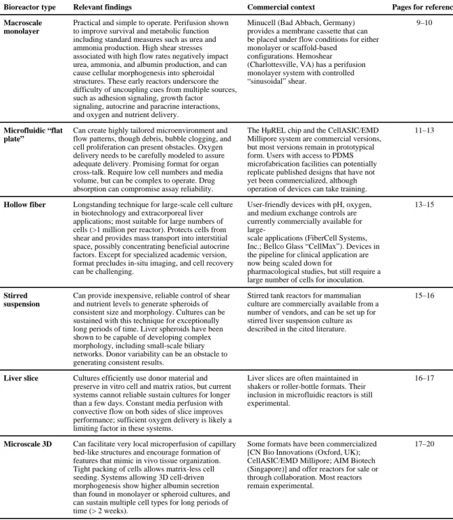
3.Bioreactors
A wide variety of bioreactor formats have been developed or adapted for liver culture over the past four decades. A summary of the major features and commercial availability is provided in Table 2, and a detailed description of the historical development and current features is provided in this section, followed by a focus on applications in Section 4.
3.1.Bioreactors to manipulate phenotype in monolayer culture
The most conceptually simple bioreactor involves inducing flow over or through a
monolayer of hepatocytes or co-cultures of hepatocytes with other cell types, which may be plated on or within matrix such as collagen gels. Such systems have undergone an evolution from early work aimed toward very large scale culture for extracorporeal liver support, to intermediate “macroscale” reactors directed toward physiology and drug development, to microfluidic systems directed at increasing throughput, improving control over
microenvironment conditions, and integrating with other organ mimics. Though
conceptually simple, reactors based on variations of monolayer culture remain an important practical use and a foundation to more complex configurations.
3.1.1.Macroscale monolayer-based reactors—The benefits of medium flow, often termed “perifusion” when slow flow medium replenishment across the cell surface is employed, have been investigated almost since the beginning of cell culture. Studies have shown improved survival and metabolic function in the form of ureagenesis of primary rat hepatocytes over 6 days when cultures were perfused compared to maintained static with daily medium changes [52]. In a continuous-flow system designed to feed petri dishes in shaker, the kinetics of insulin uptake and stimulation of glucokinase and pyruvate kinase were shown to be more physiological in the perifused system [53].
Early work directed toward extracorporeal liver support applications showed primary dog hepatocytes cultured in a monolayer on collagen-coated plates with perfusion flow across the top of the monolayer retained acceptable urea and ammonia metabolic function for at least 2 weeks, allowing support of anhepatic dogs when very large surface areas (200 plates each 200 cm2) were used [54]. Hepatocellular metabolic function relevant for liver support improved in this culture system by adapting the collagen gel sandwich format, which provides both matrix stimulatory cues and modulates mechanical shear experienced by cells [55]. A similar flat plate perfusion arrangement used to assess the effects of NPC co-cultured with hepatocytes on the metabolic functions of primary rat hepatocytes confirmed the positive benefit of co-culture and linked function to shear stress (range 1–5 dyn/cm2) as the rates of albumin secretion declined with application of flow compared to static, while rates of urea and ammonia metabolism were enhanced [56][57].
These early reactors stimulated a more systematic mathematical design-oriented approach to controlling oxygen concentration and fluid shear in low dead-volume reactors by shrinking the channel height to a 0.1–0.5 mm range, incorporating an optional oxygen-permeable upper membrane to de-couple fluid shear and oxygen gradients, and incorporating optional shallow microgrooves (0.1 mm) into the culture substrate to physically protect cells from shear [58,59]. A notable result was that for primary rat hepatocytes co-cultured with J2
NIH-PA Author Manuscript
NIH-PA Author Manuscript
fibroblasts under well-oxygenated conditions in a flat plate flow-through configuration, hepatocellular function (albumin secretion and urea synthesis) was well-maintained and comparable to the static control during 3-day perfusion for shear rates <0.33 dyne/cm2, but showed decline for shear rates in the 5–21 dyne/cm2 regime [58].
As an alternative to an oxygenation membrane, microgrooves incorporated into the flat substrate allow a high flow rate of medium to deliver oxygen while protecting cells from shear. As expected, very low flow and low shear conditions (<0.5 dyne/cm2) were associated with oxygen depletion and loss of viability for both configurations but the grooved substrates allowed viability and function to approach that of static control cultures at high flow rates, presumably by maintaining shear <1 dyne/cm2 compared to 15 dyne/cm2 for the flat configuration. A very similar flat plate configuration for rat hepatocyte cultures was implemented to create very well-defined, systematically varied oxygen gradients that allowed recapitulation of zonal expression of metabolic enzymes and toxicology of acetaminophen [60][61], with no noticeably deleterious effects on viability or gene expression using flow conditions that resulted in shear stresses of 1.25-7 dyne/cm2, and hypoxia-related death under very low flow was well-documented to occur zonally. An additional series of studies on shear stress effects on co-cultures of primary rat hepatocytes with liver non-parenchymal cells focused on the 1–5 dyne/cm2 regime using urea secretion and alanine transaminase (ALT) release as metrics of response, and found enhanced function with perfusion relative to static culture even at the high end of the range though the
magnitude of ALT release, a marker of cell damage, was elevated in perfusion compared to static culture even at the low end of the range [56,57]. These studies also showed that increasing shear induced cells to undergo morphogenesis into spheroidal structures (Figure 2).
Taken together, these studies have somewhat ambiguous implications for the effects of the magnitude of shear stress on hepatocellular function in the mid-range (∼ 1 dyne/cm2), and highlight how challenging it is to uncouple the multiple external cues that cells integrate (adhesion signaling, growth factor signaling from autocrine and paracrine sources, oxygen and other nutrients) to study any one factor in a univariate manner, especially when the phenotypic metrics are not identical. For example, the dramatic improvement in function of primary rat hepatocyte co-cultures with fibroblasts on collagen-modified
polydimethylsiloxane (PDMS, silicon rubber) substrates as a means to enhance oxygenation [62] may also arise in part due to more physiological presentation of the adhesion matrix or to partitioning of lipid-soluble hormones and nutrients into PDMS [63,64] providing a local depot regulating gene expression in a favorable manner. Similarly, culture in recessed wells may allow enhanced accumulation of autocrine factors that favorably regulate cell function [65].
Indeed, underscoring this complexity, when primary rat hepatocytes were cultured in collagen gel-covered monolayer configuration using deep cylindrical PDMS wells continuously perfused via inlet and outlet ports placed 1 cm above the culture surface, a configuration termed “MultiChamber Modular Bioreactor (McMB)” by the developers (Figure 3a,b), analysis of cell viability and albumin secretion as a function of flow rate showed a biphasic response to flow. The loss of viability at low flow compared to static
NIH-PA Author Manuscript
NIH-PA Author Manuscript
control conditions was attributed to oxygen limitations, while the loss of viability at high flow attributed to shear effects, even though the shear rates were estimated to be well below 0.01 dyne/cm2 at the highest flow rate [66]. In this configuration, the effective cell
concentration was a relatively low 0.02 × 10⁶ cells/mL (in a total volume of 10 mL) and a possible alternative explanation for lower function at higher shear is rapid washing out of autocrine factors for the high (1 mL/min) flow rate. Another approach to protecting cells from shear stress is to shield the monolayer from flow with a flat microporous membrane of a transwell sort, which has the possible additional advantage of increasing polarization via having fluid access on both sides of the monolayer. A commercial perfusion system (Minucell) provides a membrane cassette structure that can be placed in flow, and using this arrangement, Yu and coworkers used a mathematical modeling framework of nutrient and drug transport and fluid shear to guide an investigation of operational parameters (perfusion rates, in situ oxygenation method) that would optimize the viability of primary rat
hepatocytes, showing greatly improved metabolic function over 14 days of culture compared to static culture as assessed by albumin secretion, biliary structure, and maintenance of ethoxyresorufin-O-deethylase (EROD) metabolizing capacity, a measurement of cytochrome P4501A induction [67]. Additionally, culture of primary rat hepatocytes in a perfused collagen gel-overlay configuration at an estimated shear stress of 0.6 dyne/cm2 showed dramatic improvement of multiple hepatocellular functions (albumin secretion, metabolic enzymes, bile acid transport). However in this report, the fluid shear was provided by modification of the transwell culture configuration and the cell concentration was approximately an order of magnitude greater for comparable medium exchange rates in order to employ confluent cell monolayers on a large (75 mm diameter) transwell [68]. This is an intriguing approach for providing access to both sides of the monolayer while
providing a “liver sinusoidal” value of shear stress (Figure 3c).
Perfused monolayer culture configurations, such as those described above for primary rat cells, have also been applied to pig cells [69], and were shown to be beneficial for human primary cells relative to static conditions [70][71]. Interestingly, fresh primary human cells cultured in the McMB described above were very robust in long-term (2–3 weeks) culture, when cultured at flow rates of 0.25–0.5 mL/min with a 3-fold greater cell concentration (0.06 × 10⁶ cells/mL). These cells showed greatly improved hepatocellular function compared to static cultures as assessed by gene expression of a large panel of CYP450, Phase II enzymes, and transporter genes with confirmatory drug metabolism activity assays [71]. Metabolism activities for almost all genes except CYP2D6 were found to be
comparable to freshly-isolated cells. Although the authors at least partly attribute the enhanced function to shear stress, the values they estimate are several orders of magnitude lower than those in previous studies (∼ 5 × 10⁻⁷ dyne/cm2), hence it is interesting to speculate that factors such as altered autocrine accumulation of extracellular signaling molecules might play a role as well. Greatly enhanced function of cryopreserved primary human cells cultured under perfusion monolayer conditions was also observed in a reactor comprising a sandwich of two ∼20 cm2 circular gas-permeable membranes spaced ∼0.5 mm apart, with hepatocytes cultured on the surface of a collagen-coated membrane and compared to collagen gel-cultured static conditions over a period of over 4 weeks [70]. Albumin and total protein secretion rates were significantly higher in the perfused
NIH-PA Author Manuscript
NIH-PA Author Manuscript
conditions over the first 3 weeks, though interestingly total albumin secretion in the static condition, while lower than in perfused, was approximately constant over the time period. The differences in albumin may be related in part to the 10% oxygen saturation used in the gas phase as albumin secretion is positively regulated by oxygen up to highly
supraphysiological levels [72]. The cells were surprisingly robust over more than 3 weeks, as the cultures were used to analyze protective effects of IL-6 on exposure to 80 µM diclofenac over a period of 3 days; presumably due to the large number of cells used for each reactor (∼106) Untreated controls were not shown, but the results indicated that cells recovered their full function after diclofenac treatment [67].
3.1.2.Microfluidic “flat plate” configurations—The experience described above with various “macro” flat plate configurations yielded several important insights about rat and human hepatocyte and heptatocyte/NPC co-cultures. At the least, phenotypic outcomes comparable to static culture could be achieved, thus providing a way to measure metabolism and toxicity under controlled exposure. At best, reactors improved the phenotypic responses compared to control static culture systems. Cells cultured under select flow conditions can maintain certain metabolic functions at nearly the levels of freshly-isolated cells, which motivates further adaptation of the “flat plate” configuration to more accessible, user-friendly, higher throughput, and scalable technologies. Thus, we review the literature broadly, including analysis of systems that are not commercialized.
Several advances in microfabrication technologies have together yielded tremendous opportunities to create highly tailored microenvironments and flow patterns. These include (i) the application of high-resolution silicon microfabrication technologies to create fine-scale structural features in silicon, the so-called “micro-electro-mechanical systems (MEMS)” devices, coupled with the innovation of using microfabricated silicone structures as molds to cast PDMS parts that can be easily bonded to glass to create fluidic structures and (ii) photopolymerization and masking approaches.
Application of microfluidic approaches to liver cell culture began to emerge about ten years ago in several labs, both as semi-empirical feasibility studies aimed at demonstration of long-term cell survival in PDMS devices [73][74][75][76] and as design-principle based visions of how to transform physiologically based pharmacokinetic (PBPK) measurements and toxicity analysis by creating appropriately-scaled cell to medium ratios, organ cross-talk and in situ cellular activity sensors [77,78]. Leclerc and co-workers exploited the ease of fabricating intricate features in PDMS surfaces to create a device wherein the culture medium flows along the top of a PDMS slab containing square microwells (0.5 mm) and interconnecting angled channels, all recessed 100 µm beneath the top of the flow surface to provide a low shear, relatively uniform flow environment with about 2 cm2 total of culture surface area [74][73] (Figure 4). They first demonstrated basic operating parameters (flow rates, medium exchange rates, oxygen transfer) for culturing HepG2 cells for 2 weeks in an initial device [73,79]. Common challenges encountered in this microfluidic system included medium evaporation and debris bubble clogging of channels resulting in non-uniform flow, in addition to the complication not encountered with primary cells of the tendency for cell proliferation during culture. With shear stresses estimated in the range of 0.03–0.25 dyne/cm2 for flow rates in the range of 20–30 µL/min for up to 20 × 107 cells, these studies
NIH-PA Author Manuscript
NIH-PA Author Manuscript
showed that for HepG2 frequency of medium change and availability of oxygen for large cell numbers limited the growth and viability of cells, especially when several devices were concatenated in a stack to increase cell number as might be desirable for extracorporeal devices [79].
A series of subsequent papers characterizing HepG2/C3a behavior in this system, mainly in short term culture (4 days), further refined the design and operating conditions. The feasibility of carrying out transcriptomic, proteomic, metabolic and toxicologic characterization with this arrangement, was illustrated and showed that HepG2/C3a
phenotypic responses including albumin secretion, sensitivity to acetaminophen toxicity, and relative expression of CYP450 and transport genes appeared more physiologically close to primary hepatocytes in the microfluidic compared to static petri dish culture, a result that may in part due to the quasi-3D aggregate structures that form in regions of the recessed structures [74,79,80][81–84]. Initial studies analyzing primary rat [85,86] and human [74,87] cells in short term culture (4 days) indicate that these cells were less likely to form aggregates, and optimization of operating conditions for these cells is evolving. The results assessing drug metabolism with cryopreserved human cells are instructive in two key ways, first showing that these cells attach and survive in both the recessed features as well as the top of the PDMS exposed directly to flow. Secondly, experimental confirmation that after 4 h of perfusion in cell-free devices, hydrophobic drugs such as midazolam,
dextromethorphan, and desethylamodiaquine are very significantly (60–95%) depleted due to partitioning into PDMS, a phenomena reported by others for hydrophobic compounds in PDMS devices [63,64], a topic we discuss in more detail in Section 4.
Fujii and coworkers developed the concept of protecting cells from shear by perfusing the opposite side of porous flat membrane with the cultured cells using microfluidics. A bilayer PDMS device incorporated either a polyester or microfabricated PDMS membrane between two PDMS slabs containing a well above the membrane for cell monolayer culture and flow channels below the membrane [76]. They showed that in the perfused microdevice, albumin secretion rates of primary rat hepatocytes over two weeks of culture were stable, comparable to those of cells maintained in transwell-type membrane inserts in static culture, and were much higher than those in standard 2D petri dish culture. Cells in the perfusion culture demonstrated slightly superior conversion of ammonia compared to cells in static transwell culture, so this demonstration shows how a device might be used in a continuous mode. Borenstein and coworkers described a similar approach to protecting hepatocytes from shear due to high-velocity flow, while also providing a suitable culture substrate to induce cell polarization [75]. They created a bilayer PDMS device incorporating a 0.4 µm pore-size polycarbonate membrane separating the medium flow from the hepatocyte layer cultured on the membrane above the flow (Figure 5a,b). An intricate microchannel flow path inspired by vascular network geometry, essentially created a microscale version of transwells perfused on the bottom of the membrane by a “vascular network” etched into the adjacent PDMS layer, which exposed the hepatocytes to negligible levels of shear stress. Each region of the hepatocyte monolayer on the membrane was in contact with a “vascular channel” for about 25% of the monolayer surface (Figure 5b,c). Although this sparse network might seem limiting, both HepG2 and primary rat hepatocytes remained viable in the system for at least a week, provided the polycarbonate membrane culture surface was coated with collagen and
NIH-PA Author Manuscript
NIH-PA Author Manuscript
the flow rate was adjusted to accommodate the metabolic needs. Primary hepatocytes increased their per-cell rate of albumin secretion over a week of culture in the device, and secreted albumin at higher rates than in control static cultures [75]. Subsequent diffusion-reaction modeling of oxygen transport and consumption in this device suggests that oxygen may become limited at high cell densities [88], however experimental verification of oxygen gradients would shed more light on the apparent high viability of dense hepatocyte
monolayers. To our knowledge, these devices have not been commercialized.
The potential of microfluidic systems as “micro cell culture analogs (µCCA)” of humans to represent PBPK was analyzed and demonstrated in the seminal work by Shuler in co-workers [77,78], and the term µCCA is now commonly used in the in vitro tissue bioreactor community to designate the organ-cross talk system pioneered by Shuler. A detailed analysis of scaling of in vivo organ residence times and how to translate them into interlinked multi-organ crosstalk systems within constraints of low fluid shear stress and oxygen delivery was presented. The multi-organ crosstalk systems were represented by monolayer cultures of lung, liver, and fat cell lines, and the fluid shear stress was constrained to less than 5 dyne/cm2 for the liver compartment (Figure 6). Further, they provided proof of principle for naphthalene toxicity in a cross talk of human HepG2/C3a cells with lung cells[78]. While interacting micro-organ systems are discussed in more detail below, these papers made interesting observations about how the morphology and function of liver cells was exquisitely governed by cell density, flow rates, and matrix coatings in the liver
compartment where collagen or fibronectin was required to prevent cell detachment. These studies also underscored the difficulties in obtaining defect-free devices given the tendencies of bubbles and debris to clog the systems. A straightforward PDMS microfluidic system for culturing HepG2 on collagen-coated glass under shear stresses ranging from 1.4–60 dyne/cm2 confirmed observations from macroscale systems that sensitivity to shear appears to have onset around 5 dynes/cm2. In this system, shear stress was systematically varied not only by flow microchannel dimensions but addition of high molecular weight dextran to the medium to increase medium viscosity, and thus shear for a given flow rate. In 3-day culture experiments, cells at higher shear rates became progressively more rounded, detached, and reduced secretion of albumin[89] (Figure 7). A variation on this original design is being further developed in collaboration with a commercial partner under the auspices of the NIH-funded “Microphysiological Systems Program.”
The challenges in translating the benefits of perfusion culture for more sensitive primary cells from macroscopic to a microfluidic format are illustrated in a study employing the well-characterized rat hepatocyte-fibroblast micropatterned co-culture system described above to a PDMS device comprising a microperfused 8 × 8 array of interconnected (i.e., not separately addressable) 3 mm diameter micropatterned wells. Channels were fed by a flow rate constrained to keep shear well below 0.01 dyne/cm2 while providing 1.4 mL/day of medium in a one-pass configuration, and providing oxygen by a secondary network of gas flow channels above the medium flow channels [90]. In cultures maintained for 32 days, albumin and urea secretion rates, interestingly, stabilized after 5 days in both perfused and control cultures of comparably-seeded static cultures open to a medium volume of 4 mL, but the rates in the perfused system were only ∼50% of the controls. The significant decrease in absolute level of metabolic performance in the perfused microfluidic system was speculated
NIH-PA Author Manuscript
NIH-PA Author Manuscript
to arise from possible differences in local concentrations of soluble signaling factors, oxygen limitations, or nutrient limitations, as the shear stress appeared to be within an acceptable physiological range. Another implementation of the culture arrangement involves co-culture in the “liver” compartment of the HµREL® chip [91], which was designed based on the principles described by Shuler and coworkers, where the authors demonstrated clearance of several compounds by cryopreserved human hepatocytes was more physiological
compared to the chip containing hepatocytes alone. Commercial applications of the HµREL® chip are still evolving.
As outlined by Shuler, these issues, along with smallness of the volumes and cell numbers in the microfluidic systems, drives incorporation of on-board instrumentation to monitor cell activity and function noninvasively. His group illustrated this by measuring CYP450 activity non-invasively during culture of HepG2/C3a cells embedded in Matrigel in the µCCA device described earlier [92]. HepG2/C3a cells in the µCCA showed appreciable basal levels of CYP450 activity in a manner not previously observed in standard culture, although comparison to static cultures in Matrigel may have revealed contributions of Matrigel alone. This work stands out for the very detailed analysis of the reaction/diffusion effects on the conversion of CYP450 substrates in the µCCA, including a breadth of investigation into effects of cell density and substrate concentrations.
Whether various flat plate microfluidic reactor configurations focused on liver will gain significant traction in drug discovery pipelines or in more general liver physiology research is still an open question, as various implementations of traditional macroscale flat plate reactors described earlier remain accessible and can provide high levels of function. Macroscale systems in principle require more cells and reagents, which make microscale systems appear attractive for saving costs of primary cells and experimental compounds that may be expensive to synthesize early in the drug development process. Tradeoffs in
microscale systems include drug adsorption and complex operation, and other approaches are continuously being developed to address specific assays such as low clearance
compounds and reactive metabolites [93]. Thus, the advantages of microfluidic systems may be greater for more complex applications that require microscale 3D architectures to retain function, or applications where interconnection with other microscale organ mimics provides greatly enhanced information [94–96].
3.2. Evolution of hollow fiber bioreactors as prototypical 3D liver culture environments
The phenomenological advantages of 3D culture of liver cells arguably dates back to experiments culturing hepatoma cells in some of the earliest incarnations of hollow fiber reactors [97]. Since hollow fiber technologies are still being actively pursued today for 3D liver physiological culture [98], a review of the evolution of hollow fiber bioreactor technology for liver cell culture establishes a contextual foundation for other 3D bioreactor technologies.
Hollow fiber cell culture reactors emerged over 30 years ago as a means to culture mammalian cells on a large scale for biotechnology applications, such as protein and vaccine production, complementing approaches based on stirred-tank reactors and roller bottle cultures [99]. Commercial user-friendly versions implemented with pH, oxygen, and
NIH-PA Author Manuscript
NIH-PA Author Manuscript
medium exchange controls are in use today for production of proteins, vaccines, and other therapeutics [100]. Rooted in technology developed for kidney dialysis and protein purification, the classic hollow fiber mammalian cell culture reactors comprise a bundle of semipermeable hollow fiber ultrafiltration or dialysis-type membranes encased in a housing such that liquid pumped down the fiber lumens exchanges molecular components with cells cultured in the extracapillary space outside the fibers; i.e., exchange occurs in a manner that mimics facets of capillary blood-tissue molecular exchange in vivo. Because the membrane physically separates cells from fluid flow, high fluid flow rates can be maintained inside the capillary to minimize gradients along the capillary length, while protecting fragile cells from shear forces of flow. This same arrangement protects cells from the stresses of the air-liquid interface that occurs in roller bottle culture. Other major design features include small fiber dimensions on the order of 0.2–0.4 mm diameter and close packing of fibers. This affords a high surface-to volume ratio at the same time that the diffusion distances from the central fiber lumen, the source of oxygen and fresh nutrients, to and through the tissue mass outside the fibers are kept within a range of a few hundred microns to support in theory adequate oxygen and nutrient delivery to the entire cell mass.
An interesting feature and challenge of the hollow fiber configuration is the control of molecular exchange properties between the nutrient feed and tissue compartment by selection of membrane permeability properties. As a practical matter in protein production applications, appropriate choice of permeability allows the desired protein product to either be concentrated in extracapillary space which has relatively low contamination by serum proteins in medium, or to diffuse into the lumen and be automatically separated from cells on a continuous basis, thus facilitating subsequent purification in either case. One of the earliest reports of hollow fiber epithelial culture, of colon cancer cells to produce
carcinoembryonic antigen (CEA), illustrated that low-permeability (10 to 50 kDa cut-off) membranes enhanced differentiated function of cells as the tumor cells formed 3D organoid-like structures, secreted mucus, and produced higher rates of the CEA relative to 2D cultures [101]. While enhanced function is likely partially attributed to the adhesion environment driving a 3D structure, the serendipitous concentration of autocrine survival, growth and differentiation factors in the pericellular environment can be greatly enhanced in the hollow fiber arrangement compared to 2D culture.
The application to liver cell culture was almost immediate, first with a report that a rat hepatoma line could carry out bilirubin conjugation in hollow fiber culture with either 10 kDa or 50 kDa cut-off membranes [97], with speculation that the rates of conjugation may be limited by the transport of the bilirubin carrier protein albumin across the membrane. Next it was found that primary rat cells cultured up to 45 days in a 7-cm long hollow fiber reactor with 520-0.3 mm diameter fibers maintained almost half the diazepam-metabolizing capacity after 10 days provided the perfusion medium was equilibrated with 30% oxygen atmosphere [102]. Not surprisingly, given the similar design features between hollow fiber cell culture reactors and blood dialysis units, liver cell culture in hollow fiber reactors soon emerged as a potentially promising approach for extracorporeal liver support to provide metabolic capacity in synthesis of urea and metabolism of toxic metabolites like bilirubin [103][102,104][105][106][107]. Detailed reviews of early work of the approximately dozen groups using various bioreactor formats for clinical extracorporeal liver support, including
NIH-PA Author Manuscript
NIH-PA Author Manuscript
analysis of clinical constraints, can be found in a review by Rozga and co-workers [108], while later developments and clinical applications can be found in more recent reviews [109][110][111][112][113][114][115]. Here, we summarize findings and observations as context for in vitro culture models and focus on reactor configurations that have been scaled down for use in drug development.
While no precise consensus exists on the minimum number of liver cells needed in an extracorporeal device to support a liver failure patient, in part due to differences in types of cells and use of adjuvant charcoal-stripping steps to remove toxins from patient plasma, estimated needs range from 10–20% of the patient’s liver mass. The scale is unequivocally enormous – tens to hundreds of grams of cells – a scale that presents an almost
insurmountable barrier to using primary human hepatocytes on a routine basis, especially when the use of reactors is on an emergency and not pre-scheduled basis. Hence, of five reactor systems that progressed to clinical trials in the US and Europe, four were developed for use with primary porcine cells, one with the human HepG/C3a cell line [115] and nascent incorporation of HepaRG cells [116]. Translation to the clinic has been arduously slow, hampered by the high cost, difficulty in obtaining clear-cut results in clinical trials, and competition for subsets of patients by a cellular reactors that remove toxins [115]. Based on encouraging results for long-term liver cell survival in clinical-scale devices, at least two groups have developed scaled down versions of their clinical reactor for application in drug development and study of liver physiology. The system developed by Gerlach and co-workers employs an appealing design that to a certain extent uncouples oxygen delivery from transport of molecular components in the medium [98,117– 121]. The design comprises three separate sets of hollow fiber interwoven so that culture medium flows between two sets of fibers in an almost interstitial-like manner. The third set provides a flow of gas containing a pre-set oxygen concentration, so that all regions of the reactor can be fed by oxygen locally (Figure 8). Cells are seeded into the extracapillary space and maintained under perfused conditions. Early studies aimed at clinical application using a reactor volume of close to 1 L showed, promisingly, that primary porcine cells formed tissue-like structures incorporating non-parenchymal cells, and that biochemical activity was relatively stable over 30 days [118]. The principles for scaling the reactor from the clinical scale (800 mL, 1.4 × 10¹⁰ cells) to an intermediate scale (8 mL, 2.6 × 10⁸ cells) and a small scale (2 mL, 1.2 × 10⁸ cells) were illustrated using 3 week cultures of fresh primary human cells from 5 different donors, where the medium refreshment rates and perfusion flow rates were scaled for each reactor size based on computational models previously developed by the group. Immunohistochemistry revealed formation of tissue-like structures with expression of markers of hepatocytes as well as biliary cells (CK19), Kupffer cells (CD68) and endothelial cells (VWF), and polarization of key transporters. Interestingly, the specific per cell rate of secretion of albumin was greatest for the 2 mL reactor. Drug metabolizing capacity (phenacetin, Cyp 1A; midazolam, Cyp3A; diclofenac, Cyp2C9) declined 50–80% from the day 3–5 period to the day 10–23 period [122]. A subsequent study with this design further miniaturized to a 0.5 mL volume (25 × 106 cells) showed that primary human hepatocytes maintained for 10 days in culture performed comparably well in terms of protein secretion and drug metabolizing capacity across 4 drugs in serum-free medium compared to serum-containing medium, but that serum-free culture enhanced the expression
NIH-PA Author Manuscript
NIH-PA Author Manuscript
of several transporter genes [119]. This same system enhanced the differentiation of human fetal liver cells compared to static petri dish culture, especially when combined with a hyaluron-based extracellular matrix for embedding the cells [123,124], and has been extensively characterized for differentiation of the human hepatic cell line HepaRG, illustrating stable differentiation [120].
An alternate hollow fiber design combined fiber matrix sheets interspersed with hollow fibers to similarly address the high oxygen demands of hepatocytes by providing a set of hollow fibers for gas exchange in the context of adhesion to the fibers was developed by Chamuleau and coworkers for clinical application and has also been scaled down from a clinical version to a version suitable for pharmacological studies [125– 127][128,129] [116,130,131]. The large clinical-scale version of this reactor system was showed
comparable to the Gerlach 3-fiber system in maintaining liver-specific function of primary porcine cells over 7 days in culture, and both maintained >90% function of the 10 billion cells seeded [132]. The hepatocytes in this system occupy several different niches and experience a complex combination of cues where some of the cells attach within the walls of the fiber mat and are distant from the source of oxygen, and others form aggregates in spaces between the hollow fibers [127]. In the liver, glutamine synthetase (GS) is normally expressed in the pericentral zone, a region that experiences the lowest oxygen concentration, and carbamoyl phosphate synthetase I is normally expressed in the periportal and central zones. An interesting analysis of localization of expression within the bioreactor of GS and carbamoyl phosphate synthetase I showed that cells near the gas capillaries (i.e., cells experiencing the highest oxygen concentration) expressed the highest level of GS, in contrast to what would be expected in vivo. The authors suggest that combinations of substrate and autocrine factors may contribute to this zonation pattern [127]. This reactor system has also been optimized for stable differentiation and maintenance of HepaRG cells in long-term culture in the absence of DMSO for use in drug metabolism studies
[116,130,131].
Although hollow fiber reactors incorporate many elegant design principles for uncoupling fluid flow, matrix, support, and medium perfusion, others have observed that “despite these advantages, the high cost and difficult set-up of hepatocyte (hollow fiber reactors) probably limit their use in drug metabolism and transport studies” [113]. Adsorption of hydrophobic drugs to the components of hollow fiber systems may also be a limitation in some
applications, and together with challenges in in situ imaging of tissue structures and relative difficulty in accessing the tissue space, these reactor formats may need additional evolution to enter routine use. Some of the publications for the scaled-down versions of hollow-fiber reactors were co-authored by investigators in pharmaceutical companies, and these investigators are now a resource in the community for providing perspective via personal communication on the strengths and weaknesses of these systems.
3.3.Stirred suspension bioreactors for liver cell culture
The propensity of hepatocytes to spontaneously aggregate when seeded on poorly-adhesive surfaces, and for aggregates to maintain liver-specific functions to a greater degree than cells in monolayer culture, has been observed and reported for decades [133][134]. Formation
NIH-PA Author Manuscript
NIH-PA Author Manuscript
and maintenance of spheroids in static culture is almost an art, as cell seeding density, culture medium depth, and other factors all conspire to create local microenvironments that are difficult to control (particularly the concentration of oxygen) and reproduce across labs. Stirred-tank bioreactors have long been used in large-scale culture of mammalian cells to provide an environment allowing control of oxygen, pH, and continuous addition and removal of medium. With appropriate design of impellers, the fluid mechanic
microenvironment in such systems can provide relatively uniform low-shear mixing and the commercial availability of reactor systems makes them accessible for general use. The utility of such a reactor configuration for efficient (80% of cells incorporated) formation of relatively uniform, functional spheroidal aggregates of primary rat hepatocytes that exhibit evidence of appropriate polarization of bile canaliculi was illustrated over 15 years ago [135].
Recently, Alves and co-workers applied this approach to primary human hepatocytes from 3 different donors showing that appropriate choice of operating conditions such as stirring rate, maintenance of culture oxygen level at the physiologically-relevant level of 60 µM led to formation of aggregates with average dimensions of 80 µm diameter by the end of the second week of culture [136]. This length scale was expected to be well below diffusion limitations for oxygen, though steep gradients in proteins from the medium as well as autocrine factors would be expected. Interestingly, cells were maintained in serum-free medium after the initial 3-day aggregation period, and the per-cell rate of albumin secretion increased over two weeks of culture for all donors. Immunohistochemistry of the spheroids after two weeks of bioreactor culture revealed an interesting distribution of albumin concentrated around the periphery and somewhat depleted in the spheroid core and Cyp3A was strongly expressed in about half the cells with no particular localization within the spheroid (Figure 9). Similar to the previous observations with rat cells, the human spheroids exhibited strong evidence of a bile canilicular network penetrating the spheroids, as assessed by proper apical polarization of atypical protein kinase C (aPKC) and excretion of
fluorescent 5-(and-6)-carboxy-2’,7’-dichlorofluorescein diacetate (CFCDA) into the biliary structures. In general, the spheroids maintained high, stable levels of CYP gene expression and ability to be induced by rifampicin to metabolize ethoxycoumarin. This study also highlighted the high degree of variability between donors: a 10-fold difference in absolute levels of albumin secretion despite similar trends of increasing per cell rates of secretion over 2 weeks, variability in the formation of actin stress fibers and polarization, and differences in trends of gene expression of CYP enzymes as a function of time. Presumably, different donors had different initial states, and all adjusted to the environment in the bioreactor. These observations underscore the need to characterize systems with multiple different donors in order to generalize results.
The strength of using the stirred-tank bioreactor is that the conditions are well controlled potentially improving overall success given large interdonor variability, and a large number of spheroids can be maintained for extended periods, thus allowing efficient use of large numbers of cells from a single donor. Screening a large number of conditions against the spheroids would require a step of removing the spheroids and dispensing them to individual wells or flow-through systems for applying the test conditions, a process that should be entirely feasible. In previous studies, spheroidal cell aggregates seeded into a support