HAL Id: hal-01211998
https://hal.archives-ouvertes.fr/hal-01211998
Submitted on 15 Nov 2018
HAL is a multi-disciplinary open access archive for the deposit and dissemination of sci-entific research documents, whether they are pub-lished or not. The documents may come from teaching and research institutions in France or abroad, or from public or private research centers.
L’archive ouverte pluridisciplinaire HAL, est destinée au dépôt et à la diffusion de documents scientifiques de niveau recherche, publiés ou non, émanant des établissements d’enseignement et de recherche français ou étrangers, des laboratoires publics ou privés.
Copyright
mucosa and promote cell proliferation
Jennifer Raisch, Emmanuel Buc, Mathilde Bonnet, Pierre Sauvanet, Emilie
Vazeille, Amélie de Vallée, Pierre Dechelotte, Claude Darcha, Denis Pezet,
Richard Bonnet, et al.
To cite this version:
Jennifer Raisch, Emmanuel Buc, Mathilde Bonnet, Pierre Sauvanet, Emilie Vazeille, et al.. Colon cancer-associated B2 Escherichia coli colonize gut mucosa and promote cell proliferation. World Journal of Gastroenterology, Baishideng Publishing Group Co. Limited, 2014, 20 (21), pp.6560-6572. �10.3748/wjg.v20.i21.6560�. �hal-01211998�
Colon cancer-associated B2
Escherichia coli
colonize gut
mucosa and promote cell proliferation
Jennifer Raisch, Emmanuel Buc, Mathilde Bonnet, Pierre Sauvanet, Emilie Vazeille, Amélie de Vallée,
Pierre Déchelotte, Claude Darcha, Denis Pezet, Richard Bonnet, Marie-Agnès Bringer, Arlette Darfeuille-Michaud
Jennifer Raisch, Emmanuel Buc, Mathilde Bonnet, Pierre Sauvanet, Emilie Vazeille, Amélie de Vallée, Denis Pezet, Richard Bonnet, Marie-Agnès Bringer, Arlette Darfeuille-Michaud, Clermont Université, UMR1071 Inserm/Université d’ Auvergne and INRA USC2018, 63000 Clermont-Ferrand, France Emmanuel Buc, Pierre Sauvanet, Emilie Vazeille, Pierre Dé-chelotte, Claude Darcha, Denis Pezet, Richard Bonnet, Cen-tre Hospitalier Universitaire, 63000 Clermont-Ferrand, France Author contributions: Raisch J, Buc E, Bringer MA and
Dar-feuille-Michaud A conceived and designed the study, analysed data and drafted the manuscript; Bringer MA and Darfeuille-Michaud A contributed equally to the design and data analyses of this study; Raisch J, Buc E, Bonnet M, Vazeille E and Bringer MA performed experiments; Raisch J and Buc E contributed equally to this study; Buc E, Sauvanet P, de Vallée A, Pezet D and Bonnet R carried out the sample collection and the sample pro-cessing; Déchelotte P and Darcha C performed immunohistology analyses.
Supported by Ministère de l’Enseignement supérieur et de la Recherche, Inserm and Université d’Auvergne (UMR1071), INRA (USC-2018); and Grants from the Association F. Aupetit (AFA) and Ligue contre le cancer
Correspondence to: Marie-Agnès Bringer, PhD, Clermont Université, UMR1071 Inserm/Université d’Auvergne and INRA USC2018, 63000 Clermont-Ferrand,
France. m-agnes.bringer@udamail.fr
Telephone: +33-4-7317 8371 Fax: +33-4-7317 8371
Received: October 25, 2013 Revised: February 10, 2014 Accepted: March 8, 2014
Published online: June 7, 2014
Abstract
AIM: To provide further insight into the
characteriza-tion of mucosa-associated Escherichia coli (E. coli) iso-lated from the colonic mucosa of cancer patients.
METHODS: Phylogroups and the presence of
cyclo-modulin-encoding genes of mucosa-associated E. coli
from colon cancer and diverticulosis specimens were
determined by PCR. Adhesion and invasion experiments were performed with I-407 intestinal epithelial cells
using gentamicin protection assay. Carcinoembryonic
antigen-related cell adhesion molecule 6 (CEACAM6) expression in T84 intestinal epithelial cells was meas-ured by enzyme-linked immunosorbent assay and by Western Blot. Gut colonization, inflammation and pro-carcinogenic potential were assessed in a chronic infec-tion model using CEABAC10 transgenic mice. Cell prolif-eration was analyzed by real-time mRNA quantification
of PCNA and immunohistochemistry staining of Ki67.
RESULTS: Analysis of mucosa-associated E. coli from colon cancer and diverticulosis specimens showed that whatever the origin of the E. coli strains, 86% of cyclomodulin-positive E. coli belonged to B2 phylogroup
and most harbored polyketide synthase (pks) island,
which encodes colibactin, and/or cytotoxic necrotizing factor (cnf) genes. In vitro assays using I-407 intestinal
epithelial cells revealed that mucosa-associated B2 E.
coli strains were poorly adherent and invasive.
How-ever, mucosa-associated B2 E. coli similarly to Crohn’s
disease-associated E. coli are able to induce CEACAM6
expression in T84 intestinal epithelial cells. In addition,
in vivo experiments using a chronic infection model of
CEACAM6 expressing mice showed that B2 E. coli strain
11G5 isolated from colon cancer is able to highly persist in the gut, and to induce colon inflammation, epithelial damages and cell proliferation.
CONCLUSION: In conclusion, these data bring new
insights into the ability of E. coli isolated from patients with colon cancer to establish persistent colonization, exacerbate inflammation and trigger carcinogenesis. © 2014 Baishideng Publishing Group Inc. All rights reserved.
Key words: B2 Escherichia coli; Carcinoembryonic anti-gen-related cell adhesion molecule 6; Cell proliferation;
EVIDENCE-BASED MEDICINE
Colon cancer; Polyketide synthase genomic island
Core tip: Tumors and mucosa of patients with colon
cancer are abnormally colonized by Escherichia coli
(E. coli) belonging to B2 phylogroup. The aim of the present study was to provide further insight into the characterization of colon cancer-associated E. coli. Despite their poor ability to adhere to and to invade intestinal epithelial cells in vitro, we showed that colon
cancer-associated E. coli induce carcinoembryonic
an-tigen-related cell adhesion molecule 6 (CEACAM6) ex-pression, a receptor involved in adhesion of pathogenic
E. coli. These bacteria were also able to persist and promote low grade inflammation and cell proliferation, in a chronic infection model of CEACAM6 expressing mice, highlighting their oncogenic potential.
Raisch J, Buc E, Bonnet M, Sauvanet P, Vazeille E, de Vallée A, Déchelotte P, Darcha C, Pezet D, Bonnet R, Bringer MA, Darfeuille-MichaudA. Colon cancer-associated B2 Escherichia
coli colonize gut mucosa and promote cell proliferation. World J Gastroenterol 2014; 20(21): 6560-6572 Available from: URL:
http://www.wjgnet.com/1007-9327/full/v20/i21/6560.htm DOI: http://dx.doi.org/10.3748/wjg.v20.i21.6560
INTRODUCTION
Colorectal cancer (CRC) is the fourth leading cause of cancer death and is responsible for about 610000 deaths per year worldwide[1]. Although many etiologic genetic
changes are associated with progression from adenoma-tous lesions to invasive carcinoma[2], the specific
causa-tive factors in the development of sporadic CRC remain unclear. Accumulating evidence supports that inflamma-tion and gut microbial communities influence the devel-opment of colorectal carcinoma[3-5]. Two theories have
emerged to explain the contribution of bacteria in CRC: (1) the “alpha bug” concept, wherein select members of a microbial community with virulence and pro-carcino-genic features are capable of remodeling the microbiome as a whole to drive pro-inflammatory immune responses and colonic epithelial cell transformation leading to can-cer[6]; and (2) the “driver-passenger” concept, wherein
certain indigenous intestinal bacteria, termed “bacteria drivers”, initiate CRC by inducing epithelial DNA dam-ages: the resulting tumorigenesis induces intestinal niche alterations that promote the proliferation of passenger opportunistic bacteria with a growth advantage in the tu-mour microenvironment[7].
Dysbiosis of the intestinal microbiota has been ob-served in CRC patients. Recent pyrosequencing data of CRC-associated bacterial microbiota have revealed, in particular, over-representation of some bacteria such as Bacteroides/Prevotella, Faecalibacterium and Fusobacterium[8,9].
In addition, independent studies show that colonic ad-enomas, carcinomas and the mucosa of CRC patients are abnormally colonized by high numbers of adherent
Es-cherichia coli (E. coli) compared to controls[10-12]. It has been
suggested that the role of E. coli in CRC promotion and development is related to chronic inflammation. Inflam-mation can result from bacterial infection, via its effects on both the host and the microbiota, in particular by pro-moting the expansion of E. coli, which actively contribute to the accumulation of mutations resulting from DNA damages induced by genotoxins, or by downregulating host DNA mismatch repair proteins[3,11,13]. In particular,
E. coli strains harboring the polyketide synthase (pks) genotoxic island, which are found in a significantly high percentage of inflammatory bowel disease (IBD) and CRC patients, can promote invasive carcinoma in mono-colonized azoxymethane (AOM)-treated Il10-/- mice[3]. In
addition, certain pathogenic bacteria can also be involved in cancer development, like, for example enterotoxigenic Bacteroides fragilis (ETBF), a common human commensal bacterium that is associated with colon cancer[14].
ETBF-induced chronic inflammation and tumorigenesis in Ap-cMin/+ mice (a mouse model of familial adenomatous
pol-yposis) involve the induction of the polyamine catabolic enzyme spermine oxidase, which causes DNA damages and uncontrolled cell proliferation in intestinal epithelial cells[15].
Patients with IBD have an increased risk of colon cancer and small bowel adenocarcinoma[16,17]. As in colon
cancer patients, dysbiosis toward selected micro-organ-isms and decreased complexity of commensal bacteria have been observed in patients with Crohn’s disease (CD) and ulcerative colitis (UC), but it is not clear whether dysbiosis contributes to the development of IBD or is instead a consequence of the disease. Patients with IBD, compared to healthy controls, have fewer bacteria with anti-inflammatory properties and/or more bacteria with pro-inflammatory properties. Several metagenomic-based studies reported that members of the phyla Bacteroide-tes and FirmicuBacteroide-tes were reduced in patients with CD or UC[18-20]. Among the Firmicutes, Faecalibacterium
praus-nitzii has anti-inflammatory properties; its numbers are reduced in patients with CD and associated with a risk of post-resection recurrence of ileal CD[20]. In contrast,
a greater relative abundance of Enterobacteriaceae, mostly E. coli belonging to the B2 phylogenetic group, has been reported in CD patients more notably on mucosa-asso-ciated microbiota than in fecal samples[10,18,21-24]. Intestinal
colonization by E. coli correlates with bacterial adhesion of CD-associated E. coli strains to intestinal epithelial cells[10,25]. CD-associated E. coli share abilities to adhere
to and to invade intestinal epithelial cells and to survive within macrophages[26,27] and they are termed
accord-ingly adherent-invasive E. coli (AIEC). The abnormal colonization of CD mucosa by AIEC involves abnormal expression of a host receptor, the carcinoembryonic anti-gen-related cell adhesion molecule 6 (CEACAM6)[28].
In-terestingly, CEACAM6 is not only abnormally expressed in the ileum of patients with CD[28] but expression of
this molecule is also up-regulated in proliferating cells in adenomas and colorectal tumors[29,30]. However, the origin
of CEACAM6 surexpression in colon cancer is not yet clearly understood.
The aim of the present study was to provide further insight into the characterization of mucosa-associated E. coli isolated from the colonic mucosa of cancer patients. We determined their ability to interact with intestinal epi-thelial cells, with a particular focus on biofilm formation and the presence of cyclomodulin-encoding genes, and to induce CEACAM6 expression in intestinal epithelial cells. Finally, using CEABAC10 transgenic mice express-ing human CEACAMs, we assessed the effects of long-term chronic infection by the colon cancer-associated E. coli strain 11G5 for its ability to colonize the gut, to potentiate inflammation and to induce cell proliferation.
MATERIALS AND METHODS
Ethical considerations
Ethical approval for the study was granted by the Cl-ermont-Ferrand research ethics committee. This IRB allowed for the waiver of written consent and approved the process of obtaining verbal consent from potential subjects, because the research involved no procedures for which written consent is normally required outside of the research context and presents no risk of harm to subjects. The biological samples were collected from colon resections, which were required for the treatment of patients. The investigators explained the study to the potential subject verbally, providing all pertinent informa-tion such as purpose, procedures and putative risks. Fol-lowing this verbal explanation, the potential subject was provided with a study information sheet. After allowing the potential subject time to read the study information sheet, the investigators answered any additional questions the subject may have. A verbal agreement to participate in the research was obtained for all patients included in the study. The dates of verbal consent were tracked in a non-identifiable manner.
Patients
Eighty-one patients were studied between March 2007 and July 2010 at the University hospital of Clermont-Ferrand, France, 48 with colon cancer (adenocarcinoma), and 33 with diverticulosis. For ethical considerations no surgical specimens from healthy patients were included and diverticulosis specimens were used as non-neoplastic controls. Among patients with diverticulosis, we excluded those with acute or chronic inflammation at the time of surgery, and those with stenosis to avoid potential consequences of inflammation on gut microbiota. Sex ratio (M/F) was 1.22 and 0.74 for CRC and diverticulosis patients respectively. The age range was 35-95 years for cancer patients (median age, 70 years and average age, 67 years) and 34-81 years for controls (median age, 58 years and average age, 60 years). Biopsies were taken on non-involved mucosa near the site of malignant tumors in resected colon. Pathologic analysis confirmed the neo-plastic features of the samples. Bowel preparation was
by oral sodium picosulfate or oral polyethylene glycol the evening before surgery. All resection patients had received cefoxitin (2 g intravenously) at the time of inci-sion and none had received antibiotics in the 4 wk before sampling. Ethical approval for the study was granted by the Clermont-Ferrand Research Ethics Committee.
Biopsy treatment for determination of associated E. coli numbers
The mucosal biopsy specimens were transported on ice to the laboratory. The samples were weighed (50 to 100 mg each) and washed thoroughly three times in 10 mL PBS to remove most of the fecal bacteria. To determine the number of associated bacteria, samples were crushed (Ultra-Turrax, IKA) and incubated for 15 minutes in the presence of 0.1% Triton X-100. Ten-fold dilutions of the lysates were then plated on Drigalski agar and chro-mogenic agar chromID CPS3® (bioMérieux), which allow
the identification of E. coli isolates. Colony forming units (CFUs) of E. coli isolates were collected after 24 h of incubation at 37 ℃ and the identification of bacteria was
confirmed with the automated Vitek Ⅱ® (bioMérieux)
system. When possible a maximum of 96 E. coli isolates per sample were collected for molecular typing. The bac-teria were subcultured for 24 h at 37 ℃ in 96-well plates
in Luria Bertani medium, supplemented with 15% glyc-erol and then stored at -80 ℃.
Molecular phylogenetic grouping and PCR assay for cyclomodulin and adhesin-encoding genes
Ten isolates per sample were typed with molecular methods to identify the E. coli isolates (E. coli genotypes) colonizing the samples. Two genotyping methods were used: an “Enterobacterial Repetitive Intergenic Con-sensus” sequence (ERIC)-PCR using primer ERIC2 (5’ AAGTAAGTGACTGGGGTGAGCG 3’) and a “Ran-dom Amplified Polymorphic DNA” (RAPD)-PCR using primer 1283 (5’ GCGATCCCCA 3’)[31,32]. For each isolate,
one representative isolate was subsequently analysed and stored at -80 ℃ in Luria-Bertani medium supplemented
with 15% glycerol. E. coli isolates were then classified ac-cording to the E. coli Reference Collection system into phylogenetic groups A, B1, B2, and D using a multiplex PCR technique[33]. Strain RS218, which harbors all the
genes targeted by the multiplex PCR, was used as positive control. To investigate the presence of cyclomodulin (pks genomic island, cdt, cnf, and cif)-, adhesin (afa, afa/dr and aaf)-, or intimin (eae)-encoding genes, PCR assays were performed using primers listed in Table 1.
Cell culture
The intestinal epithelial cell lines T84 (ATCC, CCL-248) and Intestine-407 (I-407; ATCC, CCL-6) were maintained in an atmosphere containing 5% CO2 at 37 ℃ in the
culture medium recommended by ATCC. For infection assays, cells were seeded in 24-well plates at a density of 2 × 10⁵/cm2.
Adhesion and invasion assays
Ⅰ-407 cells were infected at a multiplicity of infection (MOI) of 10 bacteria per cell. Adhesion and invasion assays were performed as previously described[27]. For
adhesion assays, monolayers were washed five times in PBS after 3 h of incubation at 37 ℃. To determine the
numbers of intracellular bacteria (invasion assay), cell culture medium containing gentamicin at a concentration of 200 µg/mL was added for 1 h to kill extracellular bac-teria. The epithelial cells were then lysed with 1% Triton X-100 in deionized water. This concentration of Triton X-100 had no effect on bacterial viability for at least 30 min. Samples were diluted and plated onto LB agar plates to determine the number of CFU.
Biofilm formation assays
Biofilm formation assays on abiotic surface were per-formed using a previously described method[34]. Biofilm
measurements were calculated using the formula SBF = (AB-CW)/G, in which SBF is the specific biofilm forma-tion, AB the OD570nm of the attached and stained
bacte-ria, CW the OD570nm of the stained control wells
contain-ing only bacteria-free medium (to eliminate unspecific or abiotic OD values), and G is the OD630nm of cell growth
in broth. Assays were performed in triplicate.
Biofilm formation assays were also performed using PFA-fixed Ⅰ-407 cells[34]. Briefly, confluent Ⅰ-407
mono-layers were fixed for 15 min in 3.7% PFA-PBS. The fixed cells were washed and infected with bacteria in M63 min-imal medium and incubated overnight at 30 ºC without shaking. For visualization, infected epithelial cells were fixed for 15 min in 3.7% PFA-PBS and permeabilized in PBS-0.1% Triton X-100. Coverslides were incubated with goat anti-E. coli polyclonal antibodies (dilution 1/100, AbD serotec) and Alexa 488-labeled anti-goat antibod-ies (dilution 1/300, Invitrogen). Actin cytoskeleton was stained using TRITC-labelled-phalloidin (Sigma). The slides were examined with a Zeiss LSM 510 Meta con-focal microscope (ICCF platform, Clermont-Ferrand, France).
Mouse model infection
CEABAC10 transgenic mice (heterozygote[35] were
housed in specific pathogen-free conditions in the animal care facility at Université d’Auvergne, Clermont-Ferrand, France). Mice from the same generation were used for experimentation. Animal protocols were approved by the Committee for Research and Ethical Issues of the Inter-national Association for the Study of Pain.
A total of 22 female 10 wk-old CEABAC10 mice were divided into three groups: non-infected control group (n = 6), 11G5-infected group (n = 7) and AIEC LF82-infected group (n = 9). The animals were pretreated once before the first infection cycle by oral administration of the broad-spectrum antibiotic streptomycin (20 mg in-tragastric per mouse) to disrupt normal resident bacterial flora in the intestinal tract and received a dose of 0.25%
Table 1 List of primers used for PCR assays
Primer
name Sequence (5’-3’) specific forRegion Ref.
afa-f CGGCTTTTCTGCTGAACTGGCAGGC afaC [49] afa-r CCGTCAGCCCCCACGGCAGACC afa1 GCTGGGCAGCAAACTGATAACTCT C afaBC afa2 CATCAAGCTGTTTGTTCGTCCGCCG afaE-f1 TTAGACCGTACTGTTGTGTTACCCC C
afaE1-r CATCGCCCGTCGCAGAGCCCAT afaE1
afaE2-r GTTTCCCAGTAGACTGGAATGAAG C afaE2 afaE3-r CCCTATTGTTGTCGCTGATCAGGAA G afaE3 daaE-r CGGCTAGTCATATATAGATTTGTCG C daaE
afaE5-f TCAACTCACCCAGTAGCCCCAG afaE5
afaE5-r AGGAAGTGGTAGCACCGGTACG
afaE7-f GCTAAATCAACTGTTGATGTT afaE7
afaE7-r GGACAATCCAAATGGCGAATTA
afaE8-f CTAACTTGCCATGCTGTGACAGTA afaE8
afaE8-r TTATCCCCTGCGTAGTTGTGAATC
aggR1 CTAATTGTACAATCGATGTA aggR [50] aggR2 CTGAAGTAATTCTTGAA pksORF9-10.1KJ ATTCGATAGCGTCACCCAAC clbK-clbJ [51] pksORF9-10.2KJ TAAGCGTCTGGAATGCAGTG CNF-1s GGGGGAAGTACAGAAGAATTA cnf1 [51] CNF-1as TTGCCGTCCACTCTCACCAGT CNF-2s TATCATACGGCAGGAGGAAGCACC cnf2 CNF-2as GTCACAATAGACAATAATTTTCCG CNF3-3D TAACGTAATTAGCAAAGA cnf3 CNF-3as GTCTTCATTACTTACAGT CDT-s1 GAAAGTAAATGGAATATAAATGTC CG cdtB-Ⅱ, cdtB-Ⅲ, cdtB-Ⅴ [51] CDT-as1 AAATCACCAAGAATCATCCAGTTA CDT-Ⅱas2 TTTGTGTTGCCGCCGCTGGTGAAA cdtB-Ⅱ CDT-Ⅲas2 TTTGTGTCGGTGCAGCAGGGAAAA CDT-s2 GAAAATAAATGGAACACACATGTC CG cdtB-Ⅰ, cdtB-Ⅳ CDT-as2 AAATCTCCTGCAATCATCCAGTTA CDT-Ⅰs CAATAGTCGCCCACAGGA cdtB-Ⅰ CDT-Ⅰas ATAATCAAGAACACCACCAC CDT-Ⅳs CCTGATGGTTCAGGAGGCTGGTTC cdtB-Ⅳ CDT-Ⅳas TTGCTCCAGAATCTATACCT P105 GTCAACGAACATTAGATTAT cdtC-Ⅴ c2767r ATGGTCATGCTTTGTTATAT
cif-int-s AACAGATGGCAACAGACTGG cif [51] cif-int-as AGTCAATGCTTTATGCGTCAT clbQ-F TTGTATAGTTACACAACTATTTC clbQ-R CCTGTTAGCTTTCGTTC This study MIclbQaa-dA7-F CATTAAATCATCAAATTAAAC- GAATTCTATTACACAACAAGGAGT- GGGACGCACTGGCATTTAATAAC-GCGTC MIClbQaa-dA7-R GATGATGGAACAGCCATATCTATT- GCTCCTTGTATAGTTACACAAC-TATTTTTAATCACTTTACTTTTATC
1The afa-f and afa-r primers detect all afa-positive strains, irrespective of
the afaE subtype and the binding properties of the adhesins (Afa/Dr+ and Afa/Dr-). Specific detection of strains encoding Afa/Dr+ adhesins was performed using afa-1 and afa-2 primers; 2Primers used with CDT-s1
(wt/vol) of dextran sulfate sodium (DSS; molecular mass = 36000-50000 daltons; MP Biomedicals) in drinking water 3 d before infection to increase the accessibility of bacteria to the surface of the epithelial layer. The admin-istration of 0.25% DSS did not affect the body weight of mice and did not induce clinical symptoms of colitis[36].
The mice were subjected to 8 consecutive cycles of infec-tion. For each infection cycle, they were orally challenged twice a week by intra-gastric gavage with 2 × 10⁸ bacteria
for a 3-wk period. This infection period was followed by a 1-wk recovery period without infection. For each cycle, 5 d after the last oral bacterial infection, fresh fecal pellets (100-200 mg) were collected and suspended in PBS to evaluate colonization. After serial dilution, bacteria were enumerated by plating on LB agar medium containing 50 µg/mL of kanamycin and 50 µg/mL of ampicillin isolate 11G5 bacteria or 100 µg/ml of ampicillin and 20 µg/mL of erythromycin to isolate LF82 bacteria, and incubated at 37 ℃ overnight.
Histological grading of intestinal inflammation and epithelial damages
After mouse sacrifice, the entire colon was excised and rolls of the proximal colon were fixed in buffered 4% formalin, paraffin-embedded, cut into 5-µm slices, and stained with hematoxylin/eosin/safranin. The histologi-cal severity of colitis was graded in a blinded fashion by a GI pathologist. The tissue samples were assessed for
the extent and depth of inflammation and the extent of epithelial damages, as presented in Table 2. The histology score corresponds to the sum of each item.
Immunohistochemistry
For immunohistochemical staining of mouse Ki-67, heat-induced epitope retrieval was performed using sodium citrate buffer (pH 6.0). Ki-67 antigen was detected using anti-mouse Ki-67 polyclonal antibodies (Leica) and re-vealed with Vectastain ABC kit (Vector) and DAB detec-tion kit (Invitrogen). The secdetec-tions were counterstained using Gill’s hematoxylin (Vector).
Real-time mRNA quantification
Total RNAs were extracted from tissue using a Nucle-ospin® RNA/Protein extraction kit (Macherey-Nagel
GmbH & Co). RNA samples were subjected to reverse transcription using High-Capacity cDNA Reverse Tran-scription Kit and non-specific random hexamer primers (Applied Biosystems) and quantification was performed using FastStart SYBR® Green Master kit (Roche Applied
Science). The primer sequences used are given in Table 3. Gene expression values were calculated based on the ΔΔCt method.
Enzyme-linked immunosorbent assay
T84 colon epithelial cells were infected with bacteria for 6h at a MOI of 100 bacteria per cell. The amount of CEACAM6 on whole cell protein extracts was deter-mined by enzyme-linked immunosorbent assay (ELISA) according to manufacturer’s instructions (R and D sys-tems).
Western immunoblotting
T84 colon epithelial cells were infected for 6h at a MOI of 100 bacteria per cell. Whole-cell protein extracts were prepared by adding NP-40 lysis buffer. Protein
concen-Table 2 Histological grading of intestinal inflammation
Symptoms Characteristics
Infiltration of inflammatory cells
0 Rare inflammatory cells in the lamina propria
1 Increased numbers of inflammatory cells, including neutrophils in the lamina propria 2 Confluence of inflammatory cells extending into the submucosa
3 Transmural extension of the inflammatory cell infiltrate Infiltration of epithelium by polynuclear cells
0 No infiltration
1 Surface
2 Inside the crypt 3 Cryptic abscess Severity of epithelial damage
0 Absence of mucosal damage 1 Lymphoepithelial lesions 2 Mucosal erosion/ulceration
3 Extensive mucosal damage and extension through deeper structures of the bowel wall Surface of epithelial damage
0 Normal
1 Focal
2 Wide
Table 3 List of primers used for RT qPCR assays
Primer name Sequence (5’-3’) Region specific for Ref.
mmu-26s-FW TGTCATTCGGAACATTGTAG S26 This study mmu-26s-RV GGCTTTGGTGGAGGTC
mmu-PCNA-FW CCACATTGGAGATGCTGTTG PCNA
trations were determined by Bradford assay. Total pro-teins were subjected to SDS-PAGE on 12% gels.
Proteins were electroblotted onto nitrocellulose membranes (Amersham International), and the mem-branes were immunoblotted for CEACAM6 (mouse anti-CEACAM6, dilution 1/1.000, Genovac) and GAPDH (rabbit anti-GAPDH; dilution 1/1.000, Cell Signaling). Immunoreactants weredetected using horseradish perox-idase-conjugatedanti-rabbit or anti-mouse immunoglobu-lin G antibodies, ECL reagents (Amersham Biosciences) and autoradiography.
Statistical analysis
Where appropriate, nonparametric data were expressed by median value (range). Normally distributed data were expressed as means. Error bars represent SEM. Statisti-cal analysis was done using ANOVA (HistopathologiStatisti-cal score and RT-qPCR), Mann Whitney (adhesion, invasion and biofilm assays) using GraphPad prism5 software. A P value of 0.05 was considered significant.
RESULTS
Most cyclomodulin-producing E. coli strains associated with colon cancer and diverticulosis belong to B2 phylo-group
The analysis of the presence of cyclomodulin-encoding genes, pks island coding for colibactin, and/or cnf and/or cdt and/or cif genes coding for cytotoxic necrotizing fac-tor (CNF), cytolethal distending toxin (CDT), and cycle-inhibiting factor (Cif) indicated that, whatever the origin of the E. coli strains, either from colon cancer or divertic-ulosis samples, 86% of cyclomodulin-positive E. coli be-longed to B2 phylogroup (Tables 4 and 5). Among E. coli strains isolated from colon cancer specimens, B2 E. coli
strains harboring pks, cnf and cdt genes represented 26%, 18% and 11% of the total strains isolated, respectively (Tables 4 and 6). Among E. coli strains isolated from pa-tients with diverticulosis, pks-positive B2 E. coli strains represented 13%, and cnf-positive B2 E. coli strains 9% of the total strains isolated. Although a higher prevalence of B2 E. coli strains harboring pks or cnf genes was observed in colon cancer patients than in patients with diverticu-losis, this was not significant (P = 0.06 for both pks and cnf genes). Of interest, all but two cnf positive strains also harbored pks and all cnf- and pks-positive E. coli strains belonged to the B2 phylogroup.
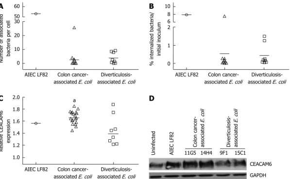
Low level of adhesion and invasion but high ability to form biofilm of B2 E. coli strains isolated from colon cancer or diverticulosis patients
The analysis of the ability of E. coli strains to adhere to and to invade intestinal epithelial cells was restricted to B2 E. coli, which were the main cyclomodulin producers in our study. Of note, due to cytolytic activity of hemo-lysin on cultured cells, hemohemo-lysin-positive E. coli strains were not tested. Results showed that B2 phylogroup E. coli strains isolated from colon cancer and from diverticu-losis displayed low levels of adhesion to Ⅰ-407 intestinal epithelial cells (Figure 1A). Compared to the adhesion level of the AIEC reference strain LF82, for which a mean adhesion index of 53.23 ± 6.63 was observed, the adhesion levels of all E. coli strains isolated from colon cancer (except E. coli strain 14H4, which had a mean adhesion level of 25.76 ± 5.06) or from diverticulosis ranged from 0.15 ± 0.02 to 4.04 ± 1.24 or from 0.10 ± 0.04 to 9.17 ± 3.40, respectively. Microscopy examination after Giemsa staining showed a diffuse adhesion pattern (data not shown), and we therefore searched for adhesive factor-encoding genes associated with diffusely adhering
Table 4 Distribution of Escherichia coli strains producing various cyclomodulins according to phylogroups and specimen origins n (%)
Phylogoups E. coli strains exhibiting cyclomodulin-encoding genes
pks cnf cdt cif
Colon cancer-associated E. coli strains (n = 88)1 A (n = 20) 0 (0) 0 (0) 0 (0) 2 (2)
B1 (n = 14) 0 (0) 1 (1) 1 (1) 1 (1) B2 (n = 38) 23 (26) 16 (18) 4 (11) 0 (0) D (n = 16) 0 (0) 1 (1) 1 (1) 0 (0) Diverticulosis-associated E. coli strains (n = 46)2 A (n = 17) 0 (0) 0 (0) 0 (0) 0 (0)
B1 (n = 3) 0 (0) 0 (0) 0 (0) 0 (0) B2 (n = 15) 6 (13) 4 (9) 0 (0) 0 (0) D (n = 11) 0 (0) 0 (0) 0 (0) 0 (0)
1Isolated from 48 colon cancer patients; 2Isolated from 33 patients with diverticulosis. E. coli: Escherichia coli.
Table 5 Phylogroup distribution of cyclomodulin-positive Escherichia coli strains according to specimen origins n (%)
Phylogroups
A B1 B2 D
Cyclomodulin-positive E. coli strains isolated from colon cancer patients 2 (6) 2 (6) 26 (84) 1 (3) Cyclomodulin-positive E. coli strains isolated from patients with diverticulosis 0 (0) 0 (0) 6 (100) 0 (0) Cyclomodulin-positive E. coli strains 2 (5) 2 (5) 32 (86) 1 (3)
E. coli (DAEC) strains (i.e., Afa and Afa/Dr adhesin-en-coding genes). None of the B2 E. coli strains tested was positive for afa or afa/dr genes except the highly adherent E. coli strain 14H4 isolated from colon cancer (Table 6). Of note, none of the B2 E. coli strains tested was positive
for eae gene coding for intimin of enteropathogenic E. coli or for aaf gene coding for the adhesive factor AAF of enteroaggregative E. coli, indicating that B2 E. coli strains studied do not belong to these E. coli pathovars. Analysis of the ability of bacteria to invade Ⅰ-407 cells showed
Table 6 Hemolysin expression and presence of cyclomodulin- and adhesin-encoding genes in B2 phylogroup Escherichia coli strains
E. coli strains Haemolytic
phenotype1 Cyclomodulin-encoding genes Adhesin-encoding genes
pks cnf cdtB cif afa dra aagR
Colon cancer 1C12 + + cnf1 - - - - 1D2 - + - - - 1F8 - - - 2D5 - - - 2F8 + + cnf1 cdtB-Ⅳ - - - 2G2 + - - - 4A9 - - - 6A8 + + cnf1 - - - - 6G8 + + cnf1 - - - - 6G10 - - - cdtB-Ⅳ - - - 6G11 - - - 7G1 - - - 7G2 + + cnf1 - - - - 8A9 + + - - - 8A10 - - - 8F1 + - cnf1 - - - - 8G8 - - - 9G5 + + cnf1 - - + - 10D12 - + - - - 10E9 + + cnf1 - - - - 11F1 - - - 11G5 - + - - - 12B1 + + cnf1 - - - - 13H2 + + cnf1 - - - - 14H4 - + - - - + + (afaE5) 15D1 + - - - 15D3 + + cnf1 - - - - 16C1 - + - - - 17G3 + - cnf1 - - - - 18C3 + + cnf1 - - - - 18C5 - - - 18H5 - + - - - 19D12 + + cnf1 - - - - 19G1 - + - - - 19H2 + + cnf1 cdtB-Ⅳ - - - 20B6 + + cnf1 - - - - 20C3 - + - cdtB-Ⅰ - - - 20D5 - - - -Diverticulosis 1D5 - - - 4D5 + + cnf1 - - - - 9D7 - - - 9F1 - - - 9F4 - - - 11D9 + + cnf1 - - - - 12H1 - + - - - 13D1 + + cnf1 - - - - 15C1 - - - 16A4 - + - - - 16A8 - - - 17C1 - - - 17F2 - - - 17E1 - - - 18E6 + + cnf1 - - - -
-1Alpha-hemolysin expression was analysed after over-night growth of bacteria on agar plates containing sheep blood. cnf: Cytotoxic necrotizing factor; cdt:
that whatever the origin of the B2 E. coli strains their in-vasion levels were very low, ranging from 0.02% to 1.49%, except strain 14H4, for which invasion level was similar to that of the AIEC strain LF82 (Figure 1B).
We also investigated the ability of B2 E. coli isolated from colon cancer to induce CEACAM6 expression as abnormal CEACAM6 expression was shown to pro-mote gut colonization by AIEC[36] and AIEC bacteria
were reported to be able to induce increased CEACAM6 expression in intestinal epithelial cells[28]. A quantitative
analysis of the level of CEACAM6 expression by T84 intestinal epithelial cells in response to B2 E. coli infection was determined by ELISA (Figure 1C) and Western blot (Figure 1D). Interestingly, we observed that most of B2 E. coli strains isolated from colon cancer induced increased expression of CEACAM6 to a level similar to that of AIEC strain LF82. Of note, B2 E. coli strains isolated from diverticulosis induced no or very low expression of CEACAM6 in T84 cells.
Another important bacterial trait involved in the colo-nization of the intestinal mucosa by gut resident bacteria is their ability to form biofilm. This property was inves-tigated both on abiotic and on fixed intestinal epithelial cells. The level of biofilm formation on abiotic surface was evaluated by calculating the specific biofilm forma-tion index (SBF). An SBF index of 3.13 ± 0.23 was ob-tained for AIEC strain LF82 compared to 0.99 ± 0.22 for the non-pathogenic K-12 E. coli strain C600 (Figure 2A). We observed that 7/19 (37%) B2 E. coli strains isolated
from colon cancer and 2/8 (25%) B2 E. coli strains isolat-ed from diverticulosis harborisolat-ed SBF index similar to that of the biofilm producer AIEC strain LF82 (P ≥ 0.05).
Biofilm formation on fixed Ⅰ-407 intestinal epithelial cells was evaluated by confocal microscopy (Figure 2B), which confirmed that 6/9 B2 E. coli strains having a high SBF index on abiotic surface were able to form a strong biofilm on fixed I-407 cultured cells. Combining the two methods of biofilm formation assessment, 16/27 B2 E. coli strains tested were able to form biofilm. This shows that even B2 E. coli strains have a low ability to adhere to intestinal epithelial cells, at least half of them were able to form biofilm to a level similar to that of CD-associated E. coli strain LF82 known to form a strong biofilm and no difference was observed between B2 E. coli strains isolated from colon cancer patients or from patients with diverticulosis.
Colonization of colon mucosa in CEACAM-expressing mice by colon cancer-associated B2 E. coli strain 11G5: induction of inflammation and enhanced epithelial intestinal cell proliferation
CEABAC10 mice harboring a bacterial artificial chromo-some that contains part of the human CEA family gene cluster including the CEACAM6 gene were infected with AIEC reference strain LF82 or B2 E. coli strain 11G5 iso-lated from colon cancer patient. To assess bacterial colo-nization, the levels of bacteria in the stools were deter-mined 5 d after the last infection of each cycle, over the
AIEC LF82 Colon cancer-associated E. coli
Diverticulosis-associated E. coli
Number of associated
bacteria per cel
l 60 50 30 20 10 0
AIEC LF82 Colon cancer-associated E. coli Diverticulosis-associated E. coli % internal iz ed bacteria/ ini tial inoculum 10 8 6 2 1 0
AIEC LF82 Colon cancer-associated E. coli Diverticulosis-associated E. coli Relativ e CEACAM6 expr ession 2.0 1.8 1.6 1.4 1.2 1.0 Uninf ected AIEC LF82 Colon cancer -associated E. coli Div erticulosis-associated E. coli 11G5 14H4 9F1 15C1 CEACAM6 GAPDH A B C D
Figure 1 Adhesion, invasion and ability to induce carcinoembryonic antigen-related cell adhesion molecule 6 expression of B2 Escherichia coli strains. A
and B: Ability of colon cancer- and diverticulosis-associated B2 Escherichia coli (E. coli) strains and AIEC strain LF82 to adhere to and to invade I-407 intestinal epi-thelial cells. A: Adhesion. Results are expressed as number of associated bacteria per cell after 3 h of infection; B: Invasion. Results are expressed as percentage of inoculum surviving after 3 h of infection and 1 h of gentamicin treatment; C and D: Induction of carcinoembryonic antigen-related cell adhesion molecule 6 (CEACAM6) expression in colon epithelial T84 cells infected for 6h with colon cancer- and diverticulosis-associated B2 E. coli strains and AIEC strain LF82. C: Quantitative dosage of CEACAM6 by ELISA. Results are expressed as amounts of CEACAM6 in stimulated or infected cells relative to untreated cells. aP ≤ 0.05 vs diverticulosis associ-ated E. coli. D: CEACAM6 expression analysis by Western Blot. E. coli strains 11G5 and 14H4 were isolassoci-ated from colon cancer patients and E. coli strains 9F1 and 15C1 from patients with diverticulosis.
8 consecutive cycles of infection. Analysis of the number of bacteria recovered in the stools at cycle 4 and cycle 8 revealed similar (P = 0.78) colonization levels for mice infected with AIEC LF82 and colon cancer-associated 11G5 bacteria (Figure 3A).
On macroscopic examination, no sign of tumor de-velopment such as neoplasia or polyp was observed in the colon of mice infected with AIEC strain LF82 or colon cancer-associated 11G5 E. coli strain. Histological analysis showed a similar colonic histological score for inflammation and epithelial damages for mice infected with AIEC strain LF82 and E. coli strain 11G5 (P ≥ 0.05)
(Figure 3B). Mice infected with E. coli strains LF82 and 11G5 exhibited infiltration of polynuclear cells in crypts, larger numbers of crypt abscesses and large and multifo-cal erosion plates (Figure 3C).
The level of proliferating cell nuclear antigen (PCNA) mRNA was measured in the colonic mucosa of infected mice to determine the proliferative index (Figure 3D). Significant (P ≤ 0.05) 2.5-fold and 2.9-fold increases in
PCNA mRNA levels were observed in the colonic muco-sa of mice infected with the E. coli strain 11G5 compared to those of control mice or mice infected with AIEC strain LF82, respectively. This finding was confirmed by Ki67 immunostaining on colonic mucosa tissue. 11G5-infected mice had higher numbers of proliferative epi-thelial cells in crypts than control mice and mice infected with AIEC strain LF82 (Figure 3E). This indicates that colonic mucosa cells undergo accelerated proliferation in response to infection by B2 E. coli strain 11G5 associated with colon cancer.
DISCUSSION
Accumulating evidence supports the involvement of in-fectious agents in the development of cancer, especially in organs that are continuously exposed to microor-ganisms such as the colon. Remodeling of the colonic microbiota due to environmental changes is thought to contribute to the pathogenesis of colon cancer by sup-pressing the growth of cancer-protective bacterial species and allowing the emergence or expansion of bacterial species with oncogenic potential. It has been suggested that the role of E. coli in CRC promotion and develop-ment is linked to chronic inflammation, which can result from bacterial infection via its effects on both the host and the microbiota, in particular that of promoting the expansion of certain bacteria, such as pro-inflammatory E. coli[37] or ETBF[38,39]. In parallel, two different studies
have reported that between 71% and 82% of patients with colonic adenoma or carcinoma[10,12] are highly
colo-nized by mucosa-associated E. coli compared to controls. The aim of the present study was to provide further insight into the characterization of the E. coli colonizing the mucosa of colon cancer patients.
It is well documented that B2 E. coli harbors genes coding for cyclomodulins such as colibactin, which is encoded by the pks genomic island, CDT, CNF or Cif, which can act as genotoxic agents and/or can modulate cellular differentiation, apoptosis, and proliferation[13,40,41].
In the present study, we observed that 86% of cyclo-modulin-positive E. coli isolated from colon cancer and diverticulosis specimens belonged to B2 phylogroup. Of interest, all but two cnf positive strains also harbored pks and all cnf- and pks-positive E. coli strains belonged to the B2 phylogroup. Our results are in good agreement with those reported by Arthur et al[3], who observed that
66.7% of patients with CRC and 20.8% of controls har-bored pks-positive E. coli.
E. coli strains belonging to B2 phylogroup have a
K-12 C600 LF82
11G5 12H1
Figure 2 Ability of B2 Escherichia coli strains to form biofilm. Biofilm
for-mation of colon cancer-associated and diverticulosis-associated B2 Escherichia coli (E. coli) were compared to that of the non-pathogenic K-12 E. coli strain C600 and the biofilm producer AIEC strain LF82. A: Biofilm formation on abiotic surface. Results are expressed as specific biofilm formation (SBF) index; B: Biofilm formation on human I-407 intestinal epithelial cells. E. coli strain 11G5 was isolated from a patient with colon cancer and E. coli strain 12H1 from a pa-tient with diverticulosis. Bacteria were stained using goat anti-E. coli polyclonal antibodies (green) and I-407 cells were labeled for actin cytoskeleton using TRITC-labeled phalloidin (red). Y- and Z-stack projections are presented.
K-12 C600 AIEC LF82 Colon
cancer-E. coli SBF index 6 4 2 0 Diverticulosis-E. coli A B
greater ability to colonize the human gut, due, at least in part, to accumulation of genes encoding fitness factors such as pili and adhesins[42,43]. In addition, an increased
proportion of mucosa-associated E. coli expressing hem-agglutinins was observed in CD patients (39%) and colon cancer patients (38%) compared to controls (4%), in cor-relation with the ability of bacteria to adhere to Ⅰ-407 and HT-29 intestinal epithelial cells[10]. However in our
study, analysis of the adhesive abilities of B2 E. coli iso-lated from colon cancer or diverticulosis revealed that the strains were poorly adherent to I-407, even if the major-ity of them were able to form biofilm. However some B2 E. coli strains isolated from colon cancer induced increased expression of CEACAM6 to a level similar to that of AIEC strain LF82 associated with CD, indicating that colon cancer-associated E. coli could influence car-cinogenesis, since CEACAM6 has been implicated in
cel-lular adhesiveness, invasiveness, and metastatic behavior of tumor cells[30,44]. In addition, this result indicates that,
in agreement with what we previously reported for AIEC strains isolated from CD patients[28], colon
cancer-associ-ated E. coli strains could have the ability to promote their own colonisation since CEACAM6 serves as a receptor for mediating adherence and/or cell entry of pathogenic
bacteria such as Neisseria bacteria[45], diffusely-adhering E.
coli (DAEC)[46] or AIEC[28].
Experiments of long-term colonization of CEA-BAC10 mice revealed that an E. coli strain isolated from colon cancer (strain 11G5) was able to persist in the gut of CEABAC10 transgenic mice expressing human CEACAMs, including CEACAM6 and to exacerbate co-lonic inflammation. Whether colonisation of the intestinal mucosa of colon cancer patients by B2 E. coli is a cause or a consequence of malignant transformation is a
ques-LF82 11G5 LF82 E. coli CFU/g of f eces 108 106 104 102 100 11G5 Cycle 4 Cycle 8 Control LF82 11G5 Histological scor e 10 8 6 4 2 a Control LF82 11G5 PCNA mRNA lev el 4 3 2 1 0 a c Control LF82 11G5 Control LF82 11G5 A B C D E
Figure 3 Impact of CEABAC10 mice colonization by B2 Escherichia coli strain on inflammation and cell proliferation. CEABAC10 mice transgenic for human
CEACAMs, including CEACAM6, were subjected to 8 consecutive cycles of infection with AIEC LF82 or B2 phylogroup Escherichia coli (E. coli) strain 11G5. Control mice received PBS. A: Quantification of the number of bacteria in the feces of mice at cycle 4 and cycle 8; B: Histopathological scoring for several parameters of in-flammation and epithelial damages (see Table 2) was performed at the end of the 8th cycle. aP ≤ 0.05 vs control; P = NS vs LF82; C: Hematoxylin/eosin/safran (HES) staining of colonic tissue sections; D: Total RNAs from colons were extracted at the end of the 8th cycle. PCNA and S26 mRNA levels were measured by RT-qPCR.
PCNA amount relative to S26 is presented. aP ≤ 0.05 vs control; cP ≤ 0.05 vs LF82; E: Immunohistochemistry examination of Ki67 on colonic tissue sections. NS:
Not significant.
40 µm 40 µm 40 µm
250 µm 250 µm
COMMENTS
Background
Colorectal cancer (CRC) is one of the most prevalent cancers worldwide, and is the fourth leading cause of cancer death worldwide. Inflammation and changes in composition and function of gut microbial communities are suspected to be causative factors in the development of sporadic CRC.
Research frontiers
The authors and other researchers have reported abnormal colonization of tu-mors and mucosa of colon cancer patients by Escherichia coli (E. coli) belong-ing to B2 phylogroup.
Innovations and breakthroughs
To date, there has been a limited number of studies analyzing interaction of colon cancer-associated E. coli to intestinal epithelial cells. The authors showed that colon cancer-associated E. coli induce expression of the carcinoembryonic antigen-related cell adhesion molecule 6 (CEACAM6) receptor in intestinal epi-thelial cells, and that these bacteria were able to persist in a chronic infection model of CEACAM6 expressing mice and had oncogenic potential.
Applications
The authors have analyzed the ability of colon cancer-associated E. coli to colo-nize gut mucosa and influence carcinogenesis. Analyses to determine whether these bacteria take advantage of the tumor microenvironment to colonize the gut or promote their own colonization may be an important step in understanding their role in carcinogenesis and in the development of therapeutic strategies.
Terminology
CEACAM6 molecule serves as a receptor for mediating mucosa colonization by pathogenic bacteria.
Peer review
This study provides evidence supporting the hypothesis that colon cancer-asso-ciated E. coli are able to colonize gut mucosa and to induce cell proliferation in a mouse model with overexpression of human CEACAM6.
REFERENCES
1 World Health Organization World. WHO Global Malaria
Programme. Switzerland: World Health Organization, 2011. Available from: URL: http://www.who.int/malaria/ world_malaria_report_2011/9789241564403_eng.pdf 2 Vogelstein B, Fearon ER, Hamilton SR, Kern SE, Preisinger
AC, Leppert M, Nakamura Y, White R, Smits AM, Bos JL. Genetic alterations during colorectal-tumor development. N Engl J Med 1988; 319: 525-532 [PMID: 2841597 DOI: 10.1056/ NEJM198809013190901]
3 Arthur JC, Perez-Chanona E, Mühlbauer M, Tomkovich
S, Uronis JM, Fan TJ, Campbell BJ, Abujamel T, Dogan B, Rogers AB, Rhodes JM, Stintzi A, Simpson KW, Hansen JJ, Keku TO, Fodor AA, Jobin C. Intestinal inflammation targets cancer-inducing activity of the microbiota. Science 2012; 338: 120-123 [PMID: 22903521 DOI: 10.1126/science.1224820] 4 Mantovani A, Allavena P, Sica A, Balkwill F. Cancer-related
inflammation. Nature 2008; 454: 436-444 [PMID: 18650914 DOI: 10.1038/nature07205]
5 Uronis JM, Jobin C. Microbes and colorectal cancer: is there
a relationship? Curr Oncol 2009; 16: 22-24 [PMID: 19672421] 6 Sears CL, Pardoll DM. Perspective: alpha-bugs, their
micro-bial partners, and the link to colon cancer. J Infect Dis 2011;
203: 306-311 [PMID: 21208921 DOI: 10.1093/jinfdis/jiq061]
7 Tjalsma H, Boleij A, Marchesi JR, Dutilh BE. A bacterial
driver-passenger model for colorectal cancer: beyond the usual suspects. Nat Rev Microbiol 2012; 10: 575-582 [PMID: 22728587 DOI: 10.1038/nrmicro2819]
8 Sobhani I, Tap J, Roudot-Thoraval F, Roperch JP, Letulle
S, Langella P, Corthier G, Tran Van Nhieu J, Furet JP. Mi-crobial dysbiosis in colorectal cancer (CRC) patients. PLoS One 2011; 6: e16393 [PMID: 21297998 DOI: 10.1371/journal. pone.0016393]
9 Kostic AD, Gevers D, Pedamallu CS, Michaud M, Duke F,
tion that has yet to be addressed. We show here that B2 phylogroup E. coli isolated from colon cancer increased the proliferative index of epithelial cells in crypts in the chronic infection model of CEABAC10 mice. This indi-cates that colonic mucosa cells undergo accelerated pro-liferation in response to infection by B2 E. coli. The ability to induce cell proliferation is a common trait of various pathogens involved in carcinogenesis. Indeed, Bacteroides fragilis enterotoxin induces c-myc transcription and transla-tion and persistent cellular proliferatransla-tion ensues, mediated in part by β-catenin/T-cell factor-dependent transcrip-tional activation[47]. Another example is Helicobacter pylori (H.
pylori), which increases the proliferation of gastric cancer cells. This process is dependent on the LPS-TLR4 path-way since H. pylori LPS induces the proliferation of gastric cancer cells and the use of neutralizing antibody against TLR4 almost completely abrogates the proliferative activi-ties of cancer cells[48]. Some cyclomodulins, such as CNF,
which are mostly produced by B2 E. coli, induce epithelial cell proliferation[40]. In our study the B2 E. coli strain 11G5
did not harbor the cnf genes and was able to promote cell proliferation as observed in infected CEABAC10 mice. This effect could be related to E. coli-derived LPS, which was previously reported to have a more remarkable cancer proliferative activity than H. pylori-derived LPS[48]. Because
E. coli inhabits the host colon as normal intestinal flora, owing to host tolerance toward E. coli, it is likely that E. coli LPS stimulates the host cellular immune response to prevent cancer progression. However, we can hypothesize that when too great a load of E. coli colonize the colonic mucosa, as observed in 11G5-infected CEABAC10 mice, potent tumor proliferative activity is no longer effectively repressed. The cell proliferation observed in 11G5-in-fected CEABAC10 mice could also result from the pres-ence of colibactin. Colibactin with its genotoxic activity promotes DNA damages, which leads to carcinogenesis and cell proliferation.
In conclusion, B2 E. coli abnormally colonized the mu-cosa of colon cancer patients, indicating that microbiota remodeling had occurred promoting their expansion. Together with previous findings reported by Arthur et al[3],
this study on a larger cohort of patients confirms the high prevalence of B2 pks-positive or pks-cnf-positive E. coli in colon cancer patients. The study also indicates that, these bacteria can promote low grade inflammation and cell proliferation, as shown in the CEABAC10 infected mouse model. Analyses to determine whether these bacteria take advantage of the tumor microenvironment to colonize the gut or promote their own colonization may be an im-portant step in understanding their role in carcinogenesis and in the development of therapeutic strategies.
ACKNOWLEDGMENTS
We thank Emmanuel Bourgeois for his help in immuno-histochemical staining and Cécile Charpy for her help in their interpretation. We thank ICCF platforms from Uni-versité d’Auvergne for confocal microscopy.
Earl AM, Ojesina AI, Jung J, Bass AJ, Tabernero J, Baselga J, Liu C, Shivdasani RA, Ogino S, Birren BW, Huttenhower C, Garrett WS, Meyerson M. Genomic analysis identifies association of Fusobacterium with colorectal carcinoma. Genome Res 2012; 22: 292-298 [PMID: 22009990 DOI: 10.1101/ gr.126573.111]
10 Martin HM, Campbell BJ, Hart CA, Mpofu C, Nayar M, Singh R, Englyst H, Williams HF, Rhodes JM. Enhanced Escherichia coli adherence and invasion in Crohn’s disease and colon cancer. Gastroenterology 2004; 127: 80-93 [PMID: 15236175]
11 Maddocks OD, Short AJ, Donnenberg MS, Bader S, Har-rison DJ. Attaching and effacing Escherichia coli down-regulate DNA mismatch repair protein in vitro and are as-sociated with colorectal adenocarcinomas in humans. PLoS One 2009; 4: e5517 [PMID: 19436735 DOI: 10.1371/journal. pone.0005517]
12 Swidsinski A, Khilkin M, Kerjaschki D, Schreiber S, Ortner M, Weber J, Lochs H. Association between intraepithelial Escherichia coli and colorectal cancer. Gastroenterology 1998;
115: 281-286 [PMID: 9679033]
13 Nougayrède JP, Homburg S, Taieb F, Boury M, Brzusz-kiewicz E, Gottschalk G, Buchrieser C, Hacker J, Dobrindt U, Oswald E. Escherichia coli induces DNA double-strand breaks in eukaryotic cells. Science 2006; 313: 848-851 [PMID: 16902142 DOI: 10.1126/science.1127059]
14 Wu S, Rhee KJ, Albesiano E, Rabizadeh S, Wu X, Yen HR, Huso DL, Brancati FL, Wick E, McAllister F, Housseau F, Pardoll DM, Sears CL. A human colonic commensal pro-motes colon tumorigenesis via activation of T helper type 17 T cell responses. Nat Med 2009; 15: 1016-1022 [PMID: 19701202 DOI: 10.1038/nm.2015]
15 Goodwin AC, Destefano Shields CE, Wu S, Huso DL, Wu X, Murray-Stewart TR, Hacker-Prietz A, Rabizadeh S, Woster PM, Sears CL, Casero RA. Polyamine catabolism contributes to enterotoxigenic Bacteroides fragilis-induced colon tumori-genesis. Proc Natl Acad Sci USA 2011; 108: 15354-15359 [PMID: 21876161 DOI: 10.1073/pnas.1010203108]
16 Bernstein CN, Blanchard JF, Kliewer E, Wajda A. Cancer risk in patients with inflammatory bowel disease: a popula-tion-based study. Cancer 2001; 91: 854-862 [PMID: 11241255] 17 Ullman TA, Itzkowitz SH. Intestinal inflammation and
can-cer. Gastroenterology 2011; 140: 1807-1816 [PMID: 21530747 DOI: 10.1053/j.gastro.2011.01.057]
18 Martinez-Medina M, Aldeguer X, Gonzalez-Huix F, Acero D, Garcia-Gil LJ. Abnormal microbiota composition in the ileocolonic mucosa of Crohn’s disease patients as revealed by polymerase chain reaction-denaturing gradient gel elec-trophoresis. Inflamm Bowel Dis 2006; 12: 1136-1145 [PMID: 17119388 DOI: 10.1097/01.mib.0000235828.09305.0c] 19 Frank DN, St Amand AL, Feldman RA, Boedeker EC,
Har-paz N, Pace NR. Molecular-phylogenetic characterization of microbial community imbalances in human inflammatory bowel diseases. Proc Natl Acad Sci USA 2007; 104: 13780-13785 [PMID: 17699621 DOI: 10.1073/pnas.0706625104]
20 Sokol H, Pigneur B, Watterlot L, Lakhdari O, Bermúdez-Humarán LG, Gratadoux JJ, Blugeon S, Bridonneau C, Furet JP, Corthier G, Grangette C, Vasquez N, Pochart P, Trugnan G, Thomas G, Blottière HM, Doré J, Marteau P, Seksik P, Langella P. Faecalibacterium prausnitzii is an anti-inflam-matory commensal bacterium identified by gut microbiota analysis of Crohn disease patients. Proc Natl Acad Sci USA 2008; 105: 16731-16736 [PMID: 18936492 DOI: 10.1073/ pnas.0804812105]
21 Swidsinski A, Ladhoff A, Pernthaler A, Swidsinski S, Loe-ning-Baucke V, Ortner M, Weber J, Hoffmann U, Schreiber S, Dietel M, Lochs H. Mucosal flora in inflammatory bowel disease. Gastroenterology 2002; 122: 44-54 [PMID: 11781279] 22 Mylonaki M, Rayment NB, Rampton DS, Hudspith BN,
Brostoff J. Molecular characterization of rectal
mucosa-asso-ciated bacterial flora in inflammatory bowel disease. Inflamm Bowel Dis 2005; 11: 481-487 [PMID: 15867588]
23 Baumgart M, Dogan B, Rishniw M, Weitzman G, Bosworth B, Yantiss R, Orsi RH, Wiedmann M, McDonough P, Kim SG, Berg D, Schukken Y, Scherl E, Simpson KW. Culture inde-pendent analysis of ileal mucosa reveals a selective increase in invasive Escherichia coli of novel phylogeny relative to depletion of Clostridiales in Crohn’s disease involving the il-eum. ISME J 2007; 1: 403-418 [PMID: 18043660 DOI: 10.1038/ ismej.2007.52]
24 Kotlowski R, Bernstein CN, Sepehri S, Krause DO. High prevalence of Escherichia coli belonging to the B2+D phylo-genetic group in inflammatory bowel disease. Gut 2007; 56: 669-675 [PMID: 17028128 DOI: 10.1136/gut.2006.099796] 25 Darfeuille-Michaud A, Neut C, Barnich N, Lederman E,
Di Martino P, Desreumaux P, Gambiez L, Joly B, Cortot A, Colombel JF. Presence of adherent Escherichia coli strains in ileal mucosa of patients with Crohn’s disease. Gastroenterol-ogy 1998; 115: 1405-1413 [PMID: 9834268]
26 Boudeau J, Glasser AL, Masseret E, Joly B, Darfeuille-Michaud A. Invasive ability of an Escherichia coli strain iso-lated from the ileal mucosa of a patient with Crohn’s disease. Infect Immun 1999; 67: 4499-4509 [PMID: 10456892]
27 Darfeuille-Michaud A, Boudeau J, Bulois P, Neut C, Glasser AL, Barnich N, Bringer MA, Swidsinski A, Beaugerie L, Co-lombel JF. High prevalence of adherent-invasive Escherichia coli associated with ileal mucosa in Crohn’s disease. Gastro-enterology 2004; 127: 412-421 [PMID: 15300573]
28 Barnich N, Carvalho FA, Glasser AL, Darcha C, Jantscheff P, Allez M, Peeters H, Bommelaer G, Desreumaux P, Colombel JF, Darfeuille-Michaud A. CEACAM6 acts as a receptor for adherent-invasive E. coli, supporting ileal mucosa coloni-zation in Crohn disease. J Clin Invest 2007; 117: 1566-1574 [PMID: 17525800 DOI: 10.1172/JCI30504]
29 Schölzel S, Zimmermann W, Schwarzkopf G, Grunert F, Ro-gaczewski B, Thompson J. Carcinoembryonic antigen fam-ily members CEACAM6 and CEACAM7 are differentially expressed in normal tissues and oppositely deregulated in hyperplastic colorectal polyps and early adenomas. Am J Pathol 2000; 156: 595-605 [PMID: 10666389 DOI: 10.1016/ S0002-9440(10)64764-5]
30 Kim KS, Kim JT, Lee SJ, Kang MA, Choe IS, Kang YH, Kim SY, Yeom YI, Lee YH, Kim JH, Kim KH, Kim CN, Kim JW, Nam MS, Lee HG. Overexpression and clinical significance of carcinoembryonic antigen-related cell adhesion molecule 6 in colorectal cancer. Clin Chim Acta 2013; 415: 12-19 [PMID: 22975528 DOI: 10.1016/j.cca.2012.09.003]
31 Versalovic J, Koeuth T, Lupski JR. Distribution of repetitive DNA sequences in eubacteria and application to finger-printing of bacterial genomes. Nucleic Acids Res 1991; 19: 6823-6831 [PMID: 1762913]
32 Wang G, Whittam TS, Berg CM, Berg DE. RAPD (arbitrary primer) PCR is more sensitive than multilocus enzyme electrophoresis for distinguishing related bacterial strains. Nucleic Acids Res 1993; 21: 5930-5933 [PMID: 8290354] 33 Bidet P, Metais A, Mahjoub-Messai F, Durand L, Dehem
M, Aujard Y, Bingen E, Nassif X, Bonacorsi S. Detection and identification by PCR of a highly virulent phylogenetic subgroup among extraintestinal pathogenic Escherichia coli B2 strains. Appl Environ Microbiol 2007; 73: 2373-2377 [PMID: 17293507 DOI: 10.1128/AEM.02341-06]
34 Chassaing B, Darfeuille-Michaud A. The σE pathway is in-volved in biofilm formation by Crohn’s disease-associated adherent-invasive Escherichia coli. J Bacteriol 2013; 195: 76-84 [PMID: 23104802 DOI: 10.1128/JB.01079-12]
35 Chan CH, Stanners CP. Novel mouse model for carcinoem-bryonic antigen-based therapy. Mol Ther 2004; 9: 775-785 [PMID: 15194045 DOI: 10.1016/j.ymthe.2004.03.009] 36 Carvalho FA, Barnich N, Sivignon A, Darcha C, Chan CH,
ad-herent-invasive Escherichia coli colonize and induce strong gut inflammation in transgenic mice expressing human CEACAM. J Exp Med 2009; 206: 2179-2189 [PMID: 19737864 DOI: 10.1084/jem.20090741]
37 Mukhopadhya I, Hansen R, El-Omar EM, Hold GL. IBD-what role do Proteobacteria play? Nat Rev Gastroenterol Hepa-tol 2012; 9: 219-230 [PMID: 22349170 DOI: 10.1038/nrgas-tro.2012.14]
38 Sears CL. Enterotoxigenic Bacteroides fragilis: a rogue among symbiotes. Clin Microbiol Rev 2009; 22: 349-369, Table of Contents [PMID: 19366918 DOI: 10.1128/CMR.00053-08] 39 Toprak NU, Yagci A, Gulluoglu BM, Akin ML, Demirkalem
P, Celenk T, Soyletir G. A possible role of Bacteroides fragilis enterotoxin in the aetiology of colorectal cancer. Clin Micro-biol Infect 2006; 12: 782-786 [PMID: 16842574 DOI: 10.1111/ j.1469-0691.2006.01494.x]
40 Nougayrède JP, Taieb F, De Rycke J, Oswald E. Cyclomodu-lins: bacterial effectors that modulate the eukaryotic cell cycle. Trends Microbiol 2005; 13: 103-110 [PMID: 15737728 DOI: 10.1016/j.tim.2005.01.002]
41 Collins D, Hogan AM, Winter DC. Microbial and viral pathogens in colorectal cancer. Lancet Oncol 2011; 12: 504-512 [PMID: 21067973 DOI: 10.1016/S1470-2045(10)70186-8] 42 Picard B, Garcia JS, Gouriou S, Duriez P, Brahimi N, Bingen
E, Elion J, Denamur E. The link between phylogeny and virulence in Escherichia coli extraintestinal infection. Infect Immun 1999; 67: 546-553 [PMID: 9916057]
43 Nowrouzian FL, Adlerberth I, Wold AE. Enhanced persis-tence in the colonic microbiota of Escherichia coli strains belonging to phylogenetic group B2: role of virulence factors and adherence to colonic cells. Microbes Infect 2006; 8: 834-840 [PMID: 16483819 DOI: 10.1016/j.micinf.2005.10.011] 44 Ilantzis C, DeMarte L, Screaton RA, Stanners CP.
Deregu-lated expression of the human tumor marker CEA and CEA family member CEACAM6 disrupts tissue architecture and blocks colonocyte differentiation. Neoplasia 2002; 4: 151-163 [PMID: 11896570 DOI: 10.1038/sj/neo/7900201]
45 Bos MP, Grunert F, Belland RJ. Differential recognition of
members of the carcinoembryonic antigen family by Opa variants of Neisseria gonorrhoeae. Infect Immun 1997; 65: 2353-2361 [PMID: 9169774]
46 Guignot J, Hudault S, Kansau I, Chau I, Servin AL. Human decay-accelerating factor and CEACAM receptor-mediated internalization and intracellular lifestyle of Afa/Dr dif-fusely adhering Escherichia coli in epithelial cells. Infect Immun 2009; 77: 517-531 [PMID: 19015254 DOI: 10.1128/ IAI.00695-08]
47 Wu S, Morin PJ, Maouyo D, Sears CL. Bacteroides fragilis enterotoxin induces c-Myc expression and cellular prolifera-tion. Gastroenterology 2003; 124: 392-400 [PMID: 12557145 DOI: 10.1053/gast.2003.50047]
48 Chochi K, Ichikura T, Kinoshita M, Majima T, Shinomiya N, Tsujimoto H, Kawabata T, Sugasawa H, Ono S, Seki S, Mo-chizuki H. Helicobacter pylori augments growth of gastric cancers via the lipopolysaccharide-toll-like receptor 4 path-way whereas its lipopolysaccharide attenuates antitumor activities of human mononuclear cells. Clin Cancer Res 2008;
14: 2909-2917 [PMID: 18483357 DOI: 10.1158/1078-0432.
CCR-07-4467]
49 Le Bouguénec C, Lalioui L, du Merle L, Jouve M, Courcoux P, Bouzari S, Selvarangan R, Nowicki BJ, Germani Y, An-dremont A, Gounon P, Garcia MI. Characterization of AfaE adhesins produced by extraintestinal and intestinal human Escherichia coli isolates: PCR assays for detection of Afa ad-hesins that do or do not recognize Dr blood group antigens. J Clin Microbiol 2001; 39: 1738-1745 [PMID: 11325983 DOI: 10.1128/JCM.39.5.1738-1745.2001]
50 Bernier C, Gounon P, Le Bouguénec C. Identification of an aggregative adhesion fimbria (AAF) type III-encoding oper-on in enteroaggregative Escherichia coli as a sensitive probe for detecting the AAF-encoding operon family. Infect Immun 2002; 70: 4302-4311 [PMID: 12117939]
51 Dubois D, Delmas J, Cady A, Robin F, Sivignon A, Oswald E, Bonnet R. Cyclomodulins in urosepsis strains of Escherichia coli. J Clin Microbiol 2010; 48: 2122-2129 [PMID: 20375237 DOI: 10.1128/JCM.02365-09]
P- Reviewers: Bommireddy R, De Nardi P, Kurniali PC, Lee KY S- Editor: Ma YJ L- Editor: A E- Editor: Wang CH
8226 Regency Drive, Pleasanton, CA 94588, USA
Telephone: +1-925-223-8242
Fax: +1-925-223-8243
E-mail: bpgoffice@wjgnet.com
Help Desk: http://www.wjgnet.com/esps/helpdesk.aspx
http://www.wjgnet.com
I S S N 1 0 0 7 - 9 3 2 7
9 7 7 1 0 07 9 3 2 0 45 2 1