HAL Id: tel-01968072
https://tel.archives-ouvertes.fr/tel-01968072
Submitted on 2 Jan 2019
HAL is a multi-disciplinary open access archive for the deposit and dissemination of sci-entific research documents, whether they are pub-lished or not. The documents may come from teaching and research institutions in France or abroad, or from public or private research centers.
L’archive ouverte pluridisciplinaire HAL, est destinée au dépôt et à la diffusion de documents scientifiques de niveau recherche, publiés ou non, émanant des établissements d’enseignement et de recherche français ou étrangers, des laboratoires publics ou privés.
The role of auxin in cell differentiation in meristems
Jekaterina Truskina
To cite this version:
Jekaterina Truskina. The role of auxin in cell differentiation in meristems. Plants genetics. Université de Lyon, 2018. English. �NNT : 2018LYSEN033�. �tel-01968072�
Numéro National de Thèse : 2018LYSEN033
THESE de DOCTORAT DE L’UNIVERSITE DE LYON
opérée par
l’Ecole Normale Supérieure de Lyon
en cotutelle avec
University of Nottingham
Ecole Doctorale N°340
Biologie Moléculaire, Intégrative et Cellulaire
Spécialité de doctorat : Biologie des plantes Discipline : Sciences de la Vie
Soutenue publiquement le 28.09.2018, par :
Jekaterina TRUSKINA
The role of auxin in cell differentiation in meristems
Rôle de l'auxine dans la différenciation des cellules au sein des méristèmes
Devant le jury composé de :
Mme Catherine BELLINI, Professeure, Umeå Plant Science Center Rapporteure Mme Zoe WILSON, Professeure, University of Nottingham Rapporteure M. Joop EM VERMEER, Professeur, University of Zurich Examinateur M. Renaud DUMAS, Directeur de recherche, Université Grenoble Alpes Examinateur M. Teva VERNOUX, Directeur de recherche, ENS de Lyon Directeur de thèse M. Anthony BISHOPP, Chercheur, University of Nottingham Co-tuteur de thèse
Thesis
submitted to the
University of Nottingham
for joint examination with the
ENS de Lyon
for the degree of
Doctor of Philosophy
by
Jekaterina Truskina
The role of auxin in cell differentiation in meristems
Rôle de l'auxine dans la différenciation des cellules au sein des méristèmes
Date of submission :
11.06.2018
Date of public defence : 28.09.2018
The thesis jury is composed of :
Mme Catherine Bellini, Professor, Umeå Plant Science Center Reporter Mme Zoe Wilson, Professor, University of Nottingham Reporter M. Joop EM Vermeer, Professor, University of Zurich Examiner M. Renaud Dumas, Research Director, Université Grenoble Alpes Examiner M. Teva Vernoux, Research Director, ENS de Lyon Thesis director M. Anthony Bishopp, University Research Fellow, University of Nottingham Thesis co-director
~ 2 ~
Abstract
Auxin regulates plant growth and development through the transcription factors of the AUXIN RESPONSE FACTOR (ARF) gene family. Most notably in Arabidopsis
thaliana ARF5, 6, 7, 8 and 19 activate expression of target genes in response to
auxin. These five ARF activators control both variable and overlapping processes during plant development including regulation of growth at the root and the shoot apical meristems, lateral root and axillary shoot formation.
Each of the five ARF activators shows unique tissue-specific expression patterns in the root and the shoot associated with their distinct functions. This tissue-specific expression is likely derived from the differences in the control of ARF activator transcription. In this study the upstream regulators of ARF5, 6, 7, 8 and 19 transcription were identified. This was achieved by utilizing a high-throughput yeast one-hybrid (Y1H) method. The transient protoplast assay revealed that each ARF activator is controlled by specific transcriptional regulators and that the majority of these regulators are repressors of ARF transcription in planta. Mutants of the regulatory transcription factors were utilized to additionally investigate the interactions
in planta. These mutants display auxin-related developmental phenotypes in the root
and the shoot including alternations in growth kinetics, emergence of lateral organs, responses to auxin and altered expression of ARF activators.
Furthermore, this study additionally focuses on cross-talk between the auxin and cytokinin signaling pathways and its role in root and shoot development. One of the interactions identified in the Y1H screen is a repression of ARF7 by CRF10, a member of the Cytokinin Response Factors gene family. The importance of this interaction in maintaining architecture of the root apical meristem, in leaf senescence and in the phototropic response to blue light in hypocotyls is studied.
~ 3 ~
Résumé
L'auxine régule la croissance et le développement des plantes grâce aux facteurs de transcription de la famille des "AUXIN RESPONSE FACTOR" (ARF). Chez
Arabidopsis thaliana en particulier, ARF5, 6, 7, 8 et 19 activent l'expression de gènes
cibles en réponse à l'auxine. Ces cinq ARF activateurs contrôlent de façon plus ou moins redondante des processus divers au cours du développement de la plante, notamment la régulation des croissances au niveau des méristèmes racinaires et aériens ainsi que la formation des racines latérales ou des méristèmes axillaires. Chacun de ces cinq ARF activateurs présente des patrons d'expression uniques dans chacun des tissus racinaires et aériens, en association avec leurs fonctions particulières. Il est probable que cette expression tissu-spécifique trouve son origine dans un contrôle différencié de leur transcription. Dans cette étude, des régulateurs amonts de la transcription de ARF5, 6, 7, 8 et 19 ont été identifiés par une méthode haut-débit de crible simple hybride en levure (Y1H). Une procédure d'expression transitoire en protoplastes a permis de confirmer que l'expression de chaque ARF activateur est contrôlée par des régulateurs spécifiques, dont la majorité se comportent comme des répresseurs de la transcription des ARF in planta. Parmi les régulateurs identifiés, les facteurs de transcription ont été étudiés grâce à des mutants pour préciser les interactions in planta. Ces mutants montrent des phénotypes développementaux typiques de perturbations de l'auxine dans les racines et les tiges : altérations des cinétiques de croissance, de l'émergence des organes latéraux ou de réponses à l'auxine et modification de l'expression des ARF activateurs.
Par ailleurs, ce travail aborde également les dialogues entre les voies de signalisation de l'auxine et des cytokinines, et en particulier le rôle de ces interactions dans le développement des racines et des tiges. Une des interactions identifiées dans le crible Y1H est la répression de ARF7 par CRF10, un gène membre de la famille des "Cytokinin Response Factors". Nous avons mis en évidence l'importance de cette interaction pour le maintien de l'architecture du méristème apical racinaire, pour la sénescence des feuille et pour la réponse phototropique à la lumière bleue dans les hypocotyles.
~ 4 ~
Acknowledgments
Teva and Tony, my most incredible supervisors, I own great gratitude to you both. You changed my life when you offered me this PhD position and since then, I spend awesome four years learning from you how to do science. When it comes to science your advices were always full of wisdom and experience guiding me through my thesis. I had amazing support from you both in my work but also in my life. I felt it from the start when you took this incredible effort to go through a huge pile of paperwork to organize this joint PhD. I felt it even more during my thesis when I was able to come up to any of you with my problems or difficulties at any time of the day, and you would always help me. Your kindness and friendliness made me feel great being a part of this project and I hope, both of you have become truly my friends. Tony, with your incredible patience, enthusiasm and kindness you are a perfect mentor that anyone can wish for. I felt your support throughout the whole thesis starting from the first day when you helped me to get to Sutton Bonington campus and all the way through. You had a young new team consisting of PhD students just starting their thesis, all fresh and inexperience like me, and it was great to see how everyone in the team had a very happy successful PhD thanks to your great supervision. Thank you for everything, Tony!
Teva, it’s your endless energy, dedication and kindness that made you the amazing mentor. You are a very busy person managing a whole laboratory, and yet you always found time for me as much as I needed. I learned so much from you and your experienced team of researchers. You have such a great team and it was amazing to feel a part of it. Thanks for everything, Teva!
Throughout my thesis I met many great people from all over the world and some of them became my friends.
In Nottingham, I was part of a great PhD student community with John, George, Ben and Nicky. Thanks a lot guys for your support! Especially, a great thank you to Nicky with whom we shared great times as friends. I also would like to thank Kamal for your friendliness and help during my stay in the lab. Jingyi, thank you for helping me with experiments, I wish you a good start on your PhD thesis with Tony, I am sure it
~ 5 ~
will be great! I would like to thank Malcolm for your kindness and gentle personality; you treat every person, even simple student like me, with such respect and patience, making them feel welcomed and a part of the great scientific community. Finally, I would like to thank all people from the lab for the friendly atmosphere.
In Lyon, I felt a true part of an incredible team. Carlos, thank you for all the great advices you gave me; you inspire everyone in the lab with your enthusiasm for science and your ingenious ideas. Stephanie, it was wonderfull to work with you together as a team. Antoine, Fabfab, Geraldine, Bihai, Jo and Guillaume, I thank you for all your support, great advices and friendliness during my thesis. I would like to thank also my friends from RDP Lucie and Claudia; the gardeners Alexis, Patrice and Justin for keeping my plants alive and well, and all the rest of the RDP for the friendly atmosphere.
My big gratitude is to my family who supported me throughout these four years, coping with me being away across the world in foreign countries. To my mom, my dad, my brother and my sister, I love you so much! And to my cat Frodo.
The biggest thank you is for one other person; you know who you are. You changed my life and supported me on this road to PhD. You made me happy like never before. I love you so much!
Finally, I would like to thank all the member of my thesis jury: Catherine Bellini, Zoe Wilson, Joop Vermeer and Renaud Dumas. You took a lot of effort to come on my defense, travelling from almost the North Pole (Umea), from England, from Switzerland and France. Thanks a lot!
~ 6 ~
Table of Contents
Abstract ... 2 Résumé ... 3 Acknowledgments ... 4 Abbreviations ... 10Introduction
... 13Plant embryonic development ... 14
Root growth and the root apical meristem ... 16
Root apical meristem morphology ... 16
Regulation of root apical meristem development ... 20
Lateral root development ... 24
Shoot apical meristem ... 27
The plant hormone auxin ... 32
The TIR1/AFB family of auxin receptors ... 37
The AUX/IAA repressors of auxin signalling ... 37
ARF transcription factors ... 38
ARF5 ... 40
ARF6 and ARF8 ... 43
ARF7 and ARF19 ... 43
The plant hormone cytokinin ... 45
Cytokinin signalling pathway ... 45
Cytokinin Response Factors (CRFs) ... 46
Aims of the thesis ... 49
Chapter I: The tissue-specific expression of ARF activators during growth and
development in the RAM and the SAM
... 51Introduction ... 51
Results ... 53
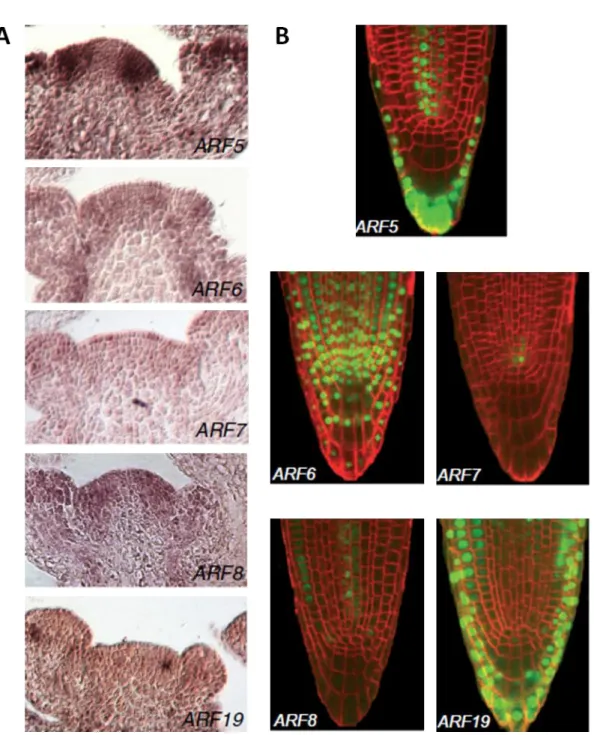
ARF activators are differentially expressed in the root and the shoot ... 53
ARF7 expression in the RAM is regulated at the first intron ... 61
ARF activator distribution can be post-transcriptionally regulated ... 64
~ 7 ~
Tissue-specific expression of ARF activators provides insights into specificity and redundancy of
their functions in plant development ... 68
Expression patterns of ARF activators fits their well-studied functions and suggests new potential functions ... 71
Which elements are important to correctly recapitulate expression patterns of genes? ... 73
Post-transcriptional movement or degradation of ARF proteins provides another level of control in the auxin signaling pathway ... 75
Material and methods ... 77
Cloning and generation of ARF reporter lines ... 77
Root microscopy ... 78
Shoot microscopy ... 79
Supplementary information ... 80
Chapter II: Auxin Response Factor (ARF) activators are transcriptionally
regulated by gene-specific repressor network
... 84Introduction ... 84
Results ... 92
Yeast one-hybrid assay revealed an elaborate network of transcription factors controlling expression of ARF activators ... 92
The majority of the ARF activator transcriptional regulators act as repressors ... 99
Publically available databases can be used to further validate and explore the interactions .... 104
Mutants of the regulatory transcription factors indicate co-regulation of multiple ARF activators ... 110
Expression of the several transcription factors is regulated by auxin ... 115
Mutants of the regulatory transcription factors show auxin-related defects in root and shoot development ... 117
Discussion... 121
Does the yeast one-hybrid gene regulatory network reflect in planta gene interactions? ... 121
ARF activator expression is predominantly regulated by transcriptional repressors ... 123
Regulation of ARF8 by multiple transcription factors might indicate its special significance in auxin signaling ... 125
Gene regulatory network motifs describe regulations that could be important during development ... 128
Expression of ARF activators is co-regulated in planta ... 132
Material and methods ... 133
Plant material ... 133
Y1H assay ... 133
~ 8 ~
Expression analysis with qRT-PCR ... 136
Expression analysis of crosses between ARF transcriptional reporter lines and T-DNA mutants ... 137
Shoot phenotype analysis of the TF mutants ... 137
Root phenotype analysis of the TF mutants ... 137
Growth on NPA for shoot phenotype analysis ... 138
Supplementary information ... 139
Chapter III: Cytokinin Response Factor 10 regulates ARF7 expression to
control plant development
... 154Introduction ... 154
Results ... 158
Genetic characterization of the crf10 mutant ... 158
CRF10 is expressed in various tissues ... 162
crf10 mutant has an ARF7-dependant early senescence phenotype ... 165
crf10 mutant shows perturbation in hypocotyl response to blue light ... 169
crf10 mutant has a defect in root apical meristem morphology ... 170
Discussion... 181
Is crf10-1 a loss-of-function mutant? ... 181
Does CRF10 act in cytokinin signaling? ... 183
CRF10 and ARF7 antagonistically control leaf senescence ... 185
CRF10 and ARF7 are acting together in hypocotyl phototropic response ... 186
CRF10 and ARF7 are involved in the maintenance of the root apical meristem ... 187
Material and methods ... 191
Plant material ... 191
Analysis of the conserved regions within the CRF10 protein across A. thaliana ecotypes ... 191
Genetic analysis of the crf10-1 mutant ... 191
Expression analysis with qRT-PCR ... 191
Cloning and generation of transgenic lines ... 192
Root microscopy ... 193
Shoot microscopy ... 193
Shoot phenotype analysis ... 194
Early senescence phenotype analysis ... 194
Chlorophyll measurement ... 194
Hypocotyl phototropism assay ... 194
~ 9 ~
Supplementary information ... 196
General discussion
... 200ARF activators in control of plant development ... 200
ARF activators diversify and specialize during evolution ... 204
Component of auxin signaling pathway show specificity in expression patterns ... 208
ARF activator expression is predominantly regulated by gene repression mechanism ... 209
Auxin and cytokinin interactions regulate many aspects of plant development ... 210
Appendix ... 212
Overview of primers used in this study ... 212
List of primers ... 214
~ 10 ~
Abbreviations
2,4-D 2,4-dichlorophenoxyacetic acid 4-Cl-IAA 4-chloroindole-3-acetic acid
A. thaliana Arabidopsis thaliana
AHK Arabidopsis histidine kinase
AHP Arabidopsis histidine phosphotransfer protein
AM Axillary meristem
AP2/ERF APETALA2/ethylene response factor
Arabidopsis Arabidopsis thaliana
ARF Auxin response factor
ARR Arabidopsis response regulator
AUX/IAA AUXIN/INDOLE-3-ACETIC ACID AUX/LAX AUXIN1/LIKE AUX1
AuxRe Auxin responsive element
bp Base pair
C. elegans Caenorhabditis elegans
CCR C-terminal conserved region CDF4 CYCLIC DOF FACTOR 4
CLE CLAVATA3/EMBRYO SURROUNDING REGION-RELATED CKX Cytokinin oxidase/dehydrogenase
CLV3 CLAVATA3
CRF Cytokinin response factor CSC Columella stem cells DII Domain II
DBD DNA-binding domain DD Dimerization domain
~ 11 ~
Dicamba 3,6-dichloro-2-methoxybenzoic acid DMAPP Dimethylallyl pyrophosphate
DNA Deoxyribonucleic acid FD Flanking domain
GFP Green fluorescent protein IAA Indole-3-acetic acid
IME intron mediated enhancement IPT Isopentenyltransferase
KAN1 KANADI 1 kb kilobase pair
L1, L2, L3 Layers 1, 2 or 3 of the shoot apical meristem LBD Lateral organ boundaries domain
LHW LONESOME HIGHWAY LOG LONELY GUY
LR Lateral root
MP MONOPTEROS
MR Middle region mRNA Messenger RNA mTQ2 mTurquoise2
NAA 1-naphthaleneacetic acid
NLS Nuclear localization signal or sequence NPA 1-N-Naphthylphthalamic acid
PAA Phenylacetic acid PB1 Phox and Bem1 pH Potential of Hydrogen
Picloram 4-amino-3,5,6-trichloropicolinic acid PIN PIN-formed protein
~ 12 ~
QC Quiescent center RAM Root apical meristem RNA Ribonucleic acid SAM Shoot apical meristem
SCR SCARECROW
SHR SHORT ROOT
SMB SOMBRERO
STM SHOOT MERISTEMLESS
TAA TRYPTOPHAN AMINOTRANSFERASE OF ARABIDOPSIS TIR1/AFB TRANSPORT INHIBITOR RESPONSE1/AUXIN SIGNALING
F-BOX PROTEIN TMO TARGET OF MP
WOX5 WUSCHEL related homeobox 5
~ 13 ~
Introduction
Among the diverse groups of living organisms land plants constitute a unique branch. Unlike animals, plants generally spend the majority of their life immobilized and restricted to a small space where they gradually progress through each stage of their development starting from a small embryo and proceeding as an adult plant capable of reproduction. During this whole developmental cycle plants have to continually adapt themselves to the ever-changing environmental conditions. They are subjected to a variety of stresses from various abiotic factors such as changes in temperature, humidity, nutrient content of the soil as well as biotic factors including attacks by pathogens and herbivore animals. To overcome these difficulties plants continue to produce new organs and modify the existing organs postembryonically. For example, phosphorus deficiency in the soil causes plants to alter their root architecture by attenuating growth of the primary root, forming more lateral roots and root hairs and this allows for more efficient forging of the available space (Bates and Lynch 1996, Bates and Lynch 2001, Brown et al. 2012, Giehl and von Wiren 2014, Lopez-Bucio et
al. 2002, Sanchez-Calderon et al. 2005, Williamson et al. 2001).
The constant growth and adaptation of the body structure throughout the entire life requires tight communication between various tissues to initiate or terminate the development of specific organs. On a cellular level this complex communication between tissues and cells is mediated by various mechanisms involving plant hormones, mobile transcription factors, small mobile peptides and small RNAs (Chaiwanon et al. 2016, Van Norman et al. 2011). Among the plant hormones, auxin and cytokinin control a large number of developmental processes in either an antagonistic or synergistic manner. For example, auxin inhibits formation of axillary branches while cytokinin acts in opposition promoting growth of axillary branches (Muller and Leyser 2011, Shimizu-Sato et al. 2009). Similarly, antagonistic interactions between auxin and cytokinin control specification of vasculature tissues in the root (Bishopp et al. 2011, Vaughan-Hirsch et al. 2018).
Auxin and cytokinin appear to be key factors that regulate the formation and maintenance of the root and the shoot apical meristems (Su et al. 2011). The root and the shoot apical meristems are important sites of growth in the plant. They contain a pool of pluripotent stem cells that are able to divide, grow and differentiate
~ 14 ~
into various specific cell types (Heidstra and Sabatini 2014). In the root apical meristem (RAM) located at the root tip the primary root tissues are produced (Petricka et al. 2012). Shoot apical meristem (SAM) is found at the shoot apex; here all new flowers, leaves and axillary shoots are formed (Barton 2010, Murray et al. 2012). How auxin and cytokinin controls plant growth in the RAM and the SAM remains a subject of intensive research with key signalling pathways being discovered in the last 20 years. Nevertheless, many aspects of auxin and cytokinin involvement in growth regulation at the RAM and the SAM still remain to be explored. This thesis focuses on genes involved in the auxin signalling pathway and their involvement in the root and shoot growth as well as on the crosstalk between auxin and cytokinin signalling pathways. For these reasons, this introduction will start with the detailed description of the plant development focusing on the growth of the root and the shoot, followed by description of the auxin and cytokinin signalling pathways. A stronger emphasis will be given to knowledge obtained using Arabidopsis thaliana, the model plant that was used in this study.
Plant embryonic development
Plants start their development from the zygote, a single totipotent cell. In Arabidopsis
thaliana, the zygote undergoes a series of highly ordered cell divisions followed by
cell differentiation that results in formation of the basic tissue types found in the mature plants including epidermis, vasculature, stem-cells and the ground tissue (Esau 1977). Several more specific tissue types develop post-embryonically including adventitious and lateral roots (Bellini et al. 2014), trichomes (Pattanaik et al. 2014) and root hair (Grierson et al. 2014).
The embryogenesis starts with an asymmetric division of the zygote cell producing the small apical and the elongated basal cells (Fig. In-1) (ten Hove et al. 2015). The apical cell will eventually give rise to the majority of the cells of the embryo whereas the lower basal cell will generate only a few specific cell types of the primary root: columella and the quiescent center (QC).
Following the initial asymmetric division, the upper apical cell will continue to divide; these divisions will partition the original cell volume without substantial cell expansion (Yoshida et al. 2014) (Fig. In-1). The lower basal cell will divide as well forming the suspensor domain (Fig. In-1). Together these divisions will form the globular stage
~ 15 ~
embryo with the roughly spherical shaped apical domain and the lower suspensor domain that looks like a vertical string of cells (Fig. In-1) (ten Howe et al. 2015). During this stage, the uppermost suspensor cell called hypophysis is specified to become the root apical meristem founder cell (Dolan et al. 1993). The hypophysis divides to produce two daughter cells: the upper cell becomes the quiescent centre cell (QC) and the lower cell gives rise to columella initials and columella proper (Yoshida et al. 2013). The QC is a part of the root stem cell niche which incorporates mitotically inactive cells (Dubrovsky and Barlow 2015). Therefore the QC and the columella cell layers below it are derived from hypophysis. On the other hand, the lower tier of the sphere-shaped apical domain eventually gives rise to the rest of the root tissues including root vasculature, cortex, endodermis and epidermis layers (Fig. In-1) (ten Hove et al. 2015; Petricka et al. 2012, Yoshida et al. 2013).
The outer cells of the sphere-shaped apical domain divide anticlinally to form the epidermal tissue. On the other hand, the inner cells divide longitudinally to produce the vasculature and the ground tissues (Fig. In-1) (ten Hove et al. 2015). The increased number of cells leads to morphological transformation from the globular to the heart-stage embryo (ten Hove et al. 2015). At this stage the future cotyledons become distinguishable as two bulges. The shoot stem cell niche forms between the emerging cotyledon primordia (Zhang et al. 2017).
~ 16 ~
The shoot- and the root stem-cell niches are already established at the heart-stage embryo and are maintained in the mature plant where they function to produce new tissues and organs. After the heart-stage embryo, the provascular tissues within the cotyledons are formed, the hypocotyl and the root cellular organization is established, and the SAM acquired the three layer structure (Capron et al. 2009). The structural complexity of the fully-formed embryo equals that of the plant seedling (Capron et al. 2009). At the end of embryogenesis the cell divisions are arrested and the mature embryo remains inside the seed through a period of dormancy until the germination phase is initiated (Bentsink and Koornneef 2008).
Root growth and the root apical meristem
Root apical meristem morphology
As pointed out earlier, roots are dynamic structures that undergo developmental changes. Studies in various plant species reveal that the root growth is not indeterminate and that the growth rate of the root is not identical throughout its development. Postembryonically, the young primary root grows at accelerated rate for the first 1-2 weeks of the seedling growth (Chapman et al. 2002, Zhu et al. 1998). This growth acceleration is followed by a period of constant growth rate with a steady increase in root length. Later, the root undergoes gradual deceleration of the growth rate which finally results in cessation of the growth altogether when the root reaches its final size (Chapman et al. 2002; Gladish and Rost 1993; Reinhardt and Rost 1995; Zhu et al. 1998). The precise final length of the root depends on growth conditions such as the temperature (Gladish and Rost 1993).
The root growth is sustained by the root apical meristem (RAM) which contains undifferentiated stem cells called initials. The cells in the RAM are able to divide and differentiate producing other tissues of the root (Petricka et al. 2012). Morphological patterns of the RAM can substantially vary depending on plant species (Heimsch and Seago 2008). In addition, root morphology undergoes changes as the root ages and in response to environmental conditions (De Tullio et al. 2010; Rost 2011). Three different types of RAM organisation have been reported: closed, intermediate and open (Groot et al. 2004). The differences between these RAM types lie in the organization of the stem cell niche consisting of the initial cells and QC. Initial cells are able to divide and differentiate into specific cell types. In the closed RAM each
~ 17 ~
cell layer (cortex, epidermis etc.) can be traced back to specific separate tiers of initials. The opposite root organization is the open type which lacks well-defined tiers of initials; here all initials look morphologically identical. The intermediate type displays to some extent organization especially for the epidermis/cortex lineage but still lacks apparent tiers of initials (Rost 2011). In many species with the closed types of RAMs the meristem structure was shown to gradually change from a well-organized closed type to a less well-organized structure resembling an intermediate type as the root became older and reached its final root length (Chapman et al. 2002). In some species the overall size of the root meristem reduces as the root ages (Chapman et al. 2002). These changes in RAM architecture over the lifespan of the root were speculated to be due to reduction or cessation of RAM function in the old root and were linked to reduced cell to cell communication evident by the reduction of plasmodesmata (Zhu et al. 1998).
As previously mentioned, this thesis has been focused on the plant model species
Arabidopsis thaliana. The root apical meristem of Arabidopsis thaliana has a closed
meristem with a very precise organization of cells that remains remarkably unchanged under different growth conditions and between individual plants. As with other species, this organization undergoes changes to a more intermediate type as the root ages, although these changes are limited (Baum et al. 2002).
The root tip of Arabidopsis can be separated into well-defined zones: the meristematic, the elongation and the differentiation zones (Fig. In-2A) (Petricka et al. 2012). The meristematic zone is made of actively dividing cells and is located above QC. In the meristematic zone cells undergo rapid divisions. The cells stop dividing, start to elongate and to increase in length when they reach the elongation zone. The area between the meristematic and the elongation zones is known as the transition zone. Finally, in the differentiation zone the cells complete their maturation (Fig. In-2A) (Dolan et al. 1993; Heidstra and Sabatini 2014; Petricka et al. 2012).
The architecture of the RAM is well-described (Fig. In-2B). The QC located at the tip of the root contains cells that divide slowly (Clowes 1956, Dubrovsky and Barlow 2015). The function of the QC is to inhibit differentiation of the surrounding initials (van den Berg et al. 1997). The QC therefore acts as an organizing center maintaining organization of the root stem cell niche. In roots younger than one week old there are 4-8 QC cells. In the 3 week old plants the QC structure changes: some
~ 18 ~
QC cells can divide forming a partial two-layered QC structure. In a 4-5 week old plants the QC becomes more disorganized (Baum et al. 2002).
Surrounding the QC are the initials that are the precursor cells of the cell types composing the root. Rootwards from QC is a single cell file of columella initials (also known as columella stem cells) followed by a few layers of mature columella cells. Shootwards from QC are vasculature initials that later produce xylem, phloem and procambium tissues. Initials of endodermis, cortex, epidermis and lateral root cap are found laterally to the QC (Fig. In-2B) (Petricka et al. 2012).
Fig. In-2. Structure of the mature primary root in Arabidopsis thaliana. (A) Zones at the root apex
(Ubeda-Thomas et al. 2012). (B) Patterning at the root apical meristem (RAM). (C) Cell divisions of the cortex/endodermal initials. (D) Cell divisions of the epidermal/lateral root cap initials (Petricka et al. 2012).
~ 19 ~
The origin of each tissue type can be easily traced to specific initials. As mentioned above columella cells originate from the columella initials which divide periclinally (Dolan et al. 1993). The cortex initial cell located laterally to the QC undergoes first anticlinal division followed by a periclinal division that results in formation of two parallel cell layers: cortex and endodermis (Fig. In-2C) (Petricka et al. 2012). This pattern of divisions changes as the plant ages; in most plants older than 1 week the cortex initial divides periclinally without first dividing anticlinally which results in two new initials: the cortex initial and the endodermis initial. These two initials give rise to the respective cell layers: cortex and endodermis. Finally, in roots older than 3 week an additional second layer of cortex, the middle cortex, forms. The second layer of cortex is formed though a periclinal division of cortex initials (Baum et al. 2002). The epidermis and lateral root cap layers are also derived from a single initial. This initial divides first anticlinally and then periclinally to produce these two cell layers (Fig. In-2D) (Petricka et al. 2012).
The vascular initials are located shootwards from the QC (Fig. In-2B). They gradually give rise to different cell types of the vasculature: the single pericycle layer, phloem, xylem and procambium (Baum et al. 2002).
The RAM structure in transverse view in a root less than 1 week old is very precise (Fig. In-2B) (Petricka et al. 2012). The root is surrounded by a single epidermis layer followed by single layers of cortex and endodermis. There are normally eight or nine cortical and endodermal cells (Baum et al. 2002). Next, a single layer of pericycle separates vasculature tissues from cortex. Xylem consists of 5 vessels which form a diarch pattern. There are two protoxylem vessels located adjacent to the pericycle layer as well as three metaxylem vessels. The two phloem sections are positioned perpendicularly to the xylem and adjacent to the pericycle layer. The vasculature cells located between the phloem and the xylem are called procambium (Fig. In-2B) (Baum et al. 2002; Dolan et al. 1993; Petricka et al. 2012).
The different cell types of the root originating from the root apical meristem have specific functions in the mature root. For example, xylem cells transport water and minerals upwards to the above-ground organs of the plant, whereas phloem cells transport nutrients from the sites of their synthesis into non-photosynthetic plant organs (Lucas et al. 2013). The columella cells are involved in gravity perception due to the presence of the starch-filled plastids in these cells (Blancaflor et al. 1998).
~ 20 ~
Overall, the structure of the RAM has been well described but the questions remain how this very precise structural organisation is achieved and maintained. New findings implement several hormone and various key genes involved in this process.
Regulation of root apical meristem development
The precise cell patterning of the root apical meristem is a result of cell division and differentiation processes which are tightly regulated by specific molecular mechanisms. Recently, a few major signals regulating these molecular mechanisms were characterized. These include auxin and cytokinin; together they manage patterning of the RAM in mostly antagonistic manner. In my thesis the focus lies on the role of auxin and, to a lesser extent, cytokinin in the growth of the root and the activity of the root apical meristem.
The levels of auxin and cytokinin signalling are relatively high in the root apical meristem, and this has been observed using appropriate in planta reporter constructs. Auxin itself and its signalling both have maximum in the QC, columella and xylem cells of the vasculature (Fig. In-3A and B) (Brunoud et al. 2012). On the other hand, cytokinin signalling is enriched in phloem and procambium domains of the vasculature as well as in the lateral root cap (Zurcher et al. 2013) (Fig. In-3C).
Fig. In-3 Auxin hormone accumulation (A) and signalling maxima (B) are visualized by the inverse
auxin sensor DII-VENUS (A) and the auxin signalling output reporter DR5 (B) (Brunoud et al. 2012). Cytokinin signalling maxima are displayed using the TCSn reporter (C) (Zurcher et al. 2013).
~ 21 ~
Auxin modulates growth of the root already at the embryonic stage through the action of the Auxin Response Factor 5 (ARF5), an essential component of the auxin signalling pathway (see later for further details on auxin signalling). ARF5 mediates specification of the hypophysis through the direct activation of the mobile transcription factor TMO7 (Schlereth et al. 2010; Weijers et al. 2006). Likewise, the establishment of the root vascular cylinder during embryonic development and its maintenance post-embryonically is also controlled by auxin via ARF5. In this process ARF5 acts through activation of its target genes including TMO5 which form heterodimers with members of the LONESOME HIGHWAY (LHW) family transcription factors and together they regulate cell divisions in vasculature initials (De Rybel et al. 2013, Ohashi-Ito and Bergmann 2007). Interestingly, the TMO5/LHW heterodimers directly induce expression of genes involved in cytokinin biosynthesis (LOG3 and LOG4) and signalling (AHP6) in xylem precursor cells of the RAM (De Rybel et al. 2014, Ohashi-Ito et al. 2014). Cytokinins produced by the activity of LOG3 and LOG4 have been proposed to move from xylem precursors into adjacent vasculature cells where they promotes vascular cell divisions resulting in the growth of the vascular cylinder (De Rybel et al. 2014, Ohashi-Ito et al. 2014). On the other hand, AHP6, an inhibitor of cytokinin signalling, acts in the xylem precursor cells where it restricts cytokinin signalling and blocks additional cell divisions maintaining an invariable number of xylem cells (De Rybel et al. 2014, Mahonen et al. 2006, Ohashi-Ito et al. 2014).
In addition to cell division, auxin and cytokinin determine specification of vasculature cells into respective cell types: proto- and metaxylem, phloem and procambium which are organized in a strict pattern (Fig. In-2B). The patterning of the vasculature cylinder is possible due separation of the cylinder into two distinct spatial domains, one with high auxin signalling in the xylem surrounded by one with the high cytokinin signalling in the phloem and procambium. This separation is achieved by two key molecular actors. On one hand, cytokinin signalling is maintained at low level in xylem through the repressive action of the auxin-induced cytokinin signalling inhibitor AHP6. Simultaneously, auxin is actively directed into xylem cells though activity of several PIN proteins whose expression and subcellular localization are regulated by cytokinin. PIN proteins are auxin efflux carriers that are localized polarly in cells and control the direction of intracellular auxin fluxes (Adamowski and Friml 2015), thus allowing for the accumulation of auxin in xylem cells and for the formation and
~ 22 ~
maintenance of the two antagonistic hormone signalling domains. The hormonal signalling output within these domains then determines the cell fate of the vasculature initial daughter cells which will be specified to become either xylem, phloem or procambium cells (Bishopp et al. 2011, Mellor et al. 2017).
Besides vasculature formation and patterning, cytokinin and auxin determine the size of the root meristem and control transition from cell division in the meristematic zone to cell differentiation in the elongation zone. Application of cytokinin reduces the size of the meristem and promotes cell differentiation; likewise, the cytokinin biosynthesis and signalling mutants exhibit increased meristematic zone (Dello Ioio et al. 2007). On the contrary, auxin increases the size of the meristematic zone and promotes cell divisions (Blilou et al. 2005, Dello Ioio et al. 2007, Dello Ioio et al. 2008). Thus these hormones together establish a boundary between two domains with different cell responses: rapid cell divisions in the meristematic zone and cell growth in the elongation zone (Di Mambro et al. 2017).
Finally, cytokinin was shown to be important in regulation of the QC function. Cytokinin is able to promote cell division in the mitotically inactive QC cells (Zhang et
al. 2013). Application of cytokinin does not alter the expression pattern of the QC
patterning marker lines such as QC46, SCR, WOX5 (Dello Ioio et al. 2007) but seems to reduce the expression levels of several of these markers (Zhang et al. 2013).
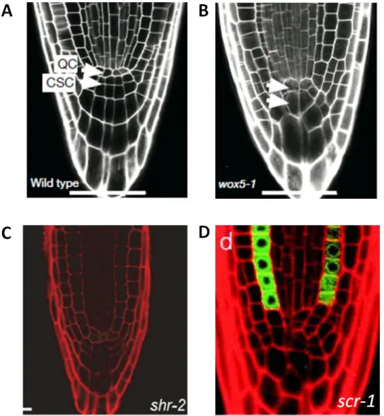
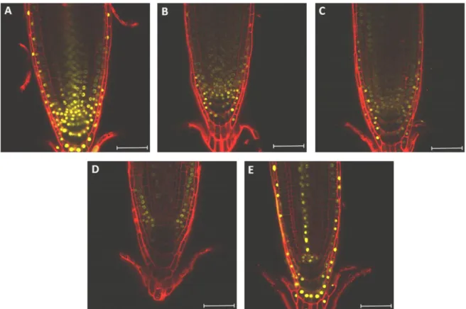
In addition to auxin and cytokinin, the RAM development is regulated by specific regulatory transcription factors. Among these is WOX5, which controls the development of the QC. WOX5 is expressed specifically in the QC starting already in the embryonic QC cell lineage (Sarkar et al. 2007). The wox5 mutant has abnormally enlarged QC and columella stem cells, reduced number of columella cells and ectopic cell divisions in the QC (Fig. In-4A and B) (Forzani et al. 2014; Sarkar et al. 2007). WOX5 is required for columella stem cell maintenance preventing their differentiation into mature columella cells (Sarkar et al. 2007). The WOX5 protein is able to move from QC into columella stem cell layer where it represses the transcription factor CDF4 thus maintaining columella stem cells in undifferentiated state (Pi et al. 2015).
The columella architecture is controlled by the two NAC-domain transcription factors SOMBRERO (SMB) and FEZ. fez mutants have a reduced number of columella
~ 23 ~
layers and lateral root cap cells whereas smb mutant has one additional columella layer. This control of the root cap patterning is independent from the other known RAM patterning regulators including WOX5, SHR, SCR or PLT genes. FEZ gene is expressed in the columella stem cells and columella proper whereas SMB is only expressed in mature columella cells. There is a negative feedback loop proposed between these two regulators: FEZ promotes cell divisions in columella stem cells and also activates SMB transcription in mature columella cells where SMB in turn represses FEZ function (Willemsen et al. 2008).
Fig. In-4. Phenotypes of mutants with defects in RAM architecture. (A) Root tip of the wild type
(Sarkar et al. 2007), (B) wox5-1 mutant (Sarkar et al. 2007), (C) shr-2 mutant (Sebastian et al. 2015),
~ 24 ~
A pair of transcription factors SHR and SCR is involved in two separate processes: they regulate both the specification of QC and the patterning of endodermis and cortex. In scr mutants the cortex/endodermis initials do not undergo asymmetric division, and this results in formation of a single cell layer with mixed cortex/endodermis identity; additionally, the QC cells have abnormal shapes and function which causes loss of meristematic activity and premature termination of root growth (Fig. In-4D) (Sabatini et al. 2003). shr mutant also shows abnormal morphology in the QC and columella regions leading to premature termination of root growth; the mutant is characterized by the presence of a single layer of ground tissue with cortex identity instead of cortex and endodermis layers (Fig. In-4C) (Helariutta et
al. 2000). Both scr and shr are required for correct QC function but the precise
mechanism of this regulation is yet to be determined (Sabatini et al. 2003). SHR mRNA is found specifically in the stele tissues of the root but the SHR protein moves from the stele to the endodermis where it activates SCR (Cui et al. 2007; Helariutta et
al. 2000; Nakajima et al. 2001). SCR and SHR interact with each other to regulate a
number of common target genes (Cui et al. 2007, Levesque et al. 2006).
Stem cell activity in the root meristem is also controlled by members of the PLETHORA (PLT) gene family. plt1 mutants have increased number of columella tiers and extra cells in tier 2 of the columella. plt2 mutants have more subtle phenotypes showing increased columella cell numbers. The plt1 plt2 double mutant displays severe defects in root development. PLT1 and PLT2 genes are AP2-domain transcription factors and their expression is induced by auxin. PLT1 and PLT2 are required for stem cell niche maintenance independently from SCR/SHR (Aida et al. 2004).
Overall, it appears that the key transcription factors such as WOX5, SCR, SHR and PLT act together with the plant hormones auxin and cytokinin to establish the RAM during embryogenesis and to regulate its functions post-embryonically.
Lateral root development
The primary root is established during embryonic development and continues to grow for a certain period post-embryonically. The origin of every cell in the root is traced back to the meristematic cells in the root apical meristem. Nevertheless, adult plants have to establish additional new roots post-embryonically in order to increase the forging surface of their root system and thus adapt themselves to changes in nutrient
~ 25 ~
and water availability of the soil. There are several types of post-embryonic roots that do not originate from the root apical meristem but are produced de novo: the lateral roots, the adventitious roots and the crown roots (Bellini et al. 2014). Lateral roots start to grow on other roots whereas the adventitious roots develop on non-root organs (for example, on hypocotyls and stems).
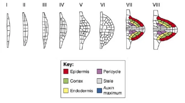
In Arabidopsis thaliana lateral roots (LR) are initiated in the differentiation zone of the primary root from pericycle cells which lie adjacent to xylem poles. These lateral root founder cells consist of a cluster of several pericycle cells (ca. 11 cells on average), but only a few of these cells contribute to the majority of the future LR body (von Wangenheim et al. 2016). The pericycle founder cells divide anticlinally to form a single file of several cells (stage I of LR formation in Fig. In-5). The sequence of these first cell divisions is strictly controlled (von Wangenheim et al. 2016). Next, the cells undergo periclinal divisions resulting in a two-layer lateral root primordium (stage II of LR formation in Fig. In-5). Subsequently, the primordium continues to divide anticlinally and periclinally creating a dome-shaped structure (stages III-VII of LR formation in Fig. In-5). The patterns of these subsequent cell divisions can vary between plants and do not follow a rigid sequence. The division planes often change depending on the cell geometry; the cells tend to divide along local minima of plane area (following the shortest wall principle) and prefer to alternate their division orientation plane between anticlinal and periclinal divisions for each subsequent
~ 26 ~
division round (von Wangenheim et al. 2016). Already at stage VI the lateral root primordium consists of distinctive cell types characteristic for a mature root tip including epidermis, cortex, endodermis and vasculature cells. At the final stage the primordium breaks through the root epidermal tissue, emerging as a new lateral root (stage VIII of LR formation in Fig. In-5) (Lavenus et al. 2013, Malamy and Benfley 1997, Peret et al. 2009, von Wangenheim et al. 2016).
The lateral roots positioning is strictly controlled: the lateral roots emerge at a regular spacing along the primary root in a left-right alternating pattern (De Smet et al. 2007). The temporal and spatial distribution of the lateral roots was argued to be regulated by an endogenous clock mechanism involving oscillating genes (Moreno-Risueno et
al. 2010). Auxin was shown to additionally contribute as a signal which primes the
xylem pericycle cells to become lateral root founder cells. The regular distribution of lateral roots correlates with an oscillating auxin signalling in the protoxylem cells which lie adjacent to the pericycle cells. When auxin response is maximum, the adjacent pericycle cells acquire a competence to divide and form a lateral root primordium (De Smet et al. 2007, Peret et al. 2009). The exact mechanism which choreographs these auxin signalling oscillations is still uncharacterized. Furthermore, exactly how an auxin signal is propagated from the xylem cells into the neighbouring pericycle cells remains undetermined.
Cytokinin appears to act antagonistically to auxin during lateral root initiation. Exogenous cytokinins perturb initiation of lateral root primordia at a very early stage by blocking asymmetric cell division in the xylem pericycle cells (Laplaze et al. 2007). This perturbation is achieved by arresting cell cycle at the G2 to M transition phase (Li
et al. 2006b). Additionally, at later stages, exogenous cytokinins induce a
morphological disorganization in the emerging lateral root primordia particularly at the root tip (Laplaze et al. 2007). Cytokinin signalling is repressed in the xylem pericycle cells in which lateral root primordia priming and initiation occurs. On the contrary, cytokinin signalling was detected in xylem pericycle cells which lie between two existing lateral root primordia where no further lateral roots are initiated (Bielach et al. 2012). Therefore cytokinin appears be a repressive signal preventing initiation of new lateral roots in close proximity to each other and thus regulating spatial distribution of lateral roots along the primary root.
~ 27 ~
The subsequent growth of the lateral root primordia appears to be regulated by auxin at all stages. Several mutants involved in auxin homeostasis, transport and signalling are impaired in lateral root growth (Peret et al. 2009). In particular, the Auxin Response Factors 7 and 19 (ARF7 and ARF19) as well as INDOLE-3-ACETIC ACID INDUCIBLE 28 and 14 (IAA28 and IAA14) are key auxin signalling pathway genes (see later for more details on auxin signalling) involved in lateral root formation at different stages (De Rybel et al. 2010; Fukaki et al. 2002; Okushima et al. 2005; Okushima et al. 2007). Auxin response is transmitted though the transcriptional activation of auxin-responsive gene by ARF7 and ARF19 transcription factors. Together ARF7 and ARF19 directly activate expression of several genes including the transcription factors Lateral Organ Boundaries Domain 16 and 29 (LBD16 and LBD29) (Okushima et al. 2007). Presumably, LBD16/LBD29 as well as other yet unidentified transcription factors are able to activate cell-specific programs in the developing lateral root primordia. These programs lead to coordinated cell divisions followed by cell specification and eventually formation of mature specialized tissues within the emerging lateral root. The de novo formed root apical meristem of the lateral root acts in the same way as the RAM of the primary root enabling post-emergence growth of the lateral root. Thus differentiated xylem pericycle cells eventually give rise to a multitude of root-specific tissues including undifferentiated stem cells.
Shoot apical meristem
The development of all above-ground organs relies almost entirely on the function of the shoot apical meristem (SAM). Two opposite processes occur in the SAM: the stem cell pool is constantly maintained and renewed whereas some cells accelerate their growth and division rate and eventually differentiate to become part of the newly forming organs (the leaves and flowers). The balance between these two processes is strictly controlled over the life of the plant and the location and timing of new organ emergence appears to be tightly regulated.
This regulation can be first seen from the organization of the SAM which is divided into functional zones with distinct cellular behaviours (division and expansion) and distinct cellular identities. In Arabidopsis thaliana the dome-shaped structure of the SAM is divided into the central, peripheral and rib zones (Fig. In-6A). The central zone is found at the apex and contains undifferentiated stem cells. The site of organ
~ 28 ~
primordia initiation occurs at the peripheral zone. The rib zone, situated below central and peripheral zones, produces the internal tissues of the stem. The SAM can be further divided into individual cell layers. The top two layers (L1 and L2; collectively referred as tunica) are able to divide only in one direction (anticlinally) whereas the deeper layers (L3 and further; collectively referred as corpus) are able to divide in any direction. This organization is largely similar in other higher plants with some variations in the number of tunica layers.
The cells in the functional zones of the SAM differ in their properties. For example, the cells in the central zone where the stem cells are located divide slower than the cells in the peripheral zone (Laufs et al. 1998; Reddy et al. 2004). The cells in the central zone frequently divide asymmetrically but maintain overall similar cell size within the zone. This is achieved by an adjustment of the cell growth rate and the
Fig. In-6. Organization of the shoot apical meristem. (A) The structure of the shoot apical meristem
with functional zones: the central zone CZ, the peripheral zone PZ, the organizing center OC, the rib zone RZ and the organ primordia P. L1, L2 and L3 indicate the cell layers (Murray et al. 2012). (B) The order of new primordia initiation from the youngest P1 to the oldest P9. I1 marks the site of the next primordia initiation (Murray et al. 2012). (C) Minimal gene interaction network controlling SAM maintenance. Circles mark the expression domains of CLV3 (red), WUS (green), KAN1 (brown) and cytokinin maximum (blue). Green dots show localization of WUS protein. X stands for a hypothetical L1-devived signal which activated CLV3 (Truskina and Vernoux 2018).
~ 29 ~
cell cycle length following an asymmetric cell division illustrating the presence of a compensatory mechanism that allows the meristem to maintain the desired overall uniform structure (Jones et al. 2017; Serrano-Mislata et al. 2015; Willis et al. 2016). The local variability of cell growth rates in the meristem plays a key role in setting the geometry of the SAM (Uyttewaal et al. 2012), highlighting the importance of cell behavior in generating a specific shape. The division of the SAM into the central and peripheral zones also correlates with differences in mechanical properties: the central zone of the SAM is characterized by increased stiffness of the tissue compared to more peripheral regions (Milani et al. 2014, Milani et al. 2011, Kierzkowski et al. 2012) or organ primordia (Braybrook and Peaucelle 2013, Peaucelle et al. 2011). Similarly to the root, these patterns associated with the functional zones of the SAM are established by both cell-autonomous factors such as the cell-specific gene regulatory networks and non-cell autonomous factors including mobile proteins and hormones (Barton 2010; Murray et al. 2012).
The functional zones of the SAM are characterized by specific expression of master regulatory genes with CLV3 in the central zone (Fletcher et al. 1999), WUS in the organizing center (Mayer et al. 1998) and KAN1 in the boundary domain (Yadav et al. 2013) amongst many others. Several publications have attempted to model SAM maintenance based on expression patterns and interactions of these regulatory genes (Adibi et al. 2016, Fujita et al. 2011, Gruel et al. 2016, Yadav et al. 2013). Computer simulations attempted to define the minimal regulatory networks required for functioning of the SAM (Fig. In-6C). The models always include the well-described WUS-CLV3 feedback loop which dynamically maintains the size of stem cell niche (Brand et al. 2000, Lenhard and Laux 2003, Schoof et al. 2000; Yadav et al. 2011). Repression of the differentiation-promoting genes such as KAN1 by WUS contributes to the entry into differentiation (Gruel et al. 2016, Yadav et al. 2013). Furthermore, this modeling work emphasizes the importance of cytokinin signalling in SAM maintenance by showing that regulation of WUS expression by cytokinin (Chickarmane et al. 2012,Gordon et al. 2009) and activation of cytokinin signalling by WUS (Leibfried et al. 2005) are fundamental for correct positioning of WUS in the SAM. Recently an additional signalling network was identified which includes a movement of a CLE peptide produced in organ primordia to the center of the SAM where it regulates stem cell activity thus providing an extra feedback regulation from
~ 30 ~
developing organs on the stem cell niche and providing an interesting mechanisms for integrating stem cell maintenance and organogenesis (Je et al. 2016).
At the periphery of the SAM, new leaf or flower primordia are initiated at predictable positions and with regular time intervals between initiation events (Fig. In-6B). In
Arabidopsis thaliana new organ primordia are initiated sequentially one after another
following a whorled phyllotaxis pattern with an angle of approximately 137.5 degrees between each newly formed primordium (Bartlett and Thompson 2014, Galvan-Ampudia et al. 2016, Kuhlemeier 2007, Traas 2013). A long-standing theory postulates that the positioning and timing of organs at the growing shoot apex is determined by the presence of inhibitory signals around developing organ primordia, these inhibitory fields preventing initiation of new primordia. The current understanding of the molecular mechanisms behind organ initiation suggests that this inhibition results from auxin depletion in the regions surrounding a local auxin accumulation that drives organ primordia (Jönsson et al. 2006, Reinhardt et al. 2003, Smith et al. 2006, Stoma et al. 2008, Vernoux et al. 2011). The developing organ primordia are characterized by high auxin signalling in primordia of all stages (Fig. In-7A). On the other hand, the cytokinin signalling is highest in the young primordia but decreases rapidly in the older primordia; cytokinin signalling is also detected in the organizing center (Fig. In-7B). Auxin and cytokinin signalling is first switched on at the site of the next primordia initiation (Besnard et al. 2014). Auxin is transported
Fig. In-7. Auxin hormone accumulation and signalling maxima (A) are visualized by the inverse auxin
sensor DII-VENUS (yellow) and the auxin signalling output reporter DR5 (blue) at the SAM (image from Carlos Galvan-Ampudia, ENS de Lyon). Cytokinin signalling maxima are displayed using the TCSn reporter at the SAM (B) (image from Fabrice Besnard, ENS de Lyon).
~ 31 ~
directionally towards these sites of the future organ primordia and this auxin accumulation leads to lateral organ initiation. The directional transport is achieved through the polar localization of the auxin efflux carriers from the PIN gene family, most notably PIN1 (Heisler et al. 2005, Reinhardt et al. 2003). The importance of the PIN1 for the shoot development is evident by the pin1 mutant phenotype; the pin1 mutants are unable to develop a functional inflorescence producing instead a “pin”-looking structure which lacks normal lateral organs (Galweiler et al. 1998). In addition to PINs, the auxin influx carriers from the AUX/LAX gene family also contribute to the spatiotemporal distribution of auxin in the SAM (Bainbridge et al. 2008, Reinhardt et
al. 2003).
Recent findings indicate that the timing of primordia initiation is regulated by an interplay between auxin and cytokinin signalling (Besnard et al. 2014). This study focuses on the role of the AHP6 protein which production is induced by auxin and enriched in organ primordia and developing flowers. The AHP6 protein produced in the primordia is then able to move to the neighboring cells where it acts as an inhibitor of cytokinin signalling. The movement of AHP6 creates a differential in cytokinin signalling activity between sites of successive organ initiation that facilitates sequential initiation of organs and thus provides robustness to the timing of organ initiation.
Differential auxin patterns continue to regulate development following the initiation of the shoot organs. In particular, the developing leaf primordia require transient low auxin zone at the adaxial (upper) side for successful establishment of leaf polarity (Qi
et al. 2014). The auxin depletion at the adaxial site is achieved by PIN1 auxin efflux
transporter which moves auxin away from the adaxial site of the developing leaf primordia towards the meristem. The same mechanisms that pattern the meristem are thus also key in establishing the symmetry of the organs.
In addition to the SAM, axillary meristems (AM) are small stem cell niches located at the upper (adaxial) side of the newly formed leaf. The AM gives rise to axillary buds which are able to remain dormant or eventually produce an axillary shoot (Bennett and Leyser 2006, Yang and Jiao 2016). Each newly formed axillary shoot contains a functional shoot apical meristem capable of producing new organs. Thus plants can increase and diversify their architecture to adapt to the changing environmental conditions.
~ 32 ~
The formation of the axillary meristem appears to be tightly controlled by patterns of hormone signalling which presents a remarkable similarity to the mechanisms behind the pattern formation in the SAM proper. The process requires initial auxin depletion at the future AM initiation site in the leaf axil closely followed by a pulse of cytokinin signalling. This auxin depletion in the leaf axil is achieved well before AM initiation at the early stages of leaf primordia formation due to directed polar auxin transport mediated by PIN1 localization (Wang et al. 2014a, Wang et al. 2014b).
Lately, the axillary meristem was shown to be regulated by the same key genes as the main SAM. Specifically, the regulators of the shoot stem-cell niche WUS and CLV3 were dynamically induced one after another during initiation of the AM creating a two-step pattern of expression. Interestingly, CLV3 was initially induced in the WUS-specific central domain before the expression shifted to the expected L1 and L2 layers at the later stages of AM formation (Xin et al. 2017). In addition, the mobile stem-cell specific gene STM was shown to be important for AM initiation (Balkunde et
al. 2017, Shi et al. 2016).
In summary, the key mechanism controlling organ initiation and maintenance of the stem-cell niche in the SAM is the contrasting hormone signalling between different regions in the SAM which results in tissue-specific gene expression patterns. In turn, the differentially expressed regulatory genes trigger cell-specific programs promoting cell fate determination. A strong emerging trend in recent research is that a similar set of signals and genes define a patterning module that is used in the SAM, the developing organs and to establish new meristems such as the AM. How this module is reused and how this allows to link organ and tissue development to the SAM activity is yet to be fully characterized but some of the key mechanisms have clearly been identified.
The plant hormone auxin
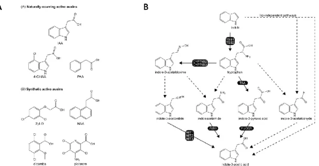
Auxin was shown to be involved in numerous developmental processes including cell division and cell expansion (Perrot-Rechenmann 2010), root and shoot growth (Overvoorde et al. 2010, Vernoux et al. 2010), phototropism (Fankhauser and Christie 2015), gravitropism (Band et al. 2012), leaf senescence (Ellis et al. 2005), response to pathogens (Kazan and Manners 2009, Fu and Wang 2011), abiotic stress (Bielach et al. 2017, Salopek-Sondi et al. 2017). The main type of auxin found
~ 33 ~
in plants is the indole-3-acetic acid (IAA) (Fig. In-8A) (Simon and Petrasek 2011). Other naturally occurring auxins include 4-chloroindole-3-acetic acid (4-Cl-IAA), and phenylacetic acid (PAA). In addition, several synthetic compounds such as 2,4-dichlorophenoxyacetic acid (2,4-D), 1-naphthaleneacetic acid (NAA), 3,6-dichloro-2-methoxybenzoic acid (dicamba), and 4-amino-3,5,6-trichloropicolinic acid (picloram) also induce auxin responses when applied externally on the plant or added to the growth medium (Fig. In-8A) (Sauer et al. 2013).
The metabolism of IAA is well-studied. IAA can be produced through two major routes using either the tryptophan (Trp)-dependent and the Trp-independent pathways (Fig. In-8B) (Korasick et al. 2013). Both these pathways might contribute to the regulation of IAA levels but the precise role of each pathway in Arabidopsis
thaliana is unclear. The IPyA pathway where tryptophan is converted to IAA via the
enzymes from the TRYPTOPHAN AMINOTRANSFERASE OF ARABIDOPSIS (TAA) family of Trp aminotransferases and the YUCCA (YUC) family of flavin monooxygenases seems to contribute the most to the active IAA (Korasick et al. 2013, Zhao 2012). Active auxins can be conjugated and transformed into inactive
Fig. In-8. Auxin structure and biosynthesis pathways. (A) Naturally occurring and synthetic active
auxins (Korasick et al. 2013). (B) Potential IAA biosynthetic pathways. Solid arrows indicate pathways for which all enzymes have been identified; dashed arrows indicate pathways for which not all enzymes have been identified (Korasick et al. 2013).
~ 34 ~
storage forms. In addition, inactive auxins can be reactivated (Korasick et al. 2013, Sauer et al. 2013).
Auxin is not synthesized ubiquitously throughout the plant; the main sites of auxin synthesis in Arabidopsis thaliana are the aerial plant parts especially the young developing leaves (Ljung et al. 2001) from which auxin is transported into the root tip though the stele (Michniewicz et al. 2007). But auxin is also synthesized in the root especially in the primary root meristem and in developing lateral roots (Ljung et al. 2005).
In addition to this, auxin is transported locally between the adjacent cells through the process of polar auxin transport. This well described process is based on the fact that IAA, as a weak organic acid, can exist in both protonated and deprotonated forms depending on the pH level of the environment (Raven 1975, Rubery and Sheldrake 1973, Rubery and Sheldrake 1974). Outside the cells, in the apoplast, the pH level is acidic which means that IAA is protonated and can diffuse into cells easily due to its neutral charge. Additionally the influx carriers of the AUX/LAX family actively pump IAA into the cells (Peret et al. 2012b). Inside the cells the pH is neutral which causes IAA to lose the proton and become electrically charged preventing it from diffusing
Fig. In-9. PIN distribution and auxin transport in the leaf (A), the shoot apex (B), in developing shoot
primordium (C) and the root apex (D) (Berkel et al. 2013). Auxin is in blue and PIN proteins in red; arrows indicate the direction of auxin flux. (E) Scheme of the auxin signalling pathway in the absence or presence of auxin (Lau et al. 2008).