HAL Id: tel-02955513
https://tel.archives-ouvertes.fr/tel-02955513
Submitted on 2 Oct 2020HAL is a multi-disciplinary open access
archive for the deposit and dissemination of sci-entific research documents, whether they are pub-lished or not. The documents may come from teaching and research institutions in France or abroad, or from public or private research centers.
L’archive ouverte pluridisciplinaire HAL, est destinée au dépôt et à la diffusion de documents scientifiques de niveau recherche, publiés ou non, émanant des établissements d’enseignement et de recherche français ou étrangers, des laboratoires publics ou privés.
Heterogeneity of polymorphonuclear neutrophils in
HIV-1 infection. Study of SIV-infected cynomolgus
macaque model.
Julien Lemaitre
To cite this version:
Julien Lemaitre. Heterogeneity of polymorphonuclear neutrophils in HIV-1 infection. Study of SIV-infected cynomolgus macaque model.. Innate immunity. Université Paris Saclay (COmUE), 2019. English. �NNT : 2019SACLS267�. �tel-02955513�
Heterogeneity of polymorphonuclear
neutrophils in HIV-1 infection
Study of SIV-infected cynomolgus
macaque model
Thèse de doctorat de l'Université Paris-Saclay préparée à l’Université Paris-Sud
École doctorale n°569 Innovation thérapeutique : du fondamental à l’appliqué (ITFA) Spécialité : Immunologie
Thèse présentée et soutenue à Fontenay-aux-Roses, le 13 septembre 2019, par
Monsieur Julien Lemaitre
Composition du Jury : Sylvie Chollet-Martin
Professeur–Praticien hospitalier, GHPNVS-INSERM (UMR-996) Présidente
Véronique Witko-Sarsat
Directeur de recherche, Institut Cochin-INSERM (UMR-1016) Rapporteur
Pierre Delobel
Professeur–Praticien hospitalier, CPTP-CHU de Toulouse Rapporteur INSERM (UMR-1043)
Mirko Paiardini
Professeur assistant, Yerkes National Primate Research Center Examinateur Emory University School of Medicine
Olivier Lambotte
Professeur–Praticien hospitalier, CHU du Kremlin Bicêtre-INSERM Directeur de thèse (UMR-1184)
Anne-Sophie Beignon
Chargé de recherche, CEA-CNRS-INSERM (UMR-1184) Co-Directeur de thèse
N N T : 2 0 19 S A C LS 2 67
1
Table of Content
Forewords: brief history of a zoonosis ___________________________________________ 8 Introduction_______________________________________________________________ 10
I. The unsolved chronic inflammation in HIV-1 infection ____________________________ 10 II. Causes of chronic inflammation in treated HIV-infection and consequences __________ 13
A. HIV infection as a chronic inflammatory disease ________________________________________ 13 B. Failure of antiretroviral treatments to resolve inflammation ______________________________ 15 1) Reservoirs and pharmacological sanctuaries maintain immune stimulation ________________ 15 i. HIV persistence in cellular and tissue reservoirs ____________________________________ 15 ii. Pharmacological sanctuaries as a source of chronic inflammation _____________________ 18 2) Maintenance of type I interferon response under cART ________________________________ 19 C. Unrestored mucosal immune response under cART _____________________________________ 20 D. Co-factors associated with amplification of inflammation _________________________________ 25 1) Co-infections as additional sources of inflammation ___________________________________ 25 i. Tuberculosis strongly activates immune cells ______________________________________ 25 ii. Cytomegalovirus modulates neutrophils responses _________________________________ 26 iii. Viral hepatitis co-infections and liver fibrosis ______________________________________ 27 2) Lifestyle and inflammation _______________________________________________________ 28 E. Consequences of chronic inflammation _______________________________________________ 30 F. Resolution of chronic inflammation by natural hosts _____________________________________ 33
III. Neutrophils plasticity in response to inflammation _______________________________ 37
A. Neutrophil life from granulopoiesis to efferocytosis _____________________________________ 37 1) Feedback loop of neutrophils production ___________________________________________ 37 2) Differentiation and maturation of neutrophils _______________________________________ 39 3) Circadian cycle orchestrates neutrophils circulation and tissue migration _________________ 41 4) Anti-microbial function of neutrophils ______________________________________________ 42 5) Neutrophil compartmentalization and death to control inflammation ____________________ 44 B. Heterogeneity and function of neutrophils in chronic inflammation ________________________ 46 1) Auto-immune diseases __________________________________________________________ 46 2) Cancers ______________________________________________________________________ 48 3) Tuberculosis ___________________________________________________________________ 49 4) Neutrophils in chronic viral infections ______________________________________________ 51 C. Neutrophils in HIV infection ________________________________________________________ 53 1) Dynamic of neutrophils in HIV infection_____________________________________________ 53 i. HIV infection and neutropenia __________________________________________________ 53
2 ii. Mucosal neutrophils in HIV infection ____________________________________________ 54 2) Neutrophil function in HIV infection _______________________________________________ 54 i. Neutrophil’s antiviral response _________________________________________________ 54 ii. Dual role in HIV acquisition and protection _______________________________________ 55 iii. Neutrophils function and chronic inflammation ____________________________________ 56 Methodology ______________________________________________________________ 59
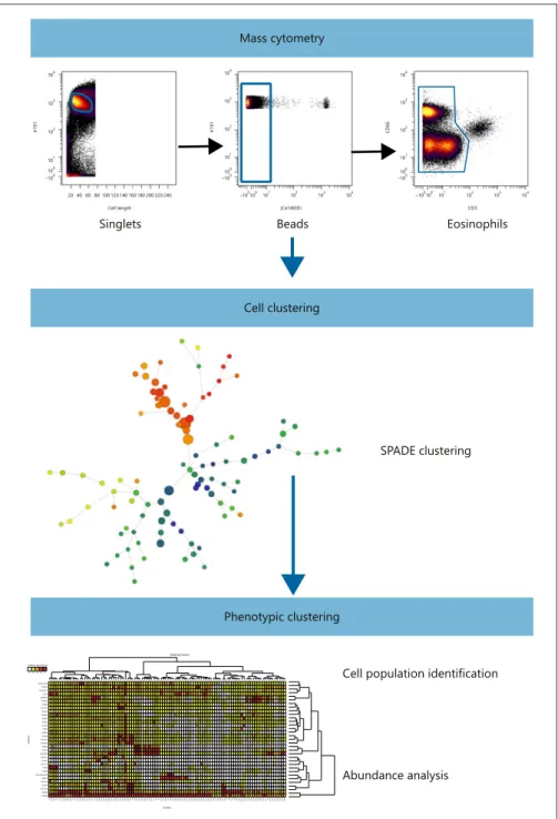
I. General objectives _________________________________________________________ 59 II. Study model ______________________________________________________________ 62 III. Principles of mass cytometry ________________________________________________ 64
Results ___________________________________________________________________ 67
I. Mass cytometry reveals the immaturity of circulating neutrophils during SIV infection __ 67
A. Summary________________________________________________________________________ 67 B. Manuscript ______________________________________________________________________ 67
II. Immature CD10- neutrophils exert immunostimulatory function during primary and chronic SIV infection __________________________________________________________________ 80
A. Summary________________________________________________________________________ 80 B. Manuscript ______________________________________________________________________ 82 1) Abstract ______________________________________________________________________ 83 2) Introduction ___________________________________________________________________ 84 3) Material and method ___________________________________________________________ 85 4) Results _______________________________________________________________________ 88 5) Discussion ____________________________________________________________________ 94
III. Unpublished results: tissue neutrophils heterogeneity in cynomolgus macaques______ 101
A. Introduction ____________________________________________________________________ 101 B. Material and method _____________________________________________________________ 101 C. Preliminary results _______________________________________________________________ 102 Discussion and perspectives _________________________________________________ 106
I. Validation of experimental approach _________________________________________ 106 II. Discussion ______________________________________________________________ 107 III. Perspectives _____________________________________________________________ 109
3
List of figures
Figure 1: Major causes of chronic inflammation in HIV-infected patients under cART and association with non-AIDS-related diseases.
Figure 2: Gut CD4+ T cell depletion and lymph node histopathology during HIV-1 infection
Figure 3: Persistence of viral reservoir and residual replication are associated to poor antiretroviral drugs distribution.
Figure 4: Neutrophils infiltrate gut during HIV infection.
Figure 5: Translocated microbial products reach secondary lymphoid tissues and drive distant inflammation.
Figure 6: Incidences of non-AIDS-related comorbidities.
Figure 7: Mechanisms involve in control of chronic inflammation by natural hosts.
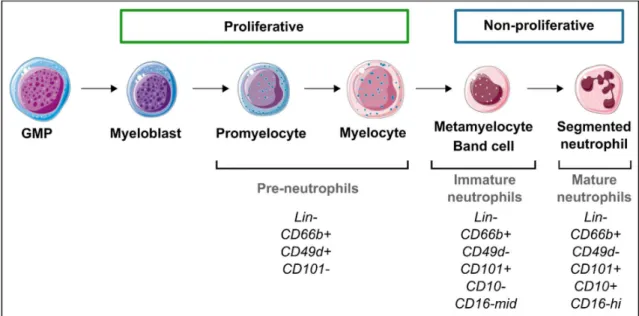
Figure 8: Regulation of neutrophils production and circulation. Figure 9: Neutrophil differentiation in human bone marrow.
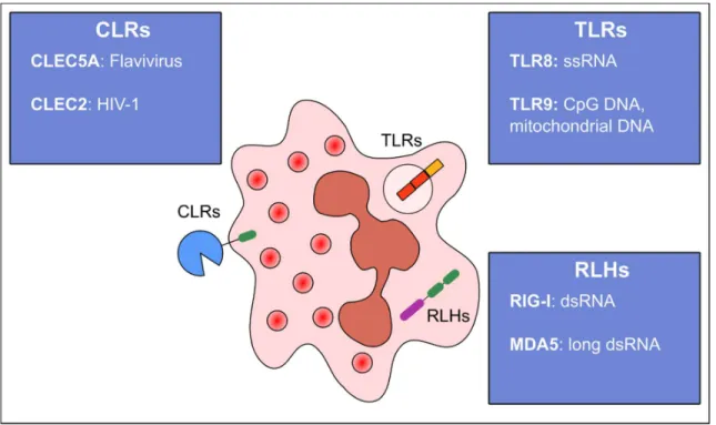
Figure 10: Pattern recognition receptors sensing viruses in human neutrophils.
Figure 11: Dynamic of viral replication along with CD4+ T cells and CD8+ T cells in SIVmac251-infected cynomolgus macaques.
4
List of abbreviations
3TC: LamivudineAAV: Antibody-associated vasculitis ACPA: Anti-citrullinated protein antibodies ADCC: Antibody-dependent cellular cytotoxicity
ADCP: Antibody-dependent cellular phagocytosis
AGM: African green monkey
AIDS : Acquired immunodeficiency syndrome ANRS: Agence nationale de recherche sur le sida et les hépatites virales
ARDS: Acute respiratory distress syndrome AZT: Azidothymidine
BAFF: B-cell activating factor
C/EBP-: CCAAT/enhancer-binding protein alpha
cART: Combination antiretroviral therapy CD: Cluster of differentiation
CLR: C-lectin receptor
CMP: Common myeloid progenitor
COPD: Chronic obstructive pulmonary disease CRP: C-reactive protein
CTLA4: Cytotoxic T-lymphocyte-associated protein 4
DAMP: Danger-associated molecular pattern DC: Dendritic cell
DNA: Deoxyribonucleic acid DRV: Darunavir
DTG: Dolutegravir
Fc-R: Fragment crystallizable receptor
FDC: Follicular dendritic cell fMLP: N-formylmethionine-leucyl-phenylalanine
FTC: Emtricitabine
G-CSF: Granulocyte-colony stimulating factor G-MDSC: Granulocytic-myeloid-derived suppressive cell
GALT: Gut-associated lymphoid tissue GM-CSF: Granulocyte-monocyte colony stimulating factor GMP: Granulocyte-monocyte progenitor HBV: Hepatitis B virus HCMV: Human cytomegalovirus HCV: Hepatitis C virus
HESN: HIV-exposed seronegative HIV: Human immunodeficiency virus HLADR: Human leukocyte antigen DR type HSC: Hematopoietic stem cells
HTLV: Human T cell leukemia/lymphoma virus I-FABP: Intestinal fatty-acid binding protein ICP-MS: Inductively coupled plasma mass spectrometry
IDV: Idinavir IFN: Interferon Ig: Immunoglobulin IL: Interleukin
IP-10: Interferon gamma-induced protein 10 ISG: Interferon-stimulated genes
ITAM: Immunoreceptor tyrosine-based activation motif
5
LDN: Low-density neutrophil LN: Lymph node
LPS: Lipopolysaccharide
MALT: Mucosa-associated lymphoid tissues MDA5: Melanoma differentiation-associated protein 5
MHC: Major histocompatibility complex MMP: Matrix metalloproteinase MPO: Myeloperoxidase
Mtb: Mycobacterium tuberculosis
NADPHox: Nicotinamide adenine dinucleotide phosphate oxidase
Nef: Negative regulatory factor NET: Neutrophil extracellular trap NK: Natural killer
NLR: Nucleotide-binding oligomerization domain-like receptor
PAMP: Pathogen-associated molecular pattern
PBMC: Peripheral blood mononuclear cell PCR: Polyclonal chain reaction
PD-1: Programmed cell death 1 pDC: Plasmacytoid dendritic cell PMA: Phorbol myristate acetate PMN: Polymorphonuclear neutrophil PPAR: Peroxisome proliferator-activated receptor gamma
QVOA: Quantitative viral outgrowth assay
RA: Rheumatoid arthritis
RIG-I: Retinoic acid-inducible gene I RNA: Ribonucleic acid
ROS: reactive oxygen species rRNA: Ribosomal ribonucleic acid SIV: Simian immunodeficiency virus SLE: Systemic lupus erythematosus SM: Sooty mangabey
SNS: sympathetic nervous system
SPADE: Spanning-tree Progression analysis of density-normalized events
ssRNA: Single-strand ribonucleic acid TAN: Tumor-associated neutrophil TB: Tuberculosis
TCM: T lymphocyte central memory TCR: T cell receptor
TDF: Tenofovir disproxil fumarate TEM: T lymphocyte effector memory Tfh: T follicular helper cell
TGF-: Transforming growth factor beta Th: T helper cell
TLR: Toll-like receptor TNF: Tumor necrosis factor
TNFR: Tumor necrosis factor receptor Treg: T regulatory cell
6
Acknowledgments
I would like to acknowledge Prof. Sylvie Chollet-Martin from Hôpital Bichat, for kindly accepting to preside over this thesis jury.
I would like to express my sincere gratitude to Dr. Véronique Witko-Sarsat, from Institut Cochin, and Prof. Pierre Delobel, from CHU de Toulouse, for giving their valuable time to review my thesis.
I am also very grateful to Dr. Mirko Paiardini, from Emory University School of Medicine, for kindly accepting to joint this thesis jury.
I thank sincerely Dr. Anne-Sophie Beignon, for her help and guidance throughout my PhD.
I would like to express my sincere gratitude to my advisors Dr. Roger Le Grand and Prof. Olivier Lambotte for the continuous support, motivation and guidance of my PhD, and for your immense knowledge. I could not have imagined having a better advisors and mentors for my PhD.
8
Forewords: brief history of a zoonosis
“There will be new diseases. It is a fatal fact. Another fact, as fatal, is that we will never be able to detect them from their origin. When we have a notion of these diseases, they will all be formed, adults could we say. They will appear as Athena appeared, coming out of the brain of Zeus. How would we recognize these new diseases, how would we suspect their existence before they donned their costume of symptoms?” – Charles Nicolle, Destin des maladies infectieuses
In 1981, first acquired immunodeficiency syndromes (AIDS) were reported among five young men affected by pneumocystis and severe T cells depletion1. Quickly more new cases were
reported and the lentivirus involved, human immunodeficiency virus-1 (HIV-1), was identified in a lymph node biopsy from a patient2. HIV-1 is a retroviridae lentivirus characterized by a
bilayer envelope containing glycoproteins and two single RNA strands genome. Gp120 glycoprotein determines the range of host cells and targets CD4 receptor present in CD4+ lymphocytes, macrophages and monocytes. Interaction of Gp120 with CD4 needs a co-receptor to enter the cell, such as CXCR4 and CCR5, which also determines virus targets. Basically, T cell tropic HIV-1 viruses use CXCR4, whereas macrophage tropic viruses use CCR5 receptor. Another important characteristic of retrovirus is the reverse transcription, allowing HIV to integrate its genome in host cells, thus generating reservoirs and long-term infection. Infection by HIV is divided in 3 different phases: primary infection, chronic phase and AIDS. Only 50% of the patients present symptoms during primary infection, especially flu-like syndrome, asthenia, lymphadenopathies and digestive symptoms3. Primary-infection lasts in
median 2 weeks with sustained viral replication and CD4+ T cells depletion. Then, with partial control of viral replication by adaptive immune response starts the chronic phase, lasting years in which clinical signs are reduced but transmission risk is elevated. As the CD4+ T cell count decreases, non-AIDS defining events appear as oral candidiasis, herpes zoster. When the CD4+ T cell count is below 200/mm3, the risk of opportunistic infection strongly increases, with an
elevated risk of bacterial infection (tuberculosis), oesophageal candidiasis, pneumocystosis, toxoplasmosis and some cancers (lymphoma, Kaposi sarcoma). AIDS is defined in the United State by a CD4+ T cell count below 200/mm3 and in France by the occurrence of various AIDS
defining conditions. Immune restoration by antiretroviral treatment with the increase of CD4+ T cell counts is fundamental to control AIDS related diseases.
9 The emergence of HIV-1 pandemic raised question about its origin. Identification of closely related lentivirus in African primates, Simian Immunodeficiency Viruses (SIV), supports the hypothesis of zoonotic transmission from primate to human4. Indeed, many African monkeys
and apes harbor SIV but rarely develop AIDS, and are so called natural hosts. On the contrary, Asian macaques are not found to be naturally infected by SIV. Experimental infection of Asian macaques with some strains of SIV results in opportunistic infections, cancers directly related to high viral loads and CD4 T cell depletion. Non-natural SIV hosts have been essential to better understand HIV-1 physiopathology and for vaccine and treatment development. Besides, natural host highlighted mechanisms involved in resistance to disease development, which basically relies on control of immune activation. Phylogenetic analyses of SIV obtained from 40 primates species reveal that HIV-1 came from SIVcpz or SIVgor isolated from Chimpanzee (Pan troglodytes) and Western gorilla (Gorilla gorilla) respectively4. Molecular clock analyses
had dated the beginning of HIV-1 infection (group M and O) epidemic in early twentieth century in west central Africa5, where cross-species transmission may occurred between apes
and human6. Bushmeat hunters being exposed to apes blood and body fluids is the best
explanation of transmission to human4. Cross-species transmission was not a matter of chance
and happened independently four time for HIV-1, giving rise to the four independent groups. HIV-1 (group M and O) spreads for 50-70 years from colonial western Africa before becoming worldwide and its first recognition in western countries. Colonial exploitation of western Africa’s resources and populations in early twentieth century had created perfect conditions for HIV-1 emergence and worldwide spreading7,8. Since start of the pandemic, UNAIDS
estimated 77.3 million people have been infected with HIV and 35.4 million died from AIDS-related illnesses9. Globally in 2017, 36.8 million people were living with HIV with 59% accessing
10
Introduction
I. The unsolved chronic inflammation in HIV-1 infection
In absence of a cure, lifelong antiretroviral treatment is necessary to control virus replication because of the inability of combinational antiretroviral treatment (cART) and host defenses to eradicate the virus. cART has increased life expectancy and the quality of life of HIV-infected patients, but is still far from a cure. Despite cART, non-AIDS related co-morbidities (metabolic, neurodegenerative and cardiovascular diseases) normally associated with aging have been shown to be more frequent in HIV-infected population than aged-matched population10. This
increased frequency of co-morbidities was soon associated with the persistence of chronic inflammation (Figure 1)11. Even under long-term cART, HIV-infected patients have a higher
level of soluble inflammatory markers (ex. IL-6) and gut epithelial dysfunction markers (ex. I-FABP)12. Several hypotheses have been explored to explain the persistence of chronic
inflammation in on-ART patients (Figure 1). First, because of their physicochemical properties, drugs generally do not distribute throughout the whole body. cART was shown to be reduced in gut and lymphoid tissues, leading to residual replication suspected to maintain local inflammation13. Second, gut mucosal damages and associated microbial translocation, which
are initiated in primary infection, were shown to be also elevated in on-ART patients. Since markers of epithelial barrier integrity (ex. zonuline), microbial translocation (ex. 16S RNA) correlate with myeloid cells activations (ex. soluble CD14) and pro-inflammatory cytokines (ex. IL-6)14–16, this phenomenon is likely to drive chronic inflammation. Third, co-infections with
Mycobaterium tuberculosis, hepatitis viruses and cytomegalovirus are common in HIV-infected patients and have been also associated with chronic inflammation17. Lastly, behavior
and lifestyle are strongly linked with morbidity and mortality, notably through inflammation10.
Nevertheless, exact mechanisms and cells involve in the persistence of increased levels of soluble mediators are not clear. Microbial translocation and residual replication are both potent to induce innate immune cells activation, which may further release pro-inflammatory mediators18. In turn, innate immune activation in tissues promote tissue fibrosis and
hypercoagulability, which are suspect to lead to non-AIDS comorbidities19. Monocytes and
macrophages were shown to be involved in chronic inflammation in HIV-infection since they are producers of IL-6 and release sCD14 under microbial product stimulation.
11
Figure 1: Major causes of chronic inflammation in HIV-infected patients under cART and association with non-AIDS-related diseases. Unrestored gut mucosal homeostasis under cART leads to persistent microbial translocation, which maintains inflammatory state. Sustain immune activation induces tissue fibrosis especially in adipose tissue, in lymphe nodes, in the liver, which reduces clearance of microbial products and leads to liver diseases. Lifestyle influences also inflammatory state and impacts organs functions such as the liver (alcool) and the lungs (tabacco). The existence of pharmacological sanctuaries allows virus persistence and residual replication inducing local and systemic inflammation and organs dysfunction. Co-infections are common in HIV-infected patients and are contributors to chronic inflammation.
12 In the first part, I will focus on causes and consequences of chronic inflammation under cART with the emphasis on the role of myeloid cells. But one of the most potent pro-inflammatory myeloid cells, polymorphonuclear neutrophil (PMN), has been overlooked in HIV-infection20.
Neutrophil have been involved in most of non-AIDS-related comorbidities as an effector of chronic inflammation21, notably potent to release IL-6 and also to modulate coagulation
cascades leading to thrombosis. In addition, PMN activation was higher in HIV-infected patients having non-AIDS-related comorbidities, suggesting a relationship between neutrophils and level of systemic inflammation22. Thus we hypothesize that PMN dysfunction
could be responsible of chronic inflammation in HIV infection23,24. In the last part, I will review
13
II. Causes of chronic inflammation in treated HIV-infection and
consequences
In HIV infection, chronic inflammation originates from viral persistence and inability of the host to resolve inflammation. Natural hosts, such as African green monkeys, demonstrate that virus-host interaction could lead to the resolution of inflammation despite viral persistence, thus protecting against AIDS. In absence of functional cure, cART is the best option to reduce disease progression, but inflammation remains elevated. In the first part, we will present HIV infection as a chronic inflammatory disease. In the second part, we will describe cause and consequences of chronic inflammation under cART.
A. HIV infection as a chronic inflammatory disease
In primary infection, immune response starts with acute phase molecules such as serum amyloid A, detectable in plasma 3 to 5 days after transmission. Increasing of viremia is associated with growing innate immune response and important inflammatory cytokines release. This inflammatory cytokine burst starts with high levels of IFN-α, interleukin -15 (IL-15), followed by a rise of IP-10, TNF-α, IL-18 and IFN-γ25. Studies conducted in both
SIV-infected macaques and HIV-SIV-infected patients showed a rapid and barely irreversible depletion of memory CD4+ T cells in mucosa, mostly in CCR5+ cells (Figure2) 26,27. HIV-1 infection leads
to progressive depletion of memory and naïve CD4+ and CD8+ T cells via direct and indirect mechanism28. Direct virus killing and NK cells or CD8 T cells cytotoxic activity were thought to
be the major mechanism of CD4 T cells depletion, but Quingsheng L. and colleages showed that only 7% of lamina propria CD4+ T cells were infected by SIV29. The virus indirectly impairs
thymus output early in SIV infection, where a decrease of thymocyte number is associated with cell apoptosis30. In addition, immune activation is an important indirect mechanism of
uninfected CD4 T cell depletion via the Fas-FasL and the TRAIL-DR5 apoptosis pathways31. The
decrease half-life of exhausted cells contributes to the reduction of the memory CD4 T cell pool32.
14
Figure 2: Gut CD4+ T cell depletion and lymph node histopathology during HIV-1 infection. A-Flow cytometry and immunohistochemical analysis showing CD4 T cells depletion in colon GALT from
chronically HIV-1 infected patients, and partial restoration under cART. B-Massive CD4 T cells depletion within lamina propria occurs during primary infection in SIV infected macaques. The peak of viral replication in GALT coincides with plasma viral load. C- LN histology associated with HIV-1 RNA in situ hybridization from patients at different stage of the disease. 1, 2- In eraly HIV disease, HIV RNA is located in follicules (1) and paracortical area (2) as indicated by white dots of silver grains. 3, 4- In chronic HIV disease, follicles are disrupted with intense signal of HIV RNA and increasing staining in paracortical zone. 5, 6- In advance HIV disease, LN architecture was lost with an absence of follicle and HIV RNA located in paracortical zone. 7- FDC network in pink (anti-CD21) coinciding with HIV RNA appearing as black dots in chronically infected patient. 8- Impaired FDC network and HIV RNA in a involute follicle. Adapted from Guadalupe, M. et al (J. Virol 2003)33, Li Q. et al . (Nature 2005)29,
15 From chronic SIV/HIV infection to AIDS, secondary lymphoid organs undergo progressive histological changes associated with CD4+ T cell depletion and chronic immune activation. Indeed, lymphadenopathy (enlarge lymph node) seen in first description of AIDS patients reflect the importance of secondary lymphoid tissues. Viral replication in LN is generally 10 to 100 fold higher than in PBMC, making lymphoid tissue the major site of viral production34.
Histological evaluation of lymph nodes (LN) demonstrated a progressive destruction of architecture. Follicular hyperplasia with focal coalescence of follicles explained lymphadenopathy observed in AIDS patients. As the disease progresses, follicles atrophy progresses and is associated with fibrosis and paracortical hyperplasia. Viral particles were found in LN, notably in germinal centers associated with follicular dendritic cells (FDC)35
(Figure 2). Disappearance of FDC plays a central role in follicular atrophy, because their organizational role in follicle architecture is lost.
B. Failure of antiretroviral treatments to resolve inflammation
1) Reservoirs and pharmacological sanctuaries maintain immune stimulation
i. HIV persistence in cellular and tissue reservoirs
Antiretroviral treatments efficiently reduce viral replication to undetectable viremia but are not curative since cART interruption mostly leads to viral replication return. Recovery of viral replication is possible thanks to viral reservoirs established in first days of infection. A reservoir is defined by persisting HIV-infected cells under treatment, regardless the mechanism allowing persistence (quiescence, cell proliferation or residual replication)36. Evaluation of HIV
reservoirs mostly relies on the detection of HIV-specific nucleic acids, which is the easiest and the most sensitive method. Presence of HIV DNA within cells is the most used reservoir quantification, but do not make the difference between replication-competent and defective virus37. Quantitative viral outgrowth assays (QVOA) makes this distinction, and represents a
gold standard to measure frequency of CD4+ T cells infected by replication-competent proviruses38. At the cellular level, researcher had identified few markers on CD4+ T cells
associated with enrichment of chromosomally integrated HIV-1 provirus such as PD-1, CTLA-4, HIV-specific TCR, some integrins and more recently CD32a. Unfortunately, these markers
16 are still unable to gather all infected cells, because none of them is universal for latently infected CD4 T cells, and they miss infected non-CD4+ T cells.
Reservoir is also defined by its anatomical localization, each tissue or organ owing its specific cell reservoir distribution. HIV/SIV DNA has been detected in several lymphoid and non-lymphoid tissues39. Primary and secondary lymphoid tissues are reservoir for HIV (Figure 2).
In bone marrow, HIV infects resident CD4 T cells and macrophages34,40. Studies on
ART-suppressed patients did not detected HIV DNA in CD34+ progenitor but there are evidence that mast cell precursor could be latently infected by the virus40–42. Spleen and lymph nodes
contain huge numbers of infected cells and free virions trapped in FDC network43. Even after
years of cART HIV RNA and DNA are still detected in lymph nodes44. Germinal centers harbored
viral particles HIV p24, p17 and gp120/gp41 abundantly detected from patients under effective cART45. In patients under ART, central memory and transitional memory represent
the largest CD4+T cells reservoir in peripheral blood, whereas effector memory CD4+ T cells had a greater contribution in lymphoid tissues36. Recently, studies on SIV-infected macaques
and HIV-infected patients have identified another important contributor of lymphoid tissue reservoir: CD4+ T cells follicular helpers (Tfh). Persistence of infected Tfh is due to the impossibility of cytotoxic T lymphocytes to enter germinal centers46. Tfh were associated with
high level of HIV-1 RNA in patients after up to 12 years of treatment duration, thus representing a source of residual replication and participating to chronic inflammation47. In
ART-suppressed patients gut, HIV-1 DNA and RNA were identified in both CD4+ T cells and CD13+ myeloid cells, mostly macrophages48,49. Indeed, in on-cART individuals, gut is one of the
organs comprising the highest number of infected cells. By analyzing 4 different regions of the gut from on-ART individuals, Yulk et al. showed HIV DNA levels per CD4+ T cells were 5-10 fold higher in gut compared to peripheral blood48(Figure3).
Myeloid cells, especially macrophages, represent an important reservoir cells in other organs. HIV DNA and RNA were detected in lung tissue, mostly in macrophages and comparison of HIV-1 sequence evolution in the lung showed a compartmentalization from the blood50,51. In
central nervous system from on-ART patients, HIV DNA and RNA are localized to perivascular macrophages and microglial cells52. Male reproductive tract, especially testis is an
17
Figure 3: Persistence of viral reservoir and residual replication are associated to poor antiretroviral drugs distribution. A-Evaluation of HIV RNA+ cells by in situ hybridization within LN and rectum from HIV infected patients before and after cART. a, HIV reservoir in LN and rectum are similar before treatment initiation. b-c, cART reduced HIV reservoir in LN but had no effect in rectum. B-Representation of SIV RNA+ cells distribution in SIV-infected macaques before and under cART. Gut represent the major site of residual replication under cART. C- Intracellular concentrations of Tenofovir-DP, emtricitabine-TP and darunavir in different tissues compared to PBMC from SIV-infected macaques. Reduce intracellular drug concentrations in LN, gut and rectum associated with residual replication support the hypothesis of pharmacological sanctuaries. Intracellular tenofovir concentration in LN explained the reduction of SIV+ cells in this compartment after cART. Adapted from Siddiqui S. et al. (Viruses 2019) and Estes, J. D. et al. (Nat. Med. 2017)55,56.
18 SIV infected macaques treated with cART had both HIV DNA and RNA positive cells in prostate, vas deferent, epididymis and seminal vesicle due to persistence in macrophages57. Lastly, skin
and adipose tissue constitute also reservoirs for HIV-1. Few studies detected competent HIV DNA in dermis, epidermis, especially Langerhans cells. Replication competent HIV was found in CD4+ T cells from adipose tissue58,59.
ii. Pharmacological sanctuaries as a source of chronic inflammation
HIV-1 RNA is still detectable in several tissues from HIV-infected patients receiving cART. HIV replication under cART, called residual replication, remains controversial since most of studies aiming to find evolution of blood HIV failed60. Using ultra-sensitive PCR, very low viremia
(median 3.1 copies/ml) was still detectable among patients under cART and for years of
follow-up61,62. A recent study showed evidence of residual viral evolution, suggesting ongoing
replication, in lymph nodes during cART63. This underline the importance to explore lymphoid
tissues, where residual replication might take place, rather than blood. The existence of residual replication indicates effective cART is not able to suppress HIV-1 replication in some compartment, called pharmacological sanctuaries.
Because of physicochemical properties, drugs generally do not distribute throughout the whole body. Depending on the molecule, absorption, distribution, metabolism and elimination affect drug concentration in tissues. In vivo imaging using positron emission tomography conducted in rats allowed to measure distribution of radiolabeled FPMPA (tenofovir analog). Spleen and submandibular lymph nodes had a two-fold lower FPMPA penetration than blood, mesenteric lymph nodes and testis 4 fold lower, and brain up to 25 fold lower than blood concentration64. Bourry et al. showed that SIV-infected macaques
treated with AZT/3TC/IDV had a persistent viral replication in peripheral lymph nodes and spleen, but efficiently suppressed viral replication in gut and GALT65. Using in situ hybridization
of viral RNA, J Estes et al. evaluate the distribution of productively infected cells within the body in SIV-infected macaques before and after cART (darunavir, ritonavir, emtricitabine and tenofovir)55. After 20 weeks under cART, virus-productive cells in tissues were still detected at
a lower level. In macaques under cART, gut comprises 98% of all viral RNA+ cells within tissues, and it was associated with a poor treatment diffusion in this compartment (Figure 3). Lamivudin concentration was reduced in secondary lymphoid tissue and correlated with level
19 of viral RNA in tissue. Another study showed presence of HIV RNA in all lymph nodes samples from HIV-infected patients receiving protease inhibitors66. HIV RNA was associated with lower
ARV concentration in lymphoid tissue compared to plasma. In fact all tissue reservoirs (lymphoid tissues, central nervous system, genital tract and liver) are also suspected to host residual replication since HIV RNA is detected13. Beside suboptimal drug distribution in tissue,
cellular mechanisms are also responsible to reduce treatment effectiveness. Transporters expressed by monocytes and tissue macrophages could decrease intracellular drug concentrations of ARV and substrates. Even if treatment reaches infected macrophages, efflux transporters make tissue macrophages pharmacological sanctuaries67,68. Multidrug resistant
proteins (MRP) and P-glycoprotein were shown to be expressed by several tissue specific macrophages, such as microglia, and decreased anti-HIV activity of lamivudine and zidovudine.
2) Maintenance of type I interferon response under cART
Type I interferons are beneficial cytokines during viral infections by inducing hundreds of interferon-stimulated genes (ISG) which protect other uninfected cells69. Nevertheless,
persistent HIV-1 replication maintains activation of ISG, leading to chronic immune activation. Higher levels of IFN correlated with ISGs up-regulation in CD4+ T cells from HIV-1 infected individuals, and was also associated with CD4+ T cell depletion70,71. Initiation of cART reduced
plasmatic inflammatory markers such as TNF- and IFN- but not IP-10 and sCD1472.
Moreover, ultrasensitive CRP (CRPus) is rarely normalized in patients on ART. Interestingly, following cART there was a lesser reduction of plasmatic inflammatory markers in women compared to men in a study73. Sex-related differences in term of inflammation and response
to cART in HIV infected patients have been explained by hormones and type I interferon production74. At the same viral load, women had a higher up-regulation of ISGs compared to
men. Under TLR7 stimulation, female’s pDC express more mRNA of all IFN and IFN. HIV-1 derived ssRNA induce higher IFN production associated with CD8+ T cells activation in women compared to men74. Hormonal cycle also influences TLR7-responsiveness and IFN
20
C. Unrestored mucosal immune response under cART
The gastrointestinal tract is an important site for HIV pathogenesis regardless the route of acquisition, since >40% of all body lymphocytes are located in gut associated lymphoid tissues (GALT)75. Lamina propria contains a large population of resident memory CD4+ T cells, which
is largely depleted in primary infection, before efficient adaptive response is generated27.
Indeed, first case reports of AIDS already described gastrointestinal symptoms with diarrhea and weigh loss76. HIV-1 preferentially depletes gut Th17 cells as shown by Paiardini, Brenchley
and colleagues77. In this study, bronchoalveolar lavages, peripheral blood and terminal ileum
biopsies obtained in HIV-1 infected patients revealed infection of Th17 cells and a tissue dependent depletion of Th17 cells. This CD4+ T helper cells producing IL-17 was first described in respiratory mucosal response to bacteria, with IL-17 known to promote rapid recruitment of monocytes and neutrophils, to induce G-CSF production and IL-878,79. Indeed, Th17 cells are
enriched in human GALT and participate to gut homeostasis by inducing epithelial cells regeneration, mucin production and by maintaining tight junction integrity80. Thus, depletion
of Th17 is thought to be responsible of epithelial barrier breakdown observed in HIV-infected patients, leading to microbial translocation.
Microbial translocation exposes lamina propria to microbial product and fuels intestinal inflammation, which in turn conducts to microbiota alteration81. Work conducted by Lynch S.
has shown an enrichment of Proteobacteria and a decrease abundance of Bacteriodetes in chronically infected patients, underlying a dysbiosis associated with HIV-1 infection82. In the
phylum of Proteobacteria there was a high representation of Enterobactericeae family, containing proinflammatory pathobionts. Dysbiosis was correlated with IL-6 and tryptophane catabolism, both linked to inflammation and unbalance immune response. So, HIV-1 infection induces dysbiosis because of inflammation and metabolism alteration, in turn altered microbiota may also participate to gut inflammation as seen in inflammatory bowel diseases. Recently, neutrophil infiltration has been associated with colonic epithelial cells apoptosis and microbial translocation in HIV and SIV infections, especially in chronic phase83,84. Regardless
HIV-infected patients were receiving cART, there was an increase PMN infiltration in colonic lamina propria83,85. PMN infiltration in colon was significantly higher in chronic phase compare
to primary infection86. (Figure 4) Whether neutrophils are a cause or a consequence of gut
21
Figure 4: Neutrophils infiltrate gut during HIV infection. A-SIV-infected RM colon stained for myeloperoxidase (brown) to quantify PMN infiltration. PMN infiltration increased with epithelial lesion and with the progression of the disease. In AIDS, neutrophils infiltration was important. Graphic showing neutrophil infiltration in few SIV infected RM. B-Neutrophil frequency in blood from HIV-infected patients under cART did not differ from healthy controls. Besides, neutrophil frequency measured in colorectal biopsies showed increased infiltration HIV-infected patients under cART. Adapted from Hensley-McBain T. et al. (PLoS Pathog. 2019).
22 In chronic inflammatory disease, increase lifespan of PMN is associated with tissue damages. A recent study shed light on the link between PMN lifespan and gut microbiota87. Using flow
cytometry, authors confirmed the increase of neutrophils in colorectal biopsies. To evaluate gut neutrophils survival, decrease of CD16 expression and increase of active Caspase-3 were used and demonstrated a higher percentage of surviving PMN in HIV-infected patients, regardless they received cART. Gut microbiota imbalance observed in HIV-infected individuals was characterized by enrichment of Prevotella and decrease abundance of Lactobacillus. Neutrophil stimulation with Prevotella was capable to increase PMN survival whereas Lactobacillus species increased PMN apoptosis. Thus, neutrophil survival is associated with pro-inflammatory microbiota and environment in HIV infection. Like inflammatory bowel diseases, gut mucosa damage in HIV infection might be mediated by neutrophil persistence and activation88. It will be necessary to evaluate neutrophil function in mucosae to better
understand their contribution to tissue damage and immune activation.
Tissue analysis of SIV-infected rhesus macaques indicate presence of microbial products which reached draining and peripheral lymph nodes and liver, where myeloid cells induce local immune responses84(Figure 5). Microbial translocation, measured directly by plasma LPS, and
indirectly by LBP or sCD14, was correlated with viral load and CD8+ T cells activation, regardless if patients were or not taking cART89,90. Therefore, microbial translocation
participates to systemic inflammation. Gut translocated microbial products arrive first to the liver from portal vein, where macrophages, dendritic cells and Kupffer cells are activated and release cytokines such as TNF, IL-6 and IL-884. The number of Kupffer cells has been shown to
decrease in HIV-infected patients, which impairs the liver’s ability to clear microbial products91,92. The resulting liver inflammation promotes hepatocyte cell death and fibrosis93.
Co-infections with hepatitis B or C viruses and alcohol consummation worsen the fibrosis. Interestingly, liver diseases in HIV-infected patients represent 13-18% causes of non-AIDS-related death94.
23 Residual replication and HIV persistence are linked with inflammation95. Despite cART, treated
HIV-infected patients have lymph nodes abnormalities such as damage of follicular structure, poor CD4+ T cell restoration and follicular abnormalities from hyperplasia to regression96–98.
The detection of microbial products in axillary and mesenteric lymph nodes from SIV-infected RM, suggests microbial translocation may participate to lymph nodes impairment84. Local
immune activation induced by LPS triggers TGF-β1 pathways, and thus collagen deposition in lymphoid tissue99,100. Collagen preferentially deposits in T cell zone and correlates with disease
stage97. Collagen deposition has dual deleterious role: by reducing niches for CD4+ T cells
reconstitution, and by limiting immunological synapses formation.
In this vicious circle, generalized fibrosis disrupts normal lymphoid tissue structure and function, and thus participates to myeloid cells activation. By inducing inflammation and fibrosis, microbial translocation might be also a mechanism to consider to explain lymphoid tissue damage in patient under cART. Nevertheless, it is still hard to conclude on whether inflammation comes from previous tissue lesions acquired before treatment initiation, or ongoing inflammatory processes occurring under cART or both.
24
Figure 5: Translocated microbial products reach secondary lymphoid tissues and drive distant inflammation. A,B,C-Images of colon stained for LPS-core antigens (brown) from SIV-uninfected and SIV-infected macaques (non-AIDS and AIDS). The two last images show the array of staining observed in AIDS. A-Histology showing an increase of LPS+ cells in lamina propria associated with epithelial damages. B-Images of paracortical T-cell zone of mesenteric LN with LPS accumulation increasing with disease progression. C-Microbial translocation reaches distant axillary LN with accumulation of LPS within germinal centers, paracortex and medullary cord. D-Co-localization of LPS and IL-18 inflammatory cytokine in mesenteric LN from SIV-uninfected and SIV-infected macaques. Adapted from Estes, J. D. et al (PLoS Pathog. 2010)84.
25
D. Co-factors associated with amplification of inflammation
1) Co-infections as additional sources of inflammation
HIV infection, by weakening the immune system, is associated with co-infections by reactivation of pathogens or increase susceptibility to newly acquired pathogens. Even after initiation of cART, co-infections fuel chronic inflammation and are associated with increase morbidity and mortality101. Tuberculosis, viral hepatitis and cytomegalovirus infection are the
most prevalent HIV-coinfections and have been studied for their impact on chronic inflammation.
i. Tuberculosis strongly activates immune cells
Tuberculosis (TB) co-infection has been largely studied because, globally, 1 in 4 individuals are living with latent TB infection102. In 2015, it has been estimated than 1.14 million of HIV-1
infected individuals suffer from active TB. Active TB accelerates HIV disease progression, increasing plasma102 and cerebrospinal103 viral loads associated with increased viral
quasispecies evolution103. In active TB infection, M. tuberculosis contributes to higher immune
activation in HIV-1 co-infected patients. In TB/HIV-1 infected patients, T-cell (HLA-DR and CD38) and macrophages activation markers (sCD163, sCD14 and IL1-RA) were elevated in both pleural fluids and plasma104,105. These inflammatory markers in pleural fluid were also higher
in TB/HIV-1 than TB mono-infected patients, suggesting a synergy on pulmonary inflammation. In the same studies, comparison of plasma LPS or IFABP in HIV/TB co-infected and HIV mono-infected patients shown similar level of microbial translocation, without impact of TB. Interestingly, even latent TB exacerbates HIV-1 associated immune activation106. TB
reactivation is increased in HIV-infected patients because of memory CD4+ T cells decline and reduced polyfunctionality107,108. In HIV infection, tissues infected by M. tuberculosis show
depletion of CD4+ T cells and large infiltration of neutrophils109 with increase of TNF-α activity.
In active tuberculosis, patients had a distinct transcriptional signature involving interferon-inducible genes expressed by neutrophils110. This neutrophil-driven signature was correlated
with lung radiographic severity, making neutrophils associated with tissue damages. Indeed, neutrophils infiltration and neutrophilia are associated with poor prognosis and are correlated with serum pro-inflammatory markers111,112. Thus, TB increases pulmonary and lymph nodes
26 Restoration of TB immune response after cART initiation is globally associated with a decreased risk of reactivation and primary TB108. Nevertheless, within the first 3 months of
cART incidence of TB reactivation is increased in patients113. This ART-associated TB relies on
the increase of Th1 response against M. tuberculosis, characterized by IFN-γ, CXCL10, CXCL9 and IL-18 production114,115. Indeed, cART initiation induces systemic inflammation because of
immune reconstitution inflammatory syndrome.
ii. Cytomegalovirus modulates neutrophils responses
Human cytomegalovirus (HCMV) is a common β-herpesvirus acquired during childhood or early adulthood, with about 70% seroprevalence in adults having good socioeconomic conditions116. Primary CMV infection induces a robust immune response and the virus persists
in latent form, or may replicate at low levels thanks to immune evasion mechanisms. HCMV is able to stimulate and maintain high level of specific T cells, with up to 50% of circulating CD8+ T cells and 30% of CD4+ T cells117. With aging, HCMV progressively selects memory CD8 T cells
(CD28- CD57+) having a poor proliferation capacities and producing large amount of IFN-γ ,TNF-α ,IL-1β and IL-6 upon stimulation118. This effect on CD8 T cells selection may contribute
to immunoscescence observed with aging117. Most of HIV-infected patients are co-infected
with CMV, with both viruses being associated with inflammation. In a recent study, HIV/CMV co-infected patients had a lower CD4+/CD8+ T cells ratio compared to mono-infected patients, maybe because of deferential impact of both virus on CD4 and CD8 T cells119. Compared to
HIV mono-infection, co-infection with HCMV significantly increases inflammatory soluble factors, such as IP-10 (CXCL10), TNF-RII and D-dimer116. Interestingly, HCMV has been shown
to modulate neutrophils at its advantage promoting viral dissemination. Production of viral CXCL-1 by HCMV was suspected to attract neutrophils at the site of infection and further attracting target cells, such as monocytes120. In a study, HCMV-stimulated neutrophils
released IL-8, IL-6, TNF and MIP-1, which promoted monocyte migration and activation to a permissive HCMV phenotype121. Neutrophils had also an increase survival after HCMV
stimulation, which is frequently associated with inflammation in other diseases. In the same study, authors hypothesize that HCMV shapes neutrophil response in a pro-inflammatory state, promoting its own persistence and dissemination.
27 In absence of treatment, HCMV accelerates HIV disease progression and also participates to AIDS-related infections122. For example HCMV retinitis, potentially leading to blindness, is one
of the most common manifestations in advanced HIV infection123. Nevertheless, the
inflammatory role of HCMV in HIV-infected patients under cART is not clear. Co-infection with HCMV has been shown to increase non-AIDS related complications124. In this large study,
HIV/CMV co-infection was associated with an increased risk of cardiovascular and cerebrovascular diseases. Treatment with valganciclovir performed in a small clinical trial reduced CD8+ T cell activation, suggesting that treatment of herpesvirus infections can reduce immune activation124.
iii. Viral hepatitis co-infections and liver fibrosis
Worldwide more than 30% of HIV infected patients are co-infected with hepatitis B (HBV) or C (HCV) virus. Sharing the same route of transmission than HIV, HBV and HCV have been both implicated to development of liver diseases, increasing morbidity and mortality. Mono-infection with HCV or HBV are also associated with microbial translocation, showing elevated levels of plasma LPS. In multiple studies on HIV/HCV co-infected patients, elevated levels of plasma sCD14, LPS or 16s rRNA were associated with higher risk of severe liver disease125–127.
Similarly, HBV/HIV co-infected patients demonstrated higher LPS and sCD14 levels than HBV mono-infected patients. Microbial translocation leads to liver fibrosis mainly through Kupffer cells activation and depletion, especially observed in HCV/HIV co-infected patients128. Both
viruses are responsible of liver inflammation by inducing IP-10 (CXCL10) production by hepatocytes, a cytokine which is known to correlate with liver fibrosis in HIV/HCV co-infection129. Neutrophils participate to liver damage by secreting matrix metalloproteinase
which remodel extracellular matrix allowing mononuclear infiltration130. In a HBV mouse
model, targeting neutrophils by matrix metalloproteinase blockade, depletion of neutrophils or neutralization of chemokines (CXCR1/2) reduce liver mononuclear infiltration and reduce liver disease130–132. CXCR1/2 chemokine axis may be a common mechanism of pathologic
neutrophils recruitment in liver cirrhosis and failure133. Without cART, co-infections of
hepatitis viruses with HIV lead to an increase of microbial translocation, acting at the same time on gut permeability and liver blood filtration of microbes from portal vein. Microbial translocation participates to liver inflammation mainly through IP-10 which increase the risk of liver disease.
28 Taken together, HBV or HCV infection in HIV-infected patients, along with aging and heavy alcohol consumption contribute to an accelerated liver disease with progression to cirrhosis129. HBV-active cART (tenofovir, emtricitabine) has reduced globally liver-related
mortality, but liver disease still progresses in some patients, especially when CD4 count is low134. cART also reduces liver-related mortality in HCV/HIV co-infected patients, but they
have still a higher risk of liver disease acquisition than matched control population135. Most of
studies analyzed immune activation and inflammation in the context of untreated co-infection136. Even if cART globally reduces immune activation, hepatitis/HIV co-infected
patients have still higher inflammatory biomarkers levels than uninfected population137. In
HIV-infected patients under cART, HCV co-infection was shown to increase T cells immune activation and to reduce immune reconstitution, which were worsen in case of liver disease138.
Thus, liver damages induced by viral hepatitis increase chronic inflammation in HIV patients.
2) Lifestyle and inflammation
Behavior and lifestyle are strongly linked with morbidity and mortality, notably through inflammation. Alcohol has been shown to induce Kupffer cells and liver macrophages to M2 polarization which trigger hepatocyte senescence. Besides, cigarette smoke leads to lung inflammation and chronic lungs diseases. Deleterious lifestyles are frequently observed in people living with HIV, with nearly 50% of which have an history or a current usage of drugs, tobacco or alcohol disorder139. These detrimental lifestyles have been associated with chronic
inflammation in HIV-infected individuals. Methamphetamine usage in HIV-infected ART-treated patients increased CD4+ and CD8+ T cells activation, exhaustion and proliferation compared to non-methamphetamine users140. In the same study, the increase of immune
activation was associated with higher HIV reservoir among PBMC. In a study on 352 HIV-infected patients, smoking was strongly correlated with an increased level of IL-6 and body mass index was related to C-reactive protein level141. Along with chronic inflammation,
HIV-infected smokers has a higher CD4+ and CD8+ T cells activation, exhaustion and an increased microbial translocation (sCD14 and LPS) compared to HIV non-smokers142. Maybe because of
liver toxicity, heavy alcohol consumption increases plasma sCD14, surrogate of myeloid cells activation in case of microbial translocation143.
29 In HIV-infected patients under cART, obesity has been shown to impair glucose metabolism and increase sensitivity to IL-6, CRP and plasma TNFR1 levels144. A recent study also
demonstrated that adipose tissue is a viral reservoir with a pro-inflammatory environment in both SIV infected macaques and HIV infected patients under cART145. Taken together, these
studies demonstrate the importance of behavior and lifestyle in chronic inflammation management.
30
E. Consequences of chronic inflammation
So even under long-term cART, HIV-infected patients have still higher chronic inflammation mostly evidenced by an increase of soluble inflammatory markers (IL-6, sTNFR1/2, CRP, sCD14, IDO) and gut epithelial dysfunction markers (zonulin and I-FABP). The increase levels of these biomarkers are associated with non-AIDS co-morbidities11. In patients under cART, non-AIDS
related diseases normally associated with aging, have been shown to be higher in HIV-infected population than aged matched population10. This comprises diabetes mellitus, cardiovascular
disease, arterial hypertension, non-AIDS-related cancers, neurocognitive dysfunction, hyperlipidemia, kidney diseases and osteoporosis146. Persistent inflammation in chronic
HIV-infection has been shown to cause premature aging of immune system147. Aging of immune
system, termed immunosenescence, characterized by reduced response to vaccine and increase sensibility to infectious diseases, was thought to be responsible of non-AIDS related diseases148. Differentiation of CD8+ T cells from naïve to effector memory is associated with
telomere shortening and CD57 expression for highly differentiated effector memory, which characterize senescent cell149. CD8 T cells CD28- CD57+ accumulates in HIV-infected patients,
demonstrating telomere shortening150. Telomere shortening, has been also linked to immune
activation in HIV disease151. But immunosenescence markers did not predict the development
of non-AIDS related disease152, and might be more a consequence of immune activation rather
than a cause of non-AIDS co-morbidities.
Myeloid cells and especially PMN, which are producers of inflammatory cytokines153,
modulators of coagulations cascades23 and effectors of tissue damages and fibrosis133, may
greatly participate to chronic inflammation21. Under cART, innate and myeloid markers were
strongly associated with non-AIDS related comorbidities, whereas T cells activation were not152. In several studies, level of coagulation markers (D-dimer, tissue factor), monocyte
activation and inflammation are predictive of cardiovascular (eg. acute myocardial infraction), thromboembolic diseases (eg. venous thromboembolism)19,154,155 and non-AIDS-defining
cancer156. Generalized tissue fibrosis observed in lymphoid tissues, adipose tissue and liver
from HIV-infected patients might be also a cause of development of non-AIDS related diseases18. Indeed, initiation of cART slow down liver fibrosis progression notably decreasing
31
Figure 6: Incidences of non-AIDS-related comorbidities (cardiovascular diseases and opportunistic infections) according to the Nadir CD4+ T cell count before cART in START and SMART studies. Adapted from Hunt P. W (J. Infect. Dis. 2016)11.
It is clear that early cART initiation is beneficial to reduce chronic inflammation and viral reservoirs. Nevertheless, START (Strategic Timing of Antiretroviral Treatment) trial showed that not all non-AIDS comorbidities are reduced in a group of patients treated during primary infection (Figure 6) 158. Compared to delay ART initiation, early treatment significantly reduced
cancer and infections but had no effect on cardiovascular diseases. The SMART (Strategies for Management of Antiretroviral Therapy) trial compared continuous treatment with intermittent treatment based on CD4+ T cells count159. Treatment interruption was associated
with an increased risk of non-AIDS related disease compare to continuous ART. But delay ART arm of START trial demonstrated 4-fold lower rates of cardiovascular compared to continuous arm ART of SMART11.
32 One explanation is maybe the follow-up duration of patients, which had not yet developed these comorbidities. Also, there was differences in term of nadir CD4+ T cells counts between studies. Thus, early cART initiation is not sufficient to reduce non-infectious comorbidities, and systemic inflammatory threshold acquired in first weeks of infection may determine long term organ diseases.
33
F. Resolution of chronic inflammation by natural hosts
HIV-1 came from zoonotic transmission occurred in the beginning of twentieth century from chimpanzee infected with SIVcpz. Chimpanzees were thought to be resistant to SIVcpz, but
natural infection leads to immunodeficiency in this specie, as observed in human160. In fact,
SIVcpz was not found in all four subspecies of chimpanzees, suggesting that SIV acquisition was
a recent event, occurring before subspecies divergence161. SIVs have been detected in most
of African primates from genus of Ceropithecus, African green monkeys (AGM, Chlorocebus), mandrills and drills (Mandrillus), the mangabeys (Cercocebus), which are natural host of their respective SIV162. For natural hosts, homologous SIV isolate lacks of virulence and the infection
rarely progress to AIDS162. Nevertheless, they are still virulent for other species and SIVsmm is
an interesting example, since it gave rise to human HIV-2 and Asian macaques SIVmac163,164.
Depending on the strain, SIV infection of Asian macaques leads to high viral load, development of progressive CD4+ T cell depletion and opportunistic infection165. Comparison of pathogenic
with nonpathogenic primate models had permit to better understand mechanisms of protection, acquired thanks to thousands years of virus-host co-evolution166. Sooty
mangabeys (SMs) and African green monkeys (AGMs) were intensively studied to understand mechanisms of nonpathogenic SIV infection, so I will mainly focus on these models.
As pathogenic infection in human and Asian macaques, natural hosts demonstrate a high viremia in primary infection and chronic phase, and depletion of CD4+ T cells occurring only in primary infection in which immune activation level is high167,168. Interestingly, AIDS resistance
mechanisms of natural hosts do not rely on immune suppression of viral replication, meaning that host evolution selected another way for protection. First, they keep healthy levels of peripheral CD4+ T cells on the long term particularly by preserving central memory CD4+ T cells (TCM)169, T cell regeneration170, and lymph node architecture and function171–173. Second,
mucosal immunity is long term preserved with normal ratio between Th17 and Treg in GALT, explaining the absence of microbial translocation in AGM and SM174,175. Lastly, despite ongoing
viral replication there is a lack of chronic immune activation173,176,177.
In natural host, primary SIV infection is similar to HIV and SIV pathogenic infection with increasing viremia reaching a peak around 12 days post-infection (Figure 7). Depletion of CD4+ T cells is observed in periphery and is more severe in gut and lung lymphoid tissues173.
34 Figure 7: Mechanisms involve in control of chronic inflammation by natural hosts.
A-Percentage of survival of SIVsmm-infected AGMs and SIVmac251-infected RMs of Indian and Chinese origins. Despite persistent viral replication, AGM did not develop AID-related symptoms. B-In situ hybridization of SIV RNA in LN from RMs or AGMs. In the peak of replication, AGMs and RMs demonstrated similar SIV RNA+ cells in T-cell zone, but at day 60 viral replication in LN was extremely low in AGMs, despite similar levels of plasma viral load. C- S. E. Bosinger et al. model of immunomodulation during SIV infection in natural and pathogenic hosts. Despite persistent viral replication, natural host demonstrated reduce ISG expression, T-cell proliferation and elevated immunoregulatory genes expression protecting from CD4+ T cells depletion and AIDS. Adapted from Cumont M.-C. et al (J. Virol. 2008) and Bosinger S. E. et al. (J. Clin. Invest. 2009)173,178. D-Mechanisms
of control of chronic inflammation and consequences for the physiopathology of SIV infection in natural hosts. Adapted from Chahroudi A. et al. (Science 2012) and Ploquin M. J. et al. (Current Opinion in HIV and AIDS 2016)12,179.
35 Cellular immune response, especially CD8+ T cells, allows a control of the viremia which is maintain during chronic phase180. In fact, strong innate and adaptive immune response are
initiated in primary infection, mostly characterized by type I interferon signatures, T cells proliferation and pro-inflammatory cytokines178. Despite ongoing viral replication mucosal
CD4+ T cells stabilize or recover in chronic phase. One explanation is the resolution of inflammation observed from 4 to 8 weeks post-infection, which protects the natural host against all immune mediated pathologies, such as T cell apoptosis, lymph node fibrosis and impaired immune functions181. Four mechanisms have been described to explain the control
of chronic inflammation: reduced microbial translocation, reduce ISG signature, down-regulation of CD3-T cell receptor, reduction of immune response against the virus and low level of replication in lymphoid tissues (Figure 7).
In natural host reduction of chronic inflammation could be explain by protection of mucosal immune response, avoiding microbial translocation observed in pathogenic infection. Even if there is a significant depletion of mucosal CD4+ T cells in acute infection77, Th17 cells are
protected and intestinal epithelial integrity is preserved in natural host84. In acute SIV infection
both natural and pathogenic host demonstrated elevated ISG signatures182. However, in
natural hosts (AGM and SM) ISG expression levels return to baseline 30 days post-infection, showing a resolution of acute inflammation. By limiting type I interferon, AGM and SM may reduce detrimental chronic immune activation. Another factor reducing immune activation relies on the capacity of Nef protein from SIVsmm and SIVagm to down-regulate CD3-TCR expression on T cell surface181. The hypothesis is that reduction CD3-TCR expression may
reduce further T cells activations. But Nef protein from SIVmac pathogenic infection is also able to down-regulate CD3-TCR, so this mechanism alone is not sufficient to explain the control of immune activation. AGM and SM have a reduced immune response to SIV infection, contributing also to limited inflammation and immune activation. Limiting viral replication in LN is another protective mechanism of natural host. The ability of NK cells to infiltrate germinal centers in primary infection, thanks to IL-15 expression by FDC, is a good evidence for the effective innate immune response183. Thus, killing CD4+ T follicular helper cells infected
by SIV reduce viral reservoir, participating to control of immune activation. The implication of neutrophils in the resolution of inflammation in natural hosts has not been studied yet.
36 Myeloid cells are important effectors of chronic inflammation and are involved in all mechanisms at its origin. Neutrophils are of particular interest in chronic inflammatory diseases since they are the largest circulating leukocyte population, equipped with deadly weapons and arriving first to injury site. Our understanding of neutrophils has been restricted for a long time by their fragility and dogma defining them as expendable phagocytic cells. As we are going to see, abundant recent studies demonstrate plasticity and several new functions carried by neutrophils.