HAL Id: tel-01782403
https://tel.archives-ouvertes.fr/tel-01782403
Submitted on 2 May 2018
HAL is a multi-disciplinary open access
archive for the deposit and dissemination of sci-entific research documents, whether they are pub-lished or not. The documents may come from teaching and research institutions in France or abroad, or from public or private research centers.
L’archive ouverte pluridisciplinaire HAL, est destinée au dépôt et à la diffusion de documents scientifiques de niveau recherche, publiés ou non, émanant des établissements d’enseignement et de recherche français ou étrangers, des laboratoires publics ou privés.
Ana Patricia Parracho Filipe Ramos
To cite this version:
Ana Patricia Parracho Filipe Ramos. Exploring Sensory Function and Evolution in the Crustacean Visual System. Neurobiology. Université de Lyon, 2017. English. �NNT : 2017LYSEN091�. �tel-01782403�
THESE de DOCTORAT DE L’UNIVERSITE DE LYON
Opérée par
L’ECOLE NORMALE SUPERIEURE DE LYON
Ecole Doctorale N° 340,
Biologie Moléculaire, Intégrative et Cellulaire Discipline : Science de la vie
Soutenue publiquement le 18/12/2017, par :
Ana Patrícia PARRACHO FILIPE RAMOS
____________________________________________________________________________________________________________
Exploring sensory function and evolution
in the crustacean visual system
Étude des fonctions sensorielles et de l'évolution du système visuel
des crustacés
_________________________________________________________________________________________________ Devant le jury composé de :
Directeur de thèse Rapporteure Rapporteur Examinatrice Averof, Michalis Dr. École Normale Supérieure de Lyon
Stollewerk, Angelika Prof. Queen Mary University of London Casares Fernandez, L.Fernando Dr. Université Pablo de Olavide, Séville Salecker, Iris Prof. The Francis Crick Institute, London
Abstract
The wide diversity of eye designs present in arthropods makes them a unique group for studying the diversity and evolution of the visual system. However, most of our knowledge on the development and the neural architecture of the visual system comes from few model organisms. My project aims to contribute to the study of the diversity and evolution of the arthropod visual system by studying the eye of the crustacean Parhyale hawaiensis; focusing on its development, neuroarchitecture and function. In particular, my work aims to characterize the structure of the visual system, to map the connections between photoreceptors and optic lobe and to understand the functional adaptations of the eye, in relation to the eyes of other arthropods.
A description of the basic anatomy of the visual system was performed by means of electron microscopy, immunostainings and by generating transgenic reporter lines. I found that Parhyale has an apposition-type compound eye composed of around 8 (in hatchlings) to 50 (in adults) ommatidia. Each ommatidium is formed by 5 photoreceptor cells (R1-R5).
Two opsins were found to be encoded in the genome and transcriptomes of Parhyale, named Ph-Opsin1 and Ph-Opsin2. Ph-Opsin 1 is most closely related to the long-wavelength opsins of crustaceans and insects, whereas Ph-Opsin2 is most closely related to crustacean mid-wavelength opsins. In situ hybridization showed that these Parhyale opsins are exclusively expressed in the retina. Using the genome sequence as a guide, I cloned upstream regulatory sequences from each opsin genes and generated transgenic reporters that recapitulate the expression patterns of Ph-Opsin1 and Ph-Opsin2. As a result, two stable transgenic lines were generates: Ops1::GFP-CAAX and Ph-Ops2::mKate-CAAX. These reporters revealed that each opsin is expressed in a different subset of photoreceptor cells: R1-R4 express Ph-Opsin1 while R5 expresses Ph-Opsin2. Immunostainings with antibodies directed against acetylated-tubulin, as well live imaging of the two transgenic lines, showed that photoreceptor cells send long projections from the retina to the optic lobe. Unlike Drosophila and other crustaceans, where the optic lobe is distinct from the central brain and located close to the retina, in Parhyale the optic lobe seems to be located away from the retina and closer to the central brain. Three optic
neuropils were identified: the lamina, the medulla and a deeper neuropil which is possibly the lobula or lobula plate. The opsin-driven reporters allowed me to follow the axonal projections of the photoreceptors into the brain, revealing that all photoreceptors project to the lamina. This differs from what has been shown in dipterans and crustaceans, where at least one photoreceptor per ommatidium projects to the medulla.
To perform a more detailed study of photoreceptor projections into the optic lobes, a Brainbow-like stochastic cell marking method is currently being adapted to label the photoreceptors and brain. This tool, still in development, will allow me to differentiate individual photoreceptor projections and to gain insights into the processing of visual signals.
Electron microscopy showed that the rhabdomeres of two pairs of photoreceptors, R1+R3 and R2+R4, are orthogonally aligned to each other in each ommatidium, and that the rhabdom does not rotate. These features render the photoreceptors intrinsically sensitive to specific directions of light polarisation and are typical of ommatidia involved in polarised light detection. Therefore, I tried to understand whether and how Parhyale respond to polarised light by means of behavioural experiments. I developed two experimental setups (a T-maze and an escape arena), to address whether Parhyale have phototactic and polarotactic responses and whether they show other behavioural responses triggered by light polarisation. The data I have collected suggest that Parhyale are phototactic to dim white light but show no response to polarised light in these specific experimental assays. Potential problems with these behavioural assays are discussed. Finally I show that the eye of Parhyale quickly adapts to different conditions of light intensity. This is achieved by movement of the shielding pigment granules, located inside the photoreceptor cells and by morphological changes of the photoreceptor basal membrane.
This project is pioneering the study of the visual system in Parhyale. It is the first time that genetic tools have been introduced to study the crustacean visual system. It establishes Parhyale as a powerful experimental system for in vivo studies of compound eye development and axonal targeting, a field currently dominated by studies in a single species of fruitfly.
Sommaire
La grande variété de morphologie de l’appareil visuel chez les arthropodes en fait un groupe unique pour l’étude de la diversité et l'évolution du système visuel. Cependant, la plupart de nos connaissances sur le développement et l'architecture neurale du système visuel provient de quelques organismes modèles. Mon projet vise à contribuer à l'étude de la diversité et de l'évolution du système visuel des arthropodes en étudiant l'œil du crustacé Parhyale hawaiensis; axé sur son développement, sa neuro-architecture et sa fonction. En particulier, mon travail vise à caractériser la structure du système visuel, à cartographier les connexions entre les photorécepteurs et le lobe optique et à comprendre les adaptations fonctionnelles de l'œil, par rapport aux yeux des autres arthropodes.
Une description de l'anatomie de base du système visuel a été réalisée au moyen de la microscopie électronique, par immunomarquage et par la production de lignées de transgénique. J'ai trouvé que Parhyale possède un œil composé de type apposition composée d'environ 8 (chez les nouveau-nés) à 50 (chez les adultes) ommatidies. Chaque ommatidie est formée par 5 cellules photoréceptrices (R1-R5).
Nous avons trouvé que deux opsines étaient codés dans le génome et transcriptome de Parhyale, nommés Ph-Opsin1 et Ph-Opsin2. Ph-Opsin1 est plus proche à des opsines des crustacés et des insectes avec une longue longueur d'onde (LWS), tandis que Ph-Opsin2 est plus étroitement apparenté aux opsines de longueur d'onde moyenne (MWS) des crustacés. L'hybridation in situ a montré que ces opsines Parhyale sont exclusivement exprimés dans la rétine. En utilisant la séquence génomique comme guide, j'ai cloné des séquences régulatrices en amont de chaque gène d’opsine et généré des rapporteurs transgéniques qui récapitulent les patterns d'expression de Ph-Opsin1 et de Ph-Opsin2. Par conséquent, deux lignées transgéniques stables ont été générées: Ph-Ops1:: GFP-CAAX et Ph-Ops2:: mKate-GFP-CAAX. Ces rapporteurs ont révélé que chaque opsine est exprimée dans un sous-ensemble différent de cellules photoréceptrices: R1-R4 exprime Ph-Opsin1 tandis que R5 exprime le Ph-Opsin2.
Les immunostainings avec des anticorps dirigés contre la tubuline acétylée, ainsi que l'imagerie des deux lignées transgéniques, ont montré que les cellules photoréceptrices envoient de longues projections depuis la rétine au lobe optique. Contrairement à Drosophila et aux autres crustacés, où le lobe optique est distinct du cerveau central et est situé près de la rétine, dans Parhyale le lobe optique semble être situé loin de la rétine et plus près du cerveau central. Trois neuropiles optiques ont été identifiés: la lamina, la medulla et un neuropile plus profond qui est probablement la lobula plate ou la lobula. Les rapporteurs opsines m'ont permis de suivre les projections axonales des
photorécepteurs dans le cerveau, révélant que tous les photorécepteurs se projettent dans la lamina. Ceci diffère de ce qui a été montré chez les diptères et les crustacés, où au moins un photorécepteur par ommatidie projette ses axones dans la medulla.
Pour effectuer une étude plus détaillée des projections des photorécepteurs dans les lobes optiques, une méthode de marquage stochastique des cellules (comme la technique ‘Brainbow’) est actuellement en cours d'adaptation pour marquer les photorécepteurs et le cerveau. Cet outil, encore en développement, me permettra de différencier les projections individuelles des photorécepteurs et d'acquérir des connaissances sur le traitement des signaux visuels.
La microscopie électronique a montré que les rhabdomères des deux paires de photorécepteurs, R1 + R3 et R2 + R4, sont orthogonalement alignés les uns aux autres dans chaque ommatidie, et que le rhabdome ne tourne pas. Ces caractéristiques rendent les photorécepteurs intrinsèquement sensibles aux directions spécifiques de la lumière polarisée; ces caractéristiques sont typiques des ommatidies impliquées dans la détection de la lumière polarisée. Par conséquent, j'ai essayé de comprendre comment réagît Parhyale à la lumière polarisée, au moyen d'expériences comportementales. J'ai développé deux configurations expérimentales (un T-Maze et une arène d'évasion), pour répondre à la question de savoir si Parhyale ont des réponses phototactiques et polarotactiques et si elles montrent d'autres réactions comportementales déclenchées par la polarisation de la lumière. Les données que j'ai recueillies suggèrent que Parhyale sont phototactiques pour la lumière blanche mais ne montrent aucune réponse à la lumière polarisée dans ces essais expérimentaux. Les problèmes potentiels liés à ces tests de comportement sont discutés.
Enfin, je montre que l'œil de Parhyale s'adapte rapidement à différentes conditions d'intensité lumineuse. Ceci est obtenu par le mouvement des granules de pigments, situés à l'intérieur des cellules photoréceptrices, et par des changements morphologiques de la membrane basale du photorécepteur.
Ce projet est pionnier dans l'étude du système visuel chez Parhyale. C'est la première fois que des outils génétiques ont été introduits pour étudier le système visuel de crustacés. Il établit Parhyale comme un puissant système expérimental pour des études in vivo de développement des yeux composé et de ciblage axonal du system visuel, un champ actuellement dominé par des études sur une seule espèce de mouche.
Acknowledgments
4 years of PhD wouldn’t have been possible without the contribution of many people, professionally and personally. It has been a long, sometimes tortuous, road and I couldn’t be more grateful to those who gave me the strength to keep going.
My first thank you goes to Michalis. Thanks for the opportunity of working and learning with you. For being always there in desperate times, for the support and enthusiasm. For giving me the needed liberty to carry on this project.
To my thesis committee members, Abdou and Iris. I’m not sure if I could have asked for a more supportive committee. A special thanks to Iris for the patience in sitting next to me in front of a computer, trying to understand the Parhyale optic lobe.
Few people need to be thanked for their participation in this project. Namely Dan-Eric Nilsson, Ola Gustafsson and Carina Rasmussen, who made the TEM imaging possible. Also a thank you to Nicola Labert for the histology preparations. A special thanks to François Lapraz (and Max Telford) for the precious help (and step by step guide) with the opsin phylogeny.
To the Averof lab, for the support, discussions and coffees. An enormous thank you to Nikos who taught me everything about Parhyale, and to Marco who, literally, spent these last 4 years by my side, listening to my complaints and offering coffee. I’m still surprised how you managed to restrain yourself, even with so many spoons around. A big thank you to Jana for embarking on the (frustrating) in situ project.
To the Neptune ‘family’. So many things shared along these years, so many places visited and wonderful times spent. It has been a pleasure to share with you this long road. I can only hope that we manage to meet again.
To my ‘Lyon Is The Best’ crew, Meli, Emilia, Ian, Marco, Leiore, Nikos, Mari, Lorenzo and Rodrigo, plus new additions Pierre, Federica, and Roberto. You have made my life in Lyon possible. So many moments that no words would do them justice. Emilia and Mari thanks for… being. To Guillaume, for being always ready to show me how important research is. Pour mes chères colocataires, pour toute la convivialité et mots de support pendant l’écriture de cette thèse.
To my dearest friends Bia, Ana, Miguel, Magda, Vitinho, for being there despite the distance.
E finalmente um obrigado à famelga, pelo apoio incondicional.
Merci, Obrigada, Grazie, Gracias, Eskerrik asko, Ευχαριστούμε,
Danke.
Table of Contents
Abstract ... I Sommaire ... III Acknowledgments ... V
Preamble ... 1
1. General introduction to visual systems ... 1
2. Vision in arthropods ... 6
3. Parhyale hawaiensis ... 8
4. Purposes of the project ... 11
Part 1 - A description of the Parhyale visual system ... 13
I. Introduction ... 15
I.1 - The compound eye ... 15
I.1.A - Three types of compound eyes ... 16
I.2 - Photoreceptors and ommatidia structure ... 18
I.2.A - Visual pigments of compound eyes ... 19
I.3 - Visual information processing – the optic lobe ... 21
I.3.A - Visual information processing – lessons from the fly ... 22
I.3.B - Optic lobe evolution in Arthropods ... 24
I.4 - Purposes of Part 1... 25
II. Results ... 27
II.1 - Structure and growth of Parhyale eyes ... 27
II.2 - Photoreceptor types ... 33
II.3 - Optic lobe structure ... 39
III. Discussion ... 47
Part 2 - Eye adaptations to the environment in Parhyale ... 53
I. Introduction ... 55
I.1 - Light intensity adaptations – pigment cells and the arthropod pupil ... 55
I.3 - Polarisation–related behaviours ... 58
I.4 - Purposes of Part 2... 59
II. Results ... 61
II.1 - Pupil adaptation in Parhyale ... 61
II.2 - Polarisation sensitivity in Parhyale ... 65
III. Discussion ... 71
Conclusions and future perspectives ... 77
Materials and Methods ... 79
In the study of this membrane I for the first time felt my faith in Darwinism weakened, being amazed and confounded by the supreme constructive ingenuity revealed not only in the retina […] of the vertebrates but even in the meanest insect eye.
Santiago Ramón y Cajal
Preamble
1. General introduction to visual systems
Eyes are probably the most exciting sensory organs that we possess. They are found in most animals; and are one of the features that distinguish animals from plants, fungi and unicellular organisms.
But what do you need to build an eye? If we think about a digital camera there are 3 main structures that contribute to the final image: the lenses, a sensor that captures the light and an electronic body that processes and transmits the information in a readable manner to the user. An eye has the same basic components: lenses to guide the light, a sensor composed of photoreceptor cells which contain light-sensing molecules, and a processor, consisting of the brain structures dedicated to the processing of the visual scene (referred to as the optic lobes). This comparison leads to the following definition of an eye: Any dioptric apparatus that focuses light on photoreceptor neurons, which convey information to retinotopically organized neural centres (Strausfeld et al., 2016).
Visually guided behaviours have shaped the evolution of eyes: more demanding behaviours (for example detection of fast moving objects or the need to discriminate colours) required more complex visuals systems. On the other hand, visual ecology – i.e. the ways by which eyes contribute to the animal’s life style – has shaped evolution of the ecosystem, influencing how animals find their food, how they escape from becoming someone else’s food, how they find their way back home, how they mate, etc. In other words, it contributes to shaping the fitness.
Eye Evolution
Most of the eye types that we know today arose during the Cambrian period, around 550 Mya. The oldest eye fossils found, which date from this period, already show a high level of complexity and there is no fossil evidence on earlier types of eye, making hard to predict the course of eye evolution. However, visual structures with different levels of complexity, adapted to the animal’s needs, can nowadays be found through the animal kingdom, giving a hint on how could have the eyes evolve. This comparative view has led to a model on eye evolution, based in four stages (Fig. 0-1) (Nilsson, 2009).
The first step to build an eye is to couple a light sensing molecule, the opsin, to a signalling system, thus forming a photoreceptor. This simple coupling, found for example in sea urchin and sea star larvae, gives the animal the opportunity to monitor ambient light intensity, which can provide information for day/night rhythms or position in the substrate/water column. The addition of a shielding pigment, which blocks light from certain directions, gives the animals the capability to know where the light is coming from, allowing for phototactic behaviour. True vision of low and high resolution arises from the multiplication of directional photoreceptors and addition of dioptric apparatus, which provides animals with spatial vision and the ability to form images.
Evolution of photoreceptors
Photoreceptor neurons carry the opsins and transmit the information that they produce to the brain. Photoreceptor cells involved in vision acquired a morphological modification which allowed them to hold a large quantity of opsin molecules, crucial for efficient detection of light: a large expansion of the cell membrane on the apical side of the cell, which forms multiple layers arranged perpendicularly to the expected direction of the incoming light. This modification was accomplished in 2 ways, giving rise to the 2 types
Fig. 0-1 Four stages of eye evolution – Major functional innovations during eye evolution allowed the organisms to perform increasingly complex tasks (from Nilsson, 2009)
of visual photoreceptors known: the rhabdomeric photoreceptors and the ciliary photoreceptors. The first type presents bundles of microvilli, extending parallel to each other, while the second type carries stacks of membrane derived from cilia. In both types, these stacks of membranes hold the transmembrane opsin molecules.
Camera eyes and compound eyes
The multiplication of photoreceptors, during the course of eye evolution, gave rise to the two major types of eyes seen in animals: camera eyes and compound eyes (Fig. 0-2). Camera eyes are characterized by the presence of a single optical unit that focuses the image into an underlying layer of photoreceptive cells that compose the retina. These eyes are mostly present in vertebrates but can also be found in molluscs, annelids, crustaceans (copepods), cnidarians, arachnids and insects (as ocelli and larval eyes). Despite this widespread distribution, camera eyes don’t have a single origin but arose by convergent evolution in the different animal groups. The most striking example of convergent camera eyes is that of the octopus eye which, compared to the vertebrate eye, presents a different kind of photoreceptor cell (rhabdomeric vs ciliary) and a retina with a different structure (photoreceptors having opposite orientations with respect to the incoming light).
Compound eyes, on the other hand, are formed by repetitive structures, the ommatidia. Each ommatidium, which is tubular in shape, is composed of an optic apparatus that guides light to rhabdomeric photoreceptor cells that lay beneath.
Compound eyes are the most common eyes in the animal kingdom, but are mostly found in arthropods. Some annelids and bivalve molluscs also present compound eyes, but in a more rudimentary form.
Fig. 0-2 Two major types of eyes - A) Compound eyes are formed by repetitive units, the ommatidia, which contain the lens (corneal lens and crystalline cone) and the rhabdomeric photoreceptors which connect to the brain. B) Camera eyes have a single lens that focus the light into the retina, composed by the photoreceptors and interneurons. Adapted from (Gehring and Ikeo, 1999)
Opsins and spectral sensitivity
The capacity of an animal to adapt the visual system to its needs and to the environment that surrounds him, is intrinsically related to the spectral sensitivity given by the opsin. Opsins are members of the G protein coupled receptors, composed of 7 transmembrane helices, which activate internal signal transduction pathways. They are covalently bound to an UV sensitive chromophore (usually a retinal) at lysine residue of the 7th helix. The
connection of the opsin to the chromophore will shift its absorbance spectrum towards the red. Fine tuning of the spectral sensitivity is then determined by specific amino acids present in the opsin, at the side chains of the binding pocket.
Based on phylogenetic analysis of opsin sequences, we can distinguish 4 major classes of opsins in the animal kingdom (Porter et al., 2012a):
- C- opsins, present in ciliary photoreceptors
- Cnidopsins, present only in cnidarians and ctenophores - R-opsins, present in rhabdomeric photoreceptors
- Group 4 opsins, less characterized opsins, including retinal G-protein-coupled receptor neuropsins and peropsins.
The most common types of opsins are the C and R opsins. They differ in their modes of function: when activated by light, C-opsins cause the hyperpolarisation of the cell, followed by a GT signalling cascade, whereas R-opsins depolarize and have a Gq signalling
transduction pathway.
For 3 of these 4 groups we can find members of all animal taxa, suggesting that multiple lineages of opsins were already present in the last common metazoan.
Opsins are expressed in a wide variety of tissues and cell types, and not all are used for image formation. Examples include the pinopsins and parapinopsin (C-opsins), found in the pineal organ of birds, lizards and lamprey, peropsin, expressed in the bee’s brain, melanopsins (R-opsins), which are present in the vertebrate retina but are responsible for setting of the circadian rhythms rather than image formation (Do et al., 2009) and the R-opsins expressed in the tube feet of sea urchins(Lesser et al., 2011).
Visual opsins can be separated into 3 classes, based on their absorbance spectra: long-wavelength (LWS), middle-long-wavelength (MWS) and short-long-wavelength (SWS), corresponding to green-yellow, blue and UV absorbance spectra respectively (Fig. 0-3).
Fig. 0-3 The electro-magnetic spectrum Visible light has a frequency from ~400 to ~750 nm
2. Vision in arthropods
Arthropods have the widest diversity of eye designs in the animal kingdom making this an exceptional group for studying eye evolution. The panoply of species, their wide range of habitats and diverse modes of living are reflected in the number of eye designs present, revealing the importance of the visuals system to the adaptation of the animals to their habitats.
In extant arthropods, we can find 4 main types of eyes (reviewed in (Nilsson and Kelber, 2007; Strausfeld et al., 2016)):
Compound retinas with fixed number of photoreceptors per ommatidium and lenses formed by crystalline cone cells
o Typical in insects, crustaceans and scutigeromorphs (Myriapoda)
Large corneal eyelets surmounting a varying number of stacked photoreceptors o Found in myriapods (except Scutigeromorpha)
Compound retinas with variable numbers of photoreceptors and corneal lenses o Found in Xiphosuran eyes
Single lens eyes
o Found in Chelicerates (except Xiphosura)
The earliest compound eyes found in the fossil record belonged to radiodontans, a lineage belonging to the arthropod stem group, whose emergence preceded arthropodization. Radiodontans are considered to be the largest predators during the Cambrian. They possessed enormous compound apposition eyes, with up to 16000 facets (Cong et al., 2014; Paterson et al., 2011).
The finding of radiontan compound eyes in the Cambrian, supports the position that high resolution apposition compound eyes, with isomorphic ommatidia and a fixed number of photoreceptor cells, are the ground pattern organization for arthropods (Strausfeld et al., 2016). From that ancestral state, we see significant conservation in crustaceans and insects, and radical divergence in chelicerates (including single lens eyes of arachnids) and myriapods (except scutigeromorphs).
The architecture of the visual system has been extensively used to reconstruct the phylogenetic relationships between arthropods. The first theory that insects and malacostracan crustaceans would share a common ancestor was based on comparisons of their retinal structures by E. Ray Lankester in 1904.
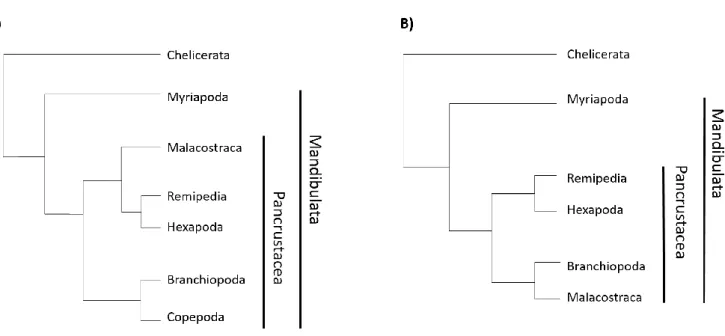
While there is an ongoing debate on the phylogenetic relationships of different arthropod groups, recent studies point clearly towards a shared ancestor of insects and crustaceans, giving rise to the monophyletic group Pancrustacea. The interrelationships within this group are still controversial (Fig. 0-4) (Budd and Telford, 2009; Cong et al., 2014; Legg et al., 2013; Regier et al., 2010).
Despite the long-lasting interest in the study of arthropod eyes, most of our knowledge on the development and neural architecture of the visual system comes from few model organisms, usually hexapods. The most profound knowledge we have comes from Drosophila melanogaster, where genetic/molecular tools allow for a careful study the architecture of the visual system and on the molecular players that give rise to it.
Fig. 0-4 Arthropod phylogeny – Two of the current views on arthropod phylogeny based in Legg et al. 2013 (A) and Regier et al. 2010 (B)
The visual systems of crustaceans have been studied largely in the context of ecology and neuro-physiology, but not in a scale comparable to hexapods. Contributions on the development of crustacean eyes are still scarce and largely descriptive. Part of the reason for this is the lack of model organisms suitable for genetic manipulation. This scenario has been changing recently with the adaptation of the small crustacean Parhyale hawaiensis to the lab life.
3. Parhyale hawaiensis
Life cycle
Parhyale is a small malacostracan crustacean of the order Amphipoda.
The generation time of Parhyale is approximately 2 to 3 months at 26ºC and animals will continue to grow throughout their lifetime (from ~1 to ~10mm in lenght). Reproduction is continuous throughout the year, as long as the conditions are favourable. For reproduction, the male grabs the female, forming a couple, until oviposition and fertilisation of the eggs. The fertilized eggs are carried by the female in a brood pouch, situated ventrally between the thoracic appendages. Hundreds of eggs at 1-cell stage can be obtained daily from anesthetized females for injections. Once the embryos hatch, they are released from the brood pouch and sexual maturation will be reached after ~7 weeks. Parhyale has a direct development; the duration of embryogenesis is 10-12 days at 26ºC and developmental stages have been described(Browne et al., 2005). Early cell lineage is stereotypical (a common feature of malacostracan embryos (Dohle and Scholtz, 1988; Dohle et al., 2004)): the first cleavage separates left from right side for most of the ectodermal and mesodermal tissues and at the 8-cell stage each blastomere will contribute to a single germ cell layer (Gerberding, 2002; Wolff and Gerberding, 2015). This characteristic allowed for studies on germ layer specification and compensation during development (Alwes et al., 2011; Gerberding, 2002; Price et al., 2010) and limb regeneration (Alwes et al., 2016; Konstantinides and Averof, 2014). Cell divisions and migration during gastrulation are also described (Alwes et al., 2011; Chaw and Patel, 2012).
Habitat
The colonies that inhabit the labs around the world today, have all come from a single population, found in the filtration system of the John G. Shedd Aquarium in Chicago in 1997. The original source of that population is unknown.
In nature, Parhyale is distributed worldwide in tropical areas, in intertidal and shallow waters such as mangroves or rocky shores. Sighting records include the Lizard Islands (Australia), the Canary Islands, Trinidad, south-eastern Brazil, Fiji Islands. Frequent changes in salinity, temperature and turbidity in these habitats have produced a robust species that can be easily kept in the lab.
Fig. 0-5 Parhyale hawaiensis life cycle – Adult Parhyale reach sexual maturation at around 2-3 months. Embryogenesis lasts 10 days at 26ºC. Embryos at one cell stage can be retrieved from dormant females and cultured in sea water. 8 hours after fertilization the egg underwent a total of three cleavages, giving rise to 4 micromeres and 4 macromeres with restricted cell fates: El, Er and Ep give rise to left, right and posterior ectoderm, respectively; Mav gives rise to the anterior and visceral mesoderm; ml, mr originate the left and right mesoderm; en gives rise to the endoderm and g to the germline. After 9 days, at stage 28, the eyes present a red pigmentation.All scale bars are 200 µm except in the adult female that is 1000 µm. Adapted from (Stamataki and Pavlopoulos 2016). Stages after (Browne et al., 2005), early cell lineage from (Gerberding 2002).
Behaviour studies on circadian clocks show some evidence for an increased activity of Parhyale during the night (B. Hunt PhD thesis), peaking at sunrise and sunset hours.
Working with Parhyale
Parhyale was introduced to the lab by Brown and Patel in late 1990s. It has been used as a research model for almost 20 years, with a community of researchers engaged in developing new experimental tools in this species. Transgenesis (Pavlopoulos and Averof, 2005), gene misexpression (Pavlopoulos et al., 2009), gene knockdown(Liubicich et al., 2009; Özhan-Kizil et al., 2009), CRISPR/Cas9-mediated gene editing(Martin et al., 2016) , a sequenced genome and other genomic and transcriptomic resources (Blythe et al., 2012; Kao et al., 2016; Nestorov et al., 2013; Parchem et al., 2010; Zeng et al., 2011) are available in this species.
These tools and the fact that crustaceans are a sister group to hexapods, make Parhyale an attractive organism to compare with Drosophila and to make inferences about the evolution of developmental, morphological and physiological traits.
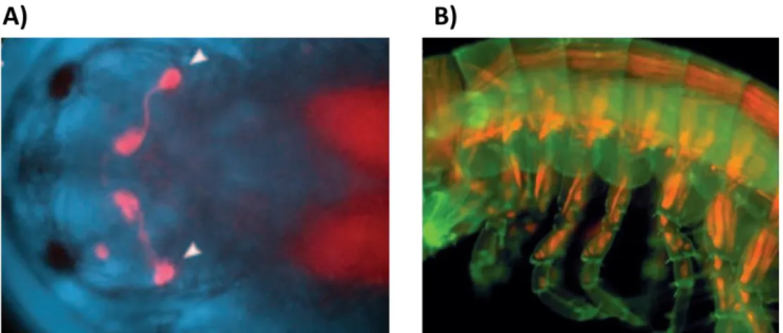
One of the most impressive features of Parhyale is its amenability for live imaging. The transparency of the embryos and of the adult cuticle allows imaging and cell tracking in embryonic, juvenile and adult stages for several days (examples in Fig. 0-6). Stunning examples are the reconstruction of the cell lineages underlying limb outgrowth using light-sheet microscopy (Wolff et al., 2017) and in the study of cell dynamics during limb regeneration (Alwes et al., 2016). Combining this characteristic with the possibility of transgenesis makes Parhyale a powerful organism for studying development, regeneration and cell behaviour in real time.
Fig. 0-6 Live imaging in Parhyale – A) Head of a Parhyale embryo seen from the dorsal side, showing dsRed expression driven by the 3xP3 regulatory sequence (white arrows) B) Trunk of a Parhyale juvenile, showing dsRed expression driven by the Ph-MuscleSpecific regulatory regions. From (Pavlopoulos 2005)
Despite the established genetic tools, many others which are routinely used in other model organisms (such as zebrafish and Drosophila) have failed to work in Parhyale. Namely we are still missing a constitutive/ubiquitous promoter despite several trials with endogenous and viral promoters (N. Konstantinides PhD thesis and A. Pavlopoulos personal communication). Also using the Cre/lox and Flp/FRT recombination systems, often employed to generate cell mosaics, proved unfruitful (N. Konstantinides PhD thesis and M. Grillo personal communication).
Cell specific markers are also still missing. One of the reasons for this is the difficulty in exploring the Parhyale’s genome for regulatory regions, due to the large intergenic regions. A gene-trapping approach has been established in Parhyale (Kontarakis et al., 2011), and few gene-trap screens have been conducted in the lab, yielding a few gene-trap lines. However, more often than expected, many of these lines proved to be unstable, both in maintenance of the trap and survival rates.
4. Purposes of the project
The diversity seen in arthropod visual systems is achieved by modifications on the optical properties of the eye and neuroanatomy. Processing of information is dependent on the connections established between the eye and the brain and within the optic neuropils. In arthropods, the knowledge on visual information processing has been largely driven by studies in a few model organisms, and principally Drosophila. The development of molecular, genetic and imaging tools in this model organism has provided great insight on the development, the neuroanatomy and sensory processing in the visual system. Outside the diptera clade, most of the studies on arthropod visual systems have relied on the usage of more classical techniques, such as transmitted electron microscopy, electrophysiology and unspecific/stochastic labelling of neuronal cells (e.g. Golgi staining). This is due, in part, on the difficulty applying modern molecular, genetic and imaging tools to non-model organisms.
Studies in Drosophila have been giving us an enormous amount of knowledge on the function of the arthropod visual system, however the lack of other organisms where genetic and imaging tools can be applied leads to a lack of knowledge on the diversity of how the visual system develops and functions, and, consequently, on its evolution. The need for new arthropod models to study the visual system has, therefore, become very important.
Parhyale has proven to be a reliable model organism where genetic and imaging tools can be applied. This provides the opportunity to compare crustacean and dipteran visual
systems in greater depth, and gain insights on the diversity and evolution of the visual system development, structure and function.
I started this project with two main (related) objectives: 1) To explore the visual system of Parhyale, focusing on a description of the eye structure, neuroarchitecture and function; 2) to develop genetic tools that allow us to label different neuronal cell types, helping to identify (and possibly manipulate in the future) different components of the visual system, starting from the primary visual sensors, the photoreceptors cells.
P
ART
1
A
DESCRIPTION OF THE
P
ARHYALE
I. Introduction
I.1 - The compound eye
The compound eye is formed by an array of multiple repetitive units, the ommatidia. Each of these units collects/senses lights from a small region in space, therefore a larger number of ommatidia results in higher image resolution. In each ommatidium we can find an optic apparatus composed of one or more lenses – usually an outer cuticular lens (or corneal lenses) and an inner lens formed by the cone cells (crystalline cone) – lying over a cluster of photoreceptor cells and their light sensing structures (the rhabdomeres). The cluster of rhabdomeres in each ommatidium is called a rhabdom; when the rhabdomeres are tightly clustered together, it is said to be a ‘fused’ rhabdom, when they are separated it is said to be ‘open’ (Fig. I-1). Open rhabdoms are found in fruit flies and house flies, while bees, mosquitos, beetles and most crustaceans have fused rhabdoms. In insects, the transition from close to open rhabdom seems to be associated with the expression of the gene Spacemaker (spam), which is only found in the eyes of insects with open rhabdom. Knock out (KO) of spam in insects with open rhabdom leads to a fused rhabdom. The opposite happens when spam is overexpressed in insects with a fused rhabdom (Zelhof et al., 2006).
Fig. I-1 The ommatidium – Schematic representation of a typical hexapod ommatidium with 8 photoreceptor cells and either an open or a fused rhabdom. Adapted from (Dan-E.Nilsson 2013)
In addition to the light focusing and sensing apparatus, pigment cells, containing granules of reflective or shielding pigment also make part of the ommatidium. This pigment, which can also be found inside the photoreceptor cells, is crucial to control the influx of light into the rhabdom, as I will discuss in Part 2 of this thesis.
Many specific modifications of this architecture occur across the arthropods.
I.1.A - Three types of compound eyes
The diversity seen in compound eyes comes from variations in the number of ommatidia, in the optic apparatus and in the neural wiring. These changes influence the resolution and sensitivity of the eye and are adapted to the animals’ needs within a specific habitat (for example, vision in water vs air or night vs day)
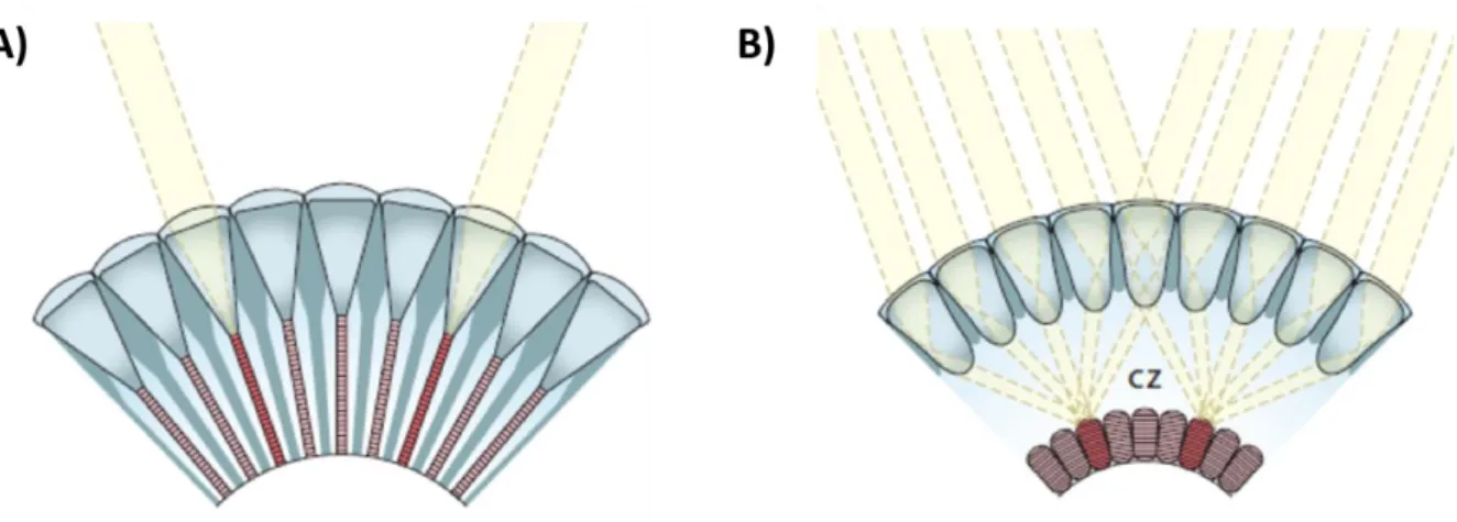
On the basis of changes in the optical arrangement and neural wiring, we can distinguish 3 main types of compound eyes: Apposition, superposition and neural superposition eyes(reviewed in(Cronin et al., 2014)) (Fig. I-2)
Apposition compound eyes
Apposition eyes are present in most diurnal insects and crustaceans. In apposition compound eyes the photoreceptors lie right beneath the lenses (crystalline cones) and extend until the proximal part of the retina. When the light enters the ommatidium through he crystalline cones, the rhabdom serves as a light guide, causing multiple reflections that will make the light to travel down until the most proximal tip. To avoid scattering of light between ommatidia, each ommatidium is surrounded by sleeves of light-absorbent screening pigments
The field of view of each ommatidium is defined by the acceptance angle, which is determined by the shape and size of the lenses. The final image perceived consists of a set of pixels, each detected by one ommatidium.
Superposition compound eyes
The main differences of superposition eyes, compared to apposition eyes, is the fact that each ommatidium is not separated by shielding pigment and the rhabdom is separated from the crystalline cone by an optically homogeneous “clear zone”.
Due to this arrangement, and to remarkable specializations of the crystalline cones, light rays entering several facets can be focused on each single rhabdom.
Since each photoreceptor receives light from many facets, the sensitivity of the eye is boosted, making the superposition eyes a common design in insects and crustaceans active under dim light conditions, such as nocturnal moths or deep sea crustaceans. However, the increase in sensitivity has costs: the absence of a shielding pigment results in decreased contrast and spatial resolution. Some species manage to overcome this trade off by employing additional specializations, allowing them to see colours at night and to follow the dim polarisation pattern of the moon light.
Neural superposition eyes
Neural superposition eyes are part of the apposition eye type, but differ by having an open rhabdom, resolving the light that enters each crystalline cone on the separate rhabdomeres. Thus, the photoreceptors within an ommatidium receive light from a slightly different point in space. Conversely, individual photoreceptors in neighbouring ommatidia receive light from exactly the same point in space; the signals of these photoreceptors are combined (superimposed) in the optic lobe, hence the name neural superposition.
Usually apposition eyes have a better resolving power than superposition eyes due to the pigment that separates each ommatidium. However, this architecture also leads to lower sensitivity. Neural superposition eyes can overcome the problem, since this design can result in a seven-fold increase in sensitivity (Kirschfeld, 1972).
Fig. I-2 Apposition and Superposition compound eyes – A) In apposition compound eyes rhabdoms are in close contact with the crystalline cone cell and each ommatidia is separated from the others by shielding pigments. Each ommatidia will form a single pixel of the final image. B) In superposition eyes, each rhabdom, which is separated from the crystalline cone cells by a clear zone (CZ), receives light focused by several lenses. From Dan-E.Nilsson.
I.2 - Photoreceptors and ommatidia structure
The cellular composition of ommatidia varies considerably when we look at the different taxa across arthropods.
The number of photoreceptor cells per ommatidium in insects and crustaceans is usually 8, but it can be as high as 10 in insects, or as low as 5 in crustaceans (reviewed in(Oakley, 2003). Important differences are also seen in the arrangement of the photoreceptors and in their spectral sensitivity. These changes can affect the sensibility of the animal to colours or to light polarisation (discussed in the Part 2 of this thesis).
In Drosophila for example, we can find a total of 8 photoreceptors per ommatidium and an open rhabdom. The 6 outer photoreceptors – R1 to R6 – span the entire length of the ommatidium, while the other two – R7 and R8 – rest in between the outer photoreceptors (therefore are called inner photoreceptors) and span only half of the length of the ommatidium. R7 is located distally, with R8 laying below (reviewed in Clandinin and Zipursky, 2002). The bee Apis mellifera has a fused rhabdom with 9 photoreceptors; of these, 8 have the same size and span the entire length of the ommatidium, whereas the 9th is a smaller cell and locates proximally (Varela, 1969).
A more complex structure is seen in butterflies, which have a so-called tiered rhabdom. Each ommatidium carries 9 photoreceptors. The distal part is a fused rhabdom with contributions from only 4 photoreceptor cells, R1-4, while the proximal part is a fused rhabdom with contributions from R5-R8. As in bees, R9 is a very small photoreceptor located proximally, and has little contribution to the rhabdom (Arikawa, 2003). Tiered receptors can also be found in stomatopod crustaceans (Cronin and Marshall, 1989b). Crabs, which have a similar structure to most other malacostran crustaceans, have a fused rhabdom formed by 8 photoreceptors. R1-R7 span the entire length of the rhabdom, while R8 sits on top, just below the cone cell (Stowe, 1977).
Fig. I-3 shows schemes for several types of ommatidia found in insects and crustaceans that can serve as a reference.
Photoreceptors also vary in the way they send their axonal projections to the part of the brain responsible for the processing of the visual stimuli, the optic lobe.
In flies, R1-R6 send projections to the first neuropil of the optic lobe – the lamina – and, therefore, have short fibers. R7 and R8 on the other hand have long projections that will cross the lamina and reach the second optic neuropil, the medulla (reviewed in Clandinin and Zipursky, 2002). Bees and butterflies have a similar arrangement with R3 to R8 projecting to the lamina, while R1, R2 and R9 project to the medulla (therefore 3 long fibers instead of 2) (Sommer and Wehner, 1975; Takemura et al., 2005; Varela, 1970). In
crabs and crayfish only R8 projects to the medulla, and R1-R7 project to the lamina (Nässel, 1976; Stowe, 1977).
A recent study was able to show that R1 and R2 of butterflies are specified similarly to R7 in Drosophila (expressing Prospero), while R9 is specified similarly to Drosophila R8 (expressing Senseless). These findings suggest that butterfly R1 and R2 are homologous to Drosophila R7, while R9 is homologous to Drosophila R8 (Perry et al., 2016).
I.2.A - Visual pigments of compound eyes
The spectral sensitivities of the visual opsins found in different animals often correlate with the spectral distribution of the light in their environment. While an insect subjected to bright day light is presented with all the visible light spectrum, marine or fresh water animals will be exposed to a narrower range of wavelengths, since long and very short wavelengths are mostly absorbed by water, and their intensity decreases rapidly with depth (reviewed in Cronin, 2006).
Phylogenetic analysis of arthropod R-opsins suggest that the ancestral pancrustacean eye had 4 visual opsin genes – LWS2, MWS1, MWS2 and SWS. Across pancrustacean evolution,
Fig. I-3 Ommatidia types in pancrustaceans – Drosophila has a total of 8 photoreceptor per ommatidium, being R7 and R8
smaller and contribute to only half of the total rhabdom length. In malacostracan crustaceans we can usually find 7
photoreceptor than contribute to almost the entire length of the rhabdom, plus an 8th photoreceptor only present at the distal
part of the ommatidium. In honeybees 8photoreceptor compose the rhabdom; a 9th, small cell is present only at the proximal
part. Butterflies have a tiered rhabdom in which the distal and proximal part are composed by 2 different sets of
photoreceptor; a 9th smaller cell is present only at the proximal tip. Adapted from (A. Kelber and M. Henze 2013 and A. Kelber
these ancestral genes were repeatedly duplicated, lost or have seen their expression change from one type of eye to another (Henze et al., 2015).
This dynamic evolution has resulted in mono, di, tri and tetra chromatic eyes, present in pancrustaceans.
Drosophila has 6 opsins in the visual system – Rh1 to Rh6. Outer photoreceptors R1 to R6 express Rh1, a blue- green (MWS) absorbing visual pigment, while R7 can express either Rh3 or Rh4 (both UV absorbing) and R8 expresses Rh5 or Rh6 (blue and green respectively). Rh2 is only expressed in the ocelli (Hardie, 1985, 1986).
Fly ommatidia can therefore differ from each other, resulting in a retinal mosaic in which we can find 2 types of ommatidia, stochastically distributed across the eye: ‘yellow’ ommatidia, where R7 expresses Rh4 and R8 expresses Rh6, and ‘pale’ ommatidia with R7 expressing Rh3 and R8 expressing Rh5 (Hardie, 1985, 1986).
Retinal mosaics are common in insects (for example, bees have 3 ommatidial types (Wakakuwa et al., 2005)) but can also occur in crustaceans, with the most extreme example found in the mantis shrimp (Cronin and Marshall, 1989b; Marshall, 1988). Besides retinal mosaics related to colour, the retina can be often regionalized, with specific areas bearing certain ommatidial types in order to perform a specific function. This is the case of the “dorsal rim area” and ‘ventral eye” in Drosophila, which specialize in the detection of polarised light, as discussed in the part 2 of the thesis (Wernet et al., 2012).
Most crustaceans possess only 2 opsins, a MWS expressed in R1-R7 and a SWS expressed in R8 (Marshall et al., 2003). There are however exceptions; we can find deep sea crustaceans with only one opsin, or others, like Daphnia, with 4 opsins (Smith and Macagno, 1990). Again, the most extreme case is seen in stomatopods, which have as many as 15 opsins (Porter et al., 2012b). The crayfish Procambarus clarkii has been shown to express different types of opsins depending on the time of the year (reviewed in (Cronin and Hariyama, 2002)).
It should also be noted that, despite the normal situation of having one rhodopsin per photoreceptor, we can also find cases where 2 opsins are expressed in the same cell. Examples include the crab Hemigrapsus sanguineos (Sakamoto et al., 1996) and some photoreceptors in butterfly eyes (Arikawa, 2003). We can also find cases where different photoreceptors are sensitive to different wavelengths even though they express the same rhodopsin. This is achieved, by the presence of different screening pigments in the ommatidium (carotenoids, ommochromes and/or pteridines), which give the eye their characteristic colour. They are used as optic filters and that constrain the spectral
sensitivity of the photoreceptor. Examples include butterflies(Arikawa, 2003), house flies(Hardie, 1986) and stomatopods (Cronin et al., 2001).
Therefore, it is difficult to deduce the spectral sensitivity of a given photoreceptor based solely on which opsin is expressed.
I.3 - Visual information processing – the optic lobe
Despite their small brains, arthropods are capable of performing complex visual tasks fast and reliably: search for food and mating partners, fight conspecifics, and avoid obstacles and predators.
The part of the brain responsible for visual information processing is collectively called the optic lobe. In arthropods, it consists of a number of neuropils which lie externally to the protocerebrum.
In pancrustaceans we can identify 4 to 5 neuropils that compose the pathway for visual information processing: lamina, medulla (outer and inner medulla in insects), lobula and lobula plate.
Each of these parts contains an ordered representation of the external space – a retinotopic map. Particular operations (spatial/temporal filtering, motion and colour detection) are processed in series or in parallel in the different layers. This ordered processing of information is a requisite for later operations that will translate into behaviours (reviewed in Strausfeld et al., 2006).
Visual processing starts when light reaches the photoreceptor cells within the ommatidia. There, light information will be transduced into neuronal signals. Photoreceptors receiving light from the same point in space, converge their axons to the same location within the first synaptic neuropil – the lamina – forming a column called the optic cartridge. While short photoreceptor fibers terminate there, long photoreceptor fibers cross the lamina and terminate in the medulla.
Each cartridge is placed in the lamina in a retinotopic manner. Thus the number of cartridges corresponds to the number of facets (ommatidia) present in the eye. In addition to photoreceptors, each cartridge also includes second order neurons, which establish connections within the cartridge and between cartridges (within or outside the neuropil).
In pancrustacean optic lobes we find two chiasmas, where the axons connecting consecutive neuropils cross each other: between the lamina and the medulla, and between the medulla and the lobula (Sinakevitch et al., 2003). The crossing of axons causes the inversion of the order of cartridges, and therefore of the retinotopic map. Chiasms are an evolutionary novelty of the pancrustacean optic lobes, not being found in
other arthropods, and are a consequence of the mode of development of the neuropils (Strausfeld, 2005).
The best studied arthropod optic lobe is undoubtedly that of Drosophila. It has given us valuable information on how visual information is computed in the brain. I will therefore use it as an example to introduce visual information processing.
I.3.A - Visual information processing – lessons from the fly
Flies have neural superposition compound eyes, in which each ommatidium is composed of 8 photoreceptor cells. Due to the open rhabdom, photoreceptors from the same ommatidium do not receive light form the same point in space. Instead, any given point is “seen” by individual photoreceptors residing in 7 neighbouring ommatidia (Fig. I-4 C). The signals received by these 7 photoreceptors converge in individual cartridges of the lamina (R1-R6) and the medulla (R7 and R8).
In addition, cartridges in the lamina contain neuronal processes from monopolar cells (L1 to L5), C2, C3 and T1 neurons, plus amacrine cells. L1 and L2 are the main post-synaptic targets of R1-R6 and have been shown to play a crucial role in motion detection. Some of those lamina interneurons leave the lamina cartridges and form connections in the medulla; in particular, L3 connects directly to the R7 and R8 axonal projections (reviewed in Zhu, 2013).
Medulla cartridges are far more complex than lamina ones, consisting of processes from around 60 neurons. Those will project either within the medulla (some connecting the outer with the inner medulla) or towards the deeper neuropils – the lobula and lobula plate (Fischbach and Dittrich, 1989).
The lobula plate contains wide-field tangential cells that integrate signals from hundreds of R1-R6 pathways. It is in this neuropil where computation of optic flow direction seems to take place. The lobula is the least studied neuropil; it is thought to be sensitive to object features such as orientation, texture and colour (Strausfeld and Lee, 1991).
Finally, visual projection neurons connect the medulla, lobula and lobula plate to the central brain. These play a dual role, conveying information from the retina to the central brain and also sending command signals back from the central brain to the optic lobe.
Colour and motion
Colour vision is the ability to discriminate between light stimuli of different spectral composition, independently of their relative intensity. It is used to detect, recognise and discriminate objects. It is mediated by colour opponent (or coding) neurons, which receive opposing inputs (excitatory and inhibitory) from receptors of different spectral types (Chittka et al., 1992; KELBER et al., 2003).
Fig. I-4 Structure of the Drosophila optic lobe – A) Scheme of the Drosophila optic lobe showing the retina Re, lamina La, medulla Me, lobula Lo and lobula plate Lop; B) Golgi staining of a Drosophila optic lobe showing the 5 neuropils and the chiasms (X1 and X2) that connect them. From (Fischbach and Dittrich, 1989) C) Scheme of the connections between R1-R6 and the lamina. Each lamina cartridge receives input from a single photoreceptor of different, neighbouring ommatidia. Adapted from (R. Sanes and S. Zipursky 2010)
In the fly medulla, intricate connections involving interneurons connected to R7 and R8 axonal projections (which have a different spectral sensitivity) point towards being part of a pathway of colour opponent neurons. Therefore, the medulla is often regarded as the neuropil responsible for the first stages of colour information processing, being fed information from R7 and R8 – the chromatic pathway.
R1-R6 have the same spectral sensitivity and make part of the achromatic pathway, providing information on motion (rather than colour), which is primarily processed at the lamina by L1 and L2 neurons.
This separation between colour and motion processing is not, however, strict. Studies on the ultrastructure of R7/R8 axons showed the existence of synapses between those and lamina monopolar neurons in the medulla, and also the existence of gap junctions and synapses between R7/R8 and R6 axons in the lamina(Shaw, 1984; Shaw et al., 1989; Takemura et al., 2008; Zheng et al., 2006). A more recent study confirmed that R7/R8 signals improve motion discrimination and adjust the sensitivity of the optomotor response (Wardill et al., 2012). These input signals are included in the motion detection circuit through interaction of R7/R8 axons with lamina monopolar cells in the medulla but also through the gap junctions between R7/R8 and R6 axons.
Conversely, it has been shown that blockage of L3 lamina monopolar cells (which connect R1-R6 in the lamina to R7-R8 in de medulla) impairs bright blue/green discrimination. This suggests that, in the medulla, there is colour opponent processing between R1-R6 and R7-R8 (Garbers and Wachtler, 2016; Schnaitmann et al., 2013a).
I.3.B - Optic lobe evolution in Arthropods
The arrangement of 4 optic neuropils is found in most pancrustaceans, independently of the type of compound eye that they use. There are however some exceptions in crustaceans and insects that underwent secondary loss or reduction of one or two neuropils. For example, branchiopod crustaceans, like Artemia and Daphnia, possess only 2 optic neuropils: a lamina, directly connected to the retina, and a tectum-like deeper neuropil. Both are connected to each other by uncrossed retinotopic axons (Harzsch and Glötzner, 2002; Nässel et al., 1978). The latter neuropil is thought to be homologous to the pancrustacean lobula plate (Strausfeld et al., 2016). In addition, the size of the lobula plate varies considerably when comparing different crustaceans, and it can be very small in many decapods(Sinakevitch et al., 2003).
Myriapods present only two optic neuropils and, both receive input from the retina. Axons connecting the two neuropils seem to cross each other, suggesting the presence of a
chiasma (Sombke and Harzsch, 2015). Similarly, xiphosurans have two optic neuropils connected through a potential chiasm (Harzsch et al., 2006).
Fossils of the euarthropod Radiondata brain show stout optic nerves associated with 2 enlarged areas, suggesting that this extinct arthropod stem group also possessed only 2 optic neuropils (Cong et al., 2014).
Taken together, these observations suggest that the ground pattern of the arthropod visual system comprises an apposition compound eye connected to two nested optic neuropils, a condition still seen today in Myriapods. The addition of two additional optic neuropils, the medulla and lobula, plus the chiasma that connects them, represents an evolutionary novelty of the pancrustacean clade (Strausfeld, 2005; Strausfeld et al., 2016).
I.4 - Purposes of Part 1
Despite the many studies on crustacean visual systems, we are still lacking models were genetic tools can be applied in a similar way has in Drosophila. Parhyale is therefore an opportunity to study visual system evolution within the Pancrustacea clade.
This part of the project, which composes the major part of my thesis, is dedicated to the description of the Parhyale visual system, providing an essential basis for future works.
II. Results
II.1 - Structure and growth of Parhyale eyes
Parhyale have been in the lab for almost 20 years but there are still no descriptive studies on the anatomy and neural connectivity of their eyes. I therefore started my project with a basic description on the structure of the Parhyale eye.
Parhyale has an apposition compound eye with 5 photoreceptors per ommatidium
Parhyale adults have one pair of dark, kidney-shaped compound eyes, located laterally in the head. As seen from the surface of the eye, the facets are arranged in rows, have a round shape and are surrounded by a white reflective pigment (Fig. II-1 A). This pattern is seen in almost all animals, with few exceptions where facets seem to have more irregular shapes (Fig. II-1 B). Around 5% of the animals (rough estimate) show this arrangement which may be due to a developmental defect. Scanning Electron Microscopy (SEM) images of Parhyale’s juveniles (data by T. Pavlopoulos and M. Averof) reveal that the cuticle above the eyes is smooth and facets are not perceptible, indicating that the cuticle is not specialized to work as a lens (Fig. II-1 C).
To further characterize the fine structure of the eye, adult heads were fixed and prepared for Transmitted Electron Microscopy (TEM). EPON-embedded samples were sectioned in semi-thin (2µm) or ultra-thin (50nm) sections and analysed with a light and transmission electron microscope respectively. Data from serial, longitudinal and transversal, semi-thin sections were collected from at least 6 different animals. For longitudinal and transversal ultra-thin sections a total of 2 animals was used (Fig. II-2 A and B, respectively). To complement the information given by the semi- and ultra-thin sections, thick sections (100µm - 300 µm) of adult heads were stained with DAPI and imaged with a confocal laser scanning microscope.
In semi-thin sections, stained with toluidine blue, each ommatidium is seen separated from the others by densely packed dark vesicles, the shielding pigment granules. The rhabdom, which has a cone shape, lays just below the dioptric apparatus (or lens, stained in dark blue) (Fig. II-1 D). The pigment-shielded ommatidia, with rhabdoms in close proximity to the lenses, are characteristic of apposition compound eyes.
The lens is formed by 2 crystalline cone cells ((Fig. II-1 E-E’). I was not able to observe the nuclei of these two cells, neither the nuclei of the 2 accessory crystalline cone cells that have been described for amphipods (Hallberg and Nilsson, 1980).
Longitudinal sectioning shows that the eye has two distinct parts: a more distal one, comprising the lens and rhabdoms; and a proximal region, comprised mainly of nuclei.
Very few nuclei are seen at the distal part of the eye, suggesting that photoreceptor nuclei are positioned at the proximal region. This separation has been noted before in amphipods (Hallberg and Nilsson, 1980) and a fenestrated membrane was described separating the 2 regions. The presumed position of the fenestrated membrane is partially marked in the figures, at the proximal end of the rhabdom (Fig. II-1D-F).
In TEM sections, rhabdoms are distinguished by their densely packed microvilli, surrounded by electron dense vesicles (shielding pigment granules). These vesicles present different TEM contrasts (white arrow heads in Fig. II-2 C) which correspond to different pigment types (probably ommochromes and carotenoids). There are no cell membranes separating the microvilli from the pigment granules, indicating that pigment granules are enclosed within the photoreceptor cells. The reflective white pigment, which appears as empty vesicles, remains outside of the photoreceptor cells, presumably within reflective pigment cells that surround the ommatidia. No dark pigment granules were found outside the photoreceptor cells.
Transversal sections of the ommatidia show a rhabdom with a rayed (star-like) shape, composed of five photoreceptor cells, named R1 to R5 (Fig. II-2 D-D’). The rhabdomeres are in close contact with each other. In all the sections observed, R1 – R4 have a similar size while R5 is considerably smaller than the others, possessing a diminutive rhabdomere. The microvilli of R1 and R3 are oriented in parallel to each other and orthogonally to R2 and R4. Interdigitation of the rhabdomeres was not seen in any of the longitudinal sections (>4 sections) (Fig. II-2 E).
Photoreceptor cells extend until the most distal part of the eye, surrounding the lens. Cytoplasmic extensions are seen between the photoreceptor and the lens (Fig. II-2 F). Some photoreceptors from neighbouring ommatidia seem to contact each other via membrane extensions (red arrows in Fig. II-2 G). It was not possible to determine, in general, which and how many photoreceptors show this connection.
From these results we can conclude that Parhyale has an apposition compound eye, with 5 photoreceptors per ommatidium. The rhabdom is fused, and, unlike other crustaceans, there is no interdigitation of the rhabdomeres.
Fig. II-1 General features of Parhyale adult eyes – A) detailed view of a Parhyale compound eye. Facets are arranged in rows. B) Few animals carry ommatidia with irregular facets C) SEM of the head. The cuticle above the eye is smooth in the surface and facets are not perceptible. D) Longitudinal semi-thin plastic section stained with toluidine blue, showing the apposition eye of Parhyale. E-E’) Detail of the dioptric apparatus composed by two cone cells, in semi-thin plastic sections. Arrowheads point to the separation between the two cells F) DAPI staining of the eye, showing the nuclei beneath the fenestrated membrane. CC- crystalline cone cell RH- rhabdom FM- fenestrated membrane
Fig. II-2 TEM details of adult Parhyale eyes – A-B) Longitudinal and transversal TEM sections of the Parhyale eye; C) Longitudinal TEM section of a single rhabdom. White arrowheads point to the different
pigment granules, black arrows point to the photoreceptor cell membrane. D-D’) TEM Cross section of a rhabdom and respective scheme. Each ommatidium is composed of 5 photoreceptor cells with a fused rhabdom. E) Longitudinal TEM section through a rhabdom. The yellow line marks the meeting point between two opposing rhabdomeres; F) Cytoplasmic connections between a photoreceptor cell and a crystalline cone cell G) Connections between 2 photoreceptor cells from neighbouring ommatidia. RH- rhabdom RHE- rhabdomere RPC – reflective pigment cell.
Eyes keep growing during the life time of Parhyale
From the moment they hatch to the end of their life, Parhyale keep growing with each molt. The size of their body increases, allowing for a rough estimate of their age based on body size. Alongside the body, the eyes also keep growing.
In embryos, the eyes are first visible at around stage 26 (S26) (Browne et al., 2005), when 3 lenses are perceptible. A red pigment is first visible at S27, where also 6 smaller, less developed ommatidia can be seen surrounding the older trio of lenses. 24h later, at S27-28, the white reflecting pigment can be seen surrounding the ommatidia. The aligned rows start to be distinguishable in 1 month old animals (Fig. II-3 B).
To document how much the eyes change, I quantified the number of ommatidia, as well the dimensions of each eye, in different aged animals (body size was used as a proxy for age) ( Fig. II-3 A). Young hatchlings present around 8 ommatidia per eye, and this number steadily increases, reaching 50 ommatidia in the oldest adults (a 6x increase in ommatidia number). In the same animals, the length of an eye can increase 7x from ~0.037 mm to ~0.27 mm and the width of the eye increases by 6x from 0.026mm to 0.16mm. Considering the “kidney” shape of the eye, the total area was calculated as an ellipse, which varies from around 800µm2 to 34000µm2, a 40x increase.
During growth, the surface area of each ommatidium also increases from 7-12 µm in hatchlings to 20-30 µm in adults (based in measurements from 2 hatchlings and 2 adults), which corresponds to ~6x increase in the surface of the ommatidium.
Based on these results, we can estimate that the increase in the area of the eye is mostly due to an increase in both ommatidial size and number.
Fig. II-3 New ommatidia are added during the life time of Parhyale - A) quantification of the number of ommatidia and eye size vs body size. Adult animals can have up to 50 ommatidia per eye, while hatchlings have around 8 ommatidia. B) Eye development from embryos to 1 month old hatchlings. At stage27 embryos have 3 big ommatidia surrounded by 6 less developed ones. The alignment pattern of into rows only starts to be distinguishable in one month old animals.