HAL Id: tel-02917978
https://tel.archives-ouvertes.fr/tel-02917978
Submitted on 20 Aug 2020HAL is a multi-disciplinary open access archive for the deposit and dissemination of sci-entific research documents, whether they are pub-lished or not. The documents may come from teaching and research institutions in France or abroad, or from public or private research centers.
L’archive ouverte pluridisciplinaire HAL, est destinée au dépôt et à la diffusion de documents scientifiques de niveau recherche, publiés ou non, émanant des établissements d’enseignement et de recherche français ou étrangers, des laboratoires publics ou privés.
Autophagy as a potential therapeutic target for
Sjögren’s syndrome
Baihui Li
To cite this version:
Baihui Li. Autophagy as a potential therapeutic target for Sjögren’s syndrome. Immunology. Uni-versité de Strasbourg, 2017. English. �NNT : 2017STRAJ042�. �tel-02917978�
Acknowledgements
Firstly, I would like to express my extraordinary gratitude to the members of my thesis jury: Dr. Benjamin Dehay, Dr. Martine Biard and Dr. Jean Sibilia for their time and effort to evaluate my study.
My sincere thanks goes to my director Prof. Sylviane Muller for supporting my study and providing me an excellent scientific atmosphere. She offered me the opportunities to conduct my research in IBMC. Her patience, insistence, and immense knowledge inspired me not only in scientific research, but also in life. She has shown me what a real scientist should be. She sacrificed her time to help me with my study and this thesis. My endless thanks is far beyond the words described here.
A heartfelt thanks goes to Dr. Hélène David-Jeltsch for her valuable suggestions and guidance in my thesis writing, and for her encouragement and kind-hearted attention on my study. She exchanged with me frequently and supported me greatly. She was always willing to help. I am very lucky to have her as a colleague.
I am especially indebted to Dr. Fengjuan Wang, the postdoc of our team and good friend of mine. She shared her experiences of research with me, and helped me to analyze the woes in my study. She was always the first one trying to defend me when misunderstandings happened on me. She helped me to refine my presentations and thesis, generously dedicated her knowledge to improve my work. A special thanks goes to Nicolas Schall, the engineer of our lab who showed me how to use the instruments and how to perform “perfect” experiments. He taught me with great patience and warm advises. We are lucky to have such a professional and highly motivated colleague in our team.
A lot of thanks goes to Hayet Dali for giving me trainings at the beginning of my study, she shared her expertise with me and showed me how to improve my experiments. She provided a lot of help for my research, and she is also my first French teacher. My deep thanks also goes to the Delphine and Monique who conducted the animal experiments in this study. The long-term treatments involve injections and bleedings
several times in dozens of mice, and they dedicated hard work to help me with this project.
This work would not have been possible without the technical support of Jean-Daniel. I am very grateful to him for showing me how to use microscopies, how to prepare samples and how to analyze data. Especially for the TEM studies, he cooperated with me to perform these experiments and tried his best to find solutions for the obstacles we have met. His patience, talent and virtuous personality deeply affected me.
I would like to thank Florent for being both my friend and colleague, encouraging me and supporting me during my study.
I would like to thank the Image center specialists Valérie Demais and Cathy Royer for their participation in the TEM experiments. They greatly supported my work and helped me to get valuable results.
I am also grateful to all of those whom helped me with this project, Astrid, Jean-Baptiste, Benjamin…
I would like to thank my friends for their wise counsel and sympathetic ear, for encouraging me and understanding me, for being with me to go through all the difficulties.
Finally, I would like to thank my family. My parents always support me and encourage me to pursue what I want. Their love and inspiration are always there for me, lighting up my life.
Content
INTRODUCTION ... 1
A. Sjögren's syndrome ... 2
A.1: Symptoms ... 2
A.1.1: Dryness ... 2
A.1.2: Chronic inflammation ... 3
A.1.3: Neuronal manifestations ... 4
A.2: Prevalence and criteria ... 5
A.2.1: Prevalence ... 5
A.2.2: Criteria ... 6
A.3: Factors and inducers ... 11
A.3.1: Environmental factors ... 12
A.3.1.1: HCV ... 13
A.3.1.2: EBV ... 14
A.3.2: Genetic factors ... 15
A.3.2.1: Human leukocyte antigen ... 15
A.3.2.2: NF-κB pathway-related genes ... 16
A.3.2.3: IFN signaling-related genes ... 17
A.3.2.4: Lymphocyte signaling pathway-related genes ... 18
A.3.3: Hormonal factors ... 19
A.4: Pathology ... 20
A.4.1: Cytokines ... 20
A.4.1.1: Th1 and Th2 balance ... 21
A.4.1.2: IL-17, IL-21 and other cytokines ... 22
A.4.2: B cells ... 23
A.4.3: T cells ... 24
A.4.4: Central role of epithelial cells ... 25
A.4.4.1: Autoantigen Production ... 27
A.4.4.2: Non-professional antigen-presentation ... 29
A.4.4.3: Stimulation of T cells and B cells ... 29
A.4.4.4: Cytokines ... 31
A.4.4.5: Toll-like receptors ... 33
A.5.1: Dryness ... 34
A.5.2: Organs regeneration ... 35
A.5.3: Chronic inflammation ... 35
B. Autophagy ... 38 B.1: Autophagy pathways ... 40 B.1.1: Macroautophagy... 40 B.1.2: Microautophagy... 40 B.1.3: Chaperone-mediated autophagy... 40 B.1.4: Mitophagy ... 42
B.1.5: Other autophagy pathways ... 44
B.2: Machinery of autophagy ... 44
B.2.1: Biogenesis and Initiation... 45
B.2.1.1: Membrane source ... 45
B.2.1.2: The initiation machinery ... 47
B.2.2: Elongation and formation of autophagosome ... 49
B.2.2.1: ATG8 modification by two ubiquitin systems ... 49
B.2.2.2: Membrane expansion ... 50
B.2.2.3: Cargo Receptors ... 51
B.2.3: Fusion ... 53
B.2.3.1: Maturation ... 53
B.2.3.2: Delivery to lysosomes ... 54
B.2.3.3: The final fusion ... 55
B.2.4: Degradation ... 56
B.2.4.1: Lysosome: the organelle for degradation ... 56
B.2.4.2: V-ATPases ... 57
B.2.4.3: Counterion pathway ... 58
B.2.4.4: Enzymes in lysosome ... 59
B.2.4.5: The regulator of lysosome: transcription factor EB ... 59
B.3: Autophagy and autoimmunity ... 60
B.3.1: Autophagy in cellular homeostasis ... 60
B.3.1.1: Autophagy in T cells ... 60
B.3.1.2: Autophagy in B cells ... 61
B.3.1.3: Non-immune cells ... 63
B.3.2: Autophagy and cytokines ... 63
B.4: Autophagy and other diseases ... 68
B.4.1: Autophagy in infection ... 68
B.4.2: Autophagy and neurodegenerative diseases ... 69
B.4.3: Autophagy and cancer ... 70
AIM OF THE STUDY ... 72
RESULTS ... 76
PUBLICATION 1... 77
Introduction ... 78
Result: Assessing Autophagy in Mouse Models and Patients with Systemic Autoimmune Diseases ... 80
Comments ... 102
Perspectives ... 102
PUBLICATION 2... 105
Introduction ... 106
Result: Critical role of autophagy processes in Sjögren’s syndrome ... 108
Comments ... 146
Perspectives ... 146
CONCLUSION AND GENERAL DISCCUSION ... 149
Mouse models of Sjögren's syndrome ... 150
Autophagy in autoimmune diseases ... 154
Various roles of autophagy in autoimmune response ... 154
The roles of autophagy in Sjögren's syndrome ... 155
Therapeutics of Sjögren's syndrome... 156
Autoimmune diseases ... 156
Therapeutics of autoimmune diseases ... 158
Therapeutic peptide P140... 159
ABBREVIATIONS
3-MA 3-methyladenine
ACR American College of Rheumatology
AD Alzheimer disease
AECG American-European Consensus Group classification criteria
AID Autoimmune disease
Aly/aly Alymphoplasia
ANA Antinuclear antibody
APCs Antigen presenting cells
ATG Autophagy related
AVs Autophagic vacuoles
BafA1 Bafilomycin A1
BAFF B cell active factor
BAG-1 BCL2-associated athanogene 1
Bax BCL2 associated X protein
BCL2 B-cell CLL/lymphoma 2
Bif-1 Endophilin B1
BNIP3L BCL2 interacting protein 3 like
C4 Complement component 4
CA6 Carbonic anhydrase VI
CALCOCO2/NDP52 Calcium binding and coiled-coil domain 2
CCL C-C motif chemokine ligand
CMA Chaperone-mediated autophagy
CMV Cytomegalovirus
CQ Chloroquine
CTLA-4 T-lymphocyte-associated protein 4
CV Coxsackie virus
CXCL C-X-C motif chemokine ligand CXCR C-X-C motif chemokine receptors
DC Dendritic cells
DFCP1 Double FYVE-containing protein 1
DHEA Dehydroepiandrosterone
DHT Dihydrotestosterone
DLBCL EBV positive diffuse large B cell lymphoma EAE Experimental autoimmune encephalomyelitis EAM Experimental autoimmune myocarditis
EBVEA Epstein Barr virus early antigen
EM Electron microscopy
ER Endoplasmic Reticulum
FAS TNF receptor superfamily, member 6
FASL FAS ligand
FcγR Fc gamma receptors
FDCs Follicular dendritic cells
FIP200 FAK family kinase-interacting protein of 200kDa Foxp3 Forkhead box protein P3
FYCO1 FYVE and coiled-coil domain containing 1
GC Germinal center
GWAS Genome-wide association study hAECs Human amniotic epithelial cells
HBV Hepatitis B virus
HCV Hepatitis C virus
HIP HSP70 interacting protein
HIV Human immunodeficiency virus
HLA Human leukocyte antigen
HOP HSC70/HSP90-organizing protein
HOPS Homotypic fusion and protein sorting HSP40 Heat shock 40kDa proteins
HSP90 Heat shock 90kDa proteins
HSPA8 Heat shock protein family A member 8 HTLV-1 Human T-lymphotropic virus Type 1 ICAM-1 Intercellular adhesion molecule-1
IFN Interferon
Ig Immunoglobulin
IKBKE Inhibitor of nuclear factor kappa B kinase subunit epsilon
IL Interleukins
ILD Interstitial lung disease IP-10 IFN-γ-induced protein 10 IRFs Interferon regulatory factors
KIR Keap1-interacting region
LAMP Lysosomal membrane protein
LAP LC3-associated phagocytosis
LD Lipid droplet
LIMP Lysosomal integral membrane proteins
LRS LC3-recognition sequence
LSG Labial salivary glands
MAP1LC3/LC3 Microtubule associated protein 1 light chain 3 MRL/lpr Murphy Roths Large/lymphoproliferation
MS Multiple sclerosis
MSGs Minor salivary glands
mTOR Mammalian target of rapamycin
NBR1 Neighbor of BRCA1 gene 1
NF-κB Nuclear factor kappa B
NOD Non-obese diabetic
NZB/W F1 New Zealand Black/White F1 Hybrid
OMM Outer mitochondrial membrane
OPTN Optineurin
PAS Pre-autophagosome structure
PB1 N-terminal Phox and Bem1p region PBMCs Peripheral blood mononuclear cells pDC Plasmacytoid dendritic cell
PE Phosphatidylethanolamine
PI3KC3 Class III PI 3-kinase complex PI3P Phosphatidylinositol-3-phosphate
PSP Parotid secretory protein
pSS Primary Sjögren's syndrome
RA Rheumatoid arthritis
RF Rheumatoid factor
RILP Rab interacting lysosomal protein. SGEC Salivary gland epithelial cell
SGP Salivary gland proteins
SGs Salivary glands
SHBG Sex hormone-binding globulin
SICCA Sjögren’s International Collaborative Clinical Alliance
SLE Systemic lupus erythematosus
SNPs Single nucleotide polymorphisms SP-1 Salivary glands protein-1
SQSTM1/p62 Sequestosome
SS Sjögren's syndrome
SSA/Ro Sjögren's syndrome-related antigen A SSB/La Sjögren's syndrome-related antigen B
SSc Systemic sclerosis
sSS Secondary Sjögren's syndrome
T1D Type 1 diabetes
TCR T cell receptor
Tfh Follicular helper T cells
Th Helper T cells
TLRs Toll-like receptors
TNF Tumor necrosis factor
TNFAIP3 TNF-α induced protein 3 TNIP1/ABIN1 TNFAIP3 interacting protein 1
TPC Two-pore complex
Treg Regulatory T cells
TRP Transient receptor potential
Ub Ubiquitin
UBA Ubiquitin-associated domain
UBDs Ub-binding domains
ULK1 Unc-51-like autophagy activating kinase 1 VCAM Vascular cell adhesion molecule
VMP1 Vacuole membrane protein 1
WB Western blotting
WIPI WD-repeat domain phosphoinositide-interacting Zinc ZZ-type zinc finger domain
1
2
A. Sjögren's syndrome
The clinical features of Sjögren's syndrome (SS) were first described in 1888 by Dr. Hadden, who reported a 65-year-old woman with severe ocular dryness to the Clinical Society of London (Hadden, 1888). In 1925, a French ophthalmologist, Dr. Gougerot found that in certain patients, ocular dryness was only a part of systemic dryness affecting mouth, larynx, nasal and vaginal mucosa, and these symptoms might occur with a possible decrease in the function of the thyroid and ovaries (Marchal et al., 1947)
In 1933, Dr. Henrik Sjögren, a Swedish ophthalmologist reported 19 clinical cases suffering from dry eyes and dry mouth. He found that most of those patients were postmenopausal women with arthritis, showing pathological manifestations of the syndrome (Gren, 1933). Since then, SS has been recognized as a chronic inflammatory disease, and numerous investigations and studies in both clinic and laboratory have been performed in this disease.
A.1: Symptoms A.1.1: Dryness
As a chronic, systemic, inflammatory autoimmune disease, SS is characterized by lymphocytic infiltration in exocrine glands. Lymphocytic infiltration in the salivary glands (SGs) and lacrimal glands (LGs) causes destruction of glandular tissues, leading to symptoms like dry eyes (xerophthalmia) and dry mouth (xerostomia). Patients with dry eyes may feel burn, itch or gritty, and continuous uncomfortable in their eyes. Dry eyes can be found in patients with SS by clinical tests that include tear break-up times, Schirmer’s-I test scores, response rates and cornea fluorescent staining (Lee et al., 2011). Dry mouth is led by reduced saliva secretion. In patients with dry mouth, the oral health is difficult to maintain as the skin in the mouth cannot be moistened, and patients can feel difficulties in speaking and eating. As saliva is the resources of epidermal growth factors that promote tissue growth, differentiation and wound healing, patients with dry mouth are vulnerable for oral
3
infections and inflammation (Furness et al., 2013). Xerophthalmia and xerostomia are found frequently in old people, and regular pharmacotherapy can be effective. However, as the glandular tissues are attacked by autoimmune responses and damaged in patients with SS, regular treatments must be continuous and relatively long lasting.
Besides SGs and LGs, SS patients also suffer from dryness in other organs or tissues such as nose, respiratory system and skin (Figure 1).
Figure 1. Tissues and organs affected by SS
Patients with SS suffer from various clinical manifestations in multiple organs and tissues. Dryness affects glandular tissues including SGs and LGs. Chronic inflammations are found in lymph nodes, lungs, kidneys, joints and muscles, causing manifestations and dysfunctions in these organs. In addition, central nervous system and respiratory system functions can also be affected by SS.
A.1.2: Chronic inflammation
SS has been recognized as a systemic rheumatic disease. It presents several rheumatic features such as chronic inflammation in kidneys and lungs, as well as
4
connective tissue arthropathy. Renal function may be affected in two different aspects, which are the tubulointerstitial nephritis caused by epithelial inflammation or glomerulopathy led by immune complex deposition (Evans et al., 2015). Compared with tubulointerstitial nephritis, glomerulonephritis is associated with higher frequency of early mortality (Kidder et al., 2015). Interstitial lung disease (ILD) is one of the major systemic manifestations of SS. Patients with ILD have pulmonary complications that are characterized by various patterns of inflammation and fibrosis in lungs (Vij et al., 2013). Impaired pulmonary functions include reduced carbon monoxide-diffusing capacity, less peak expiratory flow and forced vital capacity, which can result in significant morbidity and mortality (Zhao et al., 2013).
Around 10% of the patients develop cancers, among which B-cell lymphoma is the most frequent type and represents the most severe complications for SS patients (Brito-Zeron et al., 2017). The association between SS and lymphoma was identified as early as 1978, when a much higher incidence of lymphoma (43.8 times of same age general population) was found in SS patients (Kassan et al., 1978).
A study evaluating 25-year outcome of 152 patients with SS found that around 10% of SS patients present vasculitis (Abrol et al., 2014). Vasculitis is inflammation of blood vessels that can affect multiple organs in the body. Vasculitis, which is often caused by immune attack of blood vessels and induces systemic inflammation, leads to symptoms like fever, general aches and pains among patients.
Other organs/tissues including the liver (liver fibrosis) (Lee et al., 2016), the joints (joint pains/arthritis) (Amezcua-Guerra et al., 2013), and muscles (myositis) (Colafrancesco et al., 2015) could also be affected in SS (Figure 1).
A.1.3: Neuronal manifestations
In addition to the symptoms described above, some abnormalities in nervous system have been reported in SS patients. Prevalence of neurological manifestations in SS varies from 10-60%. This wide range is due to the criteria used for SS definition as well as methodology differences, and the methods used for detecting neuropathy (Chai et al., 2010). Several neuropathy subtypes have been found in SS, which
5
include multiple mononeuropathy, polyradiculopathy, and sensory neuropathy. Patients may have a combination of two or more neuropathies. Both peripheral and central nervous systems are involved, and the frequencies are in 66% and 44%, respectively (Jamilloux et al., 2014). Sensory neuropathies are the most frequent neuropathies among all descriptions. One of the sensory neuropathies is the small fiber sensory neuropathy. SS patients with this form of neuropathy are diagnosed with prominent neuropathic pain, accompanied by loss of pinprick or temperature discrimination and hyperesthesia (Mori et al., 2005).
A.2: Prevalence and criteria A.2.1: Prevalence
SS is classified into primary and secondary SS. In primary SS (pSS), patients show only symptoms of SS. However, as the disease develops, other autoimmune diseases (AIDs) like rheumatoid arthritis (RA), systemic sclerosis (SSc) or systemic lupus erythematosus (SLE) can occur with SS, named secondary SS (sSS). SS affects 0.01-0.72% of the whole population (Maldini et al., 2014), and 10-30% patients suffer from sSS, in which around 18% patients suffer from SLE (Yao et al., 2012), around 12% patients show SSc associated (Avouac et al., 2010), and 4-22% patients are diagnosed with RA (Haga et al., 2012; Kosrirukvongs et al., 2012) (Figure 2).
SS is considered as the third most prevalent rheumatic autoimmune disorder, following RA and SLE. Generally, the incidence and prevalence of primary pSS can be largely variable according to criteria used and study designed, as well as geographical area chosen. A study in Germany reported a yearly incidence of 4 in 100,000 by using the American-European Consensus Group classification criteria (AECG) criteria (Westhoff et al., 2010). Another five-year study in Karolinska University Hospital reported an incidence of 3.1 in 100,000 inhabitants per year (Kvarnstrom et al., 2015). A recent study using systematic literature search on PubMed and Embase involved 1880 related citations pointed out that the incidence rates for pSS was 6.92 per 100,000 person-years, and the overall prevalence rates was 60.82 per 100,000
6 (Qin et al., 2015).
Figure 2. Epidemiology of SS
SS is classified into pSS and sSS. Patients show only symptoms of SS in pSS, while patients present combined symptoms of SS and other accompanied AIDs in sSS, which present 70-90% and 10-30% of all SS patients, respectively. In sSS, the most frequent accompanied autoimmune disease is SLE, which affects approximately 18% of sSS patients. Other patients with sSS may develop SSc or RA.
SS could occur in any population and any age, but mostly in middle age and postmenopausal women. A meta-analysis confirmed the peak incidence of pSS occurs in women aged around 56 years (Qin et al., 2015). A study of systemic autoimmune disease geoepidemiology used Google search engine to collect and analyze 394,827 patients with systemic AIDs, found that pSS represents one of the most unbalanced gender ratio, with nearly the M/F ratio at 1:9 (Ramos-Casals et al., 2015).
A.2.2: Criteria
Several criteria were used for disease classification since the first case of SS, aimed to clarify the characteristics of SS as well as to differentiate SS from other symptom-like AIDs.
In 1965, Bloch et al. reported a five-year study of 65 SS patients in which dry eyes, dry mouth and other connective tissue diseases (such as RA, SSc) were used as a definition of SS (Bloch et al., 1965).
7
In 1968, Chisholm et al. suggested the labial biopsy of SGs as a useful technique to investigate SS. In this technique, the foci per 4 mm2 salivary tissue were used to
standardize the degree of histopathology (Chisholm et al., 1968). The criteria of SS have been developed and modified many times. However, this technique has been inherited and used until now.
In 1986, four classification criteria were presented during the first international symposium on SS, namely the Copenhagen, Japanese, Greek and California criteria (Fox et al., 1986b). These criteria focus on different objective and subjective components, highlighting different diagnosis sets. Although they failed to provide a widely-accepted, complete and precise criterion for SS, they provided a platform for later studies to explore and develop a profound, unified set of criteria.
In 1992, Speight et al. emphasized the value of measuring whole unstimulated salivary flow in SS patients and proposed this simple method as a diagnosis for SS (Speight et al., 1992).
A large-scale study involving 26 clinical centers from 12 countries (11 in Europe, plus Israel) tried to reach a consensus on the diagnostic procedures for SS (Vitali et al., 1993). This study established the preliminary European criteria in 1993 (Figure 3). In this set of criteria, 2 subjective and 4 objective items were proposed:
1> ocular symptom (subjective); defined by a positive response to at least 3 dry eyes related questions.
2> oral symptom (subjective); defined by a positive response to at least 3 dry mouth or swallowing problems related questions.
3> Ocular signs; defined by objective evidence of ocular involvement proved by positive result from either Schirmer’s-I test (≤ 5 mm in 5 min) or Rose bengal score (≥4, according to the van Bijsterveld scoring system) (Vitali et al., 1994). 4> Histopathologic features; refer to focus score ≥1 on biopsy of minor salivary
glands (MSGs) (focus defined as an accumulation of more than 50 mononuclear cells; focus score defined as the number of focus per 4 mm glandular tissue) 5> SGs involvement; considered as an objective evidence of SGs involvement
8
determined by positive result from either salivary scintigraphy, parotid sialography or unstimulated salivary flow (≤ 1.5 ml in 15 min).
6> Autoantibodies; in this item, autoantibodies to SS-related antigen A (SSA, also called Ro), SS-related antigen B (SSB, also called La) and antinuclear antibody (ANA), or rheumatoid factors (RF) should be at least one time positive in the serum of patients to provide an objective evidence.
The preliminary European classification criteria led to the first worldwide accepted classification criteria for SS, which have been successfully applied by the community for clinical diagnosis, epidemiologic surveys and scientific research.
However, as the knowledge of SS accumulated, the limitations of this set of criteria started to be realized. For example, the first two items related to dry eyes and dry mouth could be highly subjective and easily influenced by other factors like anxiety or medication (Manthorpe, 2002). To overcome the limitation of these criteria, a revised version of classification criteria was proposed by AECG in 2002, based on the preliminary European criteria (Vitali et al., 2002). In this version (Figure 3), the six items from the preliminary European criteria (4 subjective criteria: ocular symptoms, oral symptoms, ocular signs and oral signs; 2 objective criteria: lymphocytic foci in SGs biopsy and serum autoantibodies) were maintained. However, three revised rules were introduced to make a more accurate diagnosis of:
1> For pSS: pSS is identified by both subjective criteria and objective criteria, namely with the presence of any 4 out of 6 items described above, if either the items of histopathology or serology is positive; or that 3 out of 4 objective criteria were presented.
2> For sSS: presence of ocular symptoms or oral symptoms plus any 2 from 3 items (ocular signs, oral signs and lymphocytic focus in SGs biopsy).
3> Exclusion criteria: past head and neck radiation treatment, hepatitis C infection, use of anticholinergic drugs, etc.
9
Figure 3. Development of SS criteria
SS was reported for the first time in 1933 by Dr. Henrik Sjögren, who established the disease spectrum. Subsequent studies developed the definition of SS by dry eyes and dry mouth tests as well as labial biopsy of SGs. The first international symposium of SS in 1986 provided a platform for later studies of SS criteria. Unstimulated salivary flow was suggested as a new diagnosis criterion of SS in 1992 and promoted the generation of preliminary European criteria in the following year. At the beginning of the
21st century, AECG criteria were proposed as worldwide criteria and used for more than 10 years. The
latest criteria were built in 2017 by ACR/EULAR, based on the earlier ACR/SICCA criteria.
In addition to items listed above, the new criteria modified some items in detail. For example, in ocular signs, it was specified that the Schirmer’s-I test should be performed without anaesthesia. The presence of ANA and RF was removed from the list of autoantibodies (Baldini et al., 2012; Goules et al., 2014).
The model from AECG was used for more than 10 years as a “gold standard” for diagnosis of SS, and was refined in 2013 by the American College of Rheumatology (ACR) in association with Sjögren’s International Collaborative Clinical Alliance (SICCA) (Shiboski et al., 2012) (Figure 3). These criteria used five items to identify SS, and case definition requires at least 2 of the following 3 items:
1> positive serology of anti-Ro/SSA and/or anti-La/SSB or positive RF in association with ANA titer > 320
10 3> focus score of SG biopsy >1 focus/4 mm2
More recently, new classification criteria for SS have been proposed by the ACR in association with the European League Against Rheumatism (EULAR) to use five items as identify for SS (Shiboski et al., 2017):
1> focus score of labial SG ≥1 foci/4 mm2, scoring 3
2> anti-SSA/Ro antibody positive, scoring 3
3> an abnormal Ocular Staining Score of ≥5 (or van Bijsterveld’s score of ≥4), scoring 1
4> Schirmer's-I test result ≤5 mm/5 min, scoring 1
5> Unstimulated whole salivary flow rate ≤0.1 mL/min, scoring 1
When these criteria are used, patients are identified as having SS if the summed score from all five items is ≥4.
These criteria offer different methods and ideas to investigate SS, in both clinic and scientific research (Figure 3). Noteworthy, in most of these criteria, SS is identified by oral and ocular manifestations, while the full spectrum of disease comprises a complex series of systemic symptoms. However, none of these criteria could be able to encompass the whole spectrum of SS, that is also one of the reasons for unachieved worldwide agreement on any criteria above. Other reasons include the acquaintance of this disease is not comprehensive, and all these criteria focus different aspects of the disease which is still controversial in the debating of their importance. For example, a recent study compared the new ACR-EULAR criteria (2016) with three other criteria: the revised Japanese Ministry of Health criteria (JPN, 1999), AECG criteria (2002), ACR classification criteria (2012) in 499 Japanese patients. This study showed that ACR-EULAR criteria display higher sensitivity (95.4%) but lower specificity (72.1%) in diagnosis of pSS, compared to three other sets of criteria.
However, as the knowledge about this disease will further increase with ongoing studies, improved criteria should be proposed and should provide a better understanding of SS, that will also help to develop better therapeutic methods (Seror
11 et al., 2015).
A.3: Factors and inducers
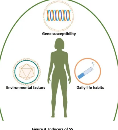
The inducers of SS could implicate diverse factors. Environmental and genetic factors are the most acknowledged inducers of SS. The triggers for SS could be attributed to genetic factors, hormonal factors and environmental factors (Figure 4). They lead to autoimmune exocrinopathy in which epithelial cells augment the levels of chemokines and pro-inflammatory cytokines, result in the activation of immune cells and finally cause the dysfunction of whole immune system.
Figure 4. Inducers of SS
SS can be induced by environmental factors like virus infections, genetic factors like genetic risk loci and daily life habits. These factors are found to be associated with SS. However, the mechanisms are largely unclear. An important theory is that SS is initiated by the combained effects of multiple factors but not single inducer.
Daily life habits like smoking are identified as other factors that induce SS. However, the precise role of cigarette smoking, for instance, is still a matter of debates. In
12
patients with pSS, ANA positivity was significantly associated with smoking (Karabulut et al., 2011). In another study, however, current tobacco smoking was found to be negatively associated with SS, and provide a protection to disease-associated humoral and cellular autoimmunity (Stone et al., 2017). This was further confirmed by a nested case-control study that involved sixty-three patients with pSS, where it was found that current smoking was associated with a reduced risk of pSS, but former smoking was associated with an increased risk (Olsson et al., 2017).
A.3.1: Environmental factors
SS can be triggered by environmental factors. Infections, especially virus infections, have been recognized as important inducers to SS since early times. Infections by hepatitis B virus (HBV), hepatitis C virus (HCV), parvovirus B19, herpesvirus-6 or human immunodeficiency virus (HIV) may trigger symptoms similar with SS (Igoe et al., 2013). Moreover, epidemiological and clinical studies suggest that viral infections, typically Epstein Barr virus (EBV), cytomegalovirus (CMV), HBV, HCV, Coxsackie virus (CV) and Human T-lymphotropic virus Type 1 (HTLV-1) are involved in the pathogenesis of SS (Sipsas et al., 2011) (Figure 5).
It is found that levels of immunoglobulin (Ig) G against above-listed viruses show association with autoantibody levels, and these viruses have been found in biopsy of patients’ SGs. The mechanisms and etiology of SS triggered by viruses are multifaceted. In the following paragraphs, we will focus on the role of HCV and EBV in SS development.
13
Figure 5. Virus as inducers of SS
Infections by EBV, CMV, HBV, HCV, CV and HTLV may cause symptoms like SS. EBV infection could induce immune responses in SGs and cause inflammation, while CMV infection is rather associated with loss of SG functions. Autoimmune hepatitis in patients with SS is mainly induced by HBV infection. HCV is found to stimulate B cell proliferation in glandular tissues, and CV infections are related to anti-Ro60 autoantibody production. HTLV-I is suggested to affect epithelial cells proliferation and survival.
A.3.1.1: HCV
According to the histological appearance of labial SGs, patients infected with HCV showed higher frequency of focal lymphocytic sialadenitis (57%) compared with controls (5%) (Haddad et al., 1992). This result was deeper developed by Brito-Zeron and colleagues, who found that 13.4% of 783 Spanish patients with SS were infected by HCV. They identified the HCV-driven autoimmune response as a lower frequency of anti-Ro/La antibodies, and an abnormal predominant anti-La antibodies, suggesting that HCV may change the immunological serum pattern of patients with SS (Brito-Zeron et al., 2015).
HCV RNA has been found in the serum and saliva of patients but not of healthy control (Toussirot et al., 2002), and in situ hybridization and immunohistochemistry
14
showed that HCV infects and replicates in the epithelial cells from SGs of patients with SS (Arrieta et al., 2001). These results indicate that HCV may propagate and reside in SG, and may promote HCV-associated sialadenitis.
Interestingly, HCV infection seems to be able to change the cytokines profile in SS patients, with increased circulating levels of interleukins (IL)-6, IL-10, IL-2, and tumor necrosis factor-alpha (TNF-α). This cytokines profile is accompanied by higher frequency of HCV-related cryoglobulinemia and hypocomplementemia (Ramos-Casals et al., 2002). However, the cause-effect relationship between HCV infection and SS still remains unclear.
A.3.1.2: EBV
The association of EBV and SS is in debate since early studies dating from 1980s. At first, it was reported that there are exaggerated immunological responses to virus in patients. This conclusion was challenged by later studies pointing out that false positive antibodies can be detected due to RF (Venables et al., 1985). Infection of EBV was re-confirmed by another study, which revealed two forms of EBV infection in the SGs of patients: the positive latency in lymphocytes, and the EBER1-negative latency in epithelial cells (Wen et al., 1996). This suggests that EBV possibly participates in the pathogenesis of SS. Similarly, a 52-year-old woman with pSS developed membranous glomerulonephritis and EBV-positive diffuse large B-cell lymphoma (DLBCL), suggesting the association between EBV infection and SS (Kim et al., 2012).
Recent studies showed more clues to this dispute, as the IgG-mediated immune response against EBV early antigen (EBVEA) was positively associated with SS, and the level of anti-EBVEA IgG was found to be anti-Ro/SSA and anti La/SSB-correlated (Kivity et al., 2014). In addition, it was found that active EBV infection selectively affects ectopic lymphoid structures (ELS) in the SGs of patients with SS. Latent EBV infection and lytic EBV infection were observed exclusively in B cells and plasma cells, respectively. In addition, the persistence of EBV is found to impair CD8-mediated cytotoxicity in ELS-containing SGs (Croia et al., 2014). These studies indicate that EBV
15
infection may play a causative role in the pathogenesis of SS.
A.3.2: Genetic factors
The pathophysiology of SS involves dysregulation of both innate and adaptive immune pathways, in which multiple processes including cellular immunity and humoral immunity have been proved to play an important role. However, the precise mechanism is largely unknown.
A.3.2.1: Human leukocyte antigen
Human leukocyte antigen (HLA) family contains hundreds of genes that are responsible for antigen presentation in immune response. The HLA class I molecules are expressed on the surface of all kinds of cells, which present intracellular antigens, while the HLA class II molecules only exist on the surface of antigen-presenting cells (APCs) and are responsible for the presenting of extracellular antigens.
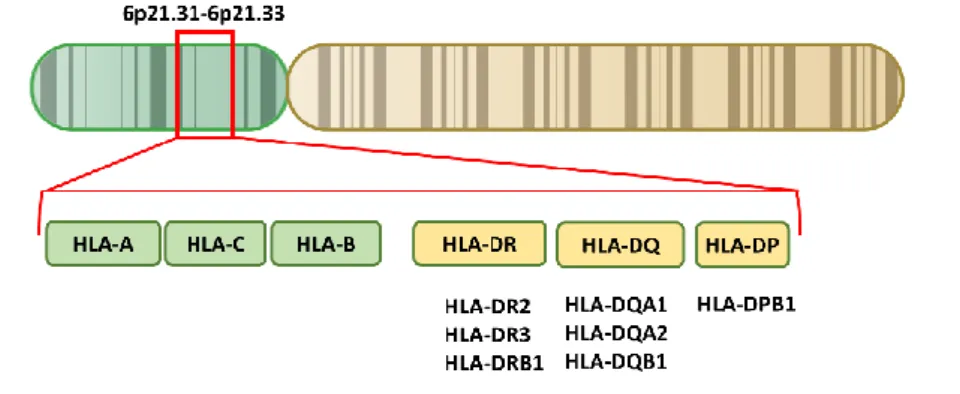
Figure 6. HLA genes associated with SS
The HLA gene complex resides on a 5×10⁶ base pairs region within chromosome 6p21. HLA class I
molecules are encoded by HLA-A, B and C, while HLA class II molecules are encoded by HLA-DR,
DQ and DP. HLA genes are highly polymorphic and present many different alleles, especially DRB1,
which contains hundreds of alleles. Some single nucleotide polymorphisms (SNPs) identified in these polymorphisms are considered to be associated with SS. These SNPs are mostly located within HLA class II regions such as HLA-DR2, DR3, DRB1; HLA-DQA1, DQA2, DQB1 and HLA-DPB1.
The HLA family genes seem to have an intimate connection with SS (Figure 6). This family of genes has been emphasized in SS for more than two decades in numerous
16
studies. The correlation of HLA family genes with SS was first identified by Gershwin et al. in 1975 by comparing the frequency of HLA8 in healthy control individuals and SS patients (Gershwin et al., 1975). The researchers found that among 25 patients, the frequency of HLA8 was 58%, which is much higher than the frequency in healthy controls (21%). Thus, they proposed the existence of a linkage between HLA gene family and SS, in addition to the previously known correlation of HLA genes with other AIDs (Fritze et al., 1974; Grumet et al., 1974).
Regarding the HLA signature in SS, other loci like HLA-Dw3, HLA-DR2 and HLA-DR3 were confirmed to display significant correlations (Chused et al., 1977; Wilson et al., 1984). A large-scale genetic study published in 2013 underscored the importance of HLA genes in innate and adaptive immunity. This study reported several genes of the HLA family, including HLA-DQA1*0501, HLA-DQB1*0201, and HLA-DRB*0301 as SS-associated genes and risk factors (Lessard et al., 2013). In addition, a very recent genome-wide association study (GWAS) analyzed a very large cohort of patients from different ethnicities (European and Asian), and found that SNPs in HLA gene family are among the strongest associations with SS. The rs9271573 in HLA–DRB1,
HLA–DQA1 regions associates very strongly (p=3×10-42) with SSA/Ro or SSB/La
autoantibodies and focus score status in the European participants (Taylor et al., 2017).
Apart from HLA genes, other genes linked with SS were progressively identified over years. Among all these genes, the most strongly associated genes focus on the nuclear factor kappa B (NF-κB) pathway, interferon (IFN) signaling pathway and lymphocyte signaling pathway (Figure 7).
A.3.2.2: NF-κB pathway-related genes
NF-κB family comprises five members, namely p50, p52, p65 (RelA), c-Rel and RelB. All the member proteins share a highly conserved DNA-binding/dimerization domain called Rel in their N-terminus, and three of them have a transactivation domain in their C-termini. As an evolutionarily conserved signaling pathway, NF-κB has a crucial role in multiple involvements of immune activities, including immune response to
17
stress and infections, development and activation of lymphocytes, cell differentiation and apoptosis, and inflammatory regulation. Nordmark and colleagues identified the association between SS and polymorphisms of four signaling members from the NF-κB pathway, namely TNF-α induced protein 3 (TNFAIP3, or A20), TNFAIP3 interacting protein 1 (TNIP1, or ABIN1), NF-κB, and inhibitor of nuclear factor kappa B kinase subunit epsilon (IKBKE) (Nordmark et al., 2013). A total of 12 SNPs were genotyped in 1,105 patients and 4,460 controls. This study demonstrated that TNIP1 risk haplotype was associated with pSS and that there were no significant associations of IKBKE, NF-κB or A20 with SS. However, a more recent study involving two cohorts, the UK cohort (308 controls and 590 patients with pSS) and the French cohort (448 controls and 589 patients with pSS) confirmed the role of A20 impairment in pSS-associated lymphoma. Interestingly, the rs2230926 missense polymorphism was not associated with pSS in both cohorts, but the rs2230926G variant was significantly associated with pSS-associated lymphoma in the UK cohort (Nocturne et al., 2016). A.3.2.3: IFN signaling-related genes
Among all the genes that have been identified to be associated with SS, the strongest one outside HLA family is IRF5 locus. IRF5 is a member of IFN regulatory factors (IRFs), a group of transcription factors that regulate the expression of target genes like proinflammatory cytokines, including type I IFN, interleukins and TNF. IRFs participate in diverse involvements of immunity such as anti-virus immune response, cell differentiation and apoptosis (Krausgruber et al., 2011). IRF5 was reported to be associated with SS (Figure 7). A 5 base pairs (CGGGG) insertion/deletion polymorphism in the promoter region of IRF5 has been proposed to be a crucial impactor to IRF5 mRNA levels in peripheral blood mononuclear cells (PBMCs) and SGs epithelial cells, correlating with expression levels of IFN-induced genes MX1 and
IFITM1 (Miceli-Richard et al., 2009). Miceli-Richard et al. used a genome-wide methylation approach to assess the role of methylation deregulation in pSS, and found that DNA methylation patterns in B cells are important to pSS (Miceli-Richard et al., 2016).
18
A.3.2.4: Lymphocyte signaling pathway-related genes
Lymphocytes play a crucial role in immunity, especially in autoimmune response. The overactive lymphocytes largely contribute to the dysregulation of immune network and lead to the overproduction of autoantibodies, as well as proinflammatory cytokines and chemokines. As described in previous context, HLA molecules, as antigen presenting molecules in immune system, are correlated to SS (Figure 7). Another key molecular in lymphocytes trafficking, CXCR5, plays a central role in guiding cell movements during immune responses. The onset of adaptive immune response needs antigen specific lymphocytes to transfer efficiently to secondary lymphoid tissues, where they encounter and respond to antigens. The migration of lymphocytes is directed by chemokines including CXCR5. CXCR5 is expressed on different cells, regulating their migration and trafficking (Muller et al., 2003). CXCR5 has been associated with SS by a large scale genotyping in 2013 (Lessard et al., 2013). Another association was identified between disease and risk locus in natural cytotoxicity triggering receptor 3 (NCR3/NKp30). NCR3/NKp30 is a natural killer (NK)-specific activating receptor, responsible for the regulation of type II IFN secretion and the cross talk between NK and dendritic cells (DCs). One of the genetic polymorphisms of the NCR3/NKp30, rs11575837 (G>A), is found to be associated with reduced gene transcription and provide a protection against pSS. (Rusakiewicz et al., 2013).
19
Figure 7. Pathways and genes associates with pathogenesis of SS
SS can be induced by genetic factors. Among the genes identified to be associated with SS, most of the risk loci for SS are found to be related with IFN signaling pathway, NK-κB pathway, HLA gene family and lymphocytic signaling-related genes. STAT4, signal transducer and activator of transcription 4; BLK, B lymphoid tyrosine kinase; CXCR5, C-X-C motif chemokine receptor 5; NCR3, natural cytotoxicity triggering receptor 3.
A.3.3: Hormonal factors
The sex bias in SS (female:male around 9:1) strongly suggests the role of sex steroids in the pathogenesis of SS. Compared with healthy controls, SS patients show significantly higher prolactin/progesterone and estrogen/progesterone ratios, suggesting a role of hormones in SS. However, Taiym and colleagues found no significant differences concerning the levels of estrogen and progesterone between patients and healthy individuals in their studies (Taiym et al., 2004). This result was confirmed by another study, which evaluated additional sex hormones including dehydroepiandrosterone (DHEA), DHEA sulfate, androstenedione, testosterone, dihydrotestosterone (DHT), estrone, estradiol, and sex hormone-binding globulin (SHBG) levels. This study was conducted on 39 patients and tried to identify the correlations between sex hormones and autoimmunity in SS. It was found that the concentrations of testosterone were associated with disease activity, but no correlation was found between disease activity and estrogens levels (Brennan et al.,
20
2003). These results may also suggest the involvement of hormone homeostasis, in addition to hormone levels in the pathogenesis of SS.
Thus, the role of estrogens in SS is still controversial. Aromatase-knockout mice were used as a model of estrogen deficiency. It was found that these mice developed severe spontaneous autoimmune manifestations resembling SS, such as proteinuria and severe leukocyte infiltration in the SGs and kidneys. These results reveal that estrogen might have a crucial role in the initiation of SS (Shim et al., 2004). In addition, upon the removal of main estrogen source by ovariectomy, healthy C57BL/6 mice (non-autoimmune prone) showed a significant increase of apoptotic epithelial cells and alpha-fodrin autoantigen in SGs (Ishimaru et al., 2003). On another hand, SS patients are mostly women in postmenopausal phase, in which estrogen level is much lower compared with other period of women’s life. Based on these facts, one of the theories proposed by Konttinen et al. is that secretary acinar cells are primarily targeted by the imbalanced sex steroids, undergo abnormal apoptosis, followed by aberrant handing of apoptotic debris, thus leading to the initiation of SS (Konttinen et al., 2012). This theory highlighted the role of acinar cells and assumed that apoptosis of glandular cells initiates autoimmune responses. Further studies are however necessary to confirm this theory.
Nowadays, the roles of hormones in autoimmune reactions are not well understood. Other pathways beside apoptosis are suggested to be involved in hormones-triggered exocrine gland dysfunction (Mavragani et al., 2012). Hormones might play significant roles in systemic homeostasis, leading to aberrant immune response.
A.4: Pathology A.4.1: Cytokines
Cytokines act as messengers of immune system, deliver and transduct signals in the immune network. Cytokines orchestrate the full panoply of immune responses and are involved in various immunological effects, such as immune cell recruitment and immune responses activation. Cytokines are functionally divided into two groups,
21
namely the proinflammatory cytokines that promote inflammation and anti-inflammatory cytokines that decrease inflammation.
A.4.1.1: Th1 and Th2 balance
A major source of cytokines is CD4 T cells, which express a cell surface marker CD4. CD4 T cells are also named helper T cells (Th) according to their capability to help B cells produce antibodies, to active macrophages, and to recruit granulocytes, including neutrophils, eosinophils, and basophils, to infection sites. Notably, these functions of Th cells depend on their production of cytokines and chemokines. Th cells can be further subdivided into Th1 and Th2, and the cytokines they produce are known as Th1 cytokines and Th2 cytokines (Berger, 2000).
Th1 cytokines tend to be proinflammatory. They facilitate the killing of intracellular invades, and perpetuate chronic inflammation in autoimmune conditions. IFN-γ is a major Th1 cytokine that can promote NK cell activity, induce antibody production of plasma B cells, and increase antigen presentation. Another Th1 cytokine, IL-2, plays a crucial role in T cell differentiation and is involved in immune tolerance generation during the process of T cells maturation in thymus. Other Th1 cytokines include IL-6, TNF-α and lymphotoxin are involved in many inflammation-mediated diseases. Excessive inflammation can lead to a breakdown of immune tolerance and even tissue damage, thus a counteract mechanism is provided by anti-inflammatory cytokines. The Th2 cytokines include IL-4, IL-5, IL-9 and IL-13, these molecules tend to be anti-inflammatory. For example, IL-4 acts as a regulator of macrophages differentiation, promotes polarization of macrophages into immune suppressive M2 state, and inhibits activation of cytotoxic M1 macrophages (Wang et al., 2014). Further secretion of anti-inflammatory cytokines IL-10 and TGF-β by M2 macrophages helps to attenuate pathological inflammation. Thus, the balance of Th1 and Th2 cytokines is crucial for immunological homeostasis.
The role of Th cells in SS has been recognized since early times when the predominant lymphocytic infiltration in glandular tissues of SS patients was found to be contributed by CD4 T cells (Fox et al., 1983). In recent years, the imbalance of Th
22
subsets has been suggested to play a role in SS (Moriyama et al., 2014). The percentage of Th1 cells is significantly higher in the PBMC of SS patients compared with healthy people. In coincidence, Th2 cells are found to be less in patients than in control group (Sudzius et al., 2013). This result was further confirmed by another study that included 66 patients with SS. Compared with normal group, the levels of IFN-γ and IL-4 were significantly increased in peripheral blood of patients (Wu et al., 2013). In saliva from SS patients, increased levels of IL-1β, IL-6, TNF-α, and IFN-γ, in association with higher ratio of Th1/Th2 CD4 T cells have been observed, and these enhanced cytokine levels have been found to be related with clinical features of pSS (Kang et al., 2011). These findings imply that a Th1-mediated inflammation may lead to pathology in SGs. Similar findings have been reported in labial SGs (LSG) from SS patients. The mRNAs of both Th1 and Th2 cytokines were found in LSG. However, the balance between Th1/Th2 was broken and shifted to a Th1 profile in patients with high infiltration scores (Mitsias et al., 2002).
A.4.1.2: IL-17, IL-21 and other cytokines
IL-17 protein expression was found to progressively increase with higher biopsy focus scores in MSGs. Th17 differentiation related cytokines such as TGF-β, IL-6 and IL-23, are highly expressed in MSGs of SS patients (Katsifis et al., 2009). In addition, IL-17A mRNA levels in PBMCs from pSS patients are increased (Fei et al., 2014), suggesting that IL-17 may play a role in the process of lymphocytic infiltration in the LSG. The roles of IL-17 and Th17 were further defined in C57BL/6 mice that are non-susceptible to SS. C57BL/6 mice develop a SS-like disease profile after receiving an adenovirus serotype 5-IL17A vector in SGs, showing the appearance of lymphocytic foci, increased cytokine levels and autoantibodies, as well as loss of saliva flow (Nguyen et al., 2010). In good agreement, in another mouse model of SS, which is generated by immunization of homogenized proteins of SGs (SGP) in C57BL/6 mice (Lin et al., 2011), the number of Th17 cells was found to be increased in SGs. In addition, knockout of IL-17A in C57BL/6 led to complete resistance for SS induction by SGP, and adoptive transfer of Th17 cells rapidly restored the onset of SS (Lin et al., 2015). These studies in different mouse models demonstrate that Th17 might be an
23 important player in SS pathology.
IL-21 is associated with B cell functions like Ig production (Ozaki et al., 2002) and Th17 cells differentiation (Wei et al., 2007). IL-21 thus seems to be involved in the pathogenesis of AIDs in multiple ways. pSS patients are found to have significantly higher serum IL-21 levels compared to control individuals, and the high-level of IL-21 is apparently correlated with autoantibody levels. In addition, lymphocytic foci and the periductal area of the LSG from SS patients showed higher ratio of IL-21/IL-21R than healthy controls. Furthermore, the extent of lymphocytic infiltration was found to be associated with IL-21 expression in LSG (Kang et al., 2011).
In addition to IL-17 and IL-21, the expression of IFN-γ-induced protein 10 (IP-10) and its receptor, CXCR3 was increased in SGs from SS patients, suggesting IP-10 and CXCR3 may contribute to the pathogenesis of SS (Ruffilli, 2014). Apart from cytokines described above, other cytokines such as CXCR2, C-X-C motif chemokine ligand (CXCL) 13 and C-C motif chemokine ligand (CCL) 11 are also found to be linked with disease activity in SS patients (Lisi et al., 2013; Nocturne et al., 2015), thus providing more clues for therapeutic targeting.
A.4.2: B cells
Hyperactivity of B cells is found in SS, and this could be the consequence of the coordinated and integrated effects of B cell activation and cytokines stimulation. It has been found that overexpressed type I IFN and B cell active factor (BAFF) are involved in the enhanced plasma cell formation in pSS patients (Brkic et al., 2013). Then, this enhancement of plasma cells results in secretion of autoantibodies and further promotes autoimmune responses. This has been observed in patients with SS, in which high levels of autoantibodies are linked with the increased frequency of autoreactive B cells and plasma cells (Kroese et al., 2014). Anti-Ro and anti-La autoantibodies-producing plasma cells have been found to reside at sites of infiltration in SGs biopsy tissues from patients with SS (Tengner et al., 1998). These plasma cells showed phenotypic characteristics of the long-lived plasma cell subtype (Szyszko et al., 2011).
24
A study aimed at characterizing the SSA-specific B cell pattern in SG biopsies showed that SSA-specific memory B cells are lacking in peripheral blood and SG. The authors supposed that this could result from activation of these cells into plasma cells at the site of inflammation (Aqrawi et al., 2012; Aqrawi et al., 2013), suggesting that the B cells differentiation process in local infiltration is imbalanced.
In recent years, lymphoid neogenesis and functional ectopic germinal center (GC) formation have been found in SGs of patients with SS. A microenvironment is required to form GC-like structures in SG, including T and B cell aggregates, follicular DCs (FDCs) and activated endothelial cells, and this microenvironment is found in patients with GC-like structures (Johnsen et al., 2014). SGs microenvironment in SS provides factors vital for plasma cell survival and facilitates the development of long-lived plasma cell subtype (Szyszko et al., 2011). These plasma cells in SG increase the production of anti-Ro/SSA and anti-La/SSB autoantibodies and drive autoimmune responses (Salomonsson et al., 2003). These findings and insights in the role of B cells in the pathogenesis of pSS offer promising targets for successful therapeutic intervention.
A.4.3: T cells
T cells are among the most important cells in the pathogenesis of SS, as they are the predominant infiltrating cells found in glandular tissues. This was noted early in 1983 by Fox and colleagues who found that the phenotype of SGs lymphocytes differs from that of peripheral blood lymphocytes in patients with pSS. They also noticed that CD4 T cells are the majority of infiltrating cells in SGs of patients (Fox et al., 1983). Parallel characterization in mouse models has been confirmed, such as in Murphy Roths Large/lymphoproliferation (MRL/lpr) mice, New Zealand Black/White F1 Hybrid (NZB/W F1) mice and Alymphoplasia (Aly/aly) mice (Soyfoo et al., 2007). The infiltrated T cells present variable activity at different disease stages, tend to be resistant to apoptosis, and lead to interstitial nephritis and peripheral CD4 lymphocytopenia (Manganelli et al., 2003). Moreover, environmental triggers of SS, such as viral infections, are considered to be mediated by CD4 T cells. As introduced
25
in previous chapters (A.2.1), HTLV-1 can be an inducer of SS. HTLV-1 is a retrovirus that predominately infects CD4 T cells. In SS, HTLV-1 can be transfected from T cells to SGs epithelial cell (SGEC) and induces specific pathological symptoms (Nakamura et al., 2016).
In addition to CD4 T cells, the role of CD8 T cells in the pathogenesis of SS is underlined by its mediation of glandular destruction. The infiltrated CD8 T cells are resistant to apoptosis by a B-cell CLL/lymphoma 2 (BCL2) mediated mechanism, thus present an overactive cytotoxic activity, and induce TNF receptor superfamily, member 6 (FAS)-mediated apoptosis in acinar epithelial cells, leading to glandular injury (Kong et al., 1997).
In recent findings, the impairment of regulatory T cells (Treg) is linked with SS. It has been found that the frequency of forkhead box protein P3 (Foxp3) expressed Treg in MSGs of SS patients is correlated with glandular inflammation grade and lymphoma risk factors such as complement component 4 (C4) hypocomplementemia and MSGs enlargement (Christodoulou et al., 2008; Sarigul et al., 2010). These findings suggest that Tregs play an important role in the pathogenesis of pSS. However, further studies are needed to explore the mechanisms that mediate the relationship between Tregs and SS.
A.4.4: Central role of epithelial cells
AIDs are recognized as the deregulation of systemic immune network, in which immune cells are found to lose self-tolerance and show features of autoreactivity. As described above, other players like inflammatory cytokines, chemokines and regulators are involved in the catastrophe of immune homeostasis. Studies of SS pathology have lasted for over one hundred years since the first case had been reported in 1888. Studies revealed large amount of evidence related to immune cells deregulation in SS. B cells were found to be overactive and produce autoantibodies, which is a key feature of SS. However, except for the formation of immune complex, which might lead to overload burden for immune clearance, the autoantibodies seem not to play direct pathogenetic role in SS. In contrast, the inducers of
26
overactive B cells are notable. It has been well recognized that several B-cell-activating cytokines promote and mediate the activation of B cells. These cytokines include BAFF, B cell growth factor IL-14, B cell promoting cytokines IL-6 and IL-10. All these cytokines are demonstrated to be produced by epithelial cells in glandular tissues (Kawanami et al., 2012; Martel et al., 2014). Crucially, autoantigens, the targets of autoantibodies, are largely produce by epithelial cells in SS (Katsiougiannis et al., 2015). Therefore, epithelial cells are thought to play a pivotal pathogenetic role in SS, highlighted by occurrence of infiltrating pathology in various epithelial tissues and the increased inflammatory cytokines produced by epithelial cells in patients (Figure 8).
Figure 8. Essential roles of epithelial cells in SS
SGs epithelial cells play central roles in SS pathogenesis. Immune-competent molecules, such as TLRs and cytokines are constitutively expressed by epthelial cells of the salivary or lacrimal glands. Activation of TLR signalling in gland epithelium facilitates production of cytokines and chemokines, including those can active B cells and T cells. Epthelial cells are recognized as targets of activated immune cells. Apoptosis and hypofunctions are then induced in epithelial cells, leading to the production of autoantigens. Autoantigens can be released from SGECs and presented to immune cells. HLA class II molecules expressed by epithelial cells enhance the autoantigen presentation and activation of T cells.
27 A.4.4.1: Autoantigen Production
The epithelial cells of the SGs differentiate into acinar cells, duct cells, and myoepithelial cells, and these cells form the secretory unites (Figure 9).
Figure 9. Structure of SGs and secretory units
Basically, two types of acinar epithelial cells exist in the secretory end of SGs: the serous cells and the mucous cells. The serous cells secrete a watery fluid, which is essential for saliva. A serous cell is pyramidal in shape, and its nucleus is round and close to the base of the cell. The mucous cells produce mucins, enzymes and other proteins, and their productions are slightly viscous. Mucous cells are pyramidal in shape (similar with serous cells), and the nucleus of mucous cells are usually very flat and lie against the basal end of the cell. Histology of SGs is represented by H&E staining of SG from a 29-week-old C57BL/6 mouse.
28
As suffering from systemic autoimmune disease, patients with SS present large amounts of various autoantigens, which trigger autoimmune responses and induce the hyperreactivity of immune system, leading to the final exhaustion of host homeostasis. In fact, our normal constituents become “autoantigen” in autoimmune patients whose immune system differentiate self from non-self less well. The most known autoantigens of SS are the SSA/Ro (includes Ro52/TRIM21 and Ro60/TROVE2), SSB/La, nuclear antigens, and RFs. Glandular epithelial cells are demonstrated to be a factory for autoantigens production. Twenty-six non-neoplastic SGEC lines established from patients with rheumatic disorders were found to release significant amounts of exosomes. These exosomes were found to contain autoantigenic Ro/SSA, La/SSB, and Sm RNPs, as well as epithelial-specific autoantigen cytokeratins (Kapsogeorgou et al., 2011).
The traditional biomarkers, SSA and SSB are widely used in diagnosis, but this specificity is doubtful since they are not always positive, especially in the initiation stage of SS and can be found in other related diseases. In recent studies, new autoantigens for SS have been identified, especially proteins specific to the salivary and lacrimal glands, like SGs protein-1 (SP-1), parotid secretory protein (PSP), carbonic anhydrase VI (CA6) (Suresh et al., 2015) and cleaved alpha-fodrin (Wang et al., 2006). On one hand, as these novel biomarkers may be detected in early stages of disease, they can be recognized as a complement to traditional biomarkers; on another hand, increased level of autoantigens identified in glandular tissues revealed the autoantigenic property of epithelial cells.
FAS and FAS ligand (FASL) were emphasized in SS, and it has been illustrated that increased apoptosis in epithelial cells leads to autoantigen production, as well as collapse clearance of apoptotic debris. It was reported that FAS and FASL levels in non-neoplastic SGEC line established from SS patients were significantly higher than in SGEC line derived from healthy individuals (Abu-Helu et al., 2001; Ping et al., 2005). In SS conditions, the increased apoptosis of epithelial cells may result from various pathways. Firstly, the imbalance between the apoptosis-inhibitor BCL2 and apoptosis-inducer BCL2 associated X protein (Bax) can lead to abnormal apoptosis.
29
Secondly, the interaction of FASL (expressed by T lymphocytes) with FAS (expressed by epithelial cells) results in the apoptosis of epithelial cells. (Manganelli et al., 2003). The consequences of apoptosis in epithelial cells could be lesion of tissues and impaired secretory functions, which cause dryness. Moreover, apoptosis acts as a perpetuation of autoantigen production, which in turn enhances autoantibody and autoimmune responses, leading to SS like inflammatory lesions (Okuma et al., 2013). A.4.4.2: Non-professional antigen-presentation
Ductal and acinal glandular epithelial cells were demonstrated to express HLA-DR antigens inappropriately, whereas rare HLA-DR molecules could be found in normal epithelial cells (Moutsopoulos et al., 1986). The expression of HLA-DR in epithelial cells seems to depend on the local production of IFN-γ by immune cells (Fox et al., 1986a). Consequently, SGs epithelial cells were suggested to act as non-professional APCs (Nagai et al., 2015), as evidenced by the expression of HLA-I antigens and HLA-II antigens after the stimulations of TNF-α (Tsunawaki et al., 2002).
Not only HLA molecules, but also costimulatory molecules like B7 and adhesion molecules including intercellular adhesion molecule-1 (ICAM-1), vascular cell adhesion molecule (VCAM), and E-selectin, are found to be expressed by SGs epithelial cells, and facilitate the non-professional antigen presenting activity (Manoussakis et al., 1999; Tsunawaki et al., 2002).
A.4.4.3: Stimulation of T cells and B cells
T cells activation involves two signals: the antigen specific signal and the co-stimulatory signal. The antigen-specific signal is mediated by the interaction of TCR and peptide-MHC molecules on the membrane of APCs. The co-stimulatory signal is provided by the interaction between T cell membrane bond receptors and co-stimulatory molecules expressed by APCs. This process is nonspecific to antigens and crucial for T cell homeostasis. It is required for T cell proliferation, differentiation and survival.
30
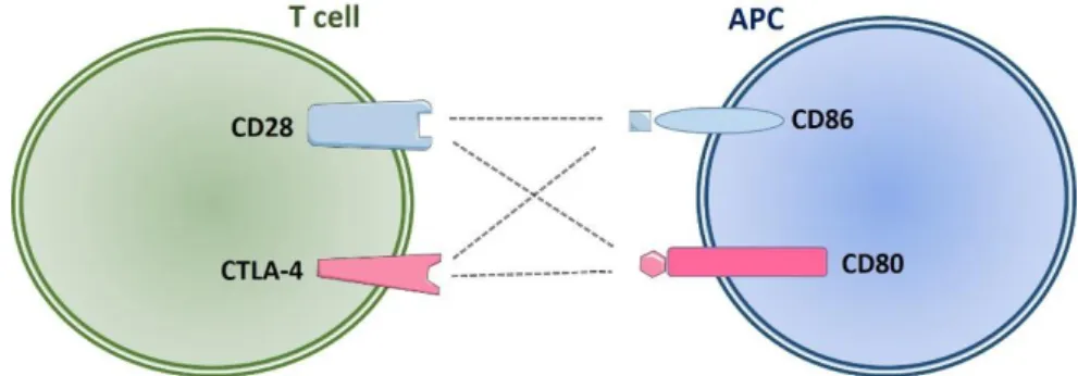
are CD80 (B7-1) and CD86 (B7-2) expressed on the membrane of APCs which interact with CD28 and cytotoxic T-lymphocyte-associated protein 4 (CTLA-4) displayed on the membrane of T cells. Activation of T cells without co-stimulation may lead to T cell anergy and even cell death, and also the development of improper immune tolerance.
Figure 10. Co-stimulatory signals between T cell and APC
CD28 co-stimulatory receptors can be ligated by CD80 or CD86. By interaction with CD80/CD86, CD28 offers stimulatory signals that are required for naive T cell activation. The CTLA-4 co-inhibitor competes with CD28 for binding to CD80 and CD86. The interaction between CD80/CD86 and CTLA-4 results in inhibition of T cell activation.
The mRNA levels of B7 costimulatory molecules CD80 and CD86 in glandular epthelial cells were found to be increased in patients with SS, indicating the active status of epithelial cells and their potential to active T cells (Manoussakis et al., 1999). CD80 and CD86 molecules were found to be efficiently expressed by salivary ductal cells in SS patients after the stimulation of IFN-γ, suggesting that these cells could have antigen presentation to T cells and cause a cascade of immune response in situ (Matsumura et al., 2001). In addition, IFN-γ stimulation increased the production of IP-10 (CXCR10) and Mig (CXCR9) in SS patient ductal epithelium, and these chemokines might act as recruiters of infiltrated T cells in SGs (Ogawa et al., 2002).
As has been described in the previous chapter (A.4.2), overexpressed BAFF is associated with the increased plasma cells and autoantibodies in SS patients. Recently, SGEC was found to be a source of BAFF. Enhanced level of BAFF was found