HAL Id: tel-03030365
https://tel.archives-ouvertes.fr/tel-03030365
Submitted on 30 Nov 2020
HAL is a multi-disciplinary open access archive for the deposit and dissemination of sci-entific research documents, whether they are pub-lished or not. The documents may come from teaching and research institutions in France or abroad, or from public or private research centers.
L’archive ouverte pluridisciplinaire HAL, est destinée au dépôt et à la diffusion de documents scientifiques de niveau recherche, publiés ou non, émanant des établissements d’enseignement et de recherche français ou étrangers, des laboratoires publics ou privés.
Development of fluorescent platforms for the design of
multifunctional compounds for in vitro and in vivo
applications in molecular imaging
Jacques Pliquett
To cite this version:
Jacques Pliquett. Development of fluorescent platforms for the design of multifunctional compounds for in vitro and in vivo applications in molecular imaging. Optics / Photonic. Université Bourgogne Franche-Comté, 2018. English. �NNT : 2018UBFCK067�. �tel-03030365�
THESE DE DOCTORAT DE L’ETABLISSEMENT UNIVERSITE BOURGOGNE FRANCHE-COMTE PREPAREE A l’INSTITUT DE CHIMIE MOLECULAIRE DE L’UNIVERSITÉ DE BOURGOGNE
(ICMUB)
Ecole doctorale n°533 Ecole Doctorale Carnot-Pasteur
Doctorat de chimie Par
Jacques Pliquett
Development of fluorescent platforms for the design of multifunctional compounds for
in vitro and in vivo applications in molecular imaging
Thèse présentée et soutenue à Dijon, le 30/11/2018
Composition du Jury:
Dr. FROCHOT, Céline Directrice de recherche
Université de Lorraine, Nancy
Rapportrice
Dr. MAURY, Olivier Directeur de recherche
École Normale Supérieure de Lyon, Lyon
Rapporteur
Pr. GASSER, Gilles Professeur des Universités
Université Paris-Sciences-et-Lettres, Paris
Examinateur
Pr. HISSLER, Muriel Professeure des Universités
Université de Rennes 1, Rennes
Présidente du Jury
Dr. BERTHET, Cyril Directeur de l’unité Pharmaco-imagerie
Société Oncodesign®, Dijon
Membre invité
Pr. DENAT, Franck Professeur des Universités
Université Bourgogne – Franche-Comté, Dijon
Directeur de thèse
Dr. BODIO, Ewen Maître de conférences
Université Bourgogne – Franche-Comté, Dijon
Co-directeur de thèse
Dr. GOZE, Christine Maître de conférences
Université Bourgogne – Franche-Comté, Dijon
Co-encadrante de thèse
Dr. PAUL, Catherine Maître de conférences
EPHE –Université de Bourgogne Franche Comté, Dijon
Co-directrice de thèse
Université Bourgogne Franche-Comté 32, avenue de l’Observatoire
Acknowledgements
This thesis has been prepared in three labs at the University of Burgundy (UB) in Dijon. The bulk of the work has been done at the “Institut de Chimie Moléculaire de l’Université de Bourgogne” (ICMUB – UMR 6302), a joint research unit between the University of Burgundy and the “Centre National de la Recherche Scientifique” (CNRS) in the two teams OCS (OrganoMetallique, Catalyse et Stéréochimie) and P2DA (Polyamines, Porphyrines, Développements et Applications). The biological investigations were performed at the LIIC-(EA 7269), the “Laboratoire d’Immunologie et Immunothérapie des Cancers”, a division of the EPHE, “École Pratique des Hautes Études” situated at the UB. All three teams are warmly acknowledged for having received me in their midst. This thesis was financed by the Conseil Régional de Bourgogne (PhD JCE grant # 2015-9205AAO033S04139 / BG0003226) with the stewardship of Oncodesign and the European Union through the PO FEDER-FSE 2014/2020 Bourgogne program, their support for this project is gratefully acknowledged.
First of all I would like to extend my sincere gratitude towards Professor Muriel Hissler of the University of Rennes 1 and Professor Gilles Gasser of the PSL University of Paris for kindly accepting to examine my work, Dr. Céline Frochot of the University of Lorraine, Nancy and Dr. Olivier Maury of the ENS Lyon for agreeing to review my thesis.
This Ph.D. thesis has of course not been prepared in solitude but is the result of great teamwork and cooperation between a multitude of individuals.
Most notably I would like to thank my four Ph.D. supervisors, Professor Franck Denat, Dr. Ewen Bodio, Dr. Christine Goze (all three ICMUB) and Dr. Catherine Paul (LIIC). All four of you are laudable supervisors and have been extremely supportive throughout my thesis. I am blessed and honored to have had such invested and caring supervisors who knew how to enable me to provide the work that I did, adapt to my style and be present, supportive and challenging in just the right, balanced amount. Christine and Ewen, especially you two have always been accessible at any time of the day or night for my silly requests and ideas, for brainstorming and fruitful discussions, both of scientific as well as personal matter.
I would also like to thank all the persons who have contributed (in a practical fashion by performing experiments for me) to this work; most notably the scientific and technical staff of the “Plateforme d’Analyse Chimique et de Synthèse Moléculaire de l’Université de Bourgogne” (PACSMUB) as well as the quantum chemistry team and other persons in various areas of expertise for their help, counsel and the numerous analyses they performed for me: Marie-Jo Penouilh, Quentin Bonnin (NMR and Mass Analyses), Myriam Laly and Laure Giancarlo (ICP-MS analyses), Yoann Rousselin (Single cristal X-ray analyses), Paul Fleurat-Lessard and Miguel Ponce-Vargas (Quantum chemical simulations).
I would like to thank Anthony Romieu for his expertise and counsel in practical matters concerning fluorescence and optical analyses.
Fluorescence imaging was performed by Souheila Amor who spent countless hours bent over culture dishes and microcopes on my behalf to provide us with great images.
I also appreciate the help and contributions of Cindy Racoeur, Nesrine Mabrouk as well as Pierre Simon Bellaye for performing the preclinical study on my bioconjugates, as well as Lucile
This thesis would probably have been a psychological nightmare without the great friendship and support that I experienced throughout my thesis by all the former and present Ph.D. students who work(ed) at the ICMUB and LIIC at the same time as me and by whom I was so well integrated. Thank you Coline, Audrey, Mario, Johan, Robin, Sarra, Antoine, Anne, Valentin, Alix, Adrien, Léa, Garence, Emma, Océane and everybody else I have forgotten for the great time we spent together both at the ICMUB and LIIC as well as outside of it; for the vacations we had together; the culinary reunions, movies, partys and sportive events. All of you always had 5 (or 50) minutes to spare to chat and joke with me. All of you are the heart and soul of both labs.
Last but not least I would like to thank my girlfriend Marie for her patience during the last three years and for supporting me through the ups and downs of this thesis.
Table of Contents
Acknowledgements ... 3 Table of Contents ... 6 Abbrevations ... 10 Preface ... 13 Introduction ... 17 0.1 Cancer ... 170.2 Imaging and Molecular imaging ... 18
0.2.1 Optical imaging ... 21
0.2.2 Metal based anti-cancer agents ... 32
0.3 Objectives of the thesis: towards the development of fluorescent platforms ... 35
Chapter I – BODIPY ... 41
1.1 Choice of the platform ... 41
1.1.1 Synthesis and functionalization of BODIPY-dyes ... 42
1.2 Gold in biology: a brief overview ... 49
1.2.1 Auranofin ... 49
1.2.2 Chemical biology of Gold... 49
1.3 BODIPY-Platform ... 53
1.3.1 Towards double functionalization ... 59
1.3.2 Synthesis of BODIPY-acid ... 60
1.3.3 Functionalization of trifunctional BODIPY-acid 8 ... 61
1.3.4 Introduction of a thioglucose tetraacetate moiety ... 63
1.3.5 Quantum chemical investigations ... 66
1.3.6 Trifunctionalization of the dichloroBODIPY acid ... 67
1.4 Characterization of the BODIPYs properties ... 73
1.4.1 Introduction of a simplified graphic representation ... 73
1.4.2 Photophysical characterization of obtained BODIPY-dyes ... 73
1.4.3 Biological investigations ... 77
1.4.4 Compounds Localization by Confocal Imaging ... 80
1.4.5 Lipophilicity and Gold uptake: generalities ... 82
1.4.6 Establishing structure-activity relationships ... 83
1.5 Summary and outlook ... 85
1.5.1 Summary and Conclusion ... 85
1.5.2 Outlook ... 86
Chapter II – AzaBODIPY ... 91
2.1.1 Synthetic access to azaBODIPYs ... 92
2.2 1,7-di(phenol)-3,5-di(phenyl)-azaBODIPY ... 94
2.2.1 Photophysical characterization ... 100
2.2.2 Activation of hydroxy-azaBODIPY 34 for bioconjugation ... 101
2.3 Hydrosolubilization of azaBODIPYs ... 104
2.3.1 Hydrosolubilization of BODIPY and azaBODIPY – an overview ... 104
2.4 E-aza-BODIPYs ... 106
2.4.1 First system: 1,7-di(phenol)-3,5-di(phenyl) E-azaBODIPY ... 108
2.4.2 New platform molecules ... 111
2.4.3 E-azaBODIPY : Second system ... 119
2.4.4 Third system: 1,7-di(phenyl)-3,5-di(methoxyphenyl) E-aza-BODIPYs... 122
2.4.5 Activation of the compounds for bioconjugation ... 126
2.4.6 Photophysical characterization of E-azaBODIPYs and precursors ... 128
2.5 Biological investigations ... 133
2.5.1 Choice of vector... 133
2.5.2 Bioconjugation ... 136
2.5.3 Characterization of in vitro-properties of the obtained bioconjugates ... 139
2.5.4 In vivo investigations ... 142
2.6 Summary and Outlook ... 150
2.6.1 Summary ... 150
2.6.2 Outlook ... 153
General Conclusion ... 159
Materials and Methods ... 163
4.1 Materials and Methods ... 163
4.1.1 NMR spectra ... 163
4.1.2 Spectroscopic properties ... 163
4.1.3 Analytical HPLC ... 164
4.1.4 Semi preparative chromatography ... 164
4.1.5 MALDI ... 164
4.1.6 Determination of cytotoxic properties ... 164
4.1.7 Determination of logP ... 165
4.1.8 Cell lines and culture conditions ... 165
4.2 Synthetic procedures ... 165
4.2.1 BODIPYs ... 165
4.2.2 AzaBODIPYs ... 184
4.3 Gold uptake ... 213
4.5 In-vitro confocal microscopy experiments ... 214 References ... 216 Table of Figures ... 235 ANNEX I ... 243 7.1 Quantum Yield ... 243 7.2 Computational Details ... 245
7.2.1 Results and Discussion ... 245
7.3 Details for the determination of distribution coefficients (logD) ... 246
ANNEX II – Photophysical Spectra ... 250
8.1 Spectra of BODIPYs ... 250
8.2 Spectra of azaBODIPYs ... 253
Annex III – single crystal X-Ray data ... 259
9.1 Compound 4b ... 259 9.1.1 Summary ... 259 9.1.2 Extended Experimental ... 259 9.1.3 Reflection Statistics ... 260 9.2 Compound 6 ... 261 9.2.1 Summary ... 261 9.2.2 Extended Experimental ... 261 9.2.3 Reflection Statistics ... 262 9.3 Compound 7 ... 263 9.3.1 Summary ... 263 9.3.2 Extended Experimental ... 263 9.3.3 Reflection Statistics ... 264 9.4 Compound 8 ... 265 9.4.1 Summary ... 265 9.4.2 Extended Experimental ... 265 9.4.3 Reflection Statistics ... 266 9.5 Compound 10 ... 267 9.5.1 Summary ... 267 9.5.2 Extended experimental ... 267 9.5.3 Reflection Statistics ... 268 Résumé ... 269 Abstract ... 270
Abbrevations
Abbrevation Meaning Abbrevation Meaning
Abs absorption MALDI-TOF
Matrix Assisted Laser Desorption Ionisation - Time of Flight
ACN acetonitrile MDSC myeloid-derived
suppressor cell
ADC antibody-drug-conjugate MICoE Molecular Imaging Center
of Excellence
anhyd. anhydrous MRI Magnetic Resonance
Imaging
approx. approximately MRS Magnetic Resonance
Spectroscopy azaBODIPY 4,4-difluoro-4-bora-3a,4a,8a-triaza-s-indazene MSI mass spectrometry imaging BBN bombesine MTS (3-(4,5-dimethylthiazol-2- yl)-5-(3- carboxymethoxyphenyl)- 2-(4-sulfophenyl)-2H-tetrazolium) Boc tert-butyloxycarbonyl n.d. none detected/unsuccessful analysis Boc-TOTA
1-(t-Butyloxycarbonyl-amino)-4,7,10-trioxa-13-tridecanamine n.t. not tested
BODIPY
4,4-difluoro-4-bora-3a,4a-diaza-s-indazene NADPH
nicotinamide adenine dinucleotide phosphate
Bz benzyl NBS N-Bromosuccinimide
cat. catalytic NCS N-Chlorosuccinimide
CEO Chief executive officer NHS N-Hydroxysuccinimide
CT computed tomography NIH National Institute of
Health CTLA4 cytotoxic T-lymphocyte-associated
protein 4 NIR near-infrared
Cy5 Cyanine 5 NMR nuclear magnetic resonance (spectroscopy)
Cys Cysteine NODAGA
1,4,7- triazacyclononane,1-glutaric acid-4,7-acetic acid
DCM dichloromethane OAc acetate
DDQ
2,3-Dichloro-5,6-dicyano-1,4-benzoquinone p.i. post injection
DEA diethylamine PBS phosphate buffered
saline
DFT density functional theory PD-1 Programmed Cell death 1
DIPEA N,N-diisopropylethylamine PDITC phenyl di(isothiocyanate)
DMAP N,N-Dimethylpyridin-4-amine PD-L1 Programmed cell death
ligand 1
11
DMSO dimethylsulfoxide PE phycoerythrin
DNA deoxyribonucleic acid PEG polyethylene glycol
DOL degree of labelling PET Positron Emission
Tomography
DOTA
1,4,7,10-Tetraazacyclododecane-1,4,7,10-tetraacetic acid ppb parts per billion
EDC
1-Ethyl-3-(3-dimethylaminopropyl)carbodiimide ppm parts per million
EGFR Epidermal growth factor receptor PSMA Prostate specific
membrane antigen
Em emission pTSA para-toluene sulfonic
acid
EN Electronegativity QY quantum yield
EPR Enhanced Permeability Retention RF radio frequency
eq equivalent RNA ribonucleic acid
ESI electrospay ionization ROS Reactive Oxygen Species
Et ethyl RP reversed phase
et al. et alius (lat.), and others rt room temperature
FA formic acid SD standard deviation
FDA US Food and Dug Administration SDS sodium dodecyl sulfate
FDG Fluorodeoxyglucose SDS-PAGE
sodium dodecyl sulfate– polyacrylamide gel electrophoresis FPLC fast protein liquid chromatography SNAr nucleophilic aromatic
substitution
Hb Hemoglobin SNM Society of Nuclear
Medicine
HbO2 oxygenated Hemoglobin SPECT Single Photon Emission
Computed Tomography HBTU (2-(1H-benzotriazol-1-yl)-1,1,3,3-tetramethyluronium hexafluorophosphate TBAF tetrabutylammonium fluoride
HER-2 Human epidermal growth factor
Receptor 2 TBDMS tert-butyl-dimethylsilane
HIV human immunodeficiency virus TEA triethylamine
HOMO Highest Occupied Molecular Orbital TEAB triethylammonium
bicarbonate
HPLC high performance liquid
chromatography TFA trifluoroacetic acid
HR-MS High resolution mass spectrometry THF tetrahydrofuran
HSAB Hard and Soft Acids and Bases tht tetrahydrothiophene
HSGlu(OAc)4 β-thioglucose tetraacetate TLC thin layer
chromatography
IC Internal Conversion TRIS
2-Amino-2- (hydroxymethyl)propane-1,3-diol
IC50 inhibitory concentration 50 TrxR Thioredoxin reductase
ICG indocyanine green TSTU
N,N,N′,N′-Tetramethyl-O-(N-succinimidyl)uronium tetrafluoroborate ICMUB Institut de chimie moléculaire de
ICP-MS inductively coupled plasma-mass
spectroscopy UK United Kingdom
IgG Immunoglobulin G UV ultraviolet
IR infrared vbr very broad
ISC Inter System Crossing VIS visible
IUPAC International Union of Pure and
Applied Chemistry WHO
World Health organization
LUMO Lowest unoccupied Molecular
13
Preface
Cancer describes a multifaceted body of diseases that originates in the unchecked and dysregulated growth and proliferation of cells. Cancerous growths impede the regular function of affected organs which, if left untreated, leads to the death of the host. [1] It is the the second leading cause of death globally (after cardiovascular diseases) with an estimated 18.07 million new patients annually in 2018, 9.55 million of which will succumb to their disease [2]. In 2010 the WHO estimated the global economic impact of cancer to at least 1.16 trillion US$, corresponding to 2% of the world’s gross domestic product [3].
Researchers around the world seek to develop new drugs and tools to improve diagnosis, response assessment and patient care in order to increase the rates of success as well lessen the impact of current treatment plans on the affected patients. Common treatment plans include operative care and chemotherapy that employes cytotoxic metal complexes, in recent years immunotherapies have been developed that recruit the immune system of the patient to fight cancer: for this pioneering approach James P. Allison and Tasuku Honjo were awarded the Nobel Prize in medicine and physiology in 2018 [4].
A cornerstone in the diagnosis of cancer and the assessment of response to treatment plans is the use of imaging and molecular imaging techniques. Molecular imaging is a set of techniques that permits the visualization, characterization and quantification of biological processes in living organisms [5]. It includes clinically established methods such as Computed Tomography (CT), Positron Emission Tomography (PET), Single Photon Emission Computed Tomography (SPECT) and Magnetic Resonance Imaging (MRI) [6,7]. In recent years optical imaging has emerged as promising modality for molecular imaging. It has proven its potential in pre-clinical applications and is being developed more and more towards clinical use, mainly because of the recent emergence of fluorescence guided surgery. However, due to the elaborateness of the compounds employed for this type of imaging the development of new probes and their modification is often difficult and time consuming. The demand for more versatile approaches and new tools for the continued advancement of this modality is thus evident.
This PhD thesis is in line with these concepts and provides new, versatile tools for the design of fluorescent probes and trackable therapeutical agents.
Introduction
0.1 Cancer
Cancer is a large, heterogeneous body of malignant diseases. It can be defined as “an
abnormal growth of cells caused by multiple changes in gene expression leading to a dysregulated balance of cell proliferation and cell death and ultimately evolving into a population of cells that can invade tissues and metastasize to distant sites, causing significant morbidity and, if untreated, death of the host.” [1]
A cancer in itself is non-lethal. However, as the abnormally growing cells gain mass (formation of tumors) and become invasive (formation of metastases), they displace healthy, normally functioning tissue. This impedes the affected organ to execute its tasks properly. The result is (multi-)organ failure and ultimately death of the host.
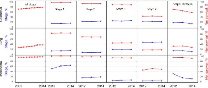
To reduce this impact and increase the rate of treatment successes many countries have installed systematic screenings as well as prevention and early-diagnosis programs. Figure 1 highlights the necessity of such screening and prevention schemes: an earlier diagnosis significantly improves the rates of success for the treatment.
Figure 1: 1-year survival vs stage of initial diagnosis for the NHS (UK) from 2012-2014 for selected cancers. Red denominates % survival, blue % of cancers diagnosed at that stage. Figure adapted from [8]
Common prevention and early diagnosis schemes include blood tests, biopsies, the use of imaging techniques (MRI, CT) and visual inspection (e.g. colonoscopy) [9]. However, as can also be seen in Figure 1 the rates of early-stage diagnosis have not significantly improved between 2012 and 2014 (for the UK), highlighting the need for continued research in early diagnosis.
In 2011 Hanahan and Weinberg defined 10 properties that cells must acquire to classify as cancerous cells: among others they include sustained proliferative signaling, angiogenesis, genomic instability and avoidance of the immune system (Figure 2).
Figure 2: The Hallmarks of cancer. Adapted from [10]
Although cancerous growths combine all of the properties displayed in Figure 2, these properties are acquired over time. Many of the above mentioned characteristics appear at specific stages of tumor development. Each characteristic is associated with deregulation of associated processes and the development of specific antigens that appear or are overexpressed in cancerous tissue. A review on the matter was published by Nair et al. in 2018 [11] and summarizes various molecular markers that are overexpressed or tumor specific and have predictive value. Examples include:
PSMA (Prostate specific membrane antigen): prostate cancer HER-2 (human epidermal growth factor receptor 2): breast cancer EGFR (Epidermal growth factor receptor): colorectal cancer
PD-L1 (Programmed cell death ligand 1): various aggressive cancers, including metastatic melanoma, renal cell carcinoma and non-small cell lung cancer
These antigens can be used for immunohistochemical determinations after a biopsy. More interesting however is their use for non-invasive techniques such as diagnostic imaging. Indeed, the antigen can be used as a target for specific probes that enable the evaluation of their presence/absence/overexpression, their systemic distribution and their quantification to verify if a certain pathology is existent. If so, it permits the evaluation of its progression and help to select the treatment plans that are likely to succeed.
0.2 Imaging and Molecular imaging
Imaging describes a set of techniques aimed at providing visual representations of a system.
For biological systems, the obtained image offers anatomical or functional insight. To physicians and researchers alike this insight is the basis to diagnose pathologies, understand biological processes and select treatment options.
In 2007 the members of the Molecular Imaging Center of Excellence (MICoE) and the Society of Nuclear Medicine (SNM) board defined Molecular imaging, a subspecialty of imaging, as follows:
“Molecular imaging is the visualization, characterization, and measurement of biological processes at the molecular and cellular levels in human and other living systems. To elaborate; molecular imaging typically includes 2- or 3- dimensional imaging as well as quantification over time. The techniques used include radiotracer imaging/nuclear medicine, magnetic resonance imaging, magnetic resonance spectroscopy, optical imaging, ultra-sound, and others.”[5]
It permits the mapping of functional differences, variations in metabolic processes or apparition of pathology-specific markers that are already present in early stages, thereby increasing a patient’s chances to be treated successfully [12]. In modern medicine molecular imaging is not only used to diagnose an illness and delineate for example a tumors’ or ulcers’ expansion, it has become a cornerstone towards the development of personalized medicine. Today a wide variety of (both classical as well as molecular) imaging techniques is available. Except for ultrasound-based techniques, they all employ electromagnetic radiation in wavelength-windows in which tissue is more or less transparent. A representation of normal attenuation of electromagnetic radiation of biological tissue as function of the employed wavelength is given in Figure 3. It shows that γ- and X-rays, the far-red/near infrared radiation and radio-waves experience little absorbtion in biological tissue, leading to wavelength-windows in which the body is mostly transparent and that are thus predestined for use in various imaging modalities.
The imaging techniques can be distinguished along several criteria. They can be differentiated by their:
energy (γ-, X-rays, VIS/IR-radiation, RF or soundwaves)
attainable spatial resolution (microscopic, mesoscopic, macroscopic) type of provided information (anatomical/structural, physiological, cellular,
molecular)
Each imaging modality possesses intrinsic advantages and disadvantages that depend on the energy that is used, the way an image is obtained and the information that is provided [6,7]. An overview of the strengths and weaknesses of some techniques are given in Table 1.
Table 1: Examples for imaging modalities. Adapted from [6] and [7] Technique spatial resolution temporal resolution depth of penetration primary contrast Contrast/Imaging agent Cost information obtained main use
CT 50 µm minutes no limit tissue density iodinated molecules, Barium infusions $$ anatomical, physiological clinical, pre-clinical SPECT 1-2mm minutes-hours no limit - 99mTc, 111In labelled molecules $$$ physiological, molecular clinical, pre-clinical PET 1-2mm minutes-hours no limit - 18F, 64Cu, 11C, (68Ga), (89Zr)-labelled molecules $$$ physiological, molecular clinical, pre-clinical MRI 10 - 100µm minutes-hours no limit water content, oxygenation paramagnetic Chelates, magnetic particles $$$ anatomical, physiological, molecular clinical, pre-clinical Ultrasound 50µm seconds-minutes mm-cm reflection, tissue interfaces microbubbles $ anatomical, physiological, (molecular) clinical, pre-clinical photoacoustic imaging 10µm – 1mm
seconds-minutes 6mm – 5cm hemoglobin chromophores $
physiological, molecular clinically translatable optical fluorescence imaging 2-3mm seconds-minutes 1-2cm - fluorophore-labelled molecules $ molecular preclinical, emerging clinical
CT (computed tomography scan) for example grants excellent spatial resolution, combined with the advantage of unlimited depth of penetration of the used X-Ray radiation. However, the main drawbacks of this technic concerns the high radiation dose, as well as the low sensitivity, which requires very often the use of contrast agent, such as iodine based systems. Due to the large administered doses, patients with damaged kidneys are excluded from such diagostics. Even with this precaution, contrast agents cause side effects in 0.15% of all patients of which 2.62% are serious (and potentially lethal) [14].
The same applies for MRI (Magnetic Resonance Imaging): it grants excellent anatomical insight but requires large doses of paramagnetic contrast agents (usually Gd-based) for physiological or molecular examinations. As for CT the large required doses of contrast agent restrict their administration and may cause severe side effects (0.3-0.5% of all patients, depending on the contrast agent [15]). SPECT (Single Photon Emission Computed Tomography) and PET (Positron Emission Tomography) are both based on the detection of decay radiation of radioactive nuclides. Both techniques adhere to the tracer-principle: very little probe is administered, which results in a very limited disturbance of the observed system and thus, from a chemical point of view, no toxicity or side effect. Both techniques instrinsically yield molecular and physiological information and permit good resolution. However, the obvious risk that is associated with handling radioactive material as well as the requirements for equipment and technical staff make these techniques extremely expensive and restrict their use to highly specialized diagnostic centers.
Currently the most commonly employed and clinically proven techniques for performing
molecular imaging are PET and MRI: 18F-FDG-PET is routinely used to detect increased glucose
uptake in the patient’s body to find metastases. MRI can be used to detect the oxygen content of tissues through changes in relaxivity. MRS (Molecular resonance imaging, comparable to NMR) is used to determine concentrations of biomolecules such as choline to N-acetylaspartate and their ratios to determine cancers’ aggressiveness [16].
In recent years optical imaging has emerged as promising technique for molecular imaging. It has proven its potential in pre-clinical applications and is being developed more and more towards clinical use, mainly because of the recent advances in fluorescence guided surgery. This thesis inscribes itself in this field of research, it is therefore worth providing a brief overview of currently existing techniques and the underlying principles they are based on.
0.2.1 Optical imaging
Optical imaging describes a set of techniques that is based on the detection of radiation in the visible and near infrared part of the electromagnetic spectrum. Various techniques have been developed that rely on its detection, examples include optical tomography, photoacoustic imaging, intravital microscopy, bioluminescence -, Cerenkov- and fluorescence imaging (Table 2).
Table 2: Exemples for imaging modalities that depend on visible and NIR-light
Optical imaging modality Image derived from
Optical tomography Backscattered light
Photoacoustic imaging Pulsed excitation and localized heating of tissue; detection by
ultra-sound device
Bioluminescence imaging Direct imaging of luciferase/luciferin couple or secondary imaging of chromophores excited by luciferase/luciferin couple
Cerenkov-imaging Excitation of a fluorophore by Cerenkov radiation from
radioactive decay.
Fluorescence imaging Excitation of a fluorophore, detection of the fluorescence light
at longer wavelengths
Among the different optical imaging modalities, optical fluorescence imaging uses photoluminescent compounds as traceable unit to obtain information about the examined system. Fluorophores may be coupled to vectors and other molecules to be used as probes for molecular imaging. The detected “light source” in tissue corresponds then to the fluorophore and thus yields information about its localization and concentration. Before diving deeper into the subject, a brief account of the photophysical processes it depends upon will be given.
0.2.1.1 Basic concepts of optical processes
Ultraviolet, visible and (near-)infrared light describe the part of the electromagnetic spectrum in which electronic transitions of outer electrons are possible (Figure 4).
Figure 4: The Electromagnetic spectrum. Adapted from [17]
When such an electronic transition occurs, an electron is excited through interaction with the electromagnetic field of light which results in its excitation from an occupied (usually the highest occupied molecular orbital, HOMO) to an unoccupied (usually the lowest unoccupied molecular orbital, LUMO) molecular orbital of the absorbing species. The energy required for such electronic transitions lies in the range of 100-600kJ mol-1 (1-6.2eV), corresponding to the
200-1200nm window.
After excitation the system has several pathways to release its energy and fall back into the ground state. The possibilities are shown in the Perrin-Jablonski diagram in Figure 5 and are
(2, 3, 4) Internal Conversion (IC) and Intersystem Crossing (ISC)
These are processes were isoenergetic, radiationless conversions of states with the same (IC) or different multiplicity (ISC) take place
(5) Vibrational relaxation (thermalization). The molecule loses its energy to the surrounding medium by collisions with other molecules
(6) Fluorescence
A Radiative process between electronic states having the same multiplicity (7) Phosphorescence
Figure 5: A Perrin-Jablonski Diagram of a given optically active system [18] 1- Absorption, 2- Internal Conversion, 3,4-Intersystem crossing, 5-vibrational relaxation, 6-Fluorescence, 7-Phosphorescence
The timescale at which the different processes occur are summarized in Table 3.
Table 3: Timescale of energetic transitions in excited molecules. Numbers refer to processes shown in Figure 5. Adapted from [18]
Process Name Time scale
τ=1/kprocess [s] 1 Absorption 10-15 2 Internal conversion 10-12-10-6 3 Intersystem crossing (ST) 10-12-10-6 4 Intersystem crossing (TS) 10-9-101 5 Vibrational relaxation 10-13-10-12 6 Fluorescence 10-9-10-7 7 Phosphorescence 10-6-10-3
The quantum efficiency (also called quantum yield) with which fluorescence occurs is defined as the ratio of emitted to absorbed photons and is proportional to the ratio of lifetimes of the excited state (𝜏𝑠) and radiative processes (𝜏𝑟) (equation (I))
𝛷 = 𝑁𝑒𝑚𝑖𝑡𝑡𝑒𝑑 𝑁𝑎𝑏𝑠𝑜𝑟𝑏𝑒𝑑 ~
𝜏𝑠
𝜏𝑟 (I)
The fluorescence quantum yield is determined either by measuring the fluorescence lifetimes (i.e. time resolved measurements) or through comparison of the fluorescence intensity of an unknown compound (uk) to a reference (k) in the steady-state regimen using equation (II)
𝛷𝑢𝑘 = 𝐴0,𝑘 𝐴0,𝑢𝑘 ∫ 𝑖𝐹,𝑢𝑘 ∫ 𝑖𝐹,𝑘 𝑛𝑢𝑘2 𝑛𝑘2 𝛷𝑘 (II)
where A is the absorption of the compound, ∫ 𝑖 is the integrated fluorescence intensity and n the refractive index of the employed solvent (a mathematical deduction can be found in ANNEX I - 7.1).
In molecular imaging, a fluorophore is characterized by the following photophysical properties:
Brightness describes the product of quantum efficiency multiplied by molar absorption coefficient (Φ x εabs). It permits the direct comparison of two fluorophores. Concerning
the fluorescence properties, a commonly encountered problem in the development of fluorophores is the quenching of fluorescence. Several phenomena can be responsible; two of them (encountered in this thesis) are photoinduced electron transfer and the aggregation of the probes.
Quenching through photoinduced electron transfer may appear in systems were an acceptor and donor are present in the same molecule. This static quenching may be caused by electron-rich groups such as amines, alcohols or phosphorous substituents with free electron pairs [19]. When the fluorophore in question is poorly soluble in the used solvent, aggregation and molecular stacking can occur. Two types of stacking interactions are possible, J and H-type stacking. Depending on the type of interaction, constructive or destructive interference of single molecule excitons may occur [20]. Compared to monomeric chromophores H-type stacks result in flattened emission peaks, lowered quantum yields and the appearance of a blue-shifted absorption band due to out-of-phase excitonic coupling. J-type stacking displays in-phase excitonic coupling, resulting in bathochromically shifted absorption peaks. Both types of stacking may coexist in solution [21].
The Stokes shift is the result of vibrational relaxation of a fluorophore into a lower excited state before the occurrence of a radiative transition. Larger Stokes shifts reduce reabsorption of emitted photons due to smaller spectral overlap. Large Stokes shifts permit the use of cheaper filters for the imaging equipement and reduce the background noise in imaging applications.
Photobleaching describes the phenomenon that fluorophores lose brightness over time under irradiation. It is caused by photochemical reactions that occur with the excited fluorophore and that lead to its permament inactivation. Reactions of triplet-states and photooxidation are possible causes for this behavior [22]
The absorption and emission wavelengths of the fluorophore represent another important property that need to be tailored for the targeted application.
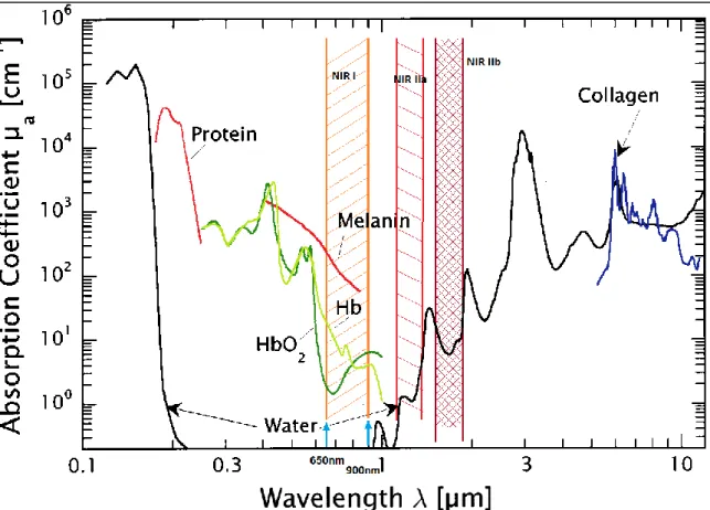
0.2.1.2 Transparency windows
Light (and thus including fluorescence light) is subject to interferences in tissues that affect the attainable depth of penetration, resolution and requirements for equipment.
Three main sources of noise or interference exist in biological tissue The absorption coefficient of tissue
Four wavelength windows are distinguished in fluorescence imaging: the visible part (350nm-650nm), the far-red- near infrared I (NIR I) window (650-900nm) and the NIR II (1000nm-1700nm) window that is subdivided into NIR IIa (1100-1300nm) and NIR IIb (1500nm-(1000nm-1700nm) (the specific values vary from source to source) [23,24]. The specific absorption of tissue and common biomolecules for various wavelengths are shown in Figure 6. Especially the NIR I window displays very little absorbance and is therefore often called optical therapeutic window.
Figure 6: Double logarithmic plot of absorption coefficients of biological tissues in the UV/Vis - infrared range. The optical therapeutic window is highlighted in orange. Hb: Hemoglobin, HbO2: oxygenated hemoglobin.
Adapted from [25]
The other two main influences are
autofluorescence of ubiquitous biomolecules such as hemoglobin, melanin and lipids and
Rayleigh-scattering on particles. Rayleigh-scattering decreases with λ-4 of the used
wavelengths (blue light scatters far stronger than red light) which leads to higher physically achievable resolutions when longer fluorescence-wavelengths are used. As mentioned before, each targeted application has optimum requirements for the employed fluorophore: for in vitro applications such as confocal/fluorescence microscopy and flow cytometry very little tissue has to be traversed; fluorescence emission in the visible spectrum is thus sufficient and desirable due to lower requirements for the used equipement. In vivo fluorescence imaging on the other hand is commonly executed in one of the transparency windows (far red-NIR I and NIR II) due to the higher relative transparency of tissue in this region.
0.2.1.3 Classes of Fluorophores and their application
Several classes of fluorophores exist; e.g. nanoparticles and quantum dots [26,27], fluorescent proteins [28,29] and organic and inorganic fluorophores. Biological, biochemical and medical research has become unimaginable without the use of fluorescence microscopy and the staining of specific organelles to establish the localization of various agents in cells and tissue alike.
A wide choice of organic fluorophores exists from which a suitable candidate can be selected, depending on the intended application and required properties (Figure 7).
Figure 7: Plot of brightness vs. emission wavelength for various fluorophores [30]
Owing to their high brightness, cyanines, xanthene dyes such as rhodamine or fluorescein and BODIPYs are highly sought after fluorophores. They can be applied as they are to stain specific organelles or be coupled to other molecules to act as a probe.
Some commercial examples for routinely used organelle-specific stains that permit the labelling of life and fixed cells alike are given in Figure 8.
Figure 8: Examples for commercial, organelle specific dyes: MitoTracker™ Green specifically labels mitochondria, Lysotracker Red DND-99 fluoresces in acidic organelles (usually Lysosomes), DRAQ5 stains DNA
through intercalation; DAPI intercalates into the minor groove of DNA and thus stains the nucleus [31–34]
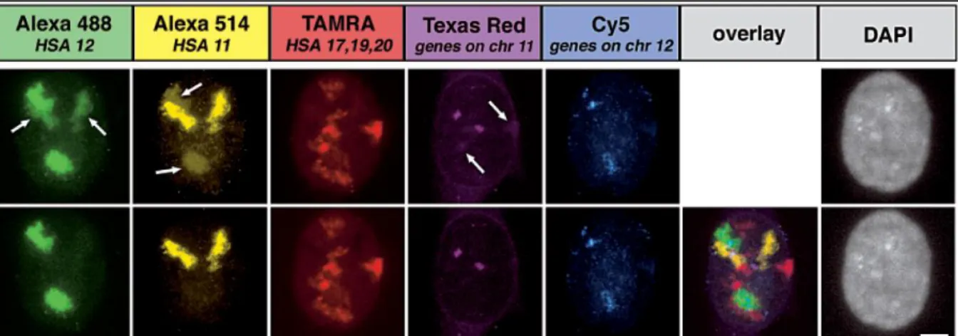
Other than organelle specific dyes Xanthene dyes such as fluorescein and Invitrogens Alexa series are the most used dyes for in vitro techniques; they are used to label antibodies, peptides, DNA/RNA sequences and many more. When selected with care to reduce spectral overlap many dyes can be used in parallel for multi-colour labelling of an analyzed sample [35] (Figure 9).
Figure 9: Multi-color fluorescence in situ hybridization of human chromosomes in the cells nucleus. Top row: untreated images for each channel; arrows indicate channel –bleed through. Bottom: spectrally unmixed
channels and overlay of unmixed channels [35]. Except for Cy5 all used dyes are Xanthene-derivates
Fluorophore-labelled probes are routinely used for the development of molecular imaging agents to gain insight into biochemical mechanisms and evaluate and quantify cellular processes. Examples include the development of β-lactamase-activated fluorophores to evaluate bacteria’s resistance to penicillin by fluorescence acitivation [36] or the evaluation of β-galactosidase activity by turn-on probes (β-galactosidase is a cellular marker for cellular senescence) [37,38] (Figure 10).
Figure 10: Turn-on fluorescent probe for the detection of β-Galactosidase activity. Image bottom left: fluorescence image of β-Gal negative HeLa cells; bottom right: fluorescence image of β-Gal-positive OVCAR-3
cells. Both were incubated with 10µM L1 for 40min [38]
In 2018 Conic et al. applied a variety of Alexa-488 labelled antibodies against nuclear protein targets on electroporated life cells. Using super resolution imaging the labelled antibodies enabled them to study cellular trafficking of newly synthesized proteins and transcription factors and their transport and organization in the cell’s nucleus as well as post translational modification events in real time [39].
Whereas in vitro appliations are dominated by xanthene-dyes such as fluorescein and rhodamine (the routinely employed and commercially available Alexa Fluor series are to ¾ xanthene derivates [40]) most fluorophores that are used in medical imaging emit in the red end of the visible spectrum or in the infrared. Due to low attenuation by the tissue and very little Rayleigh scattering the largest depth of penetration and best resolution is achieved using the NIR II window. However, the development of organic fluorophores that emit in this reagion has proven difficult; the existing examples are large polyaromatic donor-acceptor-donor assemblies that have very unfavourable behavior in solution [41]. For this reason most organic fluorophores in in vivo imaging emit in the NIR I region, the most commonly used fluorophores are cyanine derivates such as Cyanine 5 (Cy5), indocyanine green (ICG), IRDye® 800, 5-Ala/Protoporphyrin IX and xanthene-derivates such as fluorescein (Figure 11). Of the above mentioned dyes fluorescein is somewhat of an exception due to its emission far outside of a transparency window. It does however excel in superficial applications such as fluorescence guided surgery [42].
Figure 11: Some examples for fluorophores or fluorescent motives that are currently used in the clinic or in clinical trials (photophysical data of cyanines taken from [43], Fluorescein from [44], methylene blue from [45],
Protoporphyrin IX [46,47]) . n.r.= not reported
As shown in recent reviews [42,43,48] the field of in vivo applications is dominated by cyanine dyes due to their long emission wavelengths and excellent molar absorption coefficients. These translate into good brightnesses even with mediocre quantum yields as seen for the FDA approved ICG. IRDye® 800 CW-bioconjugates are being/have been evaluated in 24 clinical trials (01/2019) against various cancers (Table 4)[49].
Table 4: Status of clinical trials employing IRDye® 800 CW derivates [49]
Completed Terminated Active Recruiting/unknown
However, due to the presence of a multitude of non-aromatic double bonds, cyanine dyes are prone to photobleaching and possess limited chemical stability. Most importantly however they are difficult to functionalize multiple times once assembled. In order to obtain a stable, easy-to-functionalize dye for our purposes we took interest in another class of dyes, BODIPYs.
0.2.1.4 BODIPYs in optical fluorescence imaging
BODIPYs (boron-dipyrromethene dyes) have caught researchers’ interest due the combination of good optical properties and excellent chemical and photostability.
BODIPYs, short for 4,4-difluoro-4-bora-3a,4a-diaza-s-indazenes can be considered as annulated cyanine dyes. They possess a boron atom to rigidify the backbone and ensure the planarity of the molecule.
Figure 12: Nomenclature of BODIPYs
The base compound in itself is highly fluorescent and emits at around 500-530nm, depending on the substituents. As for fluorescein this emission is outside of the optical therapeutic window. Just like fluorescein or Alexa-488 they have found their main use in in vitro imaging or other light-depending applications such as photodynamic therapy (PDT). In terms of emission wavelength, quantum yields and absorption coefficients, both the commercial Alexa-488 and BODIPY™-FL are interchangeable; BODIPYs however have the advantage of displaying very narrow emission peaks which makes them advantageous for multicolor imaging due to easier spectral unmixing (Figure 13).
Figure 13: Spectral comparison of the commercial dyes BODIPY-Fl(cyan) and Alexa 488 (green) [50]
Besides classic labelling of compounds of interest (as would be possible using other fluorophores) BODIPYs have successfully been used to study membranes [51], lipid droplets in skeletal muscles [52] or monitor the activity of the redoxactive enzymes using cysteine-conjugated BODIPY [53], taking specifically advantage of neutral BODIPYs’ properties. For the design of non-commercial dyes they are a well liked class of dyes due to their versatile modifiability.
Besides classic in vitro applications more recently they have been been applied for in vivo imaging and to design bimodal imaging agents that permit fluorescence and PET/SPECT imaging. In these bimodal probes the BODIPY can be coupled to a polyazamacrocycle for the
use of radioactive metallic isotopes [54–57]; other examples include the use of 125I (L4)[58]
and isotopic exchange of 19F for 18F (L3)(Figure 14).
Figure 14: Examples for BODIPY based bimodal probes that combine the advantages of BODIPYs in vitro and strengths of PET in vivo: L2 [55] L3 [57], L4 [58]
Despite the short emission wavelength classical BODIPYs have been used in numerous instances to perform in vivo imaging studies (Figure 15)[59–61].
Figure 15: Examples for BODIPY-use in in vivo studies. L5: on/off-probe for studying osteoclasts in intravital microscopy on mice [59](intravital microscopic image of L5 beneath); middle: whole body imaging in mice to track anti-Chagas agent L6 in vivo [60] (whole body mice images of L6 beneath; left mouse 120min p.i. with
L6-vesicle emulsion, middle mouse 120min p.i. with L6-DMSO/H2O-solution, right mouse: control mouse injected
Other examples for their in vivo application can be found in [62–66]. Due to the limited transparency of tissue in the used wavelength-window the in vivo applications of classical BODIPYs as in vivo tracers remain scarce.
For this PhD project we selected this class of fluorophore for the development of a trackable molecular platform that mostly aims at facilitating the required in vitro investigations of metal based therapeutic drugs.
In order to overcome the spectral shortcoming of classical BODIPYs, several strategies have been developed to extend the π-system of the BODIPY core in order to reach further into the far red/infrared part of the electromagnetic spectrum. Usually this fine-tuning is done by introduction of styryl-substituents as shown in 2009 by Akkaya’s group [67] (L8-L12, Figure 16).
Figure 16: Fine-tuning of optical properties BODIPYs by Knoevenagel reaction. Photophysical data for L8 in EtOH [68], styrylBODIPYs L9-L12 in CHCl3 [67]
As shown in Figure 16 all resulting BODIPYs maintain excellent optical properties, independent of the number of styryl-substituents that were introduced.
π-extended BODIPYs have found widespread pre-clinical use as tracers [69–72], imaging agents for photoacoustic imaging [73] and especially for photodynamic therapy (a good review was recently published by Lee et al.[74]). Other strategies for the extension of the π-system are extensively reviewed in the literature (e.g. by Lu et al.[75]) and show the great versatility of this class of fluorophores.
Since 2005 azaBODIPYs, a close relative of BODIPY dyes, have received much attention: the replacement of the 8-carbon (meso-carbon) by a nitrogen atom red-shifts the absorption and emission maxima by 120-150nm to 650nm and beyond. They possess equally good optical properties (high molar absorbencies, high quantum yields and brightnesses) as their relatives while being more resistant to photobleaching than styryl-BODIPYs due to the absence of free double bonds. Their synthetic accessibility, functionalizations, properties and published applications are shown in detail in Chapter II – AzaBODIPY. Due to the great versatility we selected this class of fluorophore for the development of in vivo-trackable agents.
0.2.1.5 Theranostics and trackable drugs
Even though the usefulness of classical (non π-extended) BODIPYs as trackable agents for in
vivo purposes remains relatively limited due to the low wavelength of emission of approx.
530nm they excel during the development-stage of drugs: to gain insight into subcellular processes they are great assets.
The BODIPY-part of the molecule can play out its strength in the in vitro techniques such as confocal imaging, flow-cytometry and even intravital microscopy. This permits excellent, subcellular/submicron resolution during the development phase to gain insight into a drug’s mechanism of action to distinguish e.g. bound from unbound fluorophore (Figure 17).
Figure 17: Fluorescence anisotropy imaging of a BODIPY-Biotin derivate (L13). Binding of L13 reduces molecular movement which results in increased fluorescence anisotropy. Green: unbound fluorophore, red: bound
fluorophore [76]
The combination of therapeutically active agents with trackable units is also called “theranostics”. Theranostics is an artificial combination of the words therapy/therapeutic and diagnosis/diagnostic. This term was first coined in September 1998 as a sales-pitch by PharmaNetics CEO John Funkhouser when marketing his company’s business model: Their approach was to commercialize both the treatment and the diagnostic tool to evaluate treatment efficiency [77,78]. Both the test and the treatment were independent of each other and had to be performed separately, allowed however the personalization and adjustment of the treatment by yielding standardized information about the treatment progress.
The definition and understanding of theranostics has changed over time: nowadays it can incorporate anything between personalized medicine, trackable therapeutic agents as well as diagnostic agents that can be transformed with little effort into their therapeutic counterparts (e.g. changing the radionuclide attached to the tracer from a β+ emitter like 64Cu to a β- emitter
like 67Cu). We decided to use the theranostic approach in order to design a variety of closely
related trackable, metal based anti-cancer agents whose localization may change depending on the substitutents and substitution pattern exhibited by the molecule. It is therefore worth profiding a brief account of metal based anti cancer agents and showcase, how their trackability may facilitate the elaboration and optimization of such compounds.
0.2.2 Metal based anti-cancer agents
Nowadays the most common anti-cancer treatment plans include metal based anticancer drugs, 46% of them on the basis of Platinum [79].
The first platinum-drug, cisplatin, was introduced into cancer treatment after the accidental discovery of its cytotoxic properties by Barnett Rosenstein in 1965 [80]. In 1978 the FDA
approved its use as anti-cancer agent [81]. Treatment with cisplatin often leads to severe side effects such as nausea, vomiting, nephrotoxicity and loss of hearing [82].
However, treatment with cisplatin often leads to development of resistances [83,84]. To improve the specificity and reduce side effects a multitude of platinum-drugs has been developed over the last decades. Amongst thousands of failures a variety of compounds has been found that are now available for therapies (or in clinical trials). Some examples and interesting leads are given in Figure 18 (more examples can be found in [85]).
Figure 18: Some Platinum-based drugs that are currently used in therapy or in clinical trials [85]
Due to the frequent resistances the scope of drug development has also widened and shifted from improving platinum drugs towards the discovery of motifs based on other metals and ligands. This limits the development of resistances due to different molecular targets and grants the possibility of dual (or oligo) treatments. A multitude of leads that are based on metals such as ruthenium, titanium, iridium and gold has been discovered [85,86] (Figure 19).
Figure 19: Some lead-motives of therapeutically active metals other than Platinum that are currently in clinical trials [85,87] or showing interesting in vivo activity [88]
0.2.2.1 Metal based theranostics
Complicated experiments have to be designed to follow the drugs fate in vitro (an in vivo) and to determine their mechanism. This knowledge however is the base for a rational design and improvement of drugs. The introduction of a trackable moiety to the drug facilitates that task as it intrinsically permits to determine cellular compartments and targets. This approach is the basis of trackable metal based anticancer drugs (metal based theranostics).
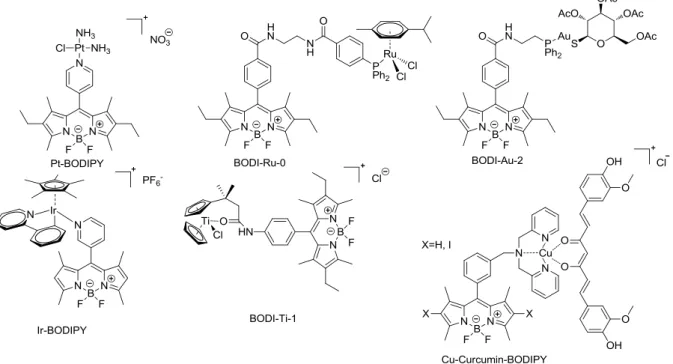
Taking the lead motifs from Figure 19 above various BODIPY analogues have been developed by our group as well as others (Figure 20)[89–94].
Figure 20: Examples of BODIPY theranostics that have been developed using the leads of Figure 19. Pt-BODIPY [89], BODI-Ru-0 [90], BODI-Au-2 [91], IrBODIPY[92], BODI-Ti-1 [93], Cu-Curcumin-BODIPY for PDT [94]
Recent advances have been thoroughly reviewed by our group [95] as well as others [96]. It is worth noting the non-innocent role of the probe, e.g. in the case of Pt-BODIPY shown in Figure 20: while cisplatin is localized and acts in the cell’s nucleus its BODIPY-analogue is localized in the mitochondria. Moreover, it does not enter the nucleus but still maintains good cytotoxicity compared to cisplatin [89]. The influence of the imaging moiety on the biological behavior becomes even more evident when different gold-based, trackable therapeutics that we developed in our group are compared to one another. Indeed, despite a very similar therapeutic moiety (phosphine-gold(I)-complex), the compounds display very different localization and antiproliferative properties (Figure 21).
Figure 21: Influence of trackable moiety on cellular localization [95]. Cellular localizations: CP-Au-1: raft domains; MC-Bio-Au-1: nucleus ; BODI-Au-0: membranes; BODI-Au-1 (in green, DAPI in blue): cytosol ;
Especially the difference between BODI-Au-0 and BODI-Au-1 is striking: The addition of a single benzamide-group results in a different localization whereas no marked change in cytotoxicity was observed.
Consequently, it is obvious that the probe needs to be introduced at the beginning of the optimization process and not when a non-labelled lead has been identified. The problem is that it is very difficult to predict, which combination of probe and therapeutic agent will give good results. Moreover, the design of syntheses providing these sophisticated compounds are often time consuming and lead to a limited number of analogues. Thus, if we want to move this theranostics from a research curiosity to a drug design strategy, it is urgent to find a solution that permits to easily fine-tune and optimize these objects. That is why we decided to conceive platforms suitable for the efficient and rapid synthesis of a large number of optical theranostics. Indeed, a stepwise, controlled and site-specific introduction of moieties of interest would enable to study the individual influence of each component on the final biological behavior. With this information in hand, it is or should be possible to predict general trends in reactivity, selectivity, photophysical properties and biological behavior of a resulting theranostic agent depending on the site of substitution. Moreover, using this information it is possible to rationally design and improve selected properties of the theranostic agent.
0.3 Objectives of the thesis: towards the development of fluorescent
platforms
“Platform approach” is a well-known strategy aiming at linking more than two compounds together thanks to an additional compound: the platform. This key element must fulfill several requirements, such as:
be commercially available or cheap to synthesize (few synthetic steps, short reaction time, simple purification, cheap starting material…)
be available in large scale be as simple as possible
be chemically and thermically stable to enable chemical functionalization enable selective stepwise functionalization by at least three substituents
In this study, we target biological applications. More precisely, we want to be able to introduce onto the platform an optical probe, a therapeutic agent, a biologically relevant vector and an additional group for either tuning the physico-chemical properties of the final object (e.g. solubility) or introducing a second modality of imaging (e.g. radioactive probe) (Figure 22).
Figure 22: Platform approach
Thus, additional characteristics are required for the platform:
enable introduction of readily available substituent in mild conditions (ideally nucleophiles such as amine, alcohol, thiol…)
create biologically stable bonds such as C-C bonds or amides between the platform and the substituents
do not quench the fluorescence
Numerous platforms are reported in the literature; among them some common examples are the use of amino acids (e.g. lysine), benzotrifuranone (L15), dichlorotetrazine (L17)… (Figure 23).
Figure 23: Examples for molecular platforms [97,98]
Most of the reported examples deal with trifunctionalizable platforms, while we envision the combination of four “modalities” (imaging, therapy, vectorization, modification of the hydrophile/lipophile balance). Moreover, it is, in our view, unfortunate that the platform skeleton does not bring any valuable property to the final compound. Thus, we decided to develop a platform, which is itself fluorescent. Nevertheless, it implies that the platform possesses very favorable optical properties (high brightness, resistance to photobleaching) and retains these properties after functionalization.
Although scarce, there are some examples of fluorophores that are used as scaffolds and can be modified or conjugated to vectors without too much trouble as shown by the use of BODIPY (L21) [99], Virginia Orange (L23) [100] and porphyrin (L26) [101] in Figure 24.
Figure 24: rare examples of fluorophores that are used as platforms: BODIPY(L21, top left) [99] Virginia Orange(L23, top right) [100] porphyrin (L26, bottom) [101]
All the work developed during my PhD was developed around this platform approach, and particularly around BODIPY-based fluorescent platforms. Specifically, we worked on two different BODIPY systems with the following objectives for my thesis:
Development of a simple and trifunctionalizable BODIPY-based fluorescent platform for in vitro application, in order to be able to test of a large family of gold(I)-based optical theranostics.
Design of a water soluble AzaBODIPY fluorescent platform for the development of an in vivo-compatible optical trackable therapeutic agent.
The first chapter of the manunscript will describe the synthesis of the first trifunctional platform, as well as its functionalization investigations, and the different applications, which could be realized, starting from this very versatile BODIPY-based compounds.
The second chapter will be dedicated to the development of a new family of very promising water soluble azaBODIPY platforms.
Chapter I – BODIPY
The work developed in this first chapter aims at proving the interest and the feasibility of using a fluorescent platform to rapidly and efficiently design a family of metal-based theranostics. We decided to use gold(I) complexes as therapeutical moiety for proving this concept. Indeed, we noticed in previous studies that gold(I) presents the great advantage to not quench the fluorescence of fluorophores [90,91,102]. First we will explain why BODIPY dyes meet all the criteria required for the conception of a fluorescent platform. This will be followed by a brief focus on the therapeutic potential of gold complexe. After this introduction an account and description of the synthesis and the use of a bi and a tri-functionalizable fluorescent platforms will be provided, as well as their use to reach a large panel of metal-based trackable therapeutics. To finish, an in vitro evaluation of the biological properties of these theranostics will be reported.
1.1 Choice of the platform
As reported earlier, the first part of this thesis aims at developing a multifunctional molecular platform that can be used for in vitro (and to a limited degree in vivo) investigations and possesses a therapeutic potential thanks to the presence of biologically active, metal based agents (Figure 25).
Figure 25: The multifunctional platform approach
The selected platform should be
- suitable for the introduction of three different substituents (in order to be able to introduce a vector, a therapeutic moiety, and another group for tuning the molecule like a water solubilizing group)
- suitable for a selective, stepwise introduction of the various substituents,
- suitable for the grafting of the substituents via nucleophilic substitution (numerous amines and thiols are commercially available, easy to synthesize, and require simple reaction conditions),
- reactive under mild reaction conditions to be suitable for the introduction of metal-based complexes and biomolecules
Additionally, for the reasons shown in the general introduction, the platform should intrinsically fulfill the task of being the trackable moiety. We have identified BODIPY dyes as potential platforms due to their versatility, various ways to access the base compound and a great variety of possible functionalizations of this class of dyes. We decided to first investigate the bifunctionalizable platform developed by Dehaen and Boens [103]: a dichloroBODIPY (Figure 26) to check if it meets the three last criteria we defined.
Figure 26: dichloro-BODIPY - the selected multifunctional platform
In addition to the bifunctional platform that we had chosen we wanted to retain the possibility of supplementing our bifunctional platform with a carboxylic acid function in order to widen its scope.
A brief overview over possible transformations as well as selected works involving the functionalization of both carboxylic acid based as well as dihalogenated BODIPYs will be provided in the next section.
1.1.1 Synthesis and functionalization of BODIPY-dyes
1.1.1.1 Accessing the base-compound
BODIPY’s were first described in 1968 by Treibs and Kreuzer [104], when they investigated
acylation techniques of pyrroles in presence of BF3 as Lewis-acid for substrate activation. To
their surprise, they obtained a brightly fluorescent compound to which they managed to attribute the structures L27 and L28 shown in Figure 27.
Figure 27: First synthesis of BODIPY-dyes [104]
Keeping in line with IUPAC rules the new compound was called BODIPY, short for 4,4-difluoro-4-bora-3a,4a-diaza-s-indazene (Figure 28).
Due to the thermal instability above -40°C of the precursor dipyrrin the unsubstituted title compound was not actually been discovered until 2008/2009 when three groups independently from one another reported the successful synthesis [105–107].
Due to said instability, BODIPYs are usually decorated with varying substituents to prevent polymerization at the dipyrrin-state. Two main synthetic protocols have been established using either aldehydes or acid chlorides as electrophiles (Figure 29).
Figure 29: General Synthesis of symmetrical BODIPYs
The strategy is divided in three steps: 1) formation of the dipyrrin/dipyrromethane, 2) oxidation of this intermediate using oxidizers such as DDQ or chloranil, and 3) complexation of the boron in presence of a base such as Et3N.
In order to access unsymmetrical BODIPYs the pyrrole unit can be acylated first and reacted with a second pyrrole in a next step as described below (Figure 30).
Figure 30: Synthesis of unsymmetrical BODIPYs
In 2007, Burgess and coworkers developed a third synthetic strategy; this strategy grants
access to symmetric BODIPYs via decarbonylation of formylpyrroles using POCl3 [108] (Figure
31).
Figure 31: Accessing symmetric BODIPYs via decarbonylation reaction [108]
This approach can be advantageous when the required substitution pattern inaccessible at the dipyrromethene or BODIPY stage as well as for substrates that do not support treatment with DDQ.
One of the main advantages of BODIPY over classic fluorophores is its versatility in terms of substitution. Indeed, almost any substituents can be envisioned for group R1-R7 either
introduced during the synthesis of the BODIPY core or during post modification. Moreover, depending on the substituents, the photophysical properties of the dye can and have been finely modulated, in particular their absorption and emission wavelengths, Stokes shift, quantum yield… This ability is key when looking at a fluorescent platform scaffold. Some important transformations will be shown in the next paragraph in order to permit an informed selection of BODIPY that can serve as a viable platform molecule.
1.1.1.2 Functionalization of the primary BODIPY
The easiest way to access BODIPYs that possess interesting photophysical properties and carry different transformable functions is to introduce the desired substituent in the meso-position during the assembly process of the BODIPY core by using commercial aromatic aldehydes such as nitro-, carboxy- or hydroxy benzaldehyde. However, the BODIPY core in itself is highly
versatile as well. Due to the electron-impoverishing character of the BF2-moiety the BODIPY
core largely behaves like an electron-poor aromatic compound and can undergo functionalizations such as Pd-catalyzed cross coupling reactions, halogenations and aromatic substitution reactions [109] (Figure 32).
Figure 32: Possible functionalizations of the BODIPY-base structure [109]
We will focus our attention on functionalizations that rely on carboxylic acid functions (for the formation of amide-bonds) and (nucleophilic aromatic) substitution reactions on the BODIPY-core.
1.1.1.3 Functionalization and reactivity of the 3,5-positions of the BODIPY
BODIPYs carrying carboxylic acid functions on a meso-phenyl substituent can be accessed through several possible pathways. They are directly accessible by use of both unprotected [110] as well as protected carboxylic acid derivatives [111] that are subsequently hydrolyzed to give the free carboxylic acid. In 2009 Ziessel used Pd(PPh3)2Cl2 catalyzed carboalkoxylations
(that were developed by Heck in 1974 [112]) of L29, followed by hydrolysis to introduce the carboxylate in the meso-phenyl [113,114] (Figure 33).
Figure 33: Accessing BODIPY-carboxylates via Pd-catalyzed carboalkoxylation [113,114]
Meso-phenyl carboxylate functionalized BODIPY derivates are commonly used as they permit
easy functionalization through the formation of amides and esters.
In previous works of our group BODIPY-carboxylic acids have found widespread use to elaborate a variety of compounds. Applications included the study of the properties and
![Figure 5: A Perrin-Jablonski Diagram of a given optically active system [18] 1- Absorption, 2- Internal Conversion, 3,4-Intersystem crossing, 5-vibrational relaxation, 6-Fluorescence, 7-Phosphorescence](https://thumb-eu.123doks.com/thumbv2/123doknet/14715429.749908/24.892.158.699.86.415/jablonski-absorption-conversion-intersystem-vibrational-relaxation-fluorescence-phosphorescence.webp)
![Figure 7: Plot of brightness vs. emission wavelength for various fluorophores [30]](https://thumb-eu.123doks.com/thumbv2/123doknet/14715429.749908/27.892.111.788.79.582/figure-plot-brightness-vs-emission-wavelength-various-fluorophores.webp)
![Figure 19: Some lead-motives of therapeutically active metals other than Platinum that are currently in clinical trials [85,87] or showing interesting in vivo activity [88]](https://thumb-eu.123doks.com/thumbv2/123doknet/14715429.749908/34.892.154.743.561.812/figure-motives-therapeutically-platinum-currently-clinical-interesting-activity.webp)
![Figure 34: Use of BODIPY-carboxylic acid in previous studies from our group [57,90,91]](https://thumb-eu.123doks.com/thumbv2/123doknet/14715429.749908/46.892.135.758.167.548/figure-use-bodipy-carboxylic-acid-previous-studies-group.webp)