HAL Id: hal-02352375
https://hal.archives-ouvertes.fr/hal-02352375
Submitted on 14 Nov 2019
HAL is a multi-disciplinary open access
archive for the deposit and dissemination of
sci-entific research documents, whether they are
pub-lished or not. The documents may come from
teaching and research institutions in France or
abroad, or from public or private research centers.
L’archive ouverte pluridisciplinaire HAL, est
destinée au dépôt et à la diffusion de documents
scientifiques de niveau recherche, publiés ou non,
émanant des établissements d’enseignement et de
recherche français ou étrangers, des laboratoires
publics ou privés.
Aline Marnef, Anne-Laure Finoux, Coline Arnould, Emmanuelle Guillou,
Virginie Daburon, Vincent Rocher, Thomas Mangeat, Philippe Mangeot,
Emiliano Ricci, Gaëlle Legube
To cite this version:
Aline Marnef, Anne-Laure Finoux, Coline Arnould, Emmanuelle Guillou, Virginie Daburon, et al..
A cohesin/HUSH- and LINC-dependent pathway controls ribosomal DNA double-strand break
re-pair. Genes and Development, Cold Spring Harbor Laboratory Press, 2019, 33 (17-18), pp.1175-1190.
�10.1101/gad.324012.119�. �hal-02352375�
HAL Id: hal-02351888
https://hal.archives-ouvertes.fr/hal-02351888
Submitted on 6 Nov 2019
HAL is a multi-disciplinary open access
archive for the deposit and dissemination of
sci-entific research documents, whether they are
pub-lished or not. The documents may come from
teaching and research institutions in France or
abroad, or from public or private research centers.
L’archive ouverte pluridisciplinaire HAL, est
destinée au dépôt et à la diffusion de documents
scientifiques de niveau recherche, publiés ou non,
émanant des établissements d’enseignement et de
recherche français ou étrangers, des laboratoires
publics ou privés.
Aline Marnef, Anne-Laure Finoux, Coline Arnould, Emmanuelle Guillou,
Virginie Daburon, Vincent Rocher, Thomas Mangeat, Philippe Mangeot,
Emiliano Ricci, Gaëlle Legube
To cite this version:
Aline Marnef, Anne-Laure Finoux, Coline Arnould, Emmanuelle Guillou, Virginie Daburon, et al..
A cohesin/HUSH- and LINC-dependent pathway controls ribosomal DNA double-strand break
re-pair. Genes and Development, Cold Spring Harbor Laboratory Press, 2019, 33 (17-18), pp.1175-1190.
�10.1101/gad.324012.119�. �hal-02351888�
A cohesin/HUSH- and LINC-dependent
pathway controls ribosomal DNA
double-strand break repair
Aline Marnef,
1Anne-Laure Finoux,
1,4Coline Arnould,
1,4Emmanuelle Guillou,
1Virginie Daburon,
1Vincent Rocher,
1Thomas Mangeat,
1Philippe E. Mangeot,
2Emiliano P. Ricci,
3and Gaëlle Legube
11
Laboratoire de Biologie Cellulaire et Moléculaire du Contrôle de la Prolifération (LBCMCP), Centre de Biologie Intégrative (CBI), Centre National de la Recherche Scientifique (CNRS), Université de Toulouse, Toulouse 31062, France;2International Center for Infectiology Research (CIRI), Ecole Normale Supérieure de Lyon (ENS), U1111, Institut National de la Santé et de la Recherche Médicale (INSERM), UMR5308, Centre National de la Recherche Scientifique (CNRS), Université Claude Bernard Lyon 1, Université Lyon, Lyon F-6900, France;3Laboratoire de Biologie et Modélisation de la Cellule (LBMC), Ecole Normale Supérieure de Lyon (ENS), U1210, Institut National de la Santé et de la Recherche Médicale (INSERM), UMR5239, Centre National de la Recherche Scientifique (CNRS), Université Claude Bernard Lyon 1, Université de Lyon, Lyon F-69007, France
The ribosomal DNA (rDNA) represents a particularly unstable locus undergoing frequent breakage. DNA
double-strand breaks (DSBs) within rDNA induce both rDNA transcriptional repression and nucleolar segregation, but the
link between the two events remains unclear. Here we found that DSBs induced on rDNA trigger transcriptional
repression in a cohesin- and HUSH (human silencing hub) complex-dependent manner throughout the cell cycle. In
S/G2 cells, transcriptional repression is further followed by extended resection within the interior of the nucleolus,
DSB mobilization at the nucleolar periphery within nucleolar caps, and repair by homologous recombination. We
showed that nuclear envelope invaginations frequently connect the nucleolus and that rDNA DSB mobilization, but
not transcriptional repression, involves the nuclear envelope-associated LINC complex and the actin pathway.
Al-together, our data indicate that rDNA break localization at the nucleolar periphery is not a direct consequence of
transcriptional repression but rather is an active process that shares features with the mobilization of persistent DSB
in active genes and heterochromatin.
[Keywords: DSB repair; HUSH; LINC; chromatin; cohesin; ribosomal DNA]
Supplemental material is available for this article.
Received January 4, 2019; revised version accepted June 26, 2019.
Double-strand breaks (DSBs) are highly harmful lesions
that arise on DNA following exposition to damaging
agents but also during normal cell metabolic activity.
Im-portantly, genome-wide mapping of endogenous DSBs has
indicated that they preferentially occur on the
transcrib-ing genome as an occasional consequence of
topoisomer-ase activity or processing of DNA secondary structures
(such as R loops and G quadruplexes) or due to replication
fork stalling and/or collapse (for review, see Marnef et al.
2017; Tubbs and Nussenzweig 2017). Multiple repair
pathways cope with such lesions, including
nonhomolo-gous end joining (NHEJ), which reseals DNA ends with
no or minimal processing throughout the cell cycle
phas-es, and homology-driven mechanisms, which take place
in S and G2 and rely on the generation of ssDNA through
a process called resection (for review, see Mladenov et al.
2016). Notably, these repair mechanisms trigger different
mutational signatures when inaccurate, such as point
mu-tations, translocations, or repeat amplification (Mladenov
et al. 2016).
Recent work revealed that the initial genomic context
in which a DSB occurs plays a critical role in the decision
that will assign a specific repair pathway to the detected
break (Clouaire and Legube 2015). We found previously
that DSBs induced in transcribing loci are channeled
to homologous recombination (HR) in G2 thanks to
H3K36me3-dependent signaling (Aymard et al. 2014),
while they tend to persist and relocate within DSB
clus-ters during the G1 cell cycle phase (Aymard et al. 2017).
Notably, movement into DSB clusters relies on ATM
ac-tivity, the MRN complex, the nuclear envelope
(NE)-4These authors contributed equally to this work.
Corresponding authors: [email protected], aline.marnef@ univ-tlse3.fr
Article published online ahead of print. Article and publication date are online at http://www.genesdev.org/cgi/doi/10.1101/gad.324012.119. Free-ly available online through the Genes & Development Open Access option.
© 2019 Marnef et al. This article, published in Genes& Development, is available under a Creative Commons License (Attribution-NonCommer-cial 4.0 International), as described at http://creativecommons.org/licens-es/by-nc/4.0/.
embedded LINC complex, and the actin network (Aymard
et al. 2017; Schrank et al. 2018; for review, see Guénolé
and Legube 2017). Additionally, repair at active genes
also necessitates prior transcriptional extinction of the
damaged gene in a manner that depends on ATM activity,
chromatin remodeling complexes (PBAF), and repressive
histone modifications (Shanbhag et al. 2010; Kakarougkas
et al. 2014; Ui et al. 2015). On the other hand, DSBs
in-duced elsewhere in euchromatin (on inactive genes and
intergenic regions) do not tend to move into clusters but
rather are repaired by NHEJ in both G1 and G2 (Aymard
et al. 2014).
DSBs located in heterochromatin that is tightly
pack-aged, transcriptionally silent, and primarily composed of
repetitive elements also display a specialized repair
behavior (Noon et al. 2010; Goodarzi et al. 2011; Frohns
et al. 2014). Notably, the use of HR involves the
reposi-tioning of the break at the periphery of the
heterochroma-tin domain (Chiolo et al. 2011; Jakob et al. 2011;
Tsouroula et al. 2016; Colmenares et al. 2017) and at the
NE (Chiolo et al. 2011; Ryu et al. 2015; Colmenares
et al. 2017; Caridi et al. 2018) in Drosophila in a manner
that involves the actin network (Caridi et al. 2018).
The ribosomal DNA (rDNA) is another genomic region
exhibiting a unique and specialized chromatin structure
due to its repetitive nature, elevated level of secondary
structures (R loops and G4), and considerable
transcrip-tional activity (for review, see Lindström et al. 2018).
In human cells, the
∼300 rDNA repeats are distributed
between the short arms of the five acrocentric
chromo-somes, each of which contains a nucleolar organizer region
(NOR), around which nucleoli form (McStay 2016). It is
well described that inhibition of rRNA synthesis triggers
a complete nucleolar reorganization, with the movement
and segregation of the inactivated rDNA at the periphery
of the nucleolus in the so-called
“nucleolar caps,” with
most caps corresponding to a single NOR (Floutsakou
et al. 2013). This strong dependency of nucleolus
morphol-ogy on active rDNA transcription led to the idea that the
nucleolus is a self-organizing compartment ensured by
the massive amount of RNAs produced and RNA-binding
proteins, potentially through a liquid-demixing process,
which is now supported by several pieces of evidence
(Németh and Grummt 2018). From yeast to humans, the
rDNA poses a challenge in terms of genome maintenance.
Previous studies established that the rDNA is particularly
susceptible to breakage, probably due to multiple
second-ary DNA structures, high RNA polymerase I (Pol I)
occu-pancy, and tightly DNA-bound regulatory proteins,
which generate a high incidence of replication fork stalling
and/or collapse (Lindström et al. 2018). Additionally, its
highly repetitive nature renders this locus particularly
prone to rearrangements, generating translocations,
extra-chromosomal circles, and repeat contractions/expansions
(Lindström et al. 2018). However, the DSB signaling and
re-pair mechanisms that cope with this peculiar locus are
only recently emerging in human cells.
Similarly to RNA Pol II transcribed genes, production of
DSBs in the nucleolus rapidly elicits a local rDNA
tran-scriptional shutdown in an ATM- and Nbs1-dependent
manner (Kruhlak et al. 2007; Larsen et al. 2014; Harding
et al. 2015; van Sluis and McStay 2015; Warmerdam
et al. 2016). Such DSB-induced transcriptional repression
is believed to trigger the segregation of the rDNA at the
periphery of the nucleolus and the formation of the
nucle-olar caps (Kruhlak et al. 2007; Larsen et al. 2014; Harding
et al. 2015; van Sluis and McStay 2015; Warmerdam et al.
2016). In terms of repair, the general picture is still blurry.
Indeed, while the induction of DSBs on the rDNA array
was reported to be largely repaired by HR within nucleolar
caps even during G1 (van Sluis and McStay 2015), another
report argues for a DNA-PK-dependent NHEJ repair
path-way (Harding et al. 2015).
Here we reveal that the nucleolar cap formation
ob-served upon rDNA damage is not the direct consequence
of transcriptional arrest but rather reflects an active
mech-anism that allows the mobilization of resected DSBs to
the nucleolar periphery to further undergo repair by HR.
We identified the cohesin and human silencing hub
(HUSH) complexes as involved in rDNA transcriptional
repression upon damage, while DSB mobilization at the
periphery of the nucleolus is ensured by an actin network
and a LINC-dependent mechanism. Strikingly, the NE
forms invaginations that contact the nucleolus, likely
pro-viding a safe environment for persistent DSB repair, as
shown in yeast and Drosophila.
Results
Uncoupling between transcriptional arrest and nucleolar
cap formation upon rDNA breakage
In order to investigate the rDNA breakage response and
repair, we used the human DIvA stable cell line, in which
relocation of the ectopic AsiSI restriction enzyme from
the cytoplasm to the nucleus can be induced upon
4-hydroxytamoxifen (OHT) addition, allowing the
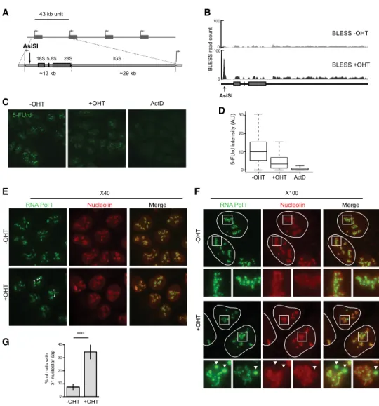
forma-tion of DSBs (Iacovoni et al. 2010). In addiforma-tion to the
DSBs induced elsewhere on the genome (174, as
deter-mined previously) (Clouaire et al. 2018), an AsiSI site
(GCGATCGC) is predicted in the 5
′external transcribed
spacer (5
′ETS) of the 47S rDNA units (Fig. 1A). We
con-firmed the appearance of a rDNA break at the
AsiSI-anno-tated site after OHT addition by direct in situ break
labeling, enrichment on streptavidin, and next-generation
sequencing (BLESS) (Fig. 1B; Crosetto et al. 2013; Clouaire
et al. 2018). Several studies showed previously that DSBs
induced in the rDNA trigger inhibition of rRNA synthesis
as well as nucleolar cap formation (Kruhlak et al. 2007;
Larsen et al. 2014; Harding et al. 2015; van Sluis and
McStay 2015; Warmerdam et al. 2016). AsiSI-induced
rDNA breaks also triggered similar responses regarding
rDNA transcriptional repression, as assessed by
5-fluo-rouridine (5-FUrd) incorporation followed by automated
high-throughput microscopy (Fig. 1C,D), and nucleolar
cap formation, as revealed by RNA Pol I (Fig. 1E
–G;
Sup-plemental Fig. S1A
;
Supplemental Movie S1
) or UBF (
Sup-plemental Fig. S1B,C
) staining.
After OHT treatment, nucleolar cap formation occurred
in only a subset of DIvA cells (
∼35%) (Fig.1G;
Supplemental Fig. S1C
), in contrast to what was reported
previously following rDNA breaks generated by CRISPR/
Cas9 (see also
Supplemental Fig. S1D
–F
; van Sluis and
McStay 2015). In addition, nucleolar segregation observed
following AsiSI was only partial (
Supplemental Fig. S1G,
H
), unlike following actinomycin D treatment (
Supple-mental Fig. S1E
–H
). Blocking AsiSI-induced rDNA repair
with an inhibitor against DNA-PK (Caron et al. 2015)
en-hanced both the number of cells displaying at least one
nucleolar cap (
Supplemental Fig. S1E,F
) and the number
of cells displaying total nucleolar segregation (
Supplemen-tal Fig. S1G,H
), hence mimicking the phenotypes
ob-served with CRISPR/Cas9. Moreover, inducing rDNA
breaks using simultaneously a CRISPR sgRNA and AsiSI
also enhanced nucleolar cap formation compared with
AsiSI alone (
Supplemental Fig. S1I,J
). Altogether, these
data suggest that the discrepancy in nucleolar segregation
observed upon AsiSI- and CRISPR/Cas9-induced breakage
is likely due to the peculiar nature of Cas9-induced breaks
(see the Discussion; Brinkman et al. 2018).
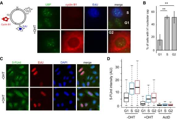
Given that only a subset of DIvA cells displayed
nucleo-lar caps, we wondered whether this could be cell
cycle-reg-ulated. EdU labeling and Cyclin B1 costaining revealed
that nucleolar caps are observed mainly in S/G2 cells
(Fig. 2A,B) even though rDNA cleavage was detected
effi-ciently by BLESS in G1 and G2 cells (
Supplemental Fig.
B
A
D
C
E
G
F
Figure 1. Transcription inhibition and cap formation following AsiSI-induced rDNA breaks. (A) Schematic representation of rDNA re-peats and the position of the AsiSI site in the 5′ETS. (B) BLESS signal at the AsiSI site in the presence or absence of OHT treatment. (C) High-content microscopy image (20× objective) of rRNA synthesis as visualized with 5-FUrd incorporation before and after OHT and actinomycin D (ActD) treatments. Actinomycin D was used as a control for the extinction of rRNA synthesis. (D) Box plot showing the quantification of 5-FUrd signal (>5000 cells) before OHT, after OHT, and after actinomycin D treatment. A representative experiment is shown. (E) DIvA cells either before or after OHT treatment were stained with antibodies against nucleolin (red) and RNA Pol I (green). Nucleolar caps are indicated with arrows. Images were acquired with a 40× objective. (F ) Same as in E, but images were acquired with a 100× objective. Enlargements (white squares) of nucleoli that display nucleolar caps (arrow) or not are also shown. (G) The number of cells with at least one nucleolar cap, as measured by RNA Pol I staining before and after OHT treatment as indicated. The average and SD of four independent experiments are shown. (∗∗∗∗) P < 0.0001.
S2A
). The fact that nucleolar caps were substantially
re-duced in G1 therefore gave us the opportunity to test
whether nucleolar cap formation is a consequence of
tran-scriptional arrest as proposed previously (van Sluis and
McStay 2015). We observed that DSB-induced
transcrip-tional arrest occurs similarly during G1, S, and G2, as
mea-sured by quantitative image-based cytometry (QIBC)
coupled with 5-FUrd incorporation (Fig. 2C,D;
Supple-mental Fig. S2B
), hence demonstrating that nucleolar cap
formation is not solely the consequence of the rDNA
tran-scriptional repression.
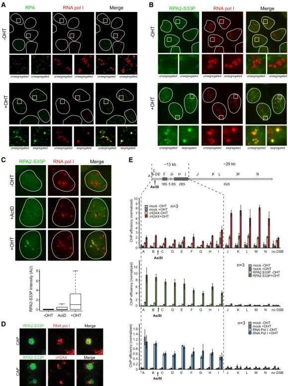
Extended resection occurs within the nucleolus
at rDNA breaks
To investigate how rDNA DSB repair is orchestrated
with-in the nucleolus, we further performed immunostawith-inwith-ing
against DSB signaling and repair proteins. As reported
pre-viously at CRISPR/Cas9- or I-PpoI-induced rDNA DSBs
(Harding et al. 2015; Franek et al. 2016; Warmerdam
et al. 2016), we found that AsiSI-rDNA breaks recruited
γH2AX, ubiquitin, MDC1, 53BP1, and RIF1 only at the
pe-riphery of the nucleolus at nucleolar caps (
Supplemental
Fig. S3A
–C,F
). Similarly, proteins involved in HR (Rad51
and BRCA1) also accumulated at nucleolar caps (
Supple-mental Fig. S3D
–F
), indicating that DSB mobilization at
the periphery of the nucleolus is a prerequisite for HR
pro-tein assembly. We therefore looked for the status of DNA
end resection, which constitutes an early step of HR
re-pair. Accumulation of RPA and phosphorylated RPA at
Ser33 (RPA2-S33P), indicative of end resection, was
clear-ly detected after DSB induction both inside the nucleolus
and in nucleolar caps (Fig. 3A,B;
Supplemental Fig. S3F
).
As expected, RPA2-S33P staining was detected mainly
in S/G2 cells (
Supplemental Fig. S3G
). In contrast,
actino-mycin D treatment only mildly induced RPA staining,
in-dicating that, in nucleoli, RPA accumulation is specific to
DSB formation (Fig. 3C).
Interestingly, superresolution microscopy showed a
mutual exclusion of RPA2-S33P and
γH2AX once
relocat-ed at nucleolar caps (Fig. 3D). In order to further
character-ize resection at rDNA breaks, we performed RPA2-S33P
chromatin immunoprecipitation (ChIP). In agreement
with superresolution microscopy, RPA2-S33P
distribu-tion was found to be antagonistic to that of
γH2AX (Fig.
3E, top and middle panels). Of note, low
γH2AX
occupan-cy on the transcribed region is likely due to decreased
nu-cleosome occupancy rather than decreased DDR kinase
activity, as indicated by ChIP against H2AX (
Supplemen-tal Fig. S3H
). Strikingly, RPA2-S33P was massively
re-cruited across the 13 kb of the transcribed region, as
indicated by RNA Pol I binding (Fig. 3E, bottom panel),
A
B
C
D
Figure 2. Decoupling of transcription inhibition and cap formation during the cell cycle. (A, left panel) Schematic representation of the cell cycle and the methods used to visualize the G2 cells (cytoplasmic cyclin B1 staining in red), S-phase cells (nuclear EdU staining in blue), or G1 cells (absence of staining). (Right panel) Images of OHT-treated cells showing the presence of nucleolar caps (assessed by UBF staining) in EdU- or cyclin B1-positive cells. (B) The number of cells with at least one nucleolar cap in G1, S, or G2 phases as counted with UBF. The mean and SD from three independent experiments are shown. (∗∗) P < 0.01. (C ) Representative images with a 20× objective from the QIBC analysis of the 5-FUrd levels (transcription) using EdU labeling (DNA synthesis) and DAPI staining to distinguish cell cycle phases. (D) A representative box plot of the quantification of the 5-FUrd level (>1500 cells) across the cell cycle phase (G1 [black, S [blue], and G2 [red]) following OHT or actinomycin D (ActD) treatment (as indicated).
B
A
E
C
D
Figure 3. Extended resection along the transcribed region occurs within the nucleolus. (A) DIvA cells left untreated or treated for 4 h with OHT (as indicated) were stained with RNA Pol I (red) and RPA2 (green). Examples of RPA2 staining in normal and segregated (nucleolus with at least one cap) nucleoli are shown in magnification at the bottom. (B) Same as in A, except that total RPA2-S33P was stained (green). (C) Untreated (−OHT), actinomycin D-treated (ActD), or OHT-treated DIvA cells stained with RPA2-S33P (green) and RNA pol I (red). Box plot of RPA2-S33P nucleolar intensity in the indicated conditions. A representative experiment is shown. (D, top panel) A schematic rep-resentation of the primers used across the rDNA unit for quantitative PCR (qPCR). Chromatin immunoprecipitation (ChIP)-qPCR were performed using antibodies against gH2AX, RPA2-S33P, and RNA Pol I in DIvA cells left untreated or treated for 4 h with OHT (as indi-cated). The mean of normalized ChIP efficiency and SEM of three independent experiments are shown. (E) Superresolution images depict-ing the presence of RPA2-S33P (green) and RNA pol I (red) within nucleolar caps (top) or the mutual exclusion of RPA2-S33P (green) in caps andγH2AX (red) surrounding the caps (bottom).
indicating that resection takes place on the entire
tran-scribed unit.
We further tested whether resection is a prerequisite for
DSB mobilization in nucleolar caps. Blocking NHEJ in G1
cells using a DNA-PK inhibitor enhanced both resection
(RPA2-S33P staining) and cap formation, suggesting a
rela-tionship between resection and DSB mobilization (
Sup-plemental Fig. S4
). Furthermore, depletion of the Mre11
component of the MRN complex (known to initiate
resec-tion), of BLM/DNA2 (mediating long-range resection)
(Bordelet and Dubrana 2019), and of RPA2 all impaired
nu-cleolar cap formation (Fig. 4A
–C).
Altogether, our data suggest that, in S/G2, rDNA
DSBs undergo extensive resection spanning the entire
transcribed unit within the interior of the nucleolus.
This resection event is further required for the
move-ment of rDNA DSBs at the periphery of the nucleolus,
where both signaling (
γH2AX, MDC1, and ubiquitin
chain formation) and HR (Rad51 and BRCA1 binding)
can occur.
The cohesin complex ensures transcriptional repression,
DSB mobilization, and HR repair
We further turned our attention toward the cohesin
com-plex, given its pleiotropic involvements in rDNA stability
and transcriptional regulation, DSB repair, and
chromo-some structure and mobility (Kobayashi et al. 2004; Caron
et al. 2012; Gelot et al. 2016a; Herdman et al. 2017; Pal
et al. 2018). Depletion of the SMC1, SCC1, and SMC3
cohesin subunits triggered a decrease in nucleolar cap
for-mation following rDNA breakage (Fig. 5A;
Supplemental
Fig. S5A,B
) without strongly affecting cell cycle
distri-bution (
Supplemental Fig. S5C
). Cohesin depletion also
prevented both resection (as measured by RPA2-S33P
foci) (Fig. 5B) and transcriptional repression following
rDNA damage (Fig. 5C;
Supplemental Fig. S5D,F
). At
rDNA genes, HR frequently triggers rDNA arrays
rear-rangement, which can be visualized using DNA FISH on
individual fibers (Fig. 5D). OHT-mediated rDNA DSB
in-duction in control cells led to an increase of noncanonical
B
A
C
Figure 4. Resection a is a prerequisite for nucleolar cap formation. (A, top panels) Western blots (left) and representative box plots of the level of RPA2-S33P nucleolar intensity (right) after Mre11 or control siRNA knockdown followed by 4 h of OHT treatment in DIvA cells (>100 nucleoli quantified). (Bottom panels) UBF staining was performed in OHT-treated Mre11 or control siRNA transfected DIvA cells. An example (left) and quantification of the percentage of cells displaying at least one nucleolar cap (right) are shown. The mean and SD from three independent experiments are shown. (∗∗∗) P < 0.001. (B) Same as in A, except that BLM/DNA2 were knocked down by siRNAs. The mean and SD from three independent experiments are shown. (∗) P < 0.05. (C) Same as in A, except that RPA2 was knocked down by siRNAs. The mean and SD from three independent experiments are shown. (∗∗∗) P < 0.001.
units indicative of rDNA rearrangements, which was
abolished upon SMC1 depletion using siRNA (Fig. 5E),
in agreement with a function of cohesin in promoting
HR at rDNA units.
The above data therefore suggest that the cohesin
com-plex acts at an early step in the rDNA DSB response and
contributes to the DSB-induced transcriptional repression
at rDNA, thereby influencing all downstream events; i.e.,
resection, DSB mobilization, and HR repair.
Transcriptional repression requires HUSH-mediated
H3K9me3 deposition
In order to identify potential effectors of the cohesin
com-plex that could mediate transcriptional repression of
damaged rDNA, we engineered a DIvA cell line
overex-pressing a Flag-GFP-tagged version of SMC1 (
Supplemen-tal Fig. S5G
). Tandem purification of SMC1 interactors in
the nucleus and nucleolus before and after DSB
produc-tion followed by mass spectroscopy analysis (
Supplemen-tal Fig. S5H
) allowed the identification of candidates,
among which was periphilin 1, a subunit of the recently
identified HUSH complex. This complex, composed of
(at least) periphilin, MPP8, and TASOR, is involved in
the epigenetic repression of retroviruses and LINE-1
retro-transposons via H3K9 trimethylation (H3K9me3)
(Tcha-sovnikarova et al. 2015; Liu et al. 2018; Robbez-Masson
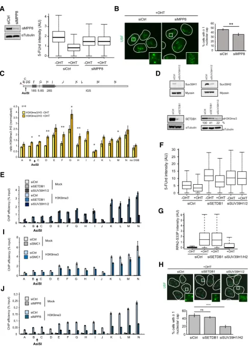
et al. 2018). Indeed, depletion of MPP8 alleviated
DSB-in-duced transcriptional repression of rDNA (Fig. 6A;
Sup-plemental Fig. S6A
), as observed after depletion of the
cohesin complex. In agreement, MPP8 depletion also
translated into decreased cap formation following rDNA
damage (Fig. 6B), with no effect on the cell cycle (
Supple-mental Fig. S6B
), suggesting that the HUSH complex
con-tributes to rDNA transcription shutdown following
rDNA breakage.
Because the HUSH complex was suggested to mediate
transcriptional silencing through H3K9me3, we assessed
the levels of this chromatin mark on rDNA following
DSB induction. An accumulation of H3K9me3 on rDNA
was clearly detected, mostly in the transcribed region after
DSB induction (Fig. 6C;
Supplemental Fig. S6C
). In
mam-malian cells, H3K9 trimethylation is catalyzed mostly
by SUV39H1/2 and SETDB1 (Rea et al. 2000; Schultz
et al. 2002); the latter is suggested to be involved in
HUSH-dependent H3K9 trimethylation
(Tchasovnikar-ova et al. 2015). Knockdown of both SUV39H1/2 and
SETDB1 decreased global H3K9me3 levels in DIvA cells,
as expected (Fig. 6D;
Supplemental Fig. S6D
). However,
on rDNA, only depletion of SUV39H1/2 led to a detectable
H3K9me3 decrease (Fig. 6E). The lack of SETDB1 effects at
the rDNA appears to be a genuine response, as H3K9me3
level decreased upon SETDB1 depletion on zinc finger
genes, as described previously (
Supplemental Fig. S6E
;
Tchasovnikarova et al. 2015). In agreement with the above
data, only SUV39H1/2 depletion, but not SETDB1,
B
A
C
D
E
Figure 5. The cohesin complex regulates rDNA transcriptional repression, resection, DSB mobilization, and HR repair following rDNA breakage. (A, left) Western blot using SMC1 and tubulin antibodies in control or SMC1 siRNA transfected DIvA cells. (Middle) Control or SMC1-depleted DIvA cells treated for 4 h with OHT were stained using an UBF antibody to detect nucleoli and nucleolar caps. (Right) The number of cells with at least one nucleolar cap was quantified. The mean and SD from three independent experiments are shown. (∗) P < 0.05. (B) Representative box plot of nucleolar RPA2-S33P intensity after SMC1 or control siRNA knockdown (>100 nucleoli quantified). (C ) Representative box plot of the 5-FUrd signal before or after 4 h of OHT treatment in control or SMC1-depleted DIvA cells (>1000 cells). (D) Schematic representation of the rDNA repeats and the two fluorescent probes (red and green) used to mea-sure rDNA rearrangements by DNA FISH combing (top), with examples of canonical and noncanonical rDNA units (indicated by an asterisk; bottom). (E) The level of noncanonical rDNA units measured by FISH combing before or after 24 h of OHT treatment in con-trol or SMC1-depleted cells. (∗) P < 0.05.
impaired DSB-induced transcription repression (Fig. 6F;
Supplemental Fig. S6F
), resection (Fig. 6G), and cap
forma-tion (Fig. 6H) without cell cycle changes (
Supplemental
Fig. S6G
). Of importance, depletion of SMC1 or MPP8
also triggered decreased levels of H3K9me3 at rDNA
(Fig. 6I,J), further strengthening the role of cohesin and
the HUSH complex in mediating H3K9me3 deposition.
Collectively, these data suggest that following DSB
pro-duction in rDNA, the cohesin complex mediates
tran-scriptional shutdown of ribosomal genes in a manner
that depends on the HUSH complex and
SUV39H1/2-me-diated H3K9me3.
The LINC complex subunit SUN1, the actin nucleator
ARP3, and the myosin regulator UNC-45A promote DSB
mobilization downstream from transcriptional
repression
We next aimed at characterizing the mechanisms that
ensure rDNA DSB mobilization at the periphery.
Previ-ous studies in yeast and mammals showed a role for
the microtubule network in the movement of DSBs
(Lot-tersberger et al. 2015; Lawrimore et al. 2017; Oshidari
et al. 2018). However, a pretreatment with the
microtu-bule-destabilizing drug nocodazole did not decrease the
A
B
C
E
I
J
D
F
G
H
Figure 6. The HUSH complex mediates transcriptional shutdown after DSB via H3K9me3 deposition. (A, left panel) West-ern blot using MPP8 and tubulin antibodies in control or MPP8 siRNA transfected DIvA cells. (Right panel) 5-FUrd level as measured by high-content microscopy (>5000 cells) in untreated or OHT-treated DIvA transfected with either control or MPP8 siRNA (as indicated). A representa-tive experiment is shown. (B) UBF staining was performed following OHT treatment in control or MPP8-depleted cells as indi-cated. The left panel shows representative images, and the right panel shows the num-ber of cells displaying at least one nucleolar cap. The mean and SD from four indepen-dent experiments are shown. (∗∗) P < 0.01. (C ) ChIP-qPCR was performed in untreated or OHT-treated DIvA cells using antibod-ies against H3K9me3 and H3. The mean and SEM of the ratio H3K9me3/H3 from four independent experiments are shown. (∗) P < 0.05; (∗∗) P < 0.01. (D) Western blots showing the level of SUV39H1 (top left), SUV39H2 (top right), or SETDB1 (bottom left) upon transfection of indicated siRNA as well as their influence on H3K9me3 lev-els (bottom right). (E) ChIP against H3K9me3 was performed in control, SETDB1, and SUV39H1/2 siRNA trans-fected untreated DIvA cells as indicated. ChIP efficiency (percentage of input) of a representative experiment is shown. (F ) Representative box plot of 5-FUrd level as measured by high-content microscopy (>2000 cells) in untreated or OHT-treated DIvA transfected with either control, SETDB1, or SUV39H1/2 siRNA (as indicat-ed). (G) Box plots of RPA2-S33P nucleolar intensity after depletion of SETDB1 or SUV39H1/2 followed by 4 h of OHT treat-ment in DIvA cells. A representative exper-iment is shown (>100 cells). (H) UBF staining was performed following OHT treatment in control, SETDB1-depleted, or SUV39H1/2-depleted cells as indicated. Representative images are shown (left panel), and the number of cells displaying at least one nu-cleolar cap was quantified (right panel). The mean and SD from four independent experiments are shown. (ns) Nonsignificant; (∗∗∗∗) P < 0.0001. (I) ChIP against H3K9me3 was performed in control and SMC1 siRNA transfected untreated DIvA cells as indicated. ChIP effi-ciency (percentage of input) of a representative experiment is shown. (J) ChIP against H3K9me3 was performed in control and MPP8 siRNA transfected untreated DIvA cells as indicated. ChIP efficiency (percentage of input) of a representative experiment is shown.
formation of nucleolar caps (
Supplemental Fig. S7A,B
).
We next focused our attention on the LINC complex,
which is embedded in the NE and mediates connections
between the nucleoskeleton and the cytoskeleton.
In-deed, the LINC complex plays roles in mobilization of
damaged telomeres and IR-induced DSBs in mouse cells
(Lottersberger et al. 2015) and also contributes to DSBs
clustering in human nuclei (Aymard et al. 2017).
Interest-ingly, SUN1, a subunit of the LINC complex localizing to
the NE, was also reported recently to be present in
nucle-oli (Matsumoto et al. 2016; Moujaber et al. 2017). In
agreement, we could indeed confirm that SUN1 forms
patches close to RNA Pol I (
Supplemental Fig. S7C
;
Sup-plemental Movie S2
). Closer inspection revealed that
SUN1-stained NE frequently forms invaginations that
connect the nucleolus (Fig. 7A;
Supplemental Fig. S7D
).
Neither DSB production nor actinomycin D treatment
affected the number of NE invaginations, as observed
using a Lamin B staining (
Supplemental Fig. S7E,F
;
Supplemental Movie S3
). However, SUN1 depletion
de-creased both invaginations and cap formation following
rDNA DSB induction (Fig. 7B;
Supplemental Fig. S7G
),
without affecting cell cycle distribution (
Supplemental
Fig. S7H
), suggesting that the LINC complex contributes
to rDNA mobilization in a manner that involves NE
invaginations.
E
A
B
C
D
F
Figure 7. The LINC complex, ARP3, and UNC-45A mediate rDNA DSB mobilization at the nucleolar periphery. (A) Example of NE in-vagination (SUN1 in green) connecting the nucleolus (RNA Pol I in red); a magnification is shown at the bottom. (B, left panel) Western blot using SUN1 and tubulin antibodies in control or SUN1 siRNA transfected DIvA cells. (Middle panel) Representative image of UBF staining in OHT-treated DIvA cells in control or SUN1-depleted cells. (Right panel) The number of cells displaying at least one nucleolar cap. The mean and SD from three independent experiments are shown. (∗) P < 0.05. (C) Same as in B, except that a siRNA against ARP3 was used. The mean and SD from three independent experiments are shown. (∗∗) P < 0.01. (D) Same as in B, except that a siRNA against UNC-45A was used. The mean and SD from five independent experiments are shown. (∗∗∗) P < 0.001. (E) 5-FUrd level measured by high-content micros-copy (>5000 cells) in untreated or OHT-treated DIvA transfected with either control or SUN1 siRNA (as indicated). A representative exper-iment is shown. (F ) Same as in E, except that siRNA against ARP3 and UNC-45A were used. A representative experexper-iment is shown.
Interestingly, persistent and heterochromatic DSBs
were shown to move toward the NE in yeast and
Droso-phila
cells, respectively (Horigome et al. 2014; Ryu et al.
2015), via nuclear actin polymerization in Drosophila
(Caridi et al. 2018). We hence further asked whether
rDNA DSB mobilization also necessitates the actin
net-work. Indeed, depletion of UNC-45A (also known as
gene-ral cell UNC-45 [GC-UNC-45], a myosin regulator)
(Lehtimäki et al. 2017) and ARP3 (an actin
nucleator-medi-ating branched actin network), both previously involved in
heterochromatic DSB mobilization in flies (Caridi et al.
2018), also decreased DSB-induced cap formation (Fig.
7C,D) without major changes to cell cycle distribution
(
Supplemental Fig. S7H
). Strikingly, depletion of all three
factors (SUN1, UNC-45A, and ARP3) did not affect
tran-scriptional repression following DSB induction (Fig. 7E,F;
Supplemental Fig. S7I
). Collectively, these data suggest
that the NE/LINC complex and actin network (ARP3
and UNC-45A) ensure rDNA DSB mobilization outside
of the nucleolus downstream from transcriptional
inhibition.
Discussion
The rDNA is one of the most fragile loci on the genome
and is also particularly prone to experience major
rear-rangements during replication and repair (Lindström
et al. 2018). Highly specialized repair mechanisms are
likely implemented in order to ensure its stability across
cell divisions. Here we uncovered that repair of DSBs
pro-duced on the rDNA requires transcriptional extinction via
a cohesin/HUSH-dependent pathway, followed in S/G2
cells by end resection in the interior of the nucleolus.
Re-sected DNA ends are further mobilized at the nucleolar
periphery, where they undergo HR repair and
accumula-tion of
γH2AX, MDC1, and ubiquitin chains.
Interesting-ly, a recent study reported that the Nbs1 component of the
MRN complex as well as low levels of
γH2AX signal can
nevertheless be observed within the interior of the
nucle-olus following rDNA break induction (Korsholm et al.
2019). Moreover, ATM activation following DSB in
rDNA precedes DSB mobilization (this study; Harding
et al. 2015; Korsholm et al. 2019). Hence, altogether,
this suggests that while DSB signaling by PI3 kinases
can take place at the interior of the nucleolus, signaling
is further amplified at the periphery of the nucleolus
once resection has occurred, in agreement with our recent
findings that
γH2AX accumulates more at DSBs repaired
by HR (Clouaire et al. 2018).
Of importance, we further found that the NE forms
in-vaginations that connect the nucleolus and that both the
LINC complex and the actin network contribute to rDNA
DSB mobilization (
Supplemental Fig. S8
).
Uncoupling DSB mobilization from transcriptional
repression in the context of rDNA damage
In agreement with previous studies using either the
CRISPR/Cas9 or the I-PpoI homing endonuclease to
in-duce DSBs on the rDNA array, we showed that
AsiSI-in-duced rDNA breaks trigger both rDNA transcriptional
arrest and nucleolar segregation as visualized by cap
for-mation (i.e., DSB movement) (Larsen et al. 2014; Harding
et al. 2015; van Sluis and McStay 2015; Pefani et al. 2018).
However, as opposed to CRISPR/Cas9- or I-PpoI-mediated
breaks that promote nucleolar segregation across all cell
cycle stages, caps are rather restricted to S/G2 cells
when induced by AsiSI despite comparable levels of DSB
formation and transcriptional repression that were
ob-served throughout the cell cycle. We could further verify
that nucleolar segregation occurred in nearly all cells
fol-lowing (1) actinomycin D treatment, (2)
CRISPR/Cas9-mediated cleavage induced using nanoblades (Mangeot
et al. 2019), and (3) AsiSI activation in presence of an
in-hibitor of DNA-PK that strongly blocks AsiSI DSB repair
(
Supplemental Fig. S1E
–H
; Caron et al. 2015). This
indi-cated that, in principle, DIvA cells are able to undergo
ro-bust nucleolar segregation and that blocking AsiSI DSB
repair somehow mimics CRISPR/Cas9- or I-PpoI-induced
rDNA cleavage. In addition, we showed that DSBs
pro-duced by AsiSI elsewhere on the genome do not compete
with the rDNA response, since CRISPR/Cas9-induced
breaks on the rDNA also triggered robust nucleolar
segre-gation in the presence of activated AsiSI (
Supplemental
Fig. S1I,J
). Altogether, this suggests that the milder
phenotypes observed upon AsiSI-mediated breakage
com-pared with Cas9-mediated break induction
—both
tran-scriptional repression (data not shown) and nucleolar
cap formation (
Supplemental Fig. S1
)
—could be due to
ei-ther a lower amount of rDNA copies cleaved by AsiSI
(sensitive to DNA methylation) compared with Cas9
and/or discrepancies in repair efficiency. Of note, recent
work on CRISPR/Cas9-induced DSBs suggested that
such breaks are particularly refractory to repair
(Brink-man et al. 2018) and may be handled by the Fanconi
ane-mia pathway (Richardson et al. 2018). In addition, in
contrast to type II restriction enzyme (such as AsiSI),
the I-PpoI homing endonuclease recognizes particularly
long DNA sequences in a manner that severely bends
and alters the DNA structure (Chevalier and Stoddard
2001), which may also affect repair efficiency. Altogether,
we propose that the inherent persistent nature of
CRISPR/Cas9- or I-PpoI-induced breaks may hence
ac-count for the extreme nucleolar segregation and HR usage
reported previously at all cell cycle stages (van Sluis and
McStay 2015) and that the restriction of DSB movement
to S and G2 cells is a bona fide rDNA DSB response.
DSB mobilization at the nucleolar periphery has been
proposed previously to be a direct consequence of
DSB-in-duced rDNA transcriptional extinction (van Sluis and
McStay 2015). Indeed, the nucleolus is a self-organizing
membrane-free organelle in which rRNAs act as seeds
ini-tiating a phase separation process leading to nucleolus
formation (Falahati et al. 2016). Extinction of rDNA
tran-scription rapidly triggers a drastic reorganization in which
rDNA is expelled at the periphery by a phase separation
event (as observed with actinomycin D). However, our
findings indicate that rDNA DSB mobilization at the
nu-cleolar periphery is not only a consequence of a phase
separation driven by rDNA transcriptional arrest. Indeed,
DSBs are mobilized only in S/G2 stage, while
transcrip-tional repression occurs throughout the cell cycle.
More-over, depletion of the LINC/actin pathway impairs
mobilization without affecting the upstream
transcrip-tional inhibition. Of note, this rDNA mobilization
path-way is specific to DSB formation, since depletion of
SMC1, Suv39H1/2, Sun1, UNC45, and Arp3 did not affect
rDNA segregation observed after actinomycin D (data not
shown). Importantly, so far, we could not identify any
con-ditions under which transcriptional arrest is impaired and
DSBs are still mobilized, which might suggest that
tran-scriptional repression is a prerequisite for DSB movement.
Mechanisms ensuring DSB mobilization: NE
invagination, LINC, and the actin network
Interestingly, the mobilization of rDNA DSBs shares
common features with the mobilization of those induced
in heterochromatin and active genes and of persistent
DSBs. First, in mammals, DSBs induced in
pericentro-meric heterochromatin display relocalization to the
pe-riphery of the heterochromatin focus during S/G2 in a
manner that depends on resection (Tsouroula et al.
2016), as shown here for a DSB induced in rDNA.
Sec-ond, in Drosophila, heterochromatic DSBs move toward
the periphery of the heterochromatic focus and further to
the NE in a manner that depends on Suv39H1-mediated
H3K9me3, on the LINC complex, and on the actin
net-work (Chiolo et al. 2011; Ryu et al. 2015; Colmenares
et al. 2017; Caridi et al. 2018). Third, studies in yeast
have shown that persistent DSBs are relocalized to the
NE via the SUN domain-containing proteins Mps3 or
Sad1 (Oza et al. 2009; Horigome et al. 2014; Swartz
et al. 2014). Fourth, we reported previously that both
the actin network and the LINC complex contribute to
clustering of DSBs induced in active genes in G1
(Aymard et al. 2017). Of importance all of the
above-mentioned DSBs were described previously as refractory
to fast repair (for review, see Marnef et al. 2017; Fortuny
and Polo 2018). We thus propose that breaks in rDNA
may also display impaired or delayed repair kinetics
—
in line with its repetitive nature, high RNA Pol I
occu-pancy, and prevalence of secondary structures (R loops
and G4)
—and hence use similar mobilization pathways
to undergo safe repair.
Notably, while the interaction of persistent DSBs with
the NE has been reported in Drosophila and yeast, there
was no evidence of such phenomena in mammals despite
a reported function of the LINC complex in DSB repair
(Lottersberger et al. 2015; Aymard et al. 2017). Here we
re-port that the NE forms invaginations that contact the
nu-cleolus (in line with previous reports showing strong
relationship with the NE and nucleolus)
(Hernandez-Verdun et al. 2010) and that depletion of the LINC
com-plex, which reduces NE invaginations (
Supplemental
Fig. S7
), also affects rDNA DSB relocalization to the
nucleolus periphery. Hence, our data suggest that
NE/per-sistent DSB anchorage also occur in mammals and
that, for rDNA breaks, this interaction contributes to
the localization of the DSB at the nucleolar periphery.
Al-ternatively, or in addition, the LINC complex and NE
in-vaginations could also directly regulate resection, which
could further foster DSB mobilization to the periphery
of the nucleolus.
Our data also indicate that rDNA movement relies on
the actin network, as reported for Drosophila
hetero-chromatic breaks (Caridi et al. 2018) and in line with
the contribution of actin regulation in DSB repair (Belin
et al. 2015; Spichal et al. 2016; Aymard et al. 2017;
Schrank et al. 2018). One possibility is that rDNA DSB
mobilization involves the formation of nuclear/nucleolar
actin filaments as found in other contexts (Belin et al.
2015; Caridi et al. 2018; Schrank et al. 2018).
Alterna-tively, since cytoplasmic filamentous actin can be found
in NE invaginations (Jorgens et al. 2017), one could
en-visage that cytoplasmic actin cables/filaments regulate
NE deformation and the dynamics of invaginations,
which could further contribute to the relocalization of
the rDNA DSBs outside of the nucleolus thanks to a
physical link between the DSBs and NE-embedded
LINC complex. Altogether our data suggest that a
con-served mechanism involving the LINC complex and
the actin network probably exists to mobilize all
“persis-tent
” or “difficult to repair” DSBs to the NE, providing a
safe recombination environment.
Cohesin/HUSH/H3K9me3-driven DSB-induced rDNA
transcriptional repression
DSB-induced rDNA transcriptional repression has been
shown to require ATM activity, Nbs1 (our data not shown;
Kruhlak et al. 2007; Larsen et al. 2014; Harding et al. 2015;
van Sluis and McStay 2015; Warmerdam et al. 2016; Pefani
et al. 2018), and, more recently, the MST2-dependent
phosphorylation of H2B on Ser14 (Pefani et al. 2018).
Here we found that the cohesin complex also regulates
RNA Pol I transcription in response to DSBs. Interestingly,
conserved mechanisms may account for RNA Pol I and Pol
II regulation in cis to DSBs, since both ATM and the
cohe-sin complex were identified as key players in RNA Pol II
transcriptional arrest in cis to DSBs (Shanbhag et al.
2010; Meisenberg et al. 2019). This raises the interesting
possibility that ATM may regulate transcription at least
in part by regulating the cohesin biology in cis to DSBs,
as the cohesin complex is an ATM target (Kim et al.
2002). Our data also indicate that the HUSH complex,
shown previously to display H3K9me3 methylation
activ-ity (Tchasovnikarova et al. 2015; Liu et al. 2018;
Robbez-Masson et al. 2018), participates in DSB-induced RNA
Pol I transcriptional repression. Notably, SUV39H1,
SETDB1, and also the histone demethylase KDM4A, all
known to regulate H3K9me3 levels, have been involved
in DSB repair by HR (Chiolo et al. 2011; Ayrapetov et al.
2014; Alagoz et al. 2015; Colmenares et al. 2017), and
loss of H3K9me3 triggers aberrant recombination between
repetitive sequence (Peng and Karpen 2007). On the other
hand, the cohesin complex is a key regulator of HR; it
pre-vents the end joining of distant DSB ends (for review, see
Dorsett and Ström 2012; Gelot et al. 2016b) and suppresses
aberrant recombination at rDNA (Kobayashi et al. 2004;
Kobayashi and Ganley 2005). Our study provides a
mecha-nistic link between the cohesin complex and the
regula-tion of H3K9me3 levels and suggests that cohesin
contributes to the regulation of H3K9me3 levels by
re-cruiting the HUSH complex, further allowing
transcrip-tional repression, resection, and DSB mobilization for
recombination.
In summary, here we report that as for heterochromatic
DSBs, rDNA DSB repair relies on a spatiotemporal
separa-tion, where resection within the nucleolus is followed by
the mobilization of the break outside its environment.
Since mobilization involves players also required for the
relocalization of other
“persistent” DSBs such as the
LINC complex, our data suggest that conserved DSB
mo-bilization-dependent mechanisms exist from yeast to
hu-mans in order to relocate harmful breaks to a NE
compartment, allowing safe repair.
Materials and methods Cell culture and treatments
DIvA cells were grown in Dulbecco’s modified Eagle’s medium (DMEM) supplemented with 10% fetal calf serum (Invitrogen) in the presence of 1 µg/mL puromycin (Invivogen). Unless stated otherwise, DSBs were induced by the addition of 300 nM OHT (Sigma, H7904) for 4 h. For DNA-PK inhibition, 2 µM NU7441 (Selleckchem) was added 1 h prior to OHT addition and main-tained during the OHT-mediated break induction. When indicat-ed, 100 ng/mL actinomycin D (Sigma) was added to cells for a total of 1 h. Nocodazole (3 µM) was added 1 h prior to OHT addi-tion. siRNA-mediated depletion of proteins (Supplemental Table S1) was performed using the cell line Nucleofector kit V (Program X-001, Amaxa) according to the manufacturer’s instructions. De-tails for CRISPR–Cas9-induced breakage and cell synchroniza-tion are in theSupplemental Material.
BLESS analysis on rDNA
BLESS sequences from asynchronized cells or G1 and G2 cell pop-ulations were retrieved from our previous work (Aymard et al. 2017; Clouaire et al. 2018; Mourad et al. 2018). Barcodes were re-moved using Cutadapt, and BLESS data were aligned on a custom-built genome composed of hg19 plus rRNA unit U13369.1 from GenBank using bwa in paired-end mode (bwa aln and bwa sampe). Fragments were reconstituted from paired reads using Rsamtools and GenomicAlignments (R packages). Fragments with lengths >500 bp were dropped, and the remaining fragments were dedupli-cated (fragments with the exact same start and end positions were considered as duplicates). For the G1 and G2 BLESS data analysis, proximal positions were extracted from fragments based on their relative positions to the DSB.
Immunofluorescence
Cells were plated on glass coverslips and fixed in 4% paraformal-dehyde/PBS for 20 min, permeabilized in 0.5% Triton X-100/PBS for 5 min unless stated otherwise (Supplemental Table S2), and blocked with 3% BSA/PBS for 30 min before incubation with pri-mary antibodies (see Supplemental Table S2) in 3% BSA/PBS overnight at 4°C. For CSK pre-extraction, cells were permeabi-lized using 0.5% Triton X-100 for 5 min followed by 10 min in
CSK buffer (100 mM NaCl, 300 mM sucrose, 3 mM MgCl2,
10 mM PIPES at pH 6.8), fixation in 4% PFA/PBS, and blocking with 3% BSA/PBS. For buffer II pre-extraction (20 mM NaCl, 0.5% NP-40, 20 mM HEPES at pH 7.5, 5 mM MgCl2, 1 mM
DTT), buffer II was added to cells for 20 min on ice followed by fixation in 4% PFA/PBS and blocking with 3% BSA/PBS. The ap-propriate Alexa-conjugated secondary antibodies diluted in 3% BSA/PBS (mouse or rabbit Alexa 594 or Alexa 488 used at a 1:1000 dilution [Invitrogen]) were then added to cells for 1 h at room temperature followed by DAPI or Hoechst 33342 staining. Coverslips were mounted in Citifluor (Citifluor, AF-1), and imag-es were acquired using a wide-field microscope (Leica, DM6000) equipped with a cooled charge-coupled device camera (Cool-SNAP HQ2) using 40× or 100× objectives and MetaMorph.
Images of total RPA and SUN1 immunostainings were pro-cessed by nonlinear deconvolution with Huygens software (Sci-entific Volume Imaging). Classical maximum likelihood estimation was used to estimate positive fluorescent signal after deconvolution. An adaptive estimation of background was done for each image, the signal to noise ratio was set to 40 (RPA) or 20 (SUN1), and 50 iterations were used during the minimization. For superresolution microscopy, we imaged cells using a fast structure illumination microscopy (SIM) setup with a binary phase optical modulator (QXGA SLM Fourth Dimension) under a 100× objective lens (CFI SR APO 100XH ON 1,49 DT 0,12 Nikon) mounted on a Nikon Eclipse Ti-E and a Hamamatsu SCMOS camera (ORCA FLASH4.0 LT). A series of 800 3D Speck-le illuminations was made on the object plane thanks to the ran-dom phase modulation from the SLM conjugate with the pupil plane of the microscope objective. A 150-nm Z interval was ac-quired on each image. Superresolution images were processed by a joint reconstruction strategy using random speckle illumina-tions (Negash et al. 2016; Labouesse et al. 2017). A positivity con-straint was applied based on the realistic hypothesis that the fluorescent signal was only positive.
Quantifications of immunofluorescence
For DSB mobilization, the number of cells displaying at least one nucleolar cap was counted manually using the 40× objective in >150 cells per experiments and for a minimum of three indepen-dent experiments using either RNA pol I or UBF as markers. The number of caps (RNA Pol I staining) per nucleus was counted manually in >80 cells. Resection was quantified using RPAS33P antibody in >100 cells acquired with a 40× objective and analyzed using Columbus software (Perkin Elmer). Nucleoli were detected thanks to the RNA Pol I (using method A), and the sum of RPA2S33 spot (method D) intensity inside the nucleolar region was determined. At least three independent experiments were performed; only one representative experiment is shown as a box plot. InSupplemental Figure S3F, the presence of repair pro-tein in either unsegregated or segregated nucleoli was quantified in cells with at least one nucleolar cap. The number of invagina-tions per cell was quantified manually using Lamin B1 staining in >70 cells and in three independent experiments.
High-throughput microscopy
The Operetta automated high-content screening microscope (PerkinElmer) was used for the rDNA transcription and/or cell cy-cle analyses. DIvA cells were plated in 96-well plates and treated with OHT for 4 h. For the visualization of nascent rRNA synthe-sis, 2 mM 5-FUrd (Sigma) was added 20–40 min before the end of the OHT treatment. For the detection of S-phase cells, EdU was added 10–20 min prior to the end of the OHT treatment. Cells
were fixed and permeabilized as mentioned above, and saturation was done in 1%BSA/PBS for 30 min. For EdU-treated cells, a Click-iT reaction (Alexa 488; Invitrogen) was performed accord-ing to the manufacturer’s instruction before the addition of an anti-BrdU primary antibody (BU-33; Sigma) at 1:500 in 1% BSA/ PBS in the presence of 160 U/m RNasinL (Promega) overnight at 4°C. Secondary antimouse Alexa 647 (1:500 in 1% BSA/PBS; Molecular Probes, A21235) was incubated for 30 min at 37°C in the presence of 80 U/m RNasinL followed by DAPI staining. Im-age acquisition with a 20× objective lens was automated to obtain at least 36 fields per well, allowing the visualization of a total of >1000 cells (three wells were acquired for each condition). Each picture was analyzed with the integrated Columbus software. Briefly, the DAPI-stained nuclei were selected (method B), and the size and roundness of nuclei were used as parameters to elim-inate false positive compounds. The 5-FUrd staining attributed to the nucleoli was delineated using the find spot method D, and the sum of the 5-FUrd intensity was measured and is represented as box plots. For cell cycle analysis, the sum of the DAPI intensity and the mean of the EdU intensity were plotted in order to sepa-rate G1, S, and G2 cells. The sum of the 5-FUrd intensity was sub-sequently determined in each of these cell population.
ChIP
ChIP experiments were performed as in Iacovoni et al. (2010). The amount of chromatin and antibodies used are detailed in Supple-mental Table S2. ChIP efficiencies were calculated as a percent-age of input of DNA immunoprecipitated. For quantitative PCR (qPCR) analyses, the input as well as immunoprecipitation sam-ples were analyzed with the primers listed inSupplemental Table S3. ForSupplemental Figs. 3E and S6C, ChIP efficiencies (per-centage of input) obtained with each primer pair were further nor-malized to the value obtained for primer B in the undamaged condition. The ratio of H3K9m3 over H3 was calculated before normalization with primer B for Figure 6C.
DNA FISH combing and analysis
The combing and FISH probing of the rDNA fibers were carried out before or after 24 h of OHT treatment (Genomic Vision). The detection and rDNA fibers and the attribution of rDNA-spe-cific probes (Caburet et al. 2005) was carried out using Genomic Vision’s provided Web interface. Analysis of rearrangements was performed using a script that was designed to count the total number of units (as defined when comprised between two“IGS” regions) and assess the occurrence of changes when comparing one unit with the next. The source code was deposited on GitHub (https://github.com/LegubeDNAREPAIR/rDNA/blob/master/ get_break_event.py).
Western blotting
Cells were incubated in RIPA buffer (0.5% deoxycholate, 1% NP-40, 50 mM Tris at pH 8, 150 mM NaCl, 0.1% SDS) for 20 min on ice and centrifuged at 13,000 rpm for 10 min before SDS loading buffer addition to the supernatant. In the case of H3 and H3K9me3 immunoblots, cells were sonicated three times for 10 sec in lysis buffer (1% SDS, 1% Triton, 10 mM Tris at pH 7.5, 450 mM NaCl) before the addition of SDS loading buffer. Protein extracts were resolved on 3%–8% NuPAGE Tris-acetate gels or 4%–12% Bis-Tris (Invitrogen) according to the manufac-turer’s instructions and transferred onto PVDF membranes (Invi-trogen). The latter were blocked in TBS containing 0.1% Tween 20 (Sigma) and 3% nonfat dry milk for 1 h followed by an
over-night incubation at 4°C with primary antibodies (Supplemental Table S2). The appropriate horseradish peroxidase-coupled sec-ondary antibodies were used to reveal the proteins (antimouse at 1:10,000 [Sigma, A2554] and antirabbit at 1:10,000 [Sigma, A0545]) using a luminol-based enhanced chemiluminescence HRP substrate (Super Signal West Dura Extended Duration Sub-strate, Thermo Scientific).
Acknowledgments
We thank T. Clouaire and N. Puget for their advice throughout the project and comments on the manuscript. We thank E. Watrin (Rennes) for the Flag-GFP-SMC1 construct. We thank the high-throughput microscopy platform of the Centre de Biolo-gie Intégrative (Toulouse), and the mass spectrometry platform from the Institut de Pharmacologie et de Biologie Structurale (Toulouse). Funding in E.P.R.’s laboratory was provided by Labex Ecofect (ANR-11-LABX-0048), Fondation Innovations en Infec-tiologie (Finovi), and the European Research Council (ERC-StG-LS6-805500) under the European Union’s Horizon 2020 research and innovation programs. Funding in G.L.’s laboratory was pro-vided by grants from the European Research Council (ERC-2014-CoG 647344), the Agence Nationale pour la Recherche (ANR-14-CE10-0002-01 and ANR-13-BSV8-0013), the Institut National Contre le Cancer (INCA), and the Ligue Nationale Contre le Cancer (LNCC).
Author contributions: A.M., A.-L.F., C.A., V.D., and E.G. per-formed experiments. V.R. perper-formed bioinformatic analyses of BLESS data sets. T.M. performed superresolution microscopy. P.E.M. and E.P.R. provided nanoblades for CRISPR/Cas9-induced breakage. A.M. and G.L. conceived, supervised, and analyzed ex-periments. A.M. and G.L. wrote the manuscript. All authors com-mented and edited the manuscript.
References
Alagoz M, Katsuki Y, Ogiwara H, Ogi T, Shibata A, Kakarougkas A, Jeggo P. 2015. SETDB1, HP1 and SUV39 promote reposi-tioning of 53BP1 to extend resection during homologous re-combination in G2 cells. Nucleic Acids Res 43: 7931–7944. doi:10.1093/nar/gkv722
Aymard F, Bugler B, Schmidt CK, Guillou E, Caron P, Briois S, Iacovoni JS, Daburon V, Miller KM, Jackson SP, et al. 2014. Transcriptionally active chromatin recruits homologous re-combination at DNA double-strand breaks. Nat Struct Mol Biol21: 366–374. doi:10.1038/nsmb.2796
Aymard F, Aguirrebengoa M, Guillou E, Javierre BM, Bugler B, Arnould C, Rocher V, Iacovoni JS, Biernacka A, Skrzypczak M, et al. 2017. Genome-wide mapping of long-range contacts unveils clustering of DNA double-strand breaks at damaged active genes. Nat Struct Mol Biol 24: 353–361. doi:10.1038/ nsmb.3387
Ayrapetov MK, Gursoy-Yuzugullu O, Xu C, Xu Y, Price BD. 2014. DNA double-strand breaks promote methylation of histone H3 on lysine 9 and transient formation of repressive chroma-tin. Proc Natl Acad Sci 111: 9169–9174. doi:10.1073/pnas .1403565111
Belin BJ, Lee T, Mullins RD. 2015. DNA damage induces nuclear actin filament assembly by Formin-2 and Spire-1/2 that pro-motes efficient DNA repair. Elife 4: e07735. doi:10.7554/ eLife.07735
Bordelet H, Dubrana K. 2019. Keep moving and stay in a good shape to find your homologous recombination partner. Curr Genet65: 29–39. doi:10.1007/s00294-018-0873-1
Brinkman EK, Chen T, de Haas M, Holland HA, Akhtar W, van Steensel B. 2018. Kinetics and fidelity of the repair of Cas9-in-duced double-strand DNA breaks. Mol Cell 70: 801–813.e6. doi:10.1016/j.molcel.2018.04.016
Caburet S, Conti C, Schurra C, Lebofsky R, Edelstein SJ, Bensi-mon A. 2005. Human ribosomal RNA gene arrays display a broad range of palindromic structures. Genome Res 15: 1079–1085. doi:10.1101/gr.3970105
Caridi CP, D’Agostino C, Ryu T, Zapotoczny G, Delabaere L, Li X, Khodaverdian VY, Amaral N, Lin E, Rau AR, et al. 2018. Nuclear F-actin and myosins drive relocalization of hetero-chromatic breaks. Nature 559: 54–60. doi:10.1038/s41586-018-0242-8
Caron P, Aymard F, Iacovoni JS, Briois S, Canitrot Y, Bugler B, Massip L, Losada A, Legube G. 2012. Cohesin protects genes againstγH2AX Induced by DNA double-strand breaks. PLoS Genet8: e1002460. doi:10.1371/journal.pgen.1002460 Caron P, Choudjaye J, Clouaire T, Bugler B, Daburon V,
Aguirre-bengoa M, Mangeat T, Iacovoni JS, Álvarez-Quilón A, Cortés-Ledesma F, et al. 2015. Non-redundant functions of ATM and DNA-PKcs in response to DNA double-strand breaks. Cell Rep13: 1598–1609. doi:10.1016/j.celrep.2015.10.024 Chevalier BS, Stoddard BL. 2001. Homing endonucleases:
struc-tural and functional insight into the catalysts of intron/intein mobility. Nucleic Acids Res 29: 3757–3774. doi:10.1093/nar/ 29.18.3757
Chiolo I, Minoda A, Colmenares SU, Polyzos A, Costes SV, Karpen GH. 2011. Double-strand breaks in heterochromatin move outside of a dynamic HP1a domain to complete recom-binational repair. Cell 144: 732–744. doi:10.1016/j.cell.2011 .02.012
Clouaire T, Legube G. 2015. DNA double strand break repair pathway choice: a chromatin based decision? Nucleus 6: 107–113. doi:10.1080/19491034.2015.1010946
Clouaire T, Rocher V, Lashgari A, Arnould C, Aguirrebengoa M, Biernacka A, Skrzypczak M, Aymard F, Fongang B, Dojer N, et al. 2018. Comprehensive mapping of histone modifications at DNA double-strand breaks deciphers repair pathway chro-matin signatures. Mol Cell 72: 250–262.e6. doi:10.1016/j .molcel.2018.08.020
Colmenares SU, Swenson JM, Langley SA, Kennedy C, Costes SV, Karpen GH. 2017. Drosophila histone demethylase KDM4A has enzymatic and non-enzymatic roles in controlling hetero-chromatin integrity. Dev Cell 42: 156–169.e5. doi:10.1016/j .devcel.2017.06.014
Crosetto N, Mitra A, Silva MJ, Bienko M, Dojer N, Wang Q, Karaca E, Chiarle R, Skrzypczak M, Ginalski K, et al. 2013. Nucleotide-resolution DNA double-strand break mapping by next-generation sequencing. Nat Methods 10: 361–365. doi:10.1038/nmeth.2408
Dorsett D, Ström L. 2012. The ancient and evolving roles of cohe-sin in gene expression and DNA repair. Curr Biol 22: R240– R250. doi:10.1016/j.cub.2012.02.046
Falahati H, Pelham-Webb B, Blythe S, Wieschaus E. 2016. Nucle-ation by rRNA dictates the precision of nucleolus assembly. Curr Biol26: 277–285. doi:10.1016/j.cub.2015.11.065 Floutsakou I, Agrawal S, Nguyen TT, Seoighe C, Ganley ARD,
McStay B. 2013. The shared genomic architecture of human nucleolar organizer regions. Genome Res 23: 2003–2012. doi:10.1101/gr.157941.113
Fortuny A, Polo SE. 2018. The response to DNA damage in het-erochromatin domains. Chromosoma 127: 291–300. doi:10 .1007/s00412-018-0669-6
Franek M, Kovaříková A, Bártová E, Kozubek S. 2016. Nucleolar reorganization upon site-specific double-strand break
induc-tion. J Histochem Cytochem 64: 669–686. doi:10.1369/ 0022155416668505
Frohns A, Frohns F, Naumann SC, Layer PG, Löbrich M. 2014. In-efficient double-strand break repair in murine rod photorecep-tors with inverted heterochromatin organization. Curr Biol 24: 1080–1090. doi:10.1016/j.cub.2014.03.061
Gelot C, Guirouilh-Barbat J, Le Guen T, Dardillac E, Chailleux C, Canitrot Y, Lopez BS. 2016a. The cohesin complex prevents the end joining of distant DNA double-strand ends. Mol Cell 61: 15–26. doi:10.1016/j.molcel.2015.11.002
Gelot C, Guirouilh-Barbat J, Lopez BS. 2016b. The cohesin com-plex prevents the end-joining of distant DNA double-strand ends in S phase: consequences on genome stability mainte-nance. Nucleus 7: 339–345. doi:10.1080/19491034.2016 .1194159
Goodarzi AA, Kurka T, Jeggo PA. 2011. KAP-1 phosphorylation regulates CHD3 nucleosome remodeling during the DNA double-strand break response. Nat Struct Mol Biol 18: 831– 839. doi:10.1038/nsmb.2077
Guénolé A, Legube G. 2017. A meeting at risk: unrepaired DSBs go for broke. Nucleus 8: 589–599. doi:10.1080/19491034 .2017.1380138
Harding SM, Boiarsky JA, Greenberg RA. 2015. ATM dependent silencing links nucleolar chromatin reorganization to DNA damage recognition. Cell Rep 13: 251–259. doi:10.1016/j .celrep.2015.08.085
Herdman C, Mars J-C, Stefanovsky VY, Tremblay MG, Sabourin-Felix M, Lindsay H, Robinson MD, Moss T. 2017. A unique enhancer boundary complex on the mouse ribosomal RNA genes persists after loss of Rrn3 or UBF and the inactivation of RNA polymerase I transcription. PLoS Genet 13: e1006899. doi:10.1371/journal.pgen.1006899
Hernandez-Verdun D, Roussel P, Thiry M, Sirri V, Lafontaine DLJ. 2010. The nucleolus: structure/function relationship in RNA metabolism. Wiley Interdiscip Rev RNA 1: 415–431. doi:10.1002/wrna.39
Horigome C, Oma Y, Konishi T, Schmid R, Marcomini I, Hauer MH, Dion V, Harata M, Gasser SM. 2014. SWR1 and INO80 chromatin remodelers contribute to DNA double-strand break perinuclear anchorage site choice. Mol Cell 55: 626– 639. doi:10.1016/j.molcel.2014.06.027
Iacovoni JS, Caron P, Lassadi I, Nicolas E, Massip L, Trouche D, Legube G. 2010. High-resolution profiling ofγH2AX around DNA double strand breaks in the mammalian genome. EMBO J29: 1446–1457. doi:10.1038/emboj.2010.38
Jakob B, Splinter J, Conrad S, Voss K-O, Zink D, Durante M, Löbrich M, Taucher-Scholz G. 2011. DNA double-strand breaks in heterochromatin elicit fast repair protein recruit-ment, histone H2AX phosphorylation and relocation to eu-chromatin. Nucleic Acids Res 39: 6489–6499. doi:10.1093/ nar/gkr230
Jorgens DM, Inman JL, Wojcik M, Robertson C, Palsdottir H, Tsai W-T, Huang H, Bruni-Cardoso A, López CS, Bissell MJ, et al. 2017. Deep nuclear invaginations are linked to cytoskeletal filaments - integrated bioimaging of epithelial cells in 3D cul-ture. J Cell Sci 130: 177–189. doi:10.1242/jcs.190967 Kakarougkas A, Ismail A, Chambers AL, Riballo E, Herbert AD,
Künzel J, Löbrich M, Jeggo PA, Downs JA. 2014. Requirement for PBAF in transcriptional repression and repair at DNA breaks in actively transcribed regions of chromatin. Mol Cell 55: 723–732. doi:10.1016/j.molcel.2014.06.028
Kim S-T, Xu B, Kastan MB. 2002. Involvement of the cohesin pro-tein, Smc1, in Atm-dependent and independent responses to DNA damage. Genes Dev 16: 560–570. doi:10.1101/gad .970602