HAL Id: tel-03230952
https://tel.archives-ouvertes.fr/tel-03230952
Submitted on 20 May 2021
HAL is a multi-disciplinary open access
archive for the deposit and dissemination of
sci-entific research documents, whether they are
pub-lished or not. The documents may come from
teaching and research institutions in France or
abroad, or from public or private research centers.
L’archive ouverte pluridisciplinaire HAL, est
destinée au dépôt et à la diffusion de documents
scientifiques de niveau recherche, publiés ou non,
émanant des établissements d’enseignement et de
recherche français ou étrangers, des laboratoires
publics ou privés.
Dissolving microneedles for an optimal transdermal
delivery of an active principle used in photodynamic
therapy : development and proof of concept
Mathilde Champeau
To cite this version:
Mathilde Champeau. Dissolving microneedles for an optimal transdermal delivery of an active
princi-ple used in photodynamic therapy : development and proof of concept. Human health and pathology.
Université de Lille, 2020. English. �NNT : 2020LILUS007�. �tel-03230952�
Université de Lille
Doctoral School Biologie Santé 446
Laboratoire Commissariat à l’énergie atomique CEA
Thesis defended by
Mathilde Champeau
Defended on 9
thDecember, 2020
In order to become Doctor from Université de Lille
Academic Field Chemistry
Speciality Biomaterials
Dissolving microneedles for an
optimal transdermal delivery of an
active principle used in
photodynamic therapy: Development
and proof of concept.
Thesis supervised by Laurent Mortier Supervisor
Dorothée Jary Co-Monitor
Séverine Vignoud Co-Monitor
Serge Mordon Co-Monitor
Committee members
Referees Nicole Basset-Seguin Professor at Hôpital Saint Louis
APHP
Sylvie Begu Professor at Institut Charles
Ger-hardt
Examiners Juergen Siepmann Professor at Inserm U1008
Con-trolled Drug Delivery Systems and Biomaterials
Julie Morell Senior Researcher at Entreprise
Allergan-AbbVie
Supervisors Laurent Mortier CHRU Oscar Lambret
Dorothée Jary Senior Researcher at CEA-Leti
Keywords: photodynamic therapy, dissolving microneedles, 5-aminolevulinic acid, transdermal drug delivery, skin cancer, hyaluronic acid
Mots clés : thérapie photodynamique, microaiguilles solubles, acide 5-aminolévulinique,
This thesis has been prepared at the following research units.
Commissariat à l’énergie atomique CEA 17, Avenue des Martyrs
38000 Grenoble France
T (33) 04 38 78 44 00
Web Site http://www.cea.fr/
INSERM Onco Thai U1189 1, avenue Oscar Lambret 59037 LILLE Cedex France
T (33) 03 20 44 67 09
v (33) 03 20 44 67 38
A mon papy Mougeot,
&
Ita fac, mi Lucili: Vindica te tibi, et tempus, quod adhuc aut auferebatur aut subripiebatur aut excidibat, collige et serva. [...] Omnia, Lucili, aliena sunt, tempus tantum nostrum est.
Abstract xv Dissolving microneedles for an optimal transdermal delivery of an active principle used in photo-dynamic therapy: Development and proof of concept.
Abstract
Non-melanoma skin cancers are on the rise with 2 to 3 million people diagnosed each year and are sometimes treated by local ablation therapy. To avoid this surgery, photodynamic therapy (PDT) appears as an advantageous treatment. Currently used in clinics, PDT consists of applying a cream containing a photosensitive precursor to the damaged skin, which, then metabolizes and under light excitation induces cell death. However, this technique is not fully effective if the skin lesion extends into the deep skin layers. To improve the therapeutic treatment of this type of skin cancer, a patch with dissolving microneedles (MNs) was develop to reach the deep layers of lesions that are difficult to treat. Hyaluronic acid, known for its biocompatibility, solubility and biodegradability, was chosen as the constituent material, and mixed with the 5-aminolevulinic acid (photosensitive precursor, 5-ALA). To ensure the best penetration without causing pain by touching the nerve endings, an optimal “pencil-tip” design was defined with MNs length going from 400 to 750 µm. A simple and robust manufacturing process called solvent casting molding method, has been set up which is an asset for potential industrialization. In absence of realistic skin lesions model, we chose to establish one on rats skin by applying daily UV-B doses. Histology and pharmacokinetic studies
validated the presence of precancerous skin lesions and the MN-patchin vivo efficiency was therefore
tested. After one hour application on the injured rat skin, the MN-patch dissolved and released the 5-ALA that further metabolized to protoporphyrin IX (PpIX). A significant level of PpIX fluorescence was recorded suggesting that after light excitation, a PDT session could be effective. In parallel, to reduce pain felt during PDT treatment, a light device with suitable optical and thermal properties was conceived and coupled to the MN-patch. The idea would be to start the illumination directly after MN-patch application in order to avoid a painful photochemical reaction. This wearable and easy to use system purpose a all-in-one PDT processing which fulfills the criterion of patient compliance, better efficiency and speed of treatment.
Keywords: photodynamic therapy, dissolving microneedles, 5-aminolevulinic acid, transdermal drug
delivery, skin cancer, hyaluronic acid
Microaiguilles solubles pour la délivrance transdermique optimale d’un principe actif utilisé en thérapie photodynamique : Mise au point et preuve de concept.
Résumé
Les cancers de la peau de type non mélanome constituent un enjeu sanitaire majeur : l’OMS en dénombre 2 à 3 millions par an. Pour les traiter, il faut parfois recourir à une résection chirurgicale localisée. Pour éviter ce geste, la thérapie photodynamique (PDT) est un traitement alternatif intéressant. Actuellement utilisée en clinique, la PDT consiste à appliquer sur la peau lésée une crème contenant un précurseur photosensible qui après métabolisation et sous excitation lumineuse induit la mort cellulaire. Néanmoins, cette technique atteint ses limites thérapeutiques lorsque la crème ne pénètre pas dans les couches lésées et profondes de la peau. Pour améliorer la délivrance de la PDT, un patch de microaiguilles (MAs) soluble dont la longueur des MAs peut être comprise entre 400 à 750 µm a été développé permettant ainsi d’atteindre l’interface épiderme/derme sans induire de douleur. L’acide hyaluronique, polymère connu pour sa biocompatibilité, solubilité et biodégradabilité, a été choisi comme matériau constitutif du patch et a été mélangé avec l’acide aminolévunique (précurseur photosensible, 5-ALA). Un procédé de fabrication simple et robuste dit de moulage a été mis en place dans une perspective de potentielle industrialisation. Du fait de l’absence d’un modèle réaliste de lésions cutanées, nous avons choisi d’en développer un sur des rats en leur appliquant des doses d’UV-B quotidiennes. Ce modèle a été validé par des études d’histologie et de pharmacocinétique et a permis de tester l’efficacité du patch in vivo. Lorsque le patch a été appliqué sur les lésions précancéreuses, il s’est dissous en 1 heure, a libéré le 5-ALA qui s’est ensuite métabolisé en protoporphyrin IX (PpIX). Un taux significatif de fluorescence dû à la PpIX a été recueilli et montre qu’après excitation lumineuse, un traitement PDT pourrait être efficace. Aussi, afin de réduire la douleur ressentie pendant une session PDT,
l’illumination commencerait juste après l’application du patch pour éviter l’accumulation de PpIX eta
fortiori une réaction photochimique douloureuse. De ce fait, un système lumineux dont nous avons contrôlé
les propriétés optiques, thermiques et temporelles a été couplé au patch de MAs. Ce système portatif et simple d’utilisation pourrait proposer un traitement PDT tout-en-un qui répond aux critères de rapidité, d’efficacité de traitement et de confort du patient.
Remerciements
Avant de lire une thèse qui m’intéresse, j’ai cette petite habitude de lire les remerciements même s’il s’agit d’un.e auteur.e que je ne connais pas. Les remerciements en disent long sur le déroulement et l’ambiance de la thèse. Alors pour celles et ceux qui ont aussi ce petit tic, j’espère par ces nombreuses lignes réussir à vous montrer que ces trois années ont été très enrichissantes tant sur le plan professionnel que personnel.
Je souhaite tout d’abord remercier Séverine VIGNOUD qui m’a donné la chance d’intégrer son laboratoire au DTBS. Merci d’avoir répondu à mes premier mails en novembre 2016, de m’avoir accueillie en stage avant de commencer cette thèse qui m’intéressait depuis le début! Malgré tes responsabilités, tu as toujours réussi à m’accorder du temps, me soutenir moralement et décrypter mes appels au secours. Un grand merci à Dorothée JARY qui est montée dans le bateau de la thèse en cours de navigation. Merci pour l’intérêt que tu as porté à ce projet, pour tous tes conseils, idées, corrections, remarques objectives, pour ton soutien sans faille et toutes les piqures de boost qui m’ont permis de progresser. Je remercie ensuite mon directeur de thèse Monsieur Laurent MORTIER pour nos échanges constants et pour m’avoir accordé sa confiance pour mener à bien ce projet. Merci à Monsieur Serge MORDON, directeur de l’unité Onco-ThAI et expert de la thérapie photodynamique pour m’avoir fait découvrir le monde lumineux.
Mes remerciements vont ensuite à l’ensemble des membres du jury pour avoir accepté d’examiner et d’évaluer mon travail de thèse. Un grand merci à Madame Nicole BASSET-SEGUIN et Madame Sylvie BEGU qui ont suivi mon travail depuis le début grâce aux deux CSIs et qui ont aussi accepté de rapporter mon travail de thèse. Merci aussi à Madame Julie MORELL et Monsieur Juergen SIEPMANN pour l’intérêt qu’ils ont porté à ce sujet de thèse en acceptant notre invitation.
Ces quelques lignes sont aussi l’occasion d’exprimer ma gratitude à toutes les personnes qui ont composé mon environnement professionnel durant ces trois dernières années. La thèse étant menée sur deux laboratoires et rattachée administrativement à la PRTT Hauts de France j’ai eu la chance de rencontrer beaucoup de collègues qui ont contribué au bon déroulement de cette thèse.
Je commence tout d’abord par remercier l’ensemble du laboratoire du L2CB à Grenoble et plus généralement toutes les personnes qui ont croisé mon chemin au DTBS. C’était un bonheur de passer les portes de l’open-space chaque jour pour vous retrouver dans la bonne humeur et la bonne entente. Merci à Véro d’être souriante et toujours là pour les thésard.e.s notamment quand il s’agit de refaire le point sur les bases de la RMN. Merci à Isabelle, qui m’a donné le goût de la recherche après le stage passé à ses côtés. Merci à l’ours Antoine, pour son aide en chimie. Merci à Dom pour sa franchise et sa présence au labo (surtout pendant les bugs UPLC). Merci à Charly d’avoir créer un solarium couplé à une salle d’anesthésie pour que mes petits rats puissent se dorer la pilule sereinement. Merci à Thomas, membre de la ginger team, pour sa bonne humeur H24 et nos premiers échanges lumineux. Merci à Megan pour son aide au texturomètre et ses délicieux cookies. Merci à l’ensemble des thésard.e.s et stagiaires que j’ai rencontré au sein du
xviii Acknowledgments laboratoire. Vous êtes devenus bien plus que des collègues ou des partisans de galère. Je pense à Morgane, Clémentine et Claire, toujours au rendez-vous pour une raclette, Paul le voisin aux chaussettes atypiques, Bastien coéquipier de run & bike, Alex qui connait la vraie campagne, Thibault qui dit toujours bonjour, Roxane qui a toujours le sourire, Juliette partenaire de piscine, Maxime, Antoine, Sakthi, Clément, Simon, Michael, Hippolyte, Thibaut toujours présents pour aller manger à la cantine. Et après il y a des dédicaces particulières par exemple pour Rafiki (Raph), curry lover, qui a toujours répondu à toutes mes interrogations et craintes à l’idée de faire
des manipsin vivo. Merci de m’avoir rassurée de nombreuses fois notamment quand mes rats
faisaient quelques cabrioles. Une autre dédicace pour mes coéquipiers de paillasse, Anouchka et Bilal avec leur sloggan viral "**it happens" ! Merci pour les discussions scientifiques et surtout pour toutes les autres (et elles furent nombreuses)! Au plaisir de vous retrouver dans notre future entreprise Tessekurle. Et enfin une autre dédicace pour Mélanie, ma co-bureau écolo qui a toujours su me (re)motiver grâce à son sourire communiquant et à de nombreuses conversations de positive-attitude.
Une mention spéciale s’impose aussi pour mafashion week team composée d’Emilie, ma petite
perruche, abordée pour la première fois par l’adage bien connu "Les collègues, c’est comme la famille : on ne les choisit pas" et composée de Prisca, ma Prisco, ma coloc chez Cendrillon, mon binôme de thèse, mon soutien permanent. Vous avez été mes deux petits rayons de soleil, toujours là pour rendre un service, aller au lac, au bar, au sport. Vous avez su égayer mes journées et je ne vais pas citer tous les bons moments passés ensemble sinon mes remerciements risqueraient d’être plus volumineux que mon chapitre 4.
Je souhaite ensuite remercier tous les membres de l’unité Onco-ThAI. Merci pour votre accueil, même si je ne venais pas souvent dans le grand Nord vous avez toujours su m’accueillir chaleureusement sous la pluie. Merci à Anne-So, grande statisticienne et passionnée de sports, merci à Greg, pro du Arduino, merci à Pascal pour qui l’impression 3D n’a plus de secret, merci à Fabienne, Laurine et Laura pour les diverses discussions à la pause-repas. Et enfin, merci à
Elise et Clément, collègues de thèse à un instantt. J’étais ravie de vous rencontrer, merci pour
votre aide, votre compassion et compréhension. L’unité est petite mais c’était une chance de vous avoir à mes côtés !
La thèse était en collaboration entre deux laboratoires géographiquement très éloignés (Grenoble-Lille) je remercie la SNCF de m’y avoir conduit avec succès et avec plus ou moins de retard à chaque fois. Plus sérieusement, je remercie Audrey d’avoir géré efficacement mes nombreuses missions et trajets. Merci aussi pour les demandes de remboursements crackers Belin et cire Veet pour mes rats. Je remercie aussi l’ensemble des personnes de la PRTT de Lille qui ont tout fait pour que je me sente à l’aise à chaque fois que je venais sur le campus d’Euratechnologies. Merci à Claire-Noëlle et Olivier qui se sont succédés à la direction du CEA Tech d’avoir été intéressés par le sujet de recherche. Merci à Boris et Pascal pour votre aide et pour m’avoir fait prendre du recul sur mon sujet de thèse.
Je voudrais aussi remercier Monsieur Jean-Jacques FEIGE pour m’avoir autorisée à venir travailler au sein de l’animalerie du BCI-IRIG. Hervé, merci de m’avoir accordé beaucoup de temps pour la rédaction du protocole à l’expérimentation animale. Je partais de rien, tu as su répondre à toutes mes questions et a toujours été présent pour essayer de faire avancer au plus vite toutes les étapes de validation. Charlène, merci de m’avoir rassurée, tu as trouvé les mots et as su me montrer comment manipuler sereinement mon cheptel de rats. Odile, merci pour les formations à l’IVIS et au microscope. Aude et Mariela, votre gentillesse et votre calme m’ont permis de découvrir les joies de l’histologie.
Je remercie l’école doctorale, d’avoir proposé des formations dans lesquelles je me suis
épanouie. J’ai en tête par exemple la formation pour"Ma thèse en 180 secondes" où j’ai eu
Acknowledgments xix
voudrais aussi remercier Denis BITOUZÈ qui m’a fait découvrir les joies de LATEX et qui me
permet aujourd’hui de rédiger (calmement) dans les normes typographiques ma thèse.
Pour finir, je voudrais remercier les personnes qui en dehors du cadre professionnel ont contribué à mon bien être et mon équilibre. Je remercie les joueur.se.s de basket du mercredi soir, Penne, (camarade de promo à l’A7) et également en thèse, pour nos repas du mercredi midi qui m’ont permis de réaliser qu’on traversait tous des galères à différents moments mais que tout finit par s’arranger. Merci à mes copines du lycée, mes ami.e.s de prépa et mes SA7 gonzs de l’école d’avoir proposé des sorties le week-end qui me permettaient de décrocher de mon sujet. Merci à ma famille de cœur composée de Tartarins, de Cluisiens et de voisins qui a toujours pris de mes nouvelles et dont j’avais un grand plaisir à retrouver pour les week-end "chasse" et fête de village. Merci à ma petite Mamie Denise d’avoir régulièrement demandé si "mon rapport" avançait bien et si "ma thèse" était bientôt finie, tes sms m’ont souvent fait sourire.
Cet avant-dernier paragraphe est un avant-dernier remerciement mais aussi et surtout des félicitations. Je voudrais les adresser à Ludo pour m’avoir supportée pendant ces trois années de thèse (et aussi pour les années avant!). Merci de m’avoir écoutée râler sans relâche, merci pour ton calme, ta sérénité, ton soutien, ta relecture en période de confinement, ta compréhension, ton humour et ton amour.
Bien évidemment, je voudrais trouver une formule de remerciement à la hauteur du soutien et de l’investissement de mes chers parents... mais ils sont tellement au top que la langue française ne me permettra pas de les remercier dignement et de faire passer toutes les émotions que j’ai traversées avec eux... alors c’est dans eul’ jargon berrichon de cheu’nous que j’ai décidé de le faire. On sait pas trop ce que j’ai raguenassé pendant toutes ces années d’études, on sait que j’ai souvent rechigner des gosses, que j’ai été mutin mais vous étiez toujours là pour me tenir le pépin au dessus de la goule quand ça s’abeurnissait et que y’avait un agat d’iau. Je vous ai beurdassé dans tous les coins de France et vous avez toujours été là pour pas me laisser dans la pigouille. Je vous ai bassiné et vous m’avez toujours écoutée. Et mon p’tit gars aujourd’hui si je l’ai fait c’est ben grâce à vous ! Je suis fière des valeurs que vous m’avez transmises et je vous aime tellement... MMM. & PPP. Epipapu !
Acronyms
13C NMR carbon nuclear magnetic resonance. xxvii, 79, 80
1H NMR proton nuclear magnetic resonance. xxvii, 79, 80, 152
5-ALA 5-aminolevulinic acid. xxiii, xxv, xxviii, 2, 9–15, 64, 68–73, 75, 76, 78, 80, 82, 83,
117–125, 148–156, 158, 161, 163, 173–175, 177, 178, 185, 186
ACN acetonitrile. 152
AK actinic keratosis. 1, 3, 5–8, 11, 69, 159, 160
BCC basal cell carcinoma. 1, 3, 5, 7, 8, 14, 156, 160, 161, 177, 178
CMC carboxymethyl cellulose. 71
ELSD Evaporating Light Scattering detector. 153
FTIR Fourier transform infrared spectroscopy. xxvii, 79
HA hyaluronic acid. xxv, xxvii, 1, 2, 75–83, 120, 122, 159, 172, 175, 177, 185
HPLC high pressure liquid chromatography. 78
HYAL 1 hyaluronoglucosaminidase 1. 77
HYAL 2 hyaluronoglucosaminidase 2. 77
LED light-emitting diode. xxviii, 157, 160–162, 165, 166, 168–173, 175, 176, 178
MAL methyl-aminolevulinate. 9, 11–15, 69, 159
MN microneedle. xxvii, xxviii, 1, 2, 4, 64–66, 68–73, 76, 78, 80, 81, 83, 117, 122–125, 149, 151,
152, 154–156, 159, 161–163, 166, 168, 170, 171, 175, 177, 178, 185, 186
MN-patch microneedles patch. xxvii, xxviii, 76, 78, 120, 122, 148, 150, 151, 154–157, 159, 163,
169–172, 175–178
xxii Acronyms
PDMS polydimethylsiloxane. xxviii, 67, 68, 78, 80, 83, 120, 166, 168, 170
PDT photodynamic therapy. xxv, 1–3, 8, 10–15, 64, 68–73, 76, 78, 124, 156–161, 163, 169,
176–178, 185
PEG polythylene glycol. 172
PLA polylactid acid. 71, 172
PLGA polylactid-co-glycolic acid. 71, 172
PpIX protoporphyrin IX. 8–10, 71, 122–124, 148, 149, 156, 158–161, 163, 169, 173–178
PS photosensitizer. 8, 10, 13
PVP polyvinylpyrrolidone. 71
PY 2,5-dipropionic acid. 124, 148
SC stratum corneum. 125, 149, 178
SCC squamous cell carcinoma. xxv, 1, 3, 5–8, 13
SEC size exclusion chromatography. 78, 79
ToF-SIMS Time-of-Flight Secondary Ion Mass Spectrometry. 2, 123, 149–152, 154–156
Contents
Abstract xv Acknowledgments xvii Acronyms xxi Contents xxiii List of Tables xxvList of Figures xxvii
Introduction 1
1 Literature Review 3
2 Dissolving microneedles containing 5-aminolevulinic acid (5-ALA) 73
3 Preclinical studies 121
4 Coupling microneedles with light 155
Conclusion 175 Bibliography 177 A Arduino code 193 B Demonstrator 195 C Communications 197 Contents 199
List of Tables
1.1 Photodynamic therapy on AK : protocols and clinical results. . . 11
1.2 Photodynamic therapy on squamous cell carcinomas (SCCs) : protocols and
clinical results. . . 12
1.3 Photodynamic therapy on BCC : protocols and clinical results. . . 13
2.1 Average molar masses and polydispersity index of the "low" and "high" hyaluronic
acid (HA). . . 76
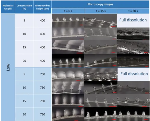
2.2 Microneedles produced with different HA at various concentrations and their
dissolution rate. . . 81
3.1 Comparison of skin thickness among species adapted from [178, 76] . . . 123
3.2 Intensities signal of 5-ALA normalized by the total ion intensity in positive and
negative modes. . . 149
4.1 Requirements specification for the light system developed in order to perform an efficient and tolerable photodynamic therapy (PDT) treatment. . . 161
List of Figures
1.1 Different types of skin cancer and the localization of their development in the skin. 5 1.2 Transversal histological section of epidermis from https://thedc.ca/ourteam/
dermatology/. . . 6
1.3 Heme cycle. . . 9
1.4 Penetration of light in skin [164]. . . 10
1.5 Excitation and emission spectra of PpIX [52]. . . 10
1.6 Microneedles obtained by shape-molding process [182].
1-Master structure. 2-Fabrication of PDMS mold. 3-Casting of the (drug loaded)
solution in the mold. . . 66
1.7 Methods of drug delivery to the skin using different kind of microneedles [86] . 67
1.8 Number of publications found in the last decades on photodynamic therapy for
dermatology and the use of microneedle (MN) in this application. . . 71
2.1 Chemical structure of hyaluronic acid. . . 75
2.2 Fourier transform infrared spectroscopy (FTIR) spectrum of HA representing its
characteristic peaks. . . 77
2.3 A: proton nuclear magnetic resonance (1H NMR) spectrum of HA. B: carbon
nuclear magnetic resonance (13C NMR) spectrum of HA. C: Summary of the13C
chemical shifts according to the chemical structure of HA in figure 2.1. . . 78
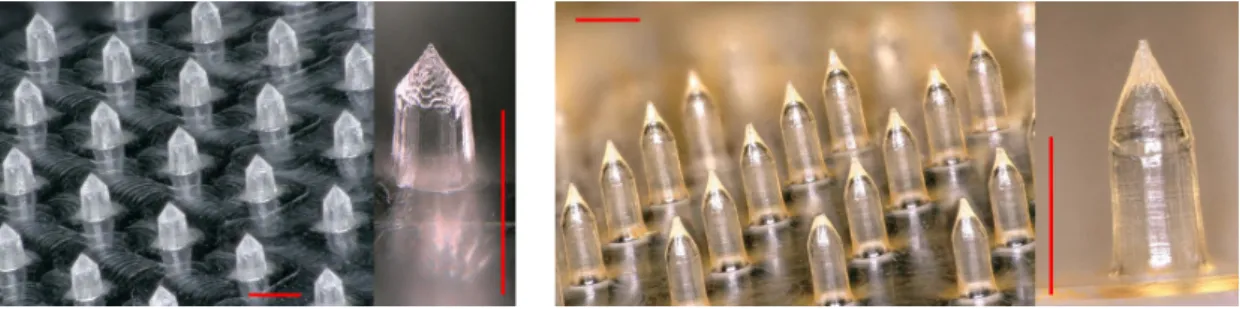
2.4 Different shapes of HA-based microneedles (HA concentration : 5% w/w). A: Conical microneedles included in a circle shaped patch (MN number : 163. Patch
surface: 2.54 cm2). B: Beveled microneedles included on a circle shaped patch
(MN number: 138 . Patch surface: 2.54 cm2). C: "Pencil-tip" shape microneedles
included on a squared shaped patch (MN number: 400. Patch surface: 20 × 20cm2). 79
2.5 HA molecular weight and concentration: Effects on dissolution time of
micronee-dles in a phantom skin. . . 80
2.6 Left: MN-100 after 1 day of storage. Right: The same MN-100 patch after 12 days
and with the same storage conditions. . . 115
2.7 Mechanical behaviour of MNtafter compression at different forces. Analyses were
performed on three different patches with at least 10 height measurements on different microneedles from each patch. Results are presented as mean ± standard
deviation. . . 116
2.8 Density of living NIH3T3 fibroblasts after 24 h incubation with MN-0, MN-20 or
MN-100. . . 117
2.9 Sterilization test after 57 days.
Tube 1: positive control. Tube 2: negative control. Tube 3: Sample containing
xxviii List of Figures 2.10 Optical density control of MN-0, MN-20 and MN-100. Positive control
sponds to a solution containing bacteria (100 CFU) and negative control
corre-sponds to TSB alone. . . 119
3.1 A: Photography of the perforated Tegaderm bandage. B: MN-patch was stuck on the perforated Tegaderm. C: The device was applied on skin rat and a drop of
water was deposited on top to ensure full dissolution. . . 146
3.2 Cross-sections of the skin of the same rat having received 40 UV-B illuminations (A), after 1 h of the cream application (B) and after 1 h of the MN-patch application
(C). . . 147
3.3 Simplified view of the ToF-SIMS analysis after bombardment of a sample by an
ion primary beam. Scheme adapted from [175]. . . 148
3.4 Mapping images in both positive and negative modes of the skin alone and the skin having been in contact with the MN-patch. Total signals obtained are in the
upper line and 5-ALA signal are displayed in the bottom line. . . 149
3.5 Simplified schema of the Franz cell apparatus with the different compartments
represented. . . 151
3.6 Calibration curve obtained with UPLC-ELSD for quantification of 5-ALA. Results
are presented as mean ± s.e.m. . . 152
3.7 Franz cell penetration profiles showing the cumulative concentration of 5-ALA in
rat skin samples after 1 h of MN-patch application. . . 153
4.1 Lighting emitting device - LED panel (Aktilite®) . . . 159
4.2 Portable light emitting system Ambulight® used for PDT treatment. . . 159
4.3 Left: Light emitting system chosen: Adafruit Dotstar High Density 8 × 8 grid.
Right: Arduino microcontroller: Adafruit Trinket. . . 160
4.4 Electronic diagram including all the components to produce the illumination light
source. . . 162
4.5 UP: Photo of the disorganized electronic circuit extending on the bench-top. Down: Scheme of the electronic circuit included in the box, each component is
well organized to avoid crowding and photo of the realisation. . . 164
4.6 Photo of the MN patch stuck to the light-emitting diode (LED) array thanks to the
double sticky layer realized with modified polydimethylsiloxane (PDMS). . . . 164
4.7 Comparison between expectation and reality. . . 165
4.8 Emission spectra in arbitrary unities (A.U) of the Dotstar LED panel (Blue), of the Dotstar LED panel covered with the double sticky layer (Red) and the whole de-veloped device including MN-patch (Black). Green line represents the maximum
obtained at 631 nm. . . 166
4.9 Photographies of the Dotstar LED panel (A), of the Dotstar LED panel covered with the double sticky layer (B) and the whole developed device including
MN-patch (C). . . 168
4.10 A, B and C: Photographies uploading in Matlab program respectively correspond-ing to the Dotstar LED panel, plus the double sticky layer, plus the MN-patch.
D, E, F: Intensity profiles in mW cm−2plotted with Matlab for the Dotstar LED
panel, plus the double sticky layer, plus the MN-patch. . . 169
4.11 Evolution of the temperature of the light emitting device during all the illumina-tion procedure. Arrows represent the decrease in temperature when the device
List of Figures xxix 4.12 Evolution of the (healthy human) skin temperature during all the illumination
procedure. The light emitting device was removed each 15 minutes to measure the skin temperature. Results are presented as mean ± standard deviation (n=3). 171 A.1 C program on Arduino platform to control the system driving the Trinket
micro-controller and the Adafruit LED array. For better understanding, comments were
added after the double slash (//). . . 193
Introduction
Skin cancer is the most common form of cancer, globally accounting for at least 40 % of cancer cases. Skin cancers include two forms: melanomas and non-melanomas ; and the most common type is the second one that occurs in at least 2–3 million people per year in the world and its incidence is still increasing [158].
Different therapies have been investigated to treat non-melanomas skin cancers such as surgery, cryotherapy or radiotherapy. PDT is another approach that had known a growing interest for the two last decades. PDT presents an acceptable efficiency at long-term follow-up and does not let scars after treatment which is positive since the lesions are usually present on face, hands or neck (sun-exposed areas) [15]. Nowadays, PDT starts to be routinely used in clinics since it is easily performed. Indeed, the principle is quite simple and rest on the combination of three major components that are: photosensitizer, light and oxygen. Therefore, for a conventional treatment in clinics, a cream containing a photosensitive precursor is applied on the lesion for at least three hours and then a precise red light dose is delivered. Afterwards, due to the presence of oxygen, a photochemical reaction occurs which induces cell death [115]. Although this treatment has been efficient on thin skin lesions, for deeper skin tumours, it may be improved. One of the major reason is due to the lack of photosensitive precursor penetration through the skin layers. Tumour cells in deep-seated lesions, such as deep basal cell carcinoma (BCC), are not destroyed because no photochemical reaction occurs due to the photosensitizer absence [41]. On the other hand, this treatment is often painful due to the important number of oxygen species created during light illumination, that are known to stimulate sensory nerve endings [8, 115]. One solution would be to to avoid excessive production of oxygen species by preventing the accumulation of the photosensitizer in the cells and consequently decrease the photochemical reaction burst.
This thesis aims to develop a biomedical device that would allow a better prodrug penetration in deep skin layers and a less painful PDT treatment. The first goal is to design MNs in a disposable patch that would contain the prodrug. When inserted into the skin lesions, the MN tip would reach the bottom of the tumour. The MNs composed of HA, a biocompatible and water-soluble polymer, will be dissolved by the skin and consequently release the prodrug on the whole volume of the skin lesion. The second objective is to couple this MN-patch to a light system that would deliver a low red light dose and starts the illumination just a few minutes after the MN insertion in order to avoid pain usually felt during treatment.
In order to answer this issue, the first chapter provides an overview of the different categories of the non melanoma skin cancers (actinic keratosis (AK), BCC, squamous cell carcinoma (SCC)) and how conventional PDT is performed on these kinds of cancers and the efficiency that is recorded. Since the lack of prodrug penetration results in a treatment efficiency decrease, different methods (chemical of physical) to overcome this issue have been considered and are described in a review. Among the different technologies suggested, the use of MNs appears as a promising approach enabling better drug penetration. Therefore, at the end of this chapter, a
2 Introduction rapid description of the fabrication process and their different types are presented.
The second chapter focuses on the production of the MN-patch in which 5-ALA is loaded. The patch development process is based on an easy one-step solvent casting molding method. The properties of fabricated MNs are characterized in terms of their ability to dissolve, to insert without fracture in the skin and their cytotoxicity. Since the prodrug, the 5-ALA, is included in the needle material (polymer HA), a special care through an analytical follow-up is taken to ensure that there is no interactions between these two products and a good stability of the drug. The results of this chapter are transformed into a publication.
The third chapter describes thein vivo experiments that are conducted to test the efficiency
of the MN-patch. For this purpose, precancerous skin lesions model are developed on rats by repetitive UV-B illumination. The photosensitizer fluorescence level is recorded after MN-patch application to highlighted if this technology could lead to a succesfull PDT session. These results are part of an article. In order to estimate the prodrug penetration profile, preliminary experiments using innovative technologies (Franz cell apparatus or Time-of-Flight Secondary
Ion Mass Spectrometry (ToF-SIMS)) onex vivo skins are also performed.
The final chapter answers the second goal of this thesis which is to couple the MN-patch to a light system less painful in order to propose a proof of concept: PDT all-in-one. The reasons that led to reduce the light dose and the time between release of 5-ALA and illumination are described. The system is then characterized in terms of temperature, light power and uniformity. Finally, a regulatory study has been conducted in order to evaluate the classification of the developed products.
Chapter
1
Literature Review
Improvement of photodynamic
therapy e
fficiency for skin cancers
Outline of the current chapter
1.1 Medical indication: skin cancers 4
1.1.1 Different types of skin cancers . . . 4
1.1.2 Non melanoma skin cancers . . . 5
1.1.2.1 Actinic keratosis (AK) . . . 5
1.1.2.2 Squamous cell carcinoma (SCC) . . . 6
1.1.2.3 Basal cell carcinoma (BCC) . . . 6
1.2 Non melanoma skin cancer and treatment perspectives 7
1.2.1 Photodynamic therapy . . . 7
1.2.1.1 Principle . . . 7
1.2.1.2 Parameters . . . 8
1.2.2 Efficiency of PDT on non melanoma skin cancers . . . 11
1.2.2.1 Treatment of AKs with PDT . . . 11
1.2.2.2 Treatment of SCCs with PDT . . . 12
1.2.2.3 Treatment of BCCs with PDT . . . 13
1.2.2.4 Assessment and perspectives . . . 14
1.3 Methods to improve drug penetration 14
1.4 Transdermal drug delivery with microneedles 63
1.4.1 Microneedles design . . . 63 1.4.1.1 Mechanical aspect . . . 63 1.4.1.2 Medical aspect . . . 64 1.4.2 Microneedles processing . . . 64 1.4.2.1 Laser-cutting . . . 64 1.4.2.2 Micromachining . . . 65 1.4.2.3 Photolithography . . . 65
4 CHAPTER 1. Literature Review
1.4.2.4 Wet or dry-etching of silicon . . . 65
1.4.2.5 Molding method or solvent casting molding method . . . . 65
1.4.3 Different kinds of MNs . . . 67
1.4.3.1 Solid microneedles for skin pretreatments . . . 67
1.4.3.2 Coated microneedles . . . 68
1.4.3.3 Hollow microneedles . . . 69
1.4.3.4 Dissolving microneedles . . . 69
Introduction: Literature review
• In this chapter, a focus on the different skin cancers and their localization in the skin layers will be presented.
• Then photodynamic therapy as treatment for non melanoma skin cancers will be reviewed with its principle, some clinical results and their limitations.
• In a review, we showed the different physical or chemical methods that can enhance prodrug penetration and consequently improve photodynamic therapy.
• Among the previous techniques presented, microneedles appear as a new technol-ogy that might be able to answer to the lack of prodrug penetration in deep skin lesions.
• Microneedles as devices enabling transdermal drug delivery will be shortly pre-sented with their different forms, advantages and production way.
• Finally, the use of microneedles for photodynamic therapy will be explained with a few examples.
1.1
Medical indication: skin cancers
Skin cancers are the most common malignancy of humans and particularly for Caucasians people with increasing incidence rate [158]. According to the World Health Organization, between 2 and 3 millions of non melanoma skin cancers and 132 000 melanomas are diagnosed each year in the world. According to the Skin Cancer Foundation (Facts and Statistics), more people are diagnosed with skin cancer each year in the U.S. than all other cancers combined.
The etiology might be divided in two categories [6, 48]:
• Individual people risks: skin, hair or eyes color (fair), UV sensitivity, skin burns antecedents,
numerous moles, family history of skin cancer.
• Environnemental risks: previous radiotherapy, phototherapy [16], indoor tanning,
UV-radiation exposure.
1.1.1
Di
fferent types of skin cancers
According to the previous risks encountered by people, different kinds of skin cancers exist and are divided in two types :
1.1. Medical indication: skin cancers 5 • Melanoma skin cancers
The different localization of skin cancers are depicted in figure 1.1.
Figure 1.1 – Different types of skin cancer and the localization of their development in the skin. As depicted in figure 1.1 the non-melanoma skin cancers are localized principally in epider-mis and deep epiderepider-mis. As for the melanoma skin cancer, it can reach lower derepider-mis and be in contact with blood vessels leading the tumors to spread in the body (metastasis). Melanoma skin cancer affects less people than the non-melanoma skin cancers. An effective treatment for melanoma is still under investigation since this disease is difficult to treat with its potential ability to create metastasis. Non-melanoma skin cancers could be treated thanks to different technologies and one of them will be presented in details in section 1.2 but before a focus on each class of non-melanoma skin cancers will be made.
1.1.2
Non melanoma skin cancers
Non-melanoma skin cancers are divided in three categories : actinic keratosis (AK), squamous cell carcinoma (SCC) and basal cell carcinoma (BCC).
1.1.2.1 Actinic keratosis (AK)
AKs also known as solar keratosis or keratinocytic intraepidermal neoplasia are premalignant tumors that take place in epithelial cells (or keratinocytes) located in epidermis and especially
in thestratum spinosum (Figure 1.2) [198]. AKs are due to the proliferation of transformed
neo-plastic keratinocytes. AKs are the most common precancerous lesions among fair-complexioned individuals [138]. A small percentage of AKs may progress to invasive SCC if they are not diag-nosed and treated on time [172]. AK could be considered as a SCC at early stage of development [138] but do not extend as deeply as SCC might do [48] . The clinical presentation of an AK is a scaling papule or a red plaque generally on sun exposed zones (head, neck, hands, foot). Diameter could reach 1 to 3 mm or may also become bigger and get several centimeters in size [122]. There are three main grades of AK [184, 212].
• Grade I: neo-plastic changes are limited to the lower third of the epidermis. Clinically, AK grade I are slightly palpable (better felt than seen). A red pseudonetwork pattern could be
6 CHAPTER 1. Literature Review
Figure 1.2 – Transversal histological section of epidermis from https://thedc.ca/ourteam/ dermatology/.
• Grade II: neo-plastic changes are located in the lower two thirds of the epidermis. Clinically,
AKgrade II are moderately thick (easily felt and seen). Dermatoscopy analysis shows
background erythema intermingled with keratotic follicular openings.
• Grade III: full-thickness atypia is observed. AK grade III are very thick and hyperkeratotic
(obvious). Observation with dermatoscope shows structureless white-yellow areas.
A fourth grade could be defined as agrade III very advanced and presenting vesiculation/erosion
in addition [148].
1.1.2.2 Squamous cell carcinoma (SCC)
SCC derives from the keratinocytes of thestratum spinosum layer of the epidermis (Figure 1.2).
SCCs are principally diagnosed after AKs and are defined as their evolution [21]. Two kinds of SCC are described in the literature:
• In situ SCC whose Bowen disease and Erythroplasia of Queyrat belong [5]
• Invasive SCC
If histological examination indicated atypical keratinocytes (hyperkeratosis and
paraker-atosis) thicker than two-thirds of the full thickness of epidermis, lesions are considered asin
situ SCC [199, 149]. Invasive SCC is a result of accumulation of mutational and cellular events
that lead to invasive growth [168]. Hence invasive SCC can be more biologically aggressive thanin situ SCC, can recur and has the potential to metastasize which could cause death in
particular case [142, 149, 123]. The clinical presentation of a SCC is a sternly demarcated and
thick erythematous or peeling red plaques on sun-exposed areas.In situ SCCs are difficult to
distinguish from AKs because there is no patho-biological difference between them [149, 5]. Moreover, lesions caused by invasive SCC can first appear as erythematous area with crusting and then as a nodule that growth over time and could become ulcerated or necrotic [168, 24]
1.1.2.3 Basal cell carcinoma (BCC)
BCC is one of the most common type of non-melanoma skin cancer (80%) [130] and its incidence is still rising even in younger people ( < 40 years old) [131]. Contrary to SCC, BCC is a locally
1.2. Non melanoma skin cancer and treatment perspectives 7 invasive malignant epidermal skin tumor that grow slowly in the area of origin. BCCs rarely spread to distant parts of the body. BCCs are also keratinocyte tumors that arise from the basal
layer of the epidermis calledstratum basale (Figure 1.2) [16, 47]. BCC has several distinctive
varieties [174, 209, 48] :
• Nodular (50-60%) (nBCC) • Superficial (15%) (sBCC)
• Infiltrative (10-20%) whose morphoeic (5%) (iBCC)
Nodular BCC is most frequently observed on the face. This kind of disease is due to tumor cells growth in rounded masses shown as solid, well-demarcated, lobulated tumor nests of various sizes. This agglomerate may be a central necrosis composed of numerous fibroblasts, mucinous material and hyaluronic acid [130, 144]. Tumor nests of superficial BCC growth is restricted to epidermis or superficial parts of hair follicles. They can be defined as small buds of proliferating basal cells that grow down from the epidermis into the superficial dermis. The most common location of the superficial BCC is the trunk [100, 138]. The third subtype of BCC is the infiltrative one and the most aggressive as it can reach deep dermis and even subcutis [48]. This kind of BCC is difficult to diagnose, however in case of morphoeic BCC, a yellow-white waxy patch with very ill-defined edges could be a proof. When not treated on time, infiltrative BCC could lead to lesions showing ulceration, crusting or fibrosis [209]. Except for superficial BCC, all others lesions thicken cutaneous surface and the diameter of these lesions may range from a few millimeters to several centimeters.
1.2
Non melanoma skin cancer and treatment perspectives
Despite non-melanoma skin cancers are the most common, they can be usually treated by surgical excision, Moh’s micrographic surgery or curettage. Recently, non-surgical methods such as cryotherapy, topical chemotherapeutics [35], immunotherapy, chemotherapy, laser treatment and photodynamic therapy have also been developed and are starting to be commonly used [16, 145].
Treatment choice depends on the type of skin lesions, number, size, location of lesions, patient’s compliance, general health conditions, and cosmetic outcomes [212]. According to the European Dermatology Forum guidelines on topical photodynamic therapy, this treatment is approved for AK, SCC, superficial and certain thin BCC [115].
1.2.1
Photodynamic therapy
1.2.1.1 Principle
The concept of PDT is a century old and has the advantages to be a selective and localized therapy [62, 46]. This therapy presents an acceptable efficacy at long-term follow-up and do not let scars after treatment which is positive since the lesions are usually present on face, hands or neck (sun-exposed areas) [15]. PDT relies on the combination of three components to induce tumor destruction :
• a photosensitive drug
• a light at a precise power and wavelength • oxygen
8 CHAPTER 1. Literature Review
None of these elements is individually toxic, but together they produce a photochemical reaction1
that generates singlet oxygen which is a highly reactive product causing cells death [2]. Clinical PDT in dermatology consists of the administration of a non-toxic drug, with a cream, known as photosensitizer (PS) to a patient presenting a skin lesion [146]. After an incubation period (∼ 3 h) the lesion is illuminated with a specific wavelength able to stimulate the PS. The excited state of the PS (PS*) is not stable and comes back to its basic state by transferring energy to surrounding oxygen that will become an active singlet oxygen O . This free radical will oxidize all tissues constituents in its close environment and destroy cells, therefore the effect will be localized [15].
1.2.1.2 Parameters
• Photosensitizer (PS)
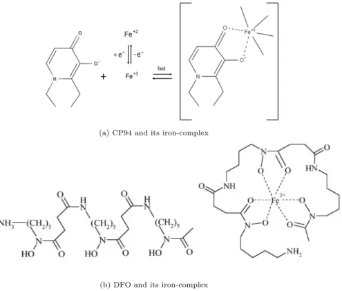
PS should respect characteristics such as being non-toxic, water soluble, chemically and physi-cally stable, selective for neoplastic tissues with the shortest time interval between administration and maximal accumulation. PS should also be eliminated by the body in a short time period in order to avoid unwanted photoreactions [146, 17]. Historically, hematoporphyrin and hemato-porphyrin derivatives (HpD) were the first PSs to be studied in details [44]. However these compounds presented a photosensitivity for a long period of time (8 weeks), so patients had to avoid sunlight which was very constraining. Moreover, tumour-localizing properties of this PS were not as pronounced as first thought [25]. These deficiencies and the fact that this product was orally administered make its use difficult for dermatology. Therefore, a new approach has been investigated consisting in the development of endogenous photosensitizers. Endogenous PS are synthetized in the body from nonphotosensitizing precursors. Precursor’s chemical conversion is established by metabolic pathways. The major endogenous PSs (protoporphyrin IX (PpIX), uroporphyrin, coproporphyrin) are products of heme cycle (Figure 1.3) [81]. Therefore, this second generation of PS is very selective and present a modest absorption on the range 600 − 700 nm that allows them to be activated until several millimeters in biological tissues (See next paragraph 1.2.1.2) [45].
Actually, the 5-aminolevulinic acid (5-ALA) and the methyl-aminolevulinate (MAL) are the most widely used molecules as precursor drugs for PpIX production by the heme-synthesis. 5-ALA or MAL could be administrated as a cream which is interesting for topical application and especially skin treatments in dermatology. They are selective and have the possibility to be
accumulated in tumours cells because individual steps of the heme cycle are disordered2and
increased the production of various intermediate leading to a PpIX accumulation [17, 81]. 5-ALA is commercially available under the pharmaceuticals products Alacare®, Alafast®, Levulan®, Ameluz® and MAL is commercially available with the Metvixia® cream.
1This reaction will be more discussed in the introduction of section 1.3.
2The heme cycle disorder leads to a high concentration in porphobilinogen deaminase and a low concentration in
1.2. Non melanoma skin cancer and treatment perspectives 9
Figure 1.3 – Heme cycle.
• Light
Light at a precise wavelength is used to stimulate the PS. The choice of light source is mainly based on PS absorption but also according to its cost, and its size [2]. Light source needs especially to present spectral characteristics that match with the absorption wavelength range of the PS in order to generate enough singulet oxygen to produce a cytotoxic effect. In dermatology (and others specialities) light should reach all the injured tissue. The interaction of light with tissue is governed by three basic processes that can occur when a photon of light reaches the skin: reflection, scattering, and absorption. In case of PDT, absorption phenomenon is privileged [17]. The higher wavelength is, the deeper light penetrates tissues as presented on figure 1.4 [164]. Blue light (400 nm) has relatively low penetration in tissue (only absorbed in the superficial epidermis of skin) whereas red light (600 − 700 nm) penetrates more deeply [7]. A smart compromise has to be found between a minimal light absorption by tissues and a maximal light absorption by the PS.
For skin cancer treatment, 5-ALA which then produces PpIX is often used and a light set at 630 nm is advantageous to activate PpIX. This wavelength is in the red light, allowing penetration in deep tissues and corresponds to the last excitation peak of PpIX (Figure 1.5) [19, 72]. The fluence rate and exposure time also affect PDT response and need to be controlled. Nowadays, research is conducted with the aim of reducing light exposure time, irradiance and fluence rate in order to improve patient compliance [188, 189]. Similarly, light fractionation significantly increases the efficiency of PDT which allows skin re-oxygenation between two exposures [191].
• Singlet oxygen and cells death
Photochemical reaction needs oxygen to occur and to destroy malignant cells. The lifetime of singlet oxygen O is very short (approximately 10 − 320 ns), so its diffusion is only limited to cell’s organelles (Distance of diffusion : 10 − 50nm) [45]. Thus, photodynamic damage will occur
10 CHAPTER 1. Literature Review
Figure 1.4 – Penetration of light in skin [164].
Figure 1.5 – Excitation and emission spectra of PpIX [52].
very close to the intracellular location of the PS [51, 109]. Two main mechanisms of tumoral cell death can occur thanks to singlet oxygen: apoptosis and necrosis [45, 163].
• Apoptosis defines the programmed cell death. When cells are subjected to aggression altering their integrity, they develop defense and immune responses but in case of abnormal
activity, cells stop their cellular division. When damages are too important, a “cellular
suicide” is engaged [160, 151].
• Necrosis defines the non-programmed cell death. It happens accidentality when cells are mistreated (burning, string compressions), they burst and their cytoplasm content is released provoking an inflammatory response [101].
Singlet oxygen is mainly responsible for cytotoxicity induced by PDT. Nevertheless, due to the large proportion of oxygen consumed by the photochemical reaction, other mechanisms can occur and kill surrounding cells. Photo-oxidative stress can lead to ischemia (restriction in blood
1.2. Non melanoma skin cancer and treatment perspectives 11 supply to tissues) or vascular response can induce vasoconstriction (narrowing of the blood vessels) that will also damage or kill cells [90].
1.2.2
E
fficiency of PDT on non melanoma skin cancers
1.2.2.1 Treatment of AKs with PDT
Topical photodynamic therapy employing 5-ALA and MAL as endogenous precursor has been widely used to treat actinic keratosis and shows quite good clinical outcomes on many patients as presented in table 1.1. Moreover, in 2019, a group of 13 European practitioners (Morton et al.)
decided to evaluate the average efficiency of conventional PDT3on AKs. Therefore, the authors
chose 6 multicentric, randomized, double-blind studies from 2004 to 2012 which represent more than 1500 patients enrolled. Then the authors reported the clearance rates 3 months after the treatments between 81 and 92% for thin and moderate thickness non-hyperkeratotic AKs localized on the face and scalp [115]. Since the last 5 years, to improve PDT outcomes, the tendency is to perform a lesion pretreatment or to combine PDT with other treatments such as cryotherapy or chemotherapy [115].
AK grade Patients number
Photosensitizer Method Light Clearance rate Ref. I to III 181 5-ALA 20 % (w/vol) Drug is applied twice to each lesion. λ = 417 nm (Blue) Intensity = 10 mW/cm2 75 % at 8 weeks 89 % at 12 weeks Piacquadio et al. 2004 [138] I to III 202 MAL 16 % (w/w) : Metvix cream © Incubation time : 3 h. λ = 570 − 670 nm (Red) Intensity = 70-200 mW/cm2 Dose = 75 J/cm2 75 % at 3 months Szeimiesa et al. 2002 [172]
I to IV 42 Metvix cream © Previous curettage of the lesion then application of the cream under occlu-sion for 3 h. λ = 570 − 670 nm (Red) Intensity = 50-200 mW/cm2 Dose = 75 J/cm2 89 % at 3 months Pariser et al. 2003 [131]
Unspecified 30 Metvix cream © MAL application and occlusion for 3 h. Daylight 79 % at 3 months Wiegell et al. 2012 [202]
Table 1.1 – Photodynamic therapy on AK : protocols and clinical results.
3Conventional PDT involves application of a topically applied photosensitizing agent, an incubation time of
12 CHAPTER 1. Literature Review
1.2.2.2 Treatment of SCCs with PDT
Topical PDT employing 5-ALA or MAL as PS has also been used to treat SCCs. Methods and results are presented in table 1.2. Most of the studies involving PDT on SCC required lesion preparation (abrasion or curettage) or two cycles of MAL-PDT [115] which was also evidenced in the following short table 1.2. Overall, the clearance rates stay satisfactory and make PDT highly recommended for this medical indication. Nevertheless, it has been evidenced that the lesion size might also impact the clearance rate. Indeed 82% of lesions up to 14 mm were cleared after a 12 months follow-up whereas when the lesion size increased up to 30 mm or even 100 mm the clearance rate dropped to 55 % [119]. This is a major drawback since SCCs could potentially metastasize if not treated on time [35, 213]. Besides, PDT is currently not indicated for treating invasive SCC for reasons indicated hereinbefore [219, 115].
SCC type Patients number
Photosensitizer Method Light Clearance rate Ref. In situ 87 MAL 16 % (w/w) Metvix cream © Lesions surface were prepared by debridement with a curette. Cream was applied to the lesion for 3 h. Treat-ment was repeated once after 1 week for a complete treatment cycle.
λ = 570 − 670 nm
(Non coherent red light) Dose = 75 J/cm2 93 % at 3 months and 80 % at 12 months Morton et al. 2006 [120] Bowen’s disease (In situ) 85 5-ALA cream prepared at 20 %
Prior to the first treatment hy-perkeratosis and crusts were gently removed with a curette. Cream was applied for 3 h and covered with a plastic film. λ = 630 nm Dose = 100 J/cm2 Intensity = 100 mW/cm2 78.6 % at 6–12 weeks Westers-Attema et al. 2015 [199] Bowen’s disease (Large lesions d > 20 mm) 40 lesions 5-ALA in oil in water 20 % (w/w) A previous abra-sion on the leabra-sion was carried. Cream was applied for 4 h under occlusive tis-sue. λ = 630 nm Dose = 100 J/cm2 78 % at 12 months Morton 2001 [118] In situ 55 and 112 lesions
Metvix cream © Cream was applied for 3 h prior to illumination. 7 days later: a second MAL-PDT session was performed. λ = 635 nm Dose = 37 J/cm2 73 % at 3 months and 56 % at 2 years. Calzavara-Pinton et al. 2008 [21]
1.2. Non melanoma skin cancer and treatment perspectives 13
1.2.2.3 Treatment of BCCs with PDT
PDT can be considered for recurrent and multiple BCCs, especially for those which are bad candidates for surgery due to co-morbid systemic disease or critical cosmetic location of the lesions [88]. A few clinical studies on BCCs are presented in table 1.3. No results on infiltrative BCCs were presented in the following table because numerous studies have excluded them from their experiments when choosing patients [31, 66]. Moreover when PDT was tried on morpheic BCCs, there was no clearance [162], so we decided to focus on the other BCCs categories. However, PDT is a recommended treatment for BCCs since superficial BCCs respond well with a clearance rate between 79 and 100 % whereas for thicker or nodular BCCs the clearance rate is decreased [4, 9] which is also displayed in table 1.3. The poor penetration of the prodrug in deep skin layers could explain the lower efficiency observed for nodular BCCs [135]. Indeed, Ahmadi et al. applied the cream (5-ALA at 20% w/w) with a thickness currently used in clinics and found
that only 36.8 % of the total dose was released during the 4 h application period (Experiments
were performed with Franz cell apparatus) [4]. To reach sufficient prodrug concentration in depth
allowing a biologic rationale for a clinical PDT, Peng et al. estimated that an application of MAL
(1 mm thick layer and 1 cm outside the lesion) at 160 mg g−1for 3 h induced the highest ratio of
porphyrin fluorescence in tumor depth [135]. To sum up, conventional PDT can not be used for all BCCs and if so, a previous lesion preparation should be ensured or several PDT sessions should take place [115].
Table 1.3 – Photodynamic therapy on BCC : protocols and clinical results.
BCC type Patients
number Photosensitizer Method Light
Clearance rate Ref. Superficial Nodular 92 36 MAL
Lesion was de-brided using a curette. Cream (1 mm thick) was applied and the lesion was covered with an adhesive occlusive dressing for 3 hours. Two PDT sessions with an interval of 1 week between each session were performed. λ = 570 − 670 nm Dose = 75 J/cm2 Intensity = 100 mW/cm2 93 % at 3 months 82 % at 3 months Vinciullo et al. 2005 [194] Nodular 168 le-sions MAL prepared at 16 % (w/w)
Lesion was de-bulked, then cream was applied for 3 to 24 hours and covered by a semipermeable film. λ = 570 − 670 nm Dose = 75 J/cm2 Intensity = 100-180 mW/cm2 At a mean period of 35 months 86 % for thick nBCC 93 % for thin nBCC Soler et al. 2001 [162] Superficial 323 5-ALA prepared at 20 % con-centration in a cream base (Neribas) Excessive scaling was removed by curettage. Then 5-ALA cream was applied and covered with a semipermeable dressing and alu-minium foil for 3.5 h.
Two light fractions
λ = 635 nm with 2 hours interval. First dose : 20 J/cm2 for 4.23 min Second dose : 80 J/cm2 for 18.8 min 77 % at 48 months Kessels et al. 2016 [82]
14 CHAPTER 1. Literature Review Superficial 87 with 128 lesions MAL 16 % (w/w) Metvix cream ©
Scales and crusts were removed. A layer of about 1 mm thick of cream was applied on the lesion and covered with an occlusive dressing for 3 hours. One cycle of two treat-ment sessions 7 days apart 4 was applied. λ = 630 nm Dose = 37 J/cm2for 7-10 min 92 % at 3 months 90 % at 3 months Szeimies et al. 2008 [171]
1.2.2.4 Assessment and perspectives
As presented in the previous sections and in the tables 1.1, 1.3 and 1.2, PDT can be used on most of the non melanoma skin cancer types and quite good clinical results were observed. To obtain these satisfactory clinical results, especially for thicker skin lesions, skin preparation prior to drug application on the lesions was necessary and consists often of a curettage or a debulking. A long prodrug incubation with a highly prodrug dose is also mandatory to allow efficient prodrug depth penetration [18, 54, 115, 161]. These supplementary steps added to conventional PDT make this treatment extended in time, painful and tedious. To overcome the pain during the treatment, an anesthetic cream may be applied, ice or cold water could be vaporized on the treated area [8] or the irradiation could also be decreased [189, 192]. Moreover to enhance prodrug penetration and reduce treatment time, different solutions have been considered and
will be presented in the next section 1.3 with a recently review published inPhotochemistry
and Photobiology and entitled: "Photodynamic therapy for skin cancers: How to enhance drug
penetration?".
1.3
Methods to improve drug penetration
4If sBCC lesions showed non-clearance 3 months later they could be retreated with a second cycle of two MAL-PDT
Photodynamic therapy for skin cancer: how to enhance drug
penetration?
Mathilde Champeaua,b,∗, S´everine Vignouda, Laurent Mortierb, Serge Mordonb
aCEA, LETI-DTBS, 17 rue des Martyrs, Grenoble Cedex, France
bUniv. Lille, Inserm, CHU Lille, U1189 - ONCO-THAI - Image Assisted Laser Therapy for Oncology, F-59000
Lille, France
Abstract
Photodynamic therapy (PDT) induced by protoporphyrin IX (PpIX) has been widely used in dermatological practices such as treatment of skin cancers. Clearance rate depends on different factors such as light irradiation, skin oxygenation and drug penetration. The poor penetration of 5-aminolevulinic acid (5-ALA) with topical application is limited and restrains the produc-tion of PpIX which could restrict PDT outcomes. This review will focus on techniques already used to enhance drug penetration in human skin, and will present their results, advantages, and drawbacks. Chemical and physical pretreatments will be discussed. Chemical pre-treatments comprise of drug formulation modification, use of agents that modify the heme cycle, enhance PpIX formation, and the combination of differentiation-promoting agent prior to PDT. On the other hand, physical pretreatments affect the skin barrier by creating holes in the skin or by removing stratum corneum. To promote drug penetration, iontophoresis and temperature mod-ulation are interesting alternative methods. Cellular mechanisms enrolled during chemical or physical pretreatments have been investigated in order to understand how 5-ALA penetrates the skin, why it is preferentially metabolized in PpIX in tumour cells, and how it could be ac-cumulated in deeper skin layers. The objective of this review is to compare clinical trials that use innovative technology to conventional PDT treatment. Most of these pretreatments present good or even better clinical outcomes than usual PDT.
Keywords: photodynamic therapy, drug penetration, skin pretreatment, skin cancer, 5-aminolevulinic acid
2010 MSC: 00-01, 99-00
∗Corresponding author
1. Introduction
Protoporphyrin IX-induced PDT has been widely used in dermatological practice such as treatment of skin cancers [1] .
Historically, treatments using light appeared during antiquity and were used by the Egyptians, Indians, Greeks and Chinese to treat skin diseases. Therapy using an exogenous substance
5
reacting with sun’s photons was observed during the 15thcentury BC in India. At the end of the
19thcentury, interaction between light and molecules was considered for medical treatment. The
concept of PDT was first introduced by Van Tappeiner who mentioned the necessity of oxygen to obtain a chemical reaction over a century ago [2]. When PDT depends on the combination of three components to induce tumour destruction: a photosensitive drug, light, and oxygen (Figure
10
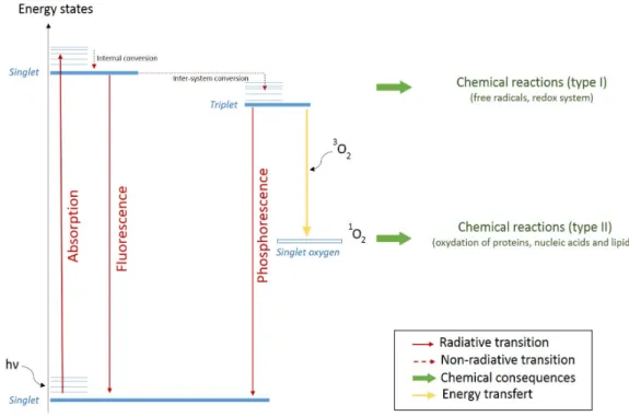
1). None of these elements is individually toxic, but together they produce a photochemical reaction that generates singlet oxygen which is a highly reactive product causing cell death [3]. Figure 2 displays the different energy states and the different pathways leading to fluorescence and other photochemical process. First the photosensitizer (PS) at its ground and singlet state absorbs a photon and moves to its excited energy state. Because of the internal conversion
15
the PS energy decreases and reaches the lower allowed transition of its excited state. Inter-system conversion causes this singlet state to become a triplet state. The singlet state relaxation produces fluorescence, and the triplet produces phosphorescence. A molecule with high triplet energy quantum yield can initiate chemical reactions (type I) that produce free radicals, and then could react with oxygen to form singlet oxygen. Singlet oxygen is a reactive oxygen species
20
that is reponsible for degradation of several cell components, and induce cell death by type II chemical reactions [4].
Figure 1: Synergy between three parameters produces PDT.
Figure 2: Jablonski diagram representing the different processes leading to singlet oxygen production responsible for cell death.
PDT treatment in dermatology consists of the administration of a non-toxic drug known as a photosensitizer (PS) to a patient presenting a lesion, which is frequently, but not always cancerous [5].
25
Photosensitizers should have characteristics such like being non-toxic, water soluble, having a chemical and physical stability, being selective for neoplastic tissues with the quickest time interval between administration and maximal accumulation, and able to be eliminated by the body in a short time in order to avoid photoreactions [5]. Historically, hematoporphyrin and hematoporphyrin derivative (HpD) were the first PSs to be studied in detail [6]. However these
30
compounds present photosensitivity after a long time (8 weeks), so patients had to avoid sunlight which was very constraining. Moreover, tumour-localizing properties of these PSs were not as pronounced as first thought [7]. These deficiencies and the fact that these products were orally administered made their use difficult for dermatology. So a new kind of PS has been developed: endogenous photosensitizers. Endogenous PS are synthetized in the body from
nonphotosen-35
sitizing precursors (also called prodrugs). A precursor's chemical conversion is established by metabolic pathways and particularly by the heme cycle.
Nowadays in the field of dermatology, topical application of 5-aminolevulinic acid (5-ALA) (a non-photosensitizing precursor) is used to induce in situ synthesis of endogenous PS according to the heme cycle [8]. Figure 3 displays the heme cycle where 5-ALA is the first metabolite
40
in the heme biosynthesis pathway in humans. 5-ALA -when not voluntary added- is originallly produced by glycine and succinyl coenzyme A, and then migrates to the cytosol. From there, porphobilinogen is formed due to the condensation of two molecules of 5-ALA. Four molecules of porphobilinogen are connected together, and after porphibilinogen deaminase they form the cyclic uroporphyrinogen III. Decarboxylation of uroporphyrinogen leads to coproporphyrinogen
45
III which then generates within mitochondria proporphyrinogen IX after coproporphyrinogen oxidase (CPO) acts upon coproporphyrinogen III. This last molecule is further oxidized to form PpIX. PpIX is subsequently chelated with iron to form heme [9]. 5-ALA is also valuable for PDT because it has the possibility to be preferentially accumulated in tumour cells. This fact could be explained by a change in mitochondrial function (lack of energy), alteration in porphyrin
50
transporters, or a perturbation in heme biosynthetic enzyme in tumour cells. Indeed individual steps of the heme cycle are delayed by an high concentration of porphobilinogen deaminase and low concentration of ferrochelatase (FC). Moreover the enzyme responsible for converting protoporphyrin IX (PpIX) in heme has been found in reduced levels in a variety of tumour tissues including liver, bladder, colorectal, esophageal, gastric, and rectal [9]. This deregulation
55
causes the accumulation of various intermediate products leading to PpIX accumulation [8]. This accumulation has been also shown in different skin lesions (psoriatic plaques, actinic keratosis, and basal cell carcinoma) where PpIX fluorescence was at higher levels as compared to healthy skin. [10, 11]
For most clinical trials [12–15], 5-ALA was administrated at a high dose (≈ 80 mg) as a
60
cream on the lesion to promote PpIX accumulation [16]. After an incubation period1, lesions
were illuminated with a special wavelength (usually 635 nm) appropriate for tissue transmission and able to stimulate the PS [1, 3]. Stimulated PS (PS*) follow the process presented in figure 2 and PDT occurs.
PDT efficiency depends on several parameters such as light dosimetry, fluence rate, time
65
between PS administration and irradiation (also called incubation period), exposure time, rate of oxygen, kinetic distribution of PS in tissues, PS selectivity, and concentration [3, 18, 19]. PDT
1Incubation time is usually 3 h in conventional PDT treatment. This period is not the perfect one to obtain
the best accumulation of PpIX in cells [17] but is the most suitable for hospital practice.
Figure 3: Heme cycle.
optimization is difficult because all parameters are correlated. Production and distribution of PS remains the most important parameters since thereafter photodynamic therapy (PDT) result on it. Photosensitizer formation and epidermal penetration depth represent basic predictors of drug
70
efficacy in dermatological PDT [20]. Concentration, distribution, aggregation, and accumulation
of PS modifies the photobleaching2 kinetic [20].
Despite progress concerning few parameters, PDT is currently limited by poor tissue pene-tration of the porphyrin precursor 5-aminolevulinic acid. This review will focus on one of the three important parameters needed for PDT. Lack of 5-ALA penetration through the skin is due
75
to molecule hydrophilicity that prevents it to cross the stratum corneum (SC) [21]. The aim of this review is to present the chemical and physical methods to increase drug penetration before PDT. To increase penetration of 5-ALA into deep epidermal regions different adaptations to standard PDT treatment have been considered. Proposals have been made concerning chemical

![Figure 5: Patch formulation and its in vivo use on nude mouse. PpIX fluorescence was observed 4 h after patch removal [53].](https://thumb-eu.123doks.com/thumbv2/123doknet/14492991.717942/54.892.150.745.474.638/figure-patch-formulation-mouse-ppix-fluorescence-observed-removal.webp)