HAL Id: tel-01127035
https://tel.archives-ouvertes.fr/tel-01127035
Submitted on 6 Mar 2015HAL is a multi-disciplinary open access archive for the deposit and dissemination of sci-entific research documents, whether they are pub-lished or not. The documents may come from teaching and research institutions in France or abroad, or from public or private research centers.
L’archive ouverte pluridisciplinaire HAL, est destinée au dépôt et à la diffusion de documents scientifiques de niveau recherche, publiés ou non, émanant des établissements d’enseignement et de recherche français ou étrangers, des laboratoires publics ou privés.
Thi-Ly Tran
To cite this version:
Thi-Ly Tran. Unraveling the muco-adhesion of Lactococcus lactis : development of biophysical ap-proaches. Biochemistry, Molecular Biology. INSA de Toulouse, 2013. English. �NNT : 2013ISAT0029�. �tel-01127035�
T
T
H
H
È
È
S
S
E
E
En vue de l'obtention du
D
D
O
O
C
C
T
T
O
O
R
R
A
A
T
T
D
D
E
E
L
L
’
’
U
U
N
N
I
I
V
V
E
E
R
R
S
S
I
I
T
T
É
É
D
D
E
E
T
T
O
O
U
U
L
L
O
O
U
U
S
S
E
E
Délivré par :Institut National des Sciences Appliquées de Toulouse (INSA de Toulouse)
Présentée et soutenue par : TRAN Thi Ly Le : jeudi 12 décembre 2013
Titre :
Unraveling the muco-adhesion of Lactococcus lactis : development of
biophysical approaches
École doctorale et discipline ou spécialité :
ED SEVAB : Ingénieries microbienne et enzymatique
Unité de recherche :
Laboratoire Ingénierie des Systèmes Biologiques et des Procédés UMR 5504 CNRS - UMR 792 INRA
Directeur(s) de Thèse :
MERCIER-BONIN Muriel
JURY :
M. SCHMITZ Philippe, Professeur INSA de Toulouse, Président M. KULAKAUSKAS Saulius, Directeur de Recherche INRA, Rapporteur M. Di MEGLIO Jean-Marc, Professeur Université Denis Diderot Paris 7, Rapporteur
Mme MERCIER-BONIN Muriel, Chargée de Recherche INRA, Directrice de thèse M. CASTELAIN Mickaël, Chargé de Recherche INRA, Co-encadrant de thèse
“Laboratoirie d'Ingénierie des Systèmes Biologiques et Procédés” of the “Institut National des Sciences Appliquées” (INSA) in Toulouse, where this work was carried out from November 2010 to December 2013, for having welcomed me to his laboratory and provided the materials and equipments that I have needed to produce and complete my thesis.
I would like to thank all members of the jury for agreeing to assess this manuscript and to participate in the thesis defence:
• Mr. Philippe SCHMITZ, Professor of the INSA in Toulouse, the chairman of the jury, for his encouragement, valuable discussions, and good humor.
• Mr. Saulius KULAKAUSKAS, Research Director of the INRA Jouy-en-Josas, for his great enthusiasm and time spent in reading, analyzing, assessing and writing the report.
• Mr. Jean-Marc Di MEGLIO, Professor of “Université Denis Diderot Paris 7”, for his particularly rigorous reading, constructive criticism and time spent in writing the report.
I would especially like to express my most sincere gratitude to my supervisor, Mrs. Muriel MERCIER-BONIN, for her excellent guidance, advice and support in all stages of this thesis. I would also like to thank her for helping me to shape my interest and ideas, painstakingly and patiently correct my writing. She cared much about my work and responded to my questions and queries so promptly. Additionally, I will forever be thankful to Mr. Mickaël CASTELAIN, who was willing to provide me a great knowledge of biophysics as well as spend his time and effort during the final year of my PhD. One simply could not wish for a better and friendlier advisor.
I would like to thank Mr. Nicolas DIETRICH, researcher of the “Transfer, Interfaces, Mixing” (TIM) group of the LISBP laboratory in Toulouse, who let me experience the Diffusion Front Tracking method and helped me analyze data using the Monte Carlo Markov Chain program. His motivating collaboration as well as his participation in my doctoral committee meetings was greatly appreciated. I would also like to thank Mrs. Dominique ANNE-ARCHARD, researcher of the “'Institut de Mécanique des Fluides de Toulouse”
The major part of this thesis is the fruitful result of collaboration with the MICALIS Institute in Jouy-en-Josas, France. I would like to thank Marie-Pierre CHAPOT-CHARTIER and Mickaël MEYRAND for their Lactococcus lactis wild-type, mutant and plasmid-cured derivative strains.
I would also like to thank Etienne DAGUE from the “Laboratoire d'Analyse et d'Architecture des Systèmes” (LAAS) in Toulouse for the Atomic Force Microscopy measurement, Yann GUERARDEL from the “Unité de Glycobiologie Structurale et Fonctionnelle” (UGSF) in Lille for pig gastric mucin fractions.
I would particularly and gratefully thank to Mrs. HO Phu Ha and Mr. CHU-KY Son from “Hanoi University of Science and Technology” (HUST), for their confidence in me, recommending me to this project and constant encouragement during my study. Additionally, I would like to thank the “Université des Sciences et Technologies de Hanoï” (USTH), for providing the funding which allowed me to undertake this research. I would also like to thank them for giving me the opportunity to attend the annual USTH workshops which provided a number of the diverse and interesting training courses as well as the career guidance as a teacher-researcher.
I am deeply grateful to Ms. Marie-Pierre DUVIAU who was there for helping me set up the Shear Stress Flow Chamber experiments when I came to the laboratory at the beginning, for her enthusiasm for discussing, caring and giving advices during my thesis.
I would like to thank all the members of my group EAD4 who are extremely friendly, humor and helpful. I would particularly like to thank Mr. Pascal LOUBIÈRE, head of the team, for receiving me to his group. I’m so glad to have worked with the wonderful colleagues. Special thanks go to my young, funny and cheerful office mates (Manon, Marie, Jade, Thomas, Arthur, Pauline, Amandine, Stéphanie, Anne-Laure and Jérôme). I just wish them the best of luck for their thesis and future career.
I would like to thank my countryman, LE Doan Thanh Lam, for his constant help and encouragement while he was doing his thesis and even after he left the laboratory. He was the one whom I wrote up for asking so many questions of science and society. I think of him as a big brother.
I especially thank my mom, dad and brothers. My hard-working parents have sacrificed their lives for their children and provided unconditional love and care. I know I always have my family to count on when times are rough.
At last, not at least, special thanks to my best friend, soul-mate and boyfriend, Jérémy, who is the only person can appreciate my quirkiness and sense of humor, and his family who all have been supportive and caring.
regarded as a viscoelastic and permeable hydrogel. This layer serves as an ecological niche for commensal and probiotic bacteria, and plays a role in the defense against pathogens. The mucus layer is described as a secreted mucin-fiber scaffold. Mucins are large glycoproteins with a serine and threonine-rich protein backbone, linked to a wide variety of O-linked oligosaccharide side chains arranged in a bottle-brush configuration. Such O-glycans are nutritive sources for bacteria and/or potential ligands for bacterial adhesins, probably contributing in this way to the selection of the species-specific microbiota.
Many studies on bacterial muco-adhesion have been carried out with commensal
Lactobacillus species, with the aim to select probiotics based on their ability to persist within
the gut. In contrast, little is known about the structural and functional factors involved in the muco-adhesion of Lactococcus lactis, the model for Lactic Acid Bacteria.
In this thesis, we focused on unraveling multi-scale interactions between a vegetal L. lactis subsp. lactis isolate, TIL448 and a model mucin, Pig Gastric Mucin (PGM). In a previous study, L. lactis TIL448 was shown to expose at its surface both pili and mucus-binding protein. In contrast to the other tested L. lactis strains, specific adhesion to Caco-2 human intestinal epithelial cells was reported. However, no data was available of the TIL448 muco-adhesive phenotype. To address such questions, different biophysical approaches were implemented. Our work was achieved in close collaboration with Micalis Institute in Jouy-en-Josas, France.
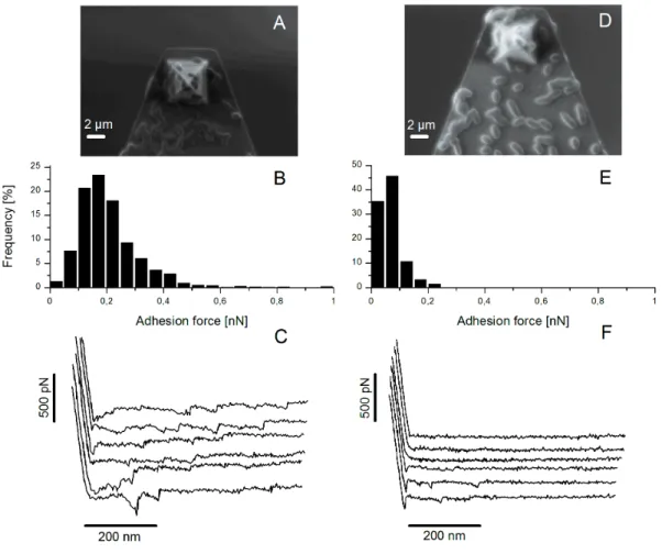
Results. In a first part, we performed single-cell scale AFM measurements with dedicated
lacto-probes and shear stress flow chamber experiments at the bacterial population level, under laminar flow conditions on the wild type L. lactis TIL448. We also tested the plasmid-cured strain and two mutants, obtained by disruption of the genes encoding the major pilin and the mucus-binding protein. Bacterial cells were put in adhesive contact with a biomimetic PGM-coated surface, under static or shear-flow conditions. AFM experiments on TIL448 revealed a high proportion of specific adhesive events (60 %) and a low level of non-adhesive ones (2 %). The strain muco-adhesive properties were confirmed by the weak detachment of
played by these two surface proteinaceous components in adhesion to PGM under static conditions. Under shear flow, a more important contribution of the mucus-binding protein than the pili one was observed. Both methods differ by the way of probing the adhesion force. AFM blocking assays with free PGM or O-glycan fractions purified from PGM demonstrated that neutral oligosaccharides played a major role in adhesion of L. lactis TIL448 to PGM. Then, the diffusion ability of L. lactis was determined by implementing a novel method, named Diffusion Front Tracking (DFT). It consists of tracking the diffusion front of stained cell suspensions over time within the PGM network. The technique involved tracking a liquid-liquid interface in a Hele-Shaw cell using a digital camera. A suspension of bacterial cells, stained with fuchsine and injected on the top, was allowed to diffuse down to the PGM hydrogel. The feasibility of the method was demonstrated. Then, the diffusion coefficient was determined for all the strains under study, which required solely the diffusing front position over time. A mathematical analysis was developed to solve this problem. Diffusion of bacterial cells follows the Fick’s law and was fitted to a model using Monte-Carlo algorithm. The latter allows checking the presence of convection or sedimentation artifacts.
In a second part, in order to have a more thorough understanding of the L. lactis muco-adhesive and diffusive ability, the microstructure and mechanical properties of PGM were determined. Gel microstructure for varying PGM concentration was probed by the analysis of diffusivities of 200-nm and 500-nm fluorescent nanoparticles with different surface properties (carboxyl-terminated, negatively charged tracers, with and without PEG coating; amine-terminated, positively charged tracers), using fluorescence Multiple-Particle Tracking. The pore size of the PGM network was evaluated between 240 and 470 nm, depending on the PGM concentration. A high heterogeneity of the mesh pore size was also highlighted. Characterization of the PGM rheological properties was achieved by combining classical bulk rheometry and one-point and two-point microrheometry approaches.
Conclusion. Our study, based on the combination of different biophysical approaches and
tools, has allowed dissecting the muco-adhesive and diffusive phenotype of L. lactis TIL448, in relation with the nature of the bacterial surface determinants and the structural, mechanical and rheological properties of the PGM network.
qui est un hydrogel perméable et viscoélastique. Cette couche sert de niche écologique pour les bactéries commensales et/ou exogènes comme les probiotiques, elle joue également un rôle dans la défense contre les pathogènes. La couche de mucus est formée d'un réseau de fibres de mucines. Ces dernières sont des glycoprotéines de haut poids moléculaire avec un squelette protéique riche en sérine et thréonine, lié à une grande variété de O-glycanes qui représentent une source nutritionnelle pour les bactéries et/ou des ligands potentiels pour les adhésines bactériennes, contribuant ainsi probablement à la sélection et l'implantation d'un microbiote régio-spécifique. De nombreuses études sur la muco-adhésion des bactéries lactiques ont été réalisées avec des lactobacilles, dans le but de sélectionner de nouveaux probiotiques, dotés d'une meilleure faculté de persistance dans l’intestin. En revanche, peu de données sont actuellement disponibles sur les facteurs structurels et fonctionnels impliqués dans la muco-adhésion de Lactococcus lactis, le modèle des bactéries lactiques. Dans ce cadre, en collaboration avec l’Institut Micalis de Jouy-en-Josas, nous nous sommes focalisés sur la quantification multi-échelles des interactions entre la souche naturelle d'origine végétale L. lactis ssp. lactis TIL448 et une mucine modèle, la mucine gastrique de porc (PGM).
Démarche expérimentale et résultats. Nous nous sommes intéressés aux capacités
muco-adhésives de L. lactis TIL448 par le couplage de (i) la microscopie à force atomique (AFM), à l'échelle de la cellule unique et en mode statique et (ii) la méthode hydrodynamique en chambre à écoulement cisaillé, à l'échelle de l’ensemble de la population bactérienne. Dans l'optique d'identifier la nature et le rôle fonctionnel des déterminants de surface mis en jeu, nous avons testé, outre la souche sauvage, la souche curée de plasmides TIL1230 et deux mutants TIL1289 et TIL1290, altérés dans la synthèse de pili et d'une protéine "mucus-binding", respectivement.
L'adhésion forte de la souche TIL448 à PGM a été démontrée par AFM avec une force de 0,18 ± 0,04 nN, accompagnée d'un pourcentage faible d'événements non adhésifs (2%) et d'un pourcentage élevé d'événements adhésifs spécifiques (60%). La viabilité des bactéries sur la pointe AFM, estimée par un marquage au CFDA, a été établie. Des distances à la rupture ont été détectées à la fois à 100-200 nm et à plus de 600-800 nm, de manière
deux mutants. Enfin, nous avons démontré, à partir d'essais AFM de blocage avec PGM ou des fractions O-glycaniques purifiées (fractions totale, acide et neutre), que les oligosaccharides neutres jouent un rôle majeur dans les interactions TIL448/PGM.
La forte adhésion de TIL448 à PGM a ensuite été confirmée par méthode hydrodynamique, en conditions d’écoulement laminaire. Dans ces conditions, la protéine "mucus-binding" semble jouer un rôle plus important que les pili dans le processus muco-adhésif de TIL448. Il faut toutefois noter que les deux méthodes diffèrent par la façon de "sonder" l'adhésion : par AFM, le contact puis le retrait de la lacto-pointe de la couche de PGM s'effectuent en mode forcé alors que, par méthode hydrodynamique, les conditions de détachement sont plus "douces".
Pour relier les propriétés muco-adhésives et diffusives de L. lactis, les capacités de migration de la souche TIL448 et de ses dérivés ont ensuite été évaluées dans des suspensions de PGM à concentration variable (0,5% et 5% (m/v)), en mettant en œuvre une nouvelle méthode "Diffusion Front Tracking" (DFT), précédemment développée au laboratoire pour mesurer le coefficient de diffusion de O2 dans les liquides. Cette méthode consiste à suivre le front de diffusion de la suspension bactérienne au cours du temps au sein du réseau de PGM, dans une chambre de Hele-Shaw, couplée à une caméra CCD. Les bactéries L. lactis sont préalablement marquées avec la fuschine pour mieux visualiser le front de diffusion. A noter que la viabilité et les propriétés de surface (hydrophobie, électronégativité) de l'ensemble des souches testées n'ont pas été modifiées de manière significative après marquage à la fuchsine. La faisabilité de la méthode DFT a tout d'abord été établie. Il a ensuite été démontré que la migration de L. lactis dans PGM est régie par un équilibre complexe entre les interactions spécifiques (liaisons du type ligand/récepteur) et non spécifiques (comme l'hydrophobie).
Par ailleurs, nous avons démontré que les bactéries L. lactis ont tendance à être plus diffusives dans PGM 0,5% (m/v) que dans PGM 5% (m/v). La microstructure du réseau de mucines a donc été caractérisée par des approches de microrhéométrie 1 point (1P) et 2 points (2P) et de suivi de particules fluorescentes. Cette technique repose sur le suivi de la position des particules en fonction du temps. Ces dernières, de diamètre 200 nm ou 500 nm, sont
milieu suspendant. Il a été montré que le coefficient de diffusion diminue significativement avec la concentration en PGM. De plus, les particules les plus petites sont avantagées ainsi que les particules neutres (i.e. greffées avec PEG). Un modèle d’obstruction, issu de la littérature, a été utilisé et permet d’accéder aux valeurs estimatives caractéristiques de la taille de pore du réseau. Les tailles ainsi obtenues varient de 470 nm à 240 nm pour des concentrations de PGM de 0,5% et 5% (m/v), respectivement. Ces valeurs sont parfaitement corrélées aux données de diffusivité et de microrhéologie. La forte hétérogénéité dans la taille de pore du réseau a toutefois été soulignée.
Conclusions. Notre étude, positionnée à l'interface Biologie/Physique et basée sur la
combinaison d'approches biophysiques innovantes, a permis de disséquer le phénotype muco- adhésif de L. lactis TIL448, en relation avec la nature des déterminants de surface impliqués et les propriétés structurales, mécaniques et rhéologiques du réseau de PGM.
Publications
• Le D.T.L*, Tran T.L*, Duviau M.P, Meyrand M., Guérardel Y., Castelain M.,
Loubière P., Chapot-Chartier M.P, Dague E., Mercier-Bonin M., Unraveling the role of surface mucus-binding protein and pili in muco-adhesion of Lactococcus lactis. Published: November 18, 2013. DOI: 10.1371/journal.pone.0079850
* co-first author
Communications
• Tran T.L, Duviau M.P, Castelain M., Loubière P., Chapot-Chartier M.P,
Mercier-Bonin M., Unravelling the muco-adhesion of Lactococcus lactis using Shear Stress Flow Chamber and Multiparticle Tracking, Journées des Microbiologistes de l’INRA, 13-15 November 2012, L'Isle-sur-la-Sorgue, France (abstract, poster).
• Tran T.L, Duviau M.P, Castelain M., Loubière P., Mercier-Bonin M., In vitro
evaluation of muco-adhesive properties of Lactococcus lactis strains, USTH workshop, 10-17 September 2012, Lyon, France (abstract, oral).
• Tran T.L, Le D.T.L, Duviau M.P, Meyrand M., Guérardel Y., Castelain M., Loubière
P., Chapot-Chartier M.P, Dague E., Mercier-Bonin M., Unraveling the role of surface mucus-binding protein and pili in muco-adhesion of Lactococcus lactis. 19ème colloque des Club des Bactéries Lactiques, 16-17-18 October 2013, Bordeaux, France (abstract, oral).
SUMMARY OF THE THESIS ... i
RESUME DE LA THESE ... iv
TABLE OF CONTENT ... x
INTRODUCTION ... 1
Chapter I. Literature review ... 7
1. Lactic Acid Bacteria ...7
1.1 Lactococcus lactis... 7
1.2 Application of L. lactis for food and health issues ... 9
2. Gastrointestinal mucus ... 14
2.1 Components, organization and functions ... 14
2.2 Rheological properties of GI mucus ... 17
2.3 Mucus microrheology and microstructure ... 20
3. Interactions between LAB and mucus ... 29
3.1 Determinants of the bacterial cell surface involved in adhesion and muco-adhesion 29 3.2 Adhesion of L. lactis to the intestinal mucosa and mucus: in vitro and in vivo approaches ... 37
4. Identification of the natural L. lactis subsp. lactis isolate TIL448: cell
surface determinants and adhesive properties to abiotic and biotic surfaces
... 40
4.1. Diversity of L. lactis strains in terms of physico-chemical properties ... 40
Chapter II. Objectives of the study ... 49
1.
Adhesion and migration of L. lactis TIL448 inside PGM-based
hydrogels: role of pili and mucus-binding protein ... 49
2.
Scrutinizing the microstructure of porcine gastric mucins by
fluorescence multiple particle tracking and microrheometry ... 50
Chapter III. Results ... 55
1.
Adhesion and migration of L. lactis TIL448 inside PGM-based
hydrogels: role of pili and mucus-binding protein ... 55
1.1. Unraveling the Role of Surface Mucus-Binding Protein and Pili in Muco-Adhesion of Lactococcus lactis ... 58
1.2. Further investigations on the migration ability of L. lactis inside PGM-based hydrogels ... 94
2. Scrutinizing the microstructure of porcine gastric mucins by
fluorescence multiple particle tracking and microrheometry ... 110
Chapter IV. Discussion - General conclusion... 146
APPENDIX. Experimental protocols for DFT measurements ... 159
REFERENCES ... 162
LIST OF TABLES ... 188
LIST OF FIGURES ... 188
The digestive epithelium is covered with a protective mucus layer that has the consistence of viscoelastic and permeable gel. This layer is the preferential habitat for commensal bacteria and plays a role in the defense against bacterial infections by expelling pathogens from the mucosal surface. Although often overlooked, mucus is also a crucial component of the innate immune system. The mucus layer is organized around secreted mucins that are large glycoproteins, based on protein backbone structures rich in serine and threonine, which are linked to a wide variety of O-linked oligosaccharide side chains, arranged in a bottle-brush configuration and constituting more than 70% of the weight of the molecule. Such O-glycans
represent nutrients and energy source for bacteria, and/or potential ligands for microbial adhesins, probably contributing in this way to the selection of the species-specific microbiota.
In the stomach and colon, mucus is divided in two adjacent layers, an easily removable outer loose layer and an inner mucus layer firmly attached to the epithelial cells. MUC5AC and MUC2 are the major secreted mucins, for the stomach and colon, respectively. Under healthy conditions, the inner mucus layer forms a physical barrier that bacteria are unable to penetrate. In contrast to this bi-layered organized structure, the mucus layer in the small intestine is rather discontinuous and less well-defined: the mucus is secreted at the top of the crypts and then moves upward between the villi. The tips of the villi are thus not always covered with mucus.
The molecular mechanisms underlying the composition and functional role of mucus remain to date poorly understood. Elucidating the barrier properties of mucus, i.e. its structure and associated trapping ability, is actually a major concern. At the nanoscale, mucus has been depicted as a heterogeneous mesh network of mucin fibers, with a fiber diameter in the range 5-10 nm, and more particularly 7 nm for intestinal mucins. Numerous works report the use of nano- or micron-sized, and non muco-adhesive nanoparticles as probes to determine the spacing between mucin fibers with Multi-Particle Tracking experiments. The mesh structure obtained (of about several hundreds of nanometers) is generally wider than the pore size expected assuming a random array of individual mucin fibers. In fact, physico-chemical interactions within the mucin network, such as hydrophobic interactions, may cause mucin
particles with smaller dimensions compared to the mucin network pore are able to migrate freely, while larger particles are obstructed. The latter mechanism is based on specific (ligand/receptor bonding) and non-specific (hydrophobic, electrostatic, Lifshitz-van der Waals) physico-chemical interactions between particles and mucins: muco-adhesive particles are trapped within mucus whereas non-muco-adhesive ones can easily pass through. These barrier properties are thus closely connected to the mucus microstructure but also to its rheological properties, which have been thoroughly investigated in the literature for various mucus sources and forms (scrapped samples vs. commercial products, crude vs. purified extracts).
In contrast to inert particles, the mechanisms underlying the migration of bacteria through mucus have been barely investigated. One striking example is given by the ulcer-causing gastric pathogen Helicobacter pylori. It is the only bacterium known to colonize the harsh acidic environment of the human stomach, by producing urease which catalyzes hydrolysis of urea to ammonia, thus elevating the pH of its local environment. Rheology of gastric mucin was found to be pH-dependent, transitioning from a viscous solution at neutral pH to a gel in acidic conditions. Bulk rheology measurements also showed that pH elevation induced by H.
pylori resulted in a dramatic decrease in mucin viscoelastic moduli. Microscopy studies of the
motility of H. pylori in gastric mucin at acidic and neutral pH revealed that bacterial cells were able to swim freely at high pH whereas they were strongly constrained at low pH. It was concluded that H. pylori moves through mucus by reducing mucin-gel viscoelasticity. Few data are currently available on the migration ability of beneficial bacteria, like Lactic Acid Bacteria (LAB), within mucus gels. However, an increasing attention is paid to their muco-adhesive phenotype, in relation with the cell surface determinants involved. A special interest is dedicated to commensal Lactobacillus species, with the aim of identifying novel probiotics based on their capacity to persist within the gut. In this framework, several cell surface proteins have been shown to act as specific mediators of Lactobacillus adhesion to mucus, such as mucus-binding proteins and pili.
In contrast to lactobacilli, little is known about the structural and functional factors involved in muco-adhesion of lactococci. Lactococcus lactis, considered as the model LAB, is
Even though lactococci are not a frequent natural element of the intestinal microbiota, they were sporadically isolated from feces of many different groups of humans. Some particular strains were also shown to transit through the stomach and survive in the gut of rodents. This adaptation ability is probably in line with the genetic, genomic and phenotypic diversity, recently highlighted for L. lactis. Such biodiversity was explored in our research group in terms of muco-adhesion by first considering the dairy strain L. lactis subsp. cremoris IBB477 (PhD thesis of D.T.L. Le, 2011), in collaboration with the Institute of Biochemistry and Biophysics of Warsaw (Poland). The muco-adhesive properties of this strain were clearly assessed by combining in vitro approaches from nanoscale with AFM force spectroscopy to multi-cellular level using quartz crystal microbalance with dissipation monitoring. In continuity with these previous studies, the present work aimed at developing a more integrative strategy through unravelling the interplay between biological, physico-chemical and mechanical mechanisms involved in the interaction(s) of L. lactis with mucins. To this aim, adhesion and migration through mucin-based gels of the vegetal L. lactis subsp. lactis isolate TIL448 were characterized, in close relation with, on the one hand, the gel rheology and microstructure and, on the other hand, the L. lactis cell surface specific determinants (i.e. pili and mucus-binding protein). To address these questions, the commercial product Pig Gastric Mucin (PGM) of type III was chosen as the model mucin. Different biophysical approaches and tools were combined, including (i) AFM force spectroscopy and shear stress flow chamber for L. lactis adhesion to PGM, (ii) Diffusion Front Tracking (DFT) for L. lactis migration through PGM-based gels and (iii) fluorescence Multi-Particle Tracking (fMPT) with functionalized nano-sized probes for the microstructure and microrheological properties of PGM-based gels, in comparison with classical bulk rheometry. This work was achieved in close collaboration with the Micalis Institute (Jouy-en-Josas) and with the "Transfert, Interface, Mélange" team of the "Laboratoire d'Ingénierie des Systèmes Biologiques et des Procédés" (Toulouse).
Chapter I. Literature review
1. Lactic Acid Bacteria
Lactic Acid Bacteria (LAB) represent a group of Gram-positive bacteria which share common characteristics of morphology, metabolism, and physiology. They are acid-tolerant, generally non-sporing, non-respiring cocci or rods, which produce lactic acid as the major end product during the fermentation of carbohydrates. LAB are generally associated with habitats rich in nutrients, such as various food products (milk, meat, vegetables, beverages), but some are members of the normal microbiota of the mouth, the gastrointestinal tract and the vagina of mammals. Based on their major contribution in food and health, LAB are generally regarded as safe (GRAS status).
1.1 Lactococcus lactis
Lactococcus lactis, which is defined as the model LAB, is one of the most important
microorganisms in the dairy industry and has been identified as a promising candidate for vaccine delivery (LeBlanc et al. 2013). L. lactis cells are non motile cocci, which group in pairs and short chains, and, depending on growth conditions, may be ovoid-shaped with typically 0.5 – 1.5 µm in length. To date, eight species of Lactococcus have been defined, consisting of L. lactis, L. garvieae, L. plantarum, L. piscium, L. raffinolactis, L. fujiensis, L.
chungangensis and L. taiwanensis. Three new species of lactococci have been recently
introduced, based on their morphological, physiological and phylogenetic features: L.
chungangensis sp. nov., isolated from activated sludge foam (Cho et al. 2008), L. fujiensis sp.
nov., isolated from the outer leaves of Chinese cabbages (Cai et al. 2010), and L. taiwanensis sp. nov., isolated from fresh cummingcordia (Chen et al. 2012). Among these species, only L.
lactis is found in the dairy industry. Four subspecies of L. lactis can be distinguished: L. lactis subsp. lactis (including the L. lactis subsp. lactis biovar diacetylactis), L. lactis subsp. cremoris, L. lactis subsp. hordniae and the recently proposed subspecies L. lactis subsp. tructae subsp. nov, isolated from the intestinal mucus of trout (Pérez et al. 2011).
The completely-sequenced genomes of some L. lactis strains (Table I.1) have been published during the last decade, which may provide some important traits in the dairy industry, as well
adaptation ability for their environment. Indeed, a number of studies have shown that the gene pool plays a pivotal role both in evolution and in the environmental adaptation of L.
lactis (Siezen et al. 2005, Górecki et al. 2011, Siezen et al. 2011). A comparative evaluation
of the genetic and genomic diversity within a collection of 36 strains isolated from different ecological sources and geographical areas revealed a high variability at both gene and genome levels and gave clues about population structure and evolution. Therefore, the authors proposed a new classification based on ecological separation corresponding to “domesticated” and “environmental” strains (Passerini et al. 2010). Similarly, an extensive whole-genome diversity analysis on 39 L. lactis strains, isolated from dairy and plant sources, showed that L. lactis has a very flexible genome: strain variability in terms of functions included proteolysis, lactose fermentation, citrate uptake, metal ion resistance and exopolysaccharides biosynthesis (Siezen et al. 2011).
Strain Resource Reference
L. lactis subsp. lactis IL1403 A plasmid-free derivative of the strain IL594, isolated from cheese
starter culture
(Bolotin et al. 2001)
L. lactis subsp. cremoris SK11 Dairy origin (Siezen et al. 2005)
L. lactis subsp. cremoris MG1363
A plasmid-free derivative of the strain L. lactis subsp. lactis
NCDO712*
(Wegmann et al. 2007)
L. lactis subsp. lactis KF147 Mung bean sprouts (Siezen et al. 2010)
L. lactis subsp. lactis A76 Dairy origin (Bolotin et al. 2012)
L. lactis subsp. lactis CV56 Vagina of healthy women (Gao et al. 2011)
L. lactis subsp. lactis IO-1 Water in the drain pit of a kitchen sink
(Kato et al. 2012)
*NCDO strains were from the National Collection of Dairy Organisms, National Institute for Research in Dairying, England.
1.2 Application of L. lactis for food and health issues
1.2.1 Application of L. lactis for food products
L. lactis is extensively used in the production of cheese, sour cream and fermented milk
(Madigan 2005), which has a significant economic value. As shown in Table I.2, which reports the global sale value of dairy products in 1998, 2003 and 2007, cheese represented about 30% of total dairy products with a 9.8% growth from 2003 to 2007 (Farkye 2004).
2004) 1998 2003 2007 % Growth 2003-2007 Total of dairy products 218 881.5 222 971.3 247 994.2 11.2 Milk 76 080.0 73 067.7 78 242.8 1.4 Cheese 66 584.5 67 810.0 74 439.1 9.8 Yoghurt 24 363.0 23 334.4 34 002.1 20.0 Cream 8 863.7 8 934.4 9 769.9 13.7 Flavored milk 7 424.2 8 367.1 9 963.1 19.1 Milk powder 6 539.3 6 003.1 7 309.1 21.8
Starter cultures consisting of L. lactis are added at the beginning of the cheese making process, which are essential for texture profile and taste of products. L. lactis subsp. lactis is used for making soft cheese while the subspecies cremoris is preferred for hard cheese. Analysis of the L. lactis strains present either in raw milk or from non-dairy source has gained interest in the dairy industry. For instance, for some wild-type lactococci from raw milk, traditional cheese ripening is faster and the flavor is more intense than the one achieved with pasteurized or micro-filtered milk (Corroler et al. 1998). Ayad et al. (Ayad et al. 1999, Ayad et al. 2001) reported the specific-flavor forming abilities of L. lactis isolates from artisanal and non-dairy origin. Wild-type strains produced relatively high levels of primary alcohols and branched aldehydes by degrading amino acids, which could explain their ability to produce distinct flavors. Other examples of L. lactis-based dairy fermented products are kefir and viili. Kefir is a cultured milk beverage from Russia, made with the milk of cow, ewes, goats and buffalos. Kefir is produced by kefir grains consisting of a symbiotic community of 90-95% bacteria (mainly L. lactis subsp. lactis and L. lactis subsp. cremoris) and 5-10% of various lactose-fermenting yeasts (Farnworth 2006). Viili is a type of traditional yogurt from Scandinavian countries and produced by the dual microbial action of
lactis biovar diacetylactis (Leporanta 2003).
Moreover, the non-dairy L. lactis strains, isolated for instance from plants, animals and soil, may also offer advantages such as bacteriocin-producing capacities, probably to withstand competition with other bacteria (Ayad et al. 2002). Among 123 strains of LAB isolated from mixed salad and fermented carrots, Uhlman et al. (Uhlman et al. 1992) identified and characterized two Lactococcus strains which produced a heat-stable bacteriocin. This bacteriocin was found to inhibit species of Listeria, Lactobacillus, Lactococcus, Pediococcus,
Leuconostoc, Carnobacterium, Bacillus and Staphylococcus. Likewise, the nisin-like
bacteriocin produced by L. lactis A164, which was isolated from Korean traditional fermented vegetables, was active against closely related LAB and some food borne pathogens (Choi et al. 2000).
1.2.2 L. lactis as a new potential probiotic
Probiotics are defined as living microorganisms that, when administered in adequate amounts, confer a health benefit on the host (according to WHO - the World Health Organization - and FAO - the Food and Agriculture Organization of the United States). Some criteria have been defined to select a probiotic strain, including its tolerance to the hostile conditions of the stomach and the small intestine, i.e. low pH and bile salts, and its ability to adhere to intestinal surfaces (Morelli 2000). Most of the probiotics belong to LAB and exert their beneficial effects within the gastrointestinal tract (Holzapfel et al. 1998). Possible mechanisms involved are the following: (i) balancing the gut ecology (e.g., reducing harmful microorganisms); (ii) protecting the underlying epithelial cells; (iii) stimulating the body immune response (Vanderpool et al. 2008). Reported beneficial effects of probiotics on host health include the treatment/prevention of some diseases like lactose intolerance (Vasiljevic and Shah 2008), diarrhea, H. pylori infection (Khulusi et al. 1995, Felley et al. 2001, Linsalata et al. 2004), Inflammatory Bowel Disease (IBD) (Gionchetti et al. 2000, Guandalini 2002), mutagenicity and carcinogenicity (Hirayama and Rafter 2000, Lo et al. 2004).
The patented probiotic strain L. lactis L1A, which is present in Scandinavian fermented milk, has been shown to have a positive effect on the immune and digestive system. Intake of this probiotic could decrease chronic bowel discomfort following radiotherapy (Henriksson et al.
women and which has been completely sequenced (Gao et al. 2011). CV56 was found to exhibit strong antimicrobial activity by producing nisin A, as well as a greater adhesion ability to vaginal epithelial cells than that of other Lactococcus strains such as L. lactis MG1363. It also contains the genes encoding the riboflavin (vitamin B2) biosynthesis (Capozzi et al. 2012).
Moreover, other studies investigated the potential use of L. lactis as a probiotic. The tolerance to harsh conditions within the gastrointestinal tract was tested for different strains (Kimoto et
al. 1999). The strains L. lactis NIAI 527 and L. lactis biovar diacetylactis N7, which are
originally from the Japanese National Institute of Animal Industry, exhibited a good ability to tolerate contact with a low-pH juice (pH 2.5), followed by exposure to bile salts at concentrations as high as 0.5 - 0.9% for 30 minutes. A high in vitro adhesion to Caco-2 cells was also observed for the NIAI 527 strain. Furthermore, the strain L. lactis HV219, isolated from human vaginal secretions, was shown to display adaptation properties, i.e. resistance to hostile conditions within the intestinal tract, adhesion capacity to Caco-2 cells and secretion of the bacteriocin HV219 (Todorov et al. 2007). In addition, using a macrophage-like cell line, Kimoto et al. (2004) (Kimoto et al. 2004) demonstrated that the strain L. lactis subsp.
lactis G50 was able to induce a high level of cytokines IL-12, IL-6 and TNF-α. Accordingly, using the same kind of cell line, Suzuki et al. (2008) investigated the probiotic immunomodulatory activity of 46 different L. lactis strains through their capacity to induce production of the same panel of cytokines (IL-6, IL-12 and TNF-α). The extent of induction of IL-6, IL-12 and TNF-α was strain-specific and was not related to subspecies, biovariety or the source of the isolates. All these findings converge on the probiotic potential of L. lactis, even though it is undoubtedly strain-specific.
1.2.3 L. lactis as a delivery vector for therapeutic molecules
The use of L. lactis in biomedical applications, as a delivery vector for therapeutic proteins, DNA and vaccine antigens, is a fast-evolving area of interest. During the two past decades, approximately 20 new articles have been published each year (LeBlanc et al. 2013), and have depicted novel strategies, notably for the treatment of human gut diseases (Nouaille et al. 2003). For instance, interleukin IL10-producing L. lactis (Steidler et al. 2003, Braat et al.
significant effects against intestinal inflammation. In addition, the use of L. lactis as a delivery vehicle at the mucosal level was reported for DNA (Guimaraes et al. 2006, de Azevedo et al. 2010) or virucide against HIV-1 (Pusch et al. 2005). The example of live lactococci for the treatment of human papilloma virus type 16 (HPV-16) infection was also depicted (Bermudez-Humaran et al. 2005).
As mentioned above, adhesion to intestinal surfaces may be viewed as an important property for the selection of probiotics. The intestinal epithelium is separated from the lumen by the mucus layer. Understanding the structural and functional properties of mucus has gained interest during the past decade. In the next section, we will present the most important findings on this “key” interface within the gastrointestinal tract.
2. Gastrointestinal mucus
2.1 Components, organization and functions
Mucus, which lines the gastrointestinal (GI) tract, is directly exposed to a potentially noxious environment and constitutes a protective barrier for the underlying epithelial cells. The mucus layer serves as a selective barrier that prevents from the translocation of toxins, viruses (e.g. herpes simplex virus) (Olmsted et al. 2001, Lai et al. 2010) and pathogens (Johansson et al. 2008), while allowing gases, ions and nutrients to diffuse (Powell 1987, McLaughlin 2002). Generally, mucus contains water (90 – 98% w/v), mucins and other components such as salts, lipids, DNA, IgA, defensins. The concentration of mucins, as obtained from the scrapping of pig mucosal surfaces, reaches ~5% (w/v) in stomach/duodenum, ~ 2% (w/v) in small intestine and ~3% (w/v) in colon (Allen 1989). Mucus is also a dynamic layer, which is continuously secreted and transported. Nearly 10 L of mucus are secreted into the GI tract each day with a flow rate of 1-100 µ L/s (Cone 2009). Most of the secreted mucus is then digested and shed in feces. Mucus turnover is balanced by continuous secretion outwards and mechanical erosion that flushes away the external layer containing potentially harmful material (pathogens, drugs, etc.).
Mucins are one of the most important components of mucus. They are extracellular and large glycoproteins. At least 19 human mucin (MUC) genes have been distinguished, cloned and partially sequenced – MUC1, MUC2, MUC3A, MUC3B, MUC4, MUC5A, MUC5B, MUC6, MUC7, MUC8, MUC12, MUC13, MUC15, MUC16, MUC17 and MUC20, and homologues to many of them have been identified in rodent models (Perez-Vilar and Hill 1999). Mucins can be classified as either secreted or membrane-bound. The secreted mucins confer to mucus its gel-forming properties due to the highly glycosylated regions (see below).
The mucin monomers consist of ~ 80% carbohydrates and the remaining 20% is the protein core, which is arranged into distinct regions. Mucin glycoproteins experience high-frequency domains composed of Pro, Thr and Ser amino acids residues (PTS domains). These domains are often made up by repetitive sequences ordered in tandem and thus referred to as tandem repeats. Flanking the PTS domains are regions that are cysteine-rich (nearly 10%), giving rise to hydrophobic patches (Turner et al. 1999). The PTS domains become highly
O-“mucin domains” that have a long and extended rod configuration, like a “bottle brush”. Glycans are composed of 5-15 monomers, including acetylgalactosamine (GalNAc), acetylglucosamine (GlcNAc), fucose (Fuc), galactose (Gal), and sialic acid or N-acetylneuraminic acid (Neu5Ac) and sulfate residues (Meyer and Silberberg 1979). The mucin O-glycan chain is initiated by a GalNAc attachment to Ser or Thr residues and consists of three regions, named the core, the backbone and the peripheral region.
Pig gastric mucin (PGM) has been widely used for investigating the properties of mucus,
such as its rheological behavior (Allen 1989, Celli et al. 2005, Celli et al. 2007). This will be detailed in the section 2.2. In terms of composition, Nordman et al. (Nordman et al. 1997) showed that PGM contains a number of different mucin populations varying in buoyant density, size, “acidity”, glycosylation, sulphation and tissue origin. Furthermore, the analysis of glycan composition showed the prevalence of LacNAc-based O-glycans partially fucosylated in α1,2 on Gal residues and sulfated in 1,6 position of GlcNAc residues (Karlsson et al. 1997, Tsubokawa et al. 2007). A schematic representation of PGM is shown in Figure I.1.
Figure I.1. A schematic drawing of the PGM monomer (a); its elementary components (b);
PGM dimer (c) and PGM polymer (d) (Bansil and Turner 2006).
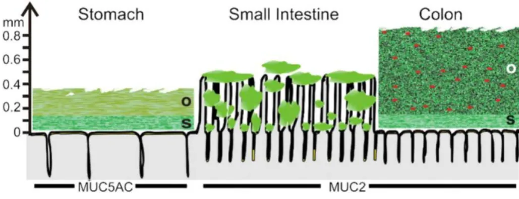
The structure and spatial organization of the mucus layer are different according to the intestinal region under study. In the colon, mucus is divided in two distinct physical layers (Figure I.2): (i) an inner layer which is firmly adhering to the epithelial cells and approximately 50-μm thick and (ii) an outer, loose layer which is approximately 100-μm
organized around the highly glycosylated MUC2 mucin, that is secreted by the goblet cells. The inner mucus layer is dense and does not allow bacteria to penetrate, thus keeping the epithelial cell surface devoid of bacteria (Johansson et al. 2008). The inner mucus layer is converted into the outer layer, which is the preferential habitat of the gut commensal bacteria. It can be speculated that bacteria contribute to dissolving the outer loose mucus since germ-free mice exhibit a thicker layer than that of conventional rodents (Johansson et al. 2011). Furthermore, the outer mucus layer has an expanded volume, due to proteolytic activities provided by the host but also probably by commensal bacteria. The numerous O-glycans on the MUC2 mucin not only serve as nutritive substrates for bacteria but also as adhesion sites, thus contributing in this way to the selection of the species-specific microbiota (Juge 2012). As in the colon, the stomach has relatively well-defined mucus layers made up by the major mucins MUC5AC and MUC6 (McGuckin et al. 2011) (Figure I.2). In the small intestine, in contrast with the stomach and the colon, the mucus layer is rather discontinuous (Figure I.2). Mucus is secreted at the top of the crypts and then moves upward between the villi. Therefore, the tips of the villi are not systematically covered with mucus (Johansson et al. 2011). The main secreted mucin is MUC2, as in the colon.
Figure I.2. Schematic representation of the mucus layer(s) along the gut (Johansson et al.
2011). Note that the thicknesses given are derived from data on rat and adapted from the work of Atuma et al. (Atuma et al. 2001). The symbols in red are representative of the bacteria trapped inside the outer mucus layer. The name of the gel-forming mucins in each intestinal region is indicated. O stands for "outer loose mucus layer" and S for "inner and firmly attached mucus layer".
Analysis of the O-glycan structures associated with Muc2 in small and large intestines of rats showed that the large intestine was enriched for sulphated residues whereas the small
glycosylation was also found in humans with a decreasing gradient of fucose from ileum to rectum and an increased gradient of sialic acid (Robbe et al. 2003). This acidic gradient was shown to be acquired after birth, perhaps due to bacterial colonization or initiation of digestive and absorptive functions within the gut (Robbe-Masselot et al. 2009).
Concerning the relationship between mucus and intestinal diseases, numerous studies described the role of alterations in the structure and/or quantity of mucins in initiating and maintaining mucosal inflammation in IBD and in driving cancer development in the intestine, as recently reviewed (Kim and Ho 2010, Sheng et al. 2012). Reduction in MUC2 synthesis and/or goblet cell number, resulting in a barrier defect, was shown to be involved in the onset or development of IBD. For instance, patients with ulcerative colitis (UC) were first characterized by depletion of goblet cells in the colonic epithelium (Theodossi et al. 1994). More recent studies in mouse models of colitis highlighted the importance of mucins in maintaining integrity of the protective mucus barrier, the breakdown of which could result in colitis. Indeed, Muc2-deficient mice, with no morphologically identifiable goblet cells and absence of Muc2 expression in the intestine, were reported to display markedly deficient mucus layers with increased permeability and enhanced bacterial adhesion to the mucosal cell surface. Moreover, these mice developed spontaneous colitis and were more susceptible to dextran sulfate sodium-induced colitis (Van der Sluis et al. 2006). Later, Johansson et al. (Johansson et al. 2013) reported that mucus secreted from human sigmoid colon was normally dense and thick whereas that in patients with active UC was thinner and more penetrable to 2-µm sized fluorescent beads and bacteria.
2.2 Rheological properties of GI mucus
In order to understand how LAB evolve within the mucus, rheology needs to be scrutinized, i.e. mucus flow behavior law has to be characterized. From a general point of view, mucus is a viscoelastic pH-dependent gel with functional consequences (Cao et al. 1999, Celli et al. 2007, Celli et al. 2009). For instance, Celli et al. (2009) reported that the pathogen H. pylori utilizes such properties to reach the stomach epithelium. To manage so, bacteria secrete urease that elevates the pH of their microenvironment, resulting in a transition gel-sol. This is a striking evidence of the interplay between biochemical and mechanical interactions that
transition of PGM with a solid-like viscoelastic behavior below pH 4 and liquid-like above. This is probably due to the neutralization of some amino acid residues with pKa values of ~4, Glu and Asp, which facilitates the cross-linking among macromolecules through hydrophobic interactions (Lee et al. 2005). Furthermore, the presence of monovalent cations, such as K+ and Na+, impacts mucin rheology (Martin et al. 1978, Celli et al. 2007). For instance, Celli et
al. (2007) reported that PGM exhibits a gel structure at a NaCl concentration of 100 mM but
evolves toward a liquid-like behavior for a higher concentration of 200 mM. This can be explained by the decrease in electrostatic interactions within the mucin network. Likewise, other substances are capable of interacting with mucins, including (phospho)lipids, albumin (Martin et al. 1978). In addition, the mechanical properties of mucus can be significantly altered by proteolytic enzymes, e.g. pepsin (Allen et al. 1984), or by reduction of disulfide bridges with mercaptoethanol, dithiothreitol or N-acetylcysteine (Allen 1989).
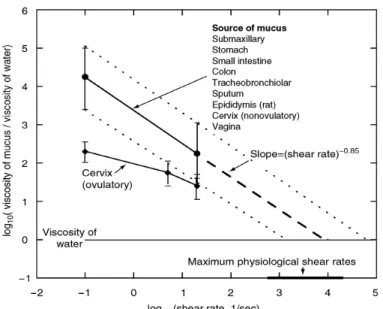
Mucus is a non-Newtonian shear-thinning fluid (Allen 1989, Zhou et al. 2004, Celli et al. 2007), i.e. its viscosity decreases with increasing shear rate (Figure I.3). Figure I.3 also shows that mucus from different sources, such as pig stomach, small intestine, colon; human lung, sputum and cervix; bovine cervix; and rat epididymis (Cone 2005), share a common behavior modeled by a power law.
Figure I.3. Flow curve (apparent viscosity (Pa.s) vs. shear rate (s-1)). The data summarized are derived from many different mucosal sites (Cone 2005). The apparent viscosity of these
viscosity of water at high physiological shear rates.
Studies of PGM secretions obtained by gentle scrapping of pig stomach showed a greater elastic modulus G’ (or storage modulus) than its viscous modulus G” (or loss modulus) throughout the frequency range studied (10-2 to 102 rad/s) (Allen et al. 1984). Delgado-Reyes
et al. (Delgado-Reyes et al. 2013) studied the viscoelasticity of two types of commercial
PGM (Sigma-Aldrich), type II and type III referred to as non-purified and purified, respectively. Flow curves were described by the shear-thinning Ostwald de Waele model that solely takes into account the flow behavior index (n), and the consistency coefficient (K) as free fitting parameters in the form
η
app=Kγ
ɺn−1, as it is the case in Figure I.3. Rheological properties of PGM were function of its concentration and varied with the types of PGM. At a shear rate of 100 s-1, non-purified mucin preparations showed a lower apparent viscosity (0.22–8.37 Pa.s) than that of purified ones (0.43–29.29 Pa.s). At a PGM concentration of 10% (w/v), the gel was a viscoelastic liquid (G”>G’) whereas at a higher concentration (40% (w/v)), it rather exhibited viscoelastic solid properties (G’>G”). The latter showed remarkable recovery during the creep compliance tests. Using atomic force microscopy, the topography of non-purified PGM was rough and regular whereas purified PGM presented a smoother, granular and inhomogeneous surface (Delgado-Reyes et al. 2013).Furthermore, these two types of PGM commercial products have been widely used as a model mucus system, for (i) muco-adhesion assays (Rojas et al. 2002, Tian et al. 2005, Dague et al. 2010), (ii) investigation of mucin composition and properties (Zenteno et al. 1995, Corfield 2000, Dawson et al. 2004, Le et al. 2011) or (ii) isolation of gut-associated LAB (Kraatz 2011). Apart from the simplicity of use (easy preparation, reproducibility, long-term stability) and reduced costs, reasons for employing such lyophilized mucins are primarily linked to the difficulties in collecting native human or animal mucins and guaranteeing stability (i.e. effect of age, healthy status and diet, etc.). However, Kocevar-Nared et al. (1997) reported a difference between the natural PGM, i.e. scraped from pig stomach and purified, and the commercial type II and type III PGM products. Asides with others (Madsen et al. 1996, Kocevar-Nared et al. 1997), Celli et al. (Celli et al. 2005, Celli et
al. 2007) concluded that commercial PGM could not resemble the natural PGM.
required.
In order to decipher the selective and filtering properties of the mucin network, many tools are currently used to connect the rheological properties with the microstructure, that both affect the migration of particles or bacteria such as pathogens, as presented for H. pylori (Celli et al. 2009). Worth mentioning is the Multi-Particle Tracking (MPT), used in this study and explained in details below.
2.3 Mucus microrheology and microstructure
2.3.1 Introduction of MPT technique
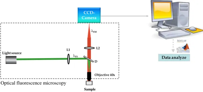
This technique consists of tracking the position of tracers over time. Tracers are beads, typically 200-500 nm in diameter (a) and termed here as nanoparticles (NPs). At the microscale level, matter is subjected to thermal agitation described by the Brownian motion. Particles are bombarded by atoms and induce stochastic displacement of them. Technically, particles suspended in a fluid are simply visualized under microscope using a high-resolution charge-coupled device (CCD) camera (Figure I.4). In crowded environment, the tendency is to use fluorescent particles to discriminate the probe with the fluid and this is commonly observed via epifluorescence. The exciting light could affect the motion by exerting an optical force but it remains small (<5%) compared to the thermally driven force of the beads (Gardel et al. 2005).
MPT has unique advantages: (i) MPT can directly measure the mechanical properties of specialized medium, such as cytoplasm (Wirtz 2009), because of the intimate contact between probing beads and material microstructure; (ii) MPT requires short times of data collection, typically 10-20s, to track mobility of tens to hundreds of embedded NPs at the same time and to obtain statistically significant data; (iii) the spatial resolution of detecting nanoparticle centers typically ranges from 3 nm to 10 nm (Wirtz 2009), allowing the investigation of the microstructure as well as the heterogeneous degree of soft matter.
Figure I.4. An example of MPT set-up is composed of an optical fluorescence microscopy
with a light source; an excitation filter L1, allowing the transfer of the short-wavelength light; an emission filter L2, allowing the transfer of the long-wavelength light; a dichroic beam splitter D; and a charge-coupled device (CDD) camera.
The important definitions in MPT method are time scale, mean-square displacement and mode of transport.
(i) Time scale. The shortest time scale achievable for a given experimental set-up is
determined by the maximum speed of the camera and the necessary acquisition hardware. Assuming a camera is able to capture images at video rate 30 frames per second (fps), the shortest time scale is 33 ms. The longest time scale depends on the length of the movie. In gels or porous networks with small mesh sizes, NPs may undergo subdiffusive transport at short time scale, however at longer time scale nanoparticle mobility may be more diffusive due to the relaxation time of polymer gel structures (Suh et al. 2005).
(ii) Mean-square displacement (MSD). MSD 2
( )
r τ
∆ describes the NP mobility in a material over time, and is given by:
(
) ( )
(
) ( )
(
) ( )
(
) ( )
2 2 2 2 2 0 1 ( ) 2 N i r t r t x t x t y t y t r r i t r i t nDt N τ τ τ τ τ = ∆ + − ∆ = ∆ + − ∆ + ∆ + − ∆ ∆ =∑
∆ + − ∆ = (I.1) Light source CY3+PGM CCD-Camera λEM Data analyze Objective 40x λEX L2 L1 DOptical fluorescence microscopy
n=2.
The embedded NPs are smaller than 1 µm so that they undergo Brownian motion, while gravity or convections are negligible. If active transport is also negligible, only two types of forces act on the NPs inside viscous liquids: (i) the thermal energy kBT and the movements of
the local structure of the material; (ii) the counteracting frictional force ξ = 6π
η
a which results from the movement of NPs driven by the thermal energy. In this case of Brownian motion, the diffusion coefficient (independent of time scale) D is also measured by the Stokes-Einstein relation:3
Bk T
D
a
πη
=
(I.2)where kB is Boltzmann constant 1.38x10⁻²³ m2.kg.s-2.K-1, T is temperature(oK),
η
is water viscosity (9.03 10-4 Pa.s), a is size of the spherical NPs in diameter (m).(iii) Mode of transport. Active and subdiffusive transport of NPs can be described by:
2
( ) 2
r τ nDtα
∆ = (I.3)
α is the anomalous exponent. For the motion of actively transported (also called “super-diffusive”) NPs, which is due to the relaxation of polymer gels, convection or gravity in systems, α equals to more than 1. In contrast, the subdiffusive transport α equals to less than 1, which describes motion impeded by obstacles or by NP binding to physical structures in the environment. NPs are classified as immobile transports when their movements are smaller than spatial resolution, i.e. their diffusion coefficients are lower than that of the same NPs attached to glass slides. Moreover, MPT allows determining the degree of heterogeneity of particle diffusion inside mucus gels by the relative width of the diffusion coefficient distribution (Lieleg et al. 2010):
rel
D
σ
σ = (I.4)
σ is the standard deviation and is the mean value of diffusion coefficients. This assumes that the distribution is normal. It will be seen later (Chapter III) that it tends to lognormal.
In addition to determine the diffusion coefficient and the anomaly of diffusion, MSD obtained by using MPT can help quantifying the dynamic rheological moduli of the microenvironment of a tracer embedded in fluid, and to some extent in complex media. In this review, two passive microrheology methods are distinguished (Levine and Lubensky 2000): one-point and two-point rheology. The first uses MSD of an individual particle, i.e. the autocorrelation of the motion of beads, and the second assumes that the motion of one tracer affects its neighbor. Therefore, two-point microrheology makes use of the cross-correlation of two beads introducing a new variable, the bead separation r.
a. One-point microrheology – conventional way
From Eq. (I.2) the viscosity of the material can be estimated by:
2 4 , 3 ( ) B k T t a r η π τ = ∆ (I.5)
In the case of purely viscous material, MSD follows the thermal limit giving rise constant viscosity that solely depends to temperature.
In the case of complex media, the time-dependent MSD of the NPs can be transformed into the frequency-dependent viscoelastic moduli G*(ω) (Mason and Weitz 1995, Gittes et al. 1997) via the Laplace transform inversion method:
2 2 ( ) , ( ) B k T r s saG s π = ɶ ɶ (I.6)
where
r s
ɶ
2( )
is the Laplace transform of the tracer’s MSD, as function of Laplace frequencys, a their diameter and
G s
ɶ
( )
the Laplace transform of the complex modulus G*=G”+iG’. Equation (I.6) is the Stokes-Einstein equation generalized (GSER) form of a frequency-dependent viscosity (Mason and Weitz 1995). Another inversion method consists of approximating the Fourier transform via an analytic continuation that handles noise arising for the Fourier transform (Mason 2000). MSD plot is fitted to a local power law and the logarithmic differential is then calculated:( ) , ln d τ α τ τ ∆ = (I.7)
This parameter, which is the anomalous exponent described above, is used in an algebraic form of the GSER:
(
)
2 1 4 ( ) , 3 ( ) 1 ( ) B k T G a r τ ω ω π τ α τ = ≈ ∆ Γ + (I.8)where ( )Γ x =(x−1)! is the gamma function. Defining ( )δ ω the phase angle as:
* ln ( ) ( ) , 2 ln d G d ω π δ ω ω = (I.9)
The storage G’ and loss G’’ moduli with respect to frequency ω can be obtained:
(
)
(
)
* * '( ) ( ) cos ( ) , ''( ) ( ) sin ( ) . G G G G ω ω δ ω ω ω δ ω = = (I.10) b. Two-point microrheologyThis method correlates the motion of two tracers separated by the distance r. It was developed to connect the microscale with the macroscopic methods that usually exhibit several orders of magnitude discrepancies (Crocker et al. 2000). Limitations of Eqs. (I.6) and (I.8) from GSER arise in majority from the inhomogeneity of the medium. Since one particle strain field will affect a second, it is therefore possible to measure the strain field by cross correlating two-particle motions. In the homogenous case, the strain field is proportional to the tracer motion and decays ~ a/r.
Recent experiments have probed such correlated motion in viscous (Crocker 1997) and elastic (Bausch et al. 1999) materials. MPT enables to measure the vector displacements of the beads
∆
r t
α( , )
τ
=
r t
α(
+ −
τ
)
r t
α( )
where t is the absolute time and τ the lag time as aforementioned. The ensemble averaged tensor product of the tracer displacements yields:,