HAL Id: tel-01927180
https://tel.archives-ouvertes.fr/tel-01927180
Submitted on 19 Nov 2018HAL is a multi-disciplinary open access archive for the deposit and dissemination of sci-entific research documents, whether they are pub-lished or not. The documents may come from teaching and research institutions in France or abroad, or from public or private research centers.
L’archive ouverte pluridisciplinaire HAL, est destinée au dépôt et à la diffusion de documents scientifiques de niveau recherche, publiés ou non, émanant des établissements d’enseignement et de recherche français ou étrangers, des laboratoires publics ou privés.
Synaptic plasticity in the lateral habenula controls
neuronal output : implications in physiology and drug
addiction
Kristina Valentinova
To cite this version:
Kristina Valentinova. Synaptic plasticity in the lateral habenula controls neuronal output : implica-tions in physiology and drug addiction. Neurons and Cognition [q-bio.NC]. Université Pierre et Marie Curie - Paris VI, 2016. English. �NNT : 2016PA066743�. �tel-01927180�
This work is licensed under a Creative Commons Attribution-NonCommercial 4.0 International License.
Université Pierre et Marie Curie
Ecole doctorale n°138
« Cerveau Cognition Comportement »
Doctoral thesis title:
Synaptic plasticity in the lateral habenula controls neuronal
output: implications in physiology and drug addiction
Presented by Kristina Valentinova
Directed by Dr. Manuel Mameli
Team « Synapses and pathophysiology of reward »
Institut du Fer à Moulin
Members of the jury:
Dr. Philippe Faure – président Pr. Mark Ungless – examinateur
Pr. Jaideep Bains –rapporteur Dr. Vivien Chevaleyre –examinateur
Pr. Camilla Bellone – rapporteur Dr. Manuel Mameli – directeur de thèse
Page | 1
Abstract
The capacity of the brain to anticipate and seek future rewards or alternatively escape aversive events allows individuals to adapt to their environment. A considerable research effort has focused on unraveling the cellular and synaptic mechanisms within the meso-cortico-limbic system underlying motivational processing both in physiological conditions and in pathologies such as addiction and depression. However, only recently we begin to understand the circuit substrates capable to control midbrain monoaminergic nuclei and their contribution to motivated behaviors.
The Lateral Habenula (LHb) has emerged in the last decade, as a major player encoding stimuli with motivational value and in controlling monoaminergic systems. The wiring of this epithalamic structure subserves discrete features of motivated behaviors, including preference and avoidance. Recent advances have also demonstrated that aberrant modifications in LHb function trigger negative emotional states in disorders including depression and addiction, highlighting the LHb as an important brain target for therapeutic intervention for these pathological states.
In my thesis work I first sought to investigate how modulation of synaptic transmission in the LHb controls neuronal activity, especially focusing on the role of metabotropic glutamate receptors. In a second study, I expanded my work examining how drug experience changes synaptic transmission in a precise habenular circuit that we discovered to be crucial for depressive states during cocaine withdrawal.
In an initial data set, we found that, in the LHb, metabotropic glutamate receptor 1 activation drives a PKC-dependent long term depression of excitatory (eLTD) and inhibitory (iLTD) synaptic transmission. Despite the common induction, eLTD and iLTD diverged in their expression mechanism. While eLTD required endocannabinoid-dependent reduction of glutamate release, iLTD expressed postsynaptically through a decrease of 2-containing GABAA receptors function. Further, eLTD and iLTD
bidirectionally controlled LHb neuronal output.
In a second study, we showed that chronic cocaine exposure leads to a persistent and projection-specific increase of excitatory synaptic transmission onto LHb neurons. This form of synaptic potentiation required membrane insertion of GluA1-containing AMPA
Page | 2 receptors and a reduction in potassium channels function ultimately leading to increased LHb neuronal excitability both in vitro and in vivo. These cocaine-driven adaptations within the LHb were instrumental for depressive-like states emerging after drug withdrawal.
Altogether this work demonstrates how synaptic plasticity in the LHb affects neuronal output and thereby contributes to behaviors associated with the pathology of motivation.
Page | 3
Résumé
La survie des individus dépend de leur capacité d’anticiper la survenue d’une récompense ou d’un danger leur permettant ainsi de s’adapter à leur environnement. De considérables efforts ont été réalisés pour identifier les mécanismes cellulaires et synaptiques ayant lieu au niveau du circuit de la récompense afin d’avoir une meilleure compréhension des processus sous tendant des états motivationnels physiologiques et pathologiques tels que l’addiction et la dépression. Pour autant, ce n’est que récemment qu’on commence à comprendre les circuits capables de contrôler les systèmes monoaminergiques mésencéphaliques et leurs contributions aux comportements motivés.
Dans les dernières décennies l’habénula latérale (LHb) a émergé comme un acteur majeur capable d’encoder des stimuli de valeur motivationnelle et de contrôler les systèmes monoaminergiques. La connectivité de cette structure épithalamique joue un rôle clé dans différents aspects des comportements motivationnels, comme l’approche et la fuite. Des avancées récentes ont aussi démontré que des altérations de la fonction de la LHb entrainent des états émotionnels négatifs caractéristiques de la dépression et l’addiction. Ces observations suggèrent que la LHb pourrait s’avérer une cible importante pour le traitement de ces pathologies.
Au cours de mon travail de thèse, j’ai d’abord cherché à comprendre comment moduler la transmission synaptique au niveau de la LHb pouvait contrôler son activité. Pour répondre à cette question, je me suis focalisée sur le rôle des récepteurs métabotropiques au glutamate (mGluRs). Dans une seconde étude, j’ai examiné les mécanismes par lesquels les drogues d’abus modifient la transmission synaptique des neurones de la LHb. Ces modifications se produisent spécifiquement dans les neurones LHb se projetant vers le noyau tegmental rostral (RMT) et sont nécessaires pour l’émergence des états dépressifs.
Dans un premier temps, nous avons démontré qu’au niveau de la LHb les mGluRs de type I sont capables d’induire une dépression à long terme de la transmission synaptique excitatrice (eLTD) et inhibitrice (iLTD). Ces deux formes de plasticité dépendent de la signalisation PKC, mais requièrent des mécanismes d’expression
Page | 4 différents. Tandis que eLTD réduit la probabilité de libération du glutamate via l’activation de récepteurs présynaptiques aux endocannabinoides (CB1), iLTD s’exprime par la réduction de la fonction des récepteurs GABAA postsynaptiques contenant la
sous-unité 2. De plus, eLTD and iLTD exercent un contrôle bidirectionnel sur la décharge des neurones de la LHb.
Dans un second temps, nous avons mis en évidence qu’une exposition chronique à la cocaïne produit une augmentation persistante de la transmission excitatrice au niveau des neurones de la LHb ciblant le RMTg. Cette forme de potentialisation synaptique nécessite l’insertion membranaire de récepteurs contenant la sous-unité GluA1, ainsi que la réduction de conductances potassiques entrainant une hyperexcitabilité neuronale in vitro et in vivo dans la LHb. Ces modifications sont nécessaires pour l’établissement d’états dépressifs émergeant lors de la période de sevrage à la cocaïne. En conclusion, ce travail a contribué à la compréhension de mécanismes de plasticité synaptique ayant lieu au niveau de la LHb et leurs répercussions pour son activité contrôlant ainsi des comportements motivationnels.
Page | 5
Acknowledgements
I would like to thank in first place the amazing lovable people in the lab. Starting with my advisor and dear friend Manuel. I thank you for your time, attention and help during all these years, for the countless days and nights you spent to guide me through my experience as a young scientist, for the discussions and fights, for the sharing and advices. I thank you for the inspiration and love for science that you are building in me with your example every single day. I thank you for making my experience in the lab an amazing adventure, overcoming difficulties and frustration and learning how to enjoy the good moments, for making me laugh when I am desperate and for showing me that there is a way out even in the most difficult moments. Thank you for being always available for me and the others in the lab. Thank you for supporting my downs and helping me get over them. Thank you for creating a positive and motivated environment where trust, mutual respect and enthusiasm are the driving forces. Thank you for challenging me and believing in me. And I thank you for all the great fun we had in the lab and outside, for all the celebrations and…… although I can continue thanking you for many other things most importantly I want to thank you for proving me that when you have positive attitude, enthusiasm and persistence everything is possible!
I want to thank also Salvatore, Frank and Anna... you guys are not only colleagues and friends for me, but my bigger brothers and sister. I have not enough words to express how much I enjoyed having you around and how grateful I am for all the amazing time we spent together. I thank you so much for all the fun and enjoyable moments we shared, for all the laughing, for all the dinners, celebrations and birrettas, for all the foosball games, for the Italian and Dutch lessons, for the adventures in Sardinia, Sizel Montpellier and Copenhagen. I thank you also for being so understanding, caring and attentive, for listening and giving me your precious advices when I most need them. I thank you for being so creative and funny, for all the jokes you make, for being so positive and pleasant, I always enjoy your company. I thank you for sharing with me your knowledge and experience, for all the discussions we had, for your constructive feedback, for sharing your ideas, for being so much passionate and implicated in our work, you keep me motivated and make my days happier.
Page | 6 Finally, I want to thank all of you Manuel, Salvatore, Anna and Frank for keeping this unique family ambiance alive every day.
With all my love and respect a big THANK YOU!!!!!
I also want to thank many people from the IFM institute with who I shared countless nice moments: Tamar, Quentin, Martin, Sana, Marie, Ferran, Jessica, Emily, Enrica, Assunta, Nicolas, Benoit, Mariano, Alfredo, Sebastian for all the enjoyable moments we spent together, sharing work space, but also knowledge and ideas, for creating friendly and convivial ambiance in the institute and for all the parties and celebrations. I thank Jean-Christophe, Sabine, Nicolas and Corentin for helpful discussions and advices. I want to thank also Jean-Antoine and Fiona for making the effort to keep this institute a dynamic and friendly place.
I want to thank my friend Silvana, who has been side by side with me throughout all these years (8!!!) I spent in Paris. I want to thank you for sharing all these precious moments and being around when I most needed you. You are very very special friend.
Finally, I want to thank my family, mom, dad and sis, the people who are always around me even thousands of kilometers away, for their endless love, understanding and support throughout my life. I feel lucky and extremely grateful to have such a loving and caring family. I love you!
Page | 7
Contents
ABSTRACT ... 1 RESUME ... 3 ACKNOWLEDGEMENTS ... 5 TABLE OF FIGURES ... 8 LIST OF ABBREVIATIONS ... 9 INTRODUCTION ... 13THE CIRCUITS OF MOTIVATION - IS IT ALL ABOUT DOPAMINE? ... 16
NEUROBIOLOGICAL SUBSTRATES OF AVERSION ... 18
THE LATERAL HABENULA: A CONTROL STATION OF AVERSION ... 20
PHYLOGENY AND ANATOMY OF THE LHB ... 20
INPUTS TO THE LHB ... 22
OUTPUT CONNECTIVITY OF THE LHB ... 24
FUNCTION OF THE LHB IN REWARD AND AVERSION ENCODING ... 25
ROLE OF LHB OUTPUT FOR AVERSION PROCESSING ... 29
ROLE OF INPUTS TO THE LHB FOR AVERSION PROCESSING ... 32
DYSFUNCTION OF THE LHB: IMPLICATIONS IN DEPRESSION AND ADDICTION ... 37
LHB IN DEPRESSION ... 37
LHB IN ADDICTION ... 38
PROPERTIES OF LATERAL HABENULA NEURONS ... 42
CELL MORPHOLOGY AND ELECTROPHYSIOLOGY ... 42
FAST EXCITATORY TRANSMISSION VIA IONOTROPIC GLUTAMATE RECEPTORS ... 44
FAST INHIBITORY SYNAPTIC TRANSMISSION VIA IONOTROPIC GABAA RECEPTORS ... 47
MODULATION OF FAST EXCITATORY AND INHIBITORY TRANSMISSION: ROLE OF MGLURS ... 49
SYNAPTIC PLASTICITY IN THE LHB: A CELLULAR SUBSTRATE FOR MOTIVATED STATES IN DISEASE ... 52
CELLULAR MECHANISMS IN THE LHB IN DEPRESSION ... 52
CELLULAR MECHANISMS IN THE LHB IN ADDICTION ... 53
CONTEXT AND OBJECTIVES FOR THE STUDIES ... 56
I. MGLUR-LTD AT EXCITATORY AND INHIBITORY SYNAPSES CONTROLS LATERAL HABENULA OUTPUT ... 56
II.COCAINE-EVOKED NEGATIVE SYMPTOMS REQUIRE AMPA RECEPTOR TRAFFICKING IN THE LATERAL HABENULA ... 100
DISCUSSION ... 111
WHICH INPUTS UNDERGO MGLUR-ELTD AND ILTD? ... 111
IS THERE ANY OUTPUT-SPECIFICITY FOR MGLUR-ELTD AND ILTD? ... 113
WHAT IS THE BEHAVIORAL RELEVANCE OF MGLUR-LTD IN THE LHB? ... 113
INDUCTION MECHANISMS AND CIRCUIT SPECIFICITY OF COCAINE-EVOKED PLASTICITY IN THE LHB ... 116
Page | 8
DISTINCT SYNAPTIC ADAPTATIONS CONVERGE TO INCREASE LHB NEURONAL AND BEHAVIORAL OUTPUT ... 119
CONCLUDING REMARKS ... 120
ADDITIONAL PUBLICATIONS AND CONTRIBUTIONS………...123
REFERENCE LIST ... 125
Table of figures
Figure 1 Simplified schematic of the meso-cortico-limbic and associated circuits in the rodent brain ... 15Figure 2 Cell-type specific modulation in the VTA for distinct motivational states... 19
Figure 3 Anatomical localization of the habenular complex ... 20
Figure 4 Comparative analysis of human and rat LHb and LHb subnuclear organization ... 22
Figure 5 Afferents and their territorial distribution within the LHb ... 23
Figure 6 LHb efferents and territorial distribution of their cell bodies throughout the LHb ... 25
Figure 7 LHb function in aversion encoding and negative prediction error ... 28
Figure 8 Contribution of LHb output to motivated behaviors ... 31
Figure 9 Contribution of inputs to the LHb to different motivational states ... 36
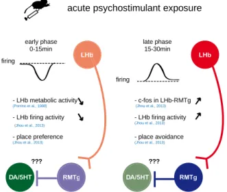
Figure 10 Acute effects of psychostimulants in the LHb ... 41
Figure 11 AMPA receptors trafficking and channel properties ... 45
Figure 12 Excitatory and inhibitory synapses in the LHb ... 49
Figure 13 mGluR1/5 signaling and mechanisms of long term depression ... 51
Figure 14 GqPCR model of inhibitory LTD ... 112
Figure 15 Input and cell type-specific model for mGluR-LTD in the LHb ... 115
Page | 9
List of abbreviations
5HT 5-hydroxytriptamine AHP afterhyperpolarization
AMPA -amino-3-hydroxy-5methyl-4-isoxazolepropionic acid AP action potential
BNST bed nucleus of the stria terminalis Ca2+ calcium ion
CaMKII calcium calmoduline-dependent protein kinase type II cAMP cyclic adenosine monophosphate
CB1 cannabinoid receptor type 1 ChR2 channel rhodopsin 2
CNS central nervous system D1R dopamine type 1 receptor D2R dopamine type 2 receptor DAT dopamine transporter DBB diagonal band of Broca DBS deep brain stimulation
eLTD long term depression of excitatory transmission EPCS excitatory postsynaptic current
EPN entopeduncular nucleus
fMRI functional magnetic resonance imaging FST forced swim test
GABA -aminobutyric acid
Page | 10
GABAB R -aminobutyric acid type B receptor
GAD67 glutamate decarboxylase
GIRK G-protein inwardly-rectifying potassium channel GluA1-4 glutamate AMPA receptor subunits type 1-4 GluN1-3 glutamate NMDA receptor subunits type 1-3 GPCR G-protein coupled receptor
HCN hyperpolarization-activated cyclic nucleotide-gated channel HFS high frequency stimulation
iLTD long term depression of inhibitory transmission IPSC inhibitory postsynaptic current
K+ potassium ion
LDT laterodorsal tegmentum LFS low frequency stimulation LH lateral hypothalamus LHb lateral habenula
LHbLB basal nucleus of the lateral lateral habenula
LHbLMc magnocellular nucleus of the lateral lateral habenula LHbLMg marginal nucleus of the lateral lateral habenula LHbLO oval nucleus of the lateral lateral habenula
LHbLPc parvocellular nucleus of the lateral lateral habenula LHbMA anterior nucleus of the medial lateral habenula LHbMC central nucleus of the medial lateral habenula LHbMMg marginal nucleus of the medial lateral habenula LHbMPc parvocellular nucleus of the medial lateral habenula LHbMS superior nucleus of the medial lateral habenula
Page | 11
LPO lateral preoptic area LS lateral septum
LTD long term depression LTP long term potentiation
mAChR muscarinic acetylcholine receptor MCH melanin concentrating hormone MDD major depression disorder
mEPSC miniature excitatory postsynaptic current mGluR metabotropic glutamate receptor
MHb medial habenula
mIPSC miniature inhibitory postsynaptic current mPFC medial prefrontal cortex
mTOR mechanistic target of rapamycin Na+ sodium ion
NAc nucleus accumbens NMDA N-methyl-D-aspartic acid
NpHR halorhodopsin from Natronomonas PFC prefrontal cortex
PICK1 protein interacting with C kinase type 1 PKA protein kinase A
PKC protein kinase C
PLC phospholipase type C PP2A protein phosphatase type 2 PSP postsynaptic potential
Page | 12
SK small conductance calcium-activated potassium channel SNc substantia nigra pars compacta
TH tyrosine hydroxylase TST tail suspension test TTX tetrodotoxin
Vgat vesicular GABA transporter
Vglut2 vesicular glutamate transporter type 2 Vmat2 vesicular monoamine transporter type 2 VP ventral pallidum
Page | 13
Introduction
The central nervous system (CNS) is designed to promote at best the survival of the species. Indeed the brain is able to encode the valence of environmental stimuli, which allow the individual to pursue rewards essential for survival or to avoid potential threats. Single neurons within complex brain circuits represent such environmental information through changes in their spiking activity. During my PhD training I developed a strong interest in understanding how neurons encode and process valenced information at different space and time scales – from the single cell to the integrated circuit and from the millisecond duration of an action potential to the long-lasting synaptic remodeling. My particular interest is to understand how reward and aversion-related experience modifies the synaptic properties of neurons and how experience-induced synaptic plasticity controls neuronal activity to drive motivational states crucial for survival.
In the course of evolution, highly organized circuits have emerged to promote survival through reward-seeking and threat-avoiding behaviors. The meso-cortico-limbic circuit has gained a central role in the processing of rewarding and aversive environmental information and in orchestrating adaptive behavioral responses. It also refers as a ‘reward’ circuit since a large body of evidence collected over the years has revealed its critical role in appetitive and reinforcing behaviors (Berridge and Robinson, 1998;
Robbins and Everitt, 1996; Schultz, 2007a; Wise and Rompre, 1989). It comprises a
meso-limbic part represented by the ventral tegmental area (VTA) dopamine neurons projections to the nucleus accumbens (NAc) of the ventral striatum and a meso-cortical part represented by the VTA projection to the prefrontal cortex (PFC). However this system is highly interconnected and is tightly controlled by a much wider network of brain structures, including the lateral hypothalamus (LH), the amygdala and the lateral habenula (LHb) among others (Fig 1), which altogether operate to encode valenced stimuli and to transform them into behavioral output (Ikemoto, 2007; Ikemoto et al., 2015; LeDoux, 2000; Matsumoto and Hikosaka, 2007; Nieh et al., 2013; Stuber and Wise, 2016; Wise, 2005).
Page | 14 A key feature of the meso-cortico-limbic circuit is the attribution of motivational value to otherwise neutral stimuli. Indeed, a stimulus can acquire positive or negative valence through innate or learned mechanisms. Stimuli assigned with a positive value (rewards) are generally beneficial for survival and elicit approach behaviors. On the contrary, life-threatening or harmful stimuli are ascribed with a negative value (aversion) and the corresponding behavioral reaction is to avoid or escape them. When animals are repetitively exposed to valenced stimuli in a particular environmental context (i.e. in presence of visual or auditory cues), cue-stimulus associations are formed allowing to predict the timing of occurrence and magnitude of the stimulus, a process necessary to orchestrate an appropriate behavioral response. Indeed, the ability to predict the outcome of a stimulus is at the base of conditioned behaviors, such as conditioned place preference and avoidance, paradigms widely used to assess motivational states
(Hollerman and Schultz, 1998; Schultz, 1998; Schultz et al., 1997; Tzschentke, 2007). In
physiological conditions behaviors leading to obtaining a reward or escaping a danger can be reinforced, positively, when an animal actively works to obtain a reward or negatively, when the animal works to stop an aversive stimulus (Valenstein & Valenstein
1964; Ferster 1957). These behaviors are compromised in pathologies associated with
deficits in reward and aversion processing such as depression or addiction (Lüscher,
2016; Russo and Nestler, 2013).
Over the last decades a great deal of attention has focused on the synaptic and cellular substrates of reward encoding, learning and behaviors within the meso-cortico-limbic system. However, it remains less known how this system processes aversion and which particular structures provide it with aversion informative signals. The lateral habenula is now emerging as a key structure capable to provide aversive information to the meso-cortico-limbic system likewise contributing to avoidance behaviors.
In this thesis I will first briefly summarize key functional features of the reward system to provide a general context of positive and negative motivational processing. Next, I will introduce how the lateral habenula interacts with this system in the context of aversion. Finally I will show how modulation of synaptic transmission in the lateral habenula can
Page | 15 tune neuronal output potentially driving opposing motivational states and how drug-experience changes lateral habenula function to promote negative emotional states.
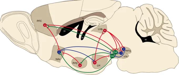
Figure 1Simplified schematic of the meso-cortico-limbic and associated circuits in the rodent brain
The meso-cortico-limbic system comprises dopaminergic VTA-to-NAc and VTA-to-PFC projections, which release dopamine upon rewarding or aversive stimuli (Abercrombie et al., 1989; Hernandez and Hoebel, 1988; Kalivas and Duffy, 1995). The VTA also contains GABAergic interneurons which inhibit dopamine neurons. Apart from the input to the NAc and PFC, dopamine neurons also project to the amygdala (Amy), which in turn sends glutamatergic input to the NAc. VTA dopamine neurons receive GABAergic inhibitory inputs from local interneurons or from other structures including the rostromedial tegmental nucleus (RMTg) and the NAc. Excitatory glutamatergic afferents to the VTA arise from the LHb. LH and PFC. The VTA receives also both GABAergic and glutamatergic connections from the BNST (see Fig2).The LHb also projects to local GABA neurons in the VTA and RMTg, likewise exerting inhibitory control over dopamine neurons. The LH sends efferents to the LHb and VTA among others (not shown). The NAc receives glutamatergic innervation from the medial prefrontal cortex (mPFC) and amygdala (Amy) and projects to the LH and VTA. These various glutamatergic and GABAergic inputs to the VTA control aspects of reward and aversion-related behaviors (adapted from Russo and Nestler, 2013).
3V NAc cc 4V 3V 3V PFC LHb VTA RMTg Amy LH + + + + - - -DA DA
Page | 16
The circuits of motivation - is it all about dopamine?
In an attempt to understand the molecular bases of motivation, early studies in the 1970s and 1980s have proposed that the neuromodulator dopamine has a central role in controlling reward-related behaviors (Gerber et al., 1981; Ungerstedt, 1971; Wise et al.,
1978). Dopamine is a monoamine produced mainly by neurons within the VTA and
substantia nigra pars compacta (SNc) which innervate the cerebral cortex and limbic forebrain regions (Björklund and Dunnett, 2007). Initial studies using microdialysis measurements have suggested that dopamine release within the NAc (meso-limbic projection) is associated to self-stimulation reward (Fiorino et al., 1993), while stress specifically activates the meso-cortical projection (Thierry et al., 1976). Indeed, the function of dopamine in reward and aversive behaviors largely depend on the circuit connectivity of individual dopamine neurons (Bromberg-Martin et al., 2010; Lammel et
al., 2011). Furthermore, depending on their specific identity, dopamine neurons undergo
different experience-driven synaptic modifications which are instrumental for behavioral adaptations underlying opposing motivational states (Pignatelli and Bonci, 2015; Volman et al., 2013).
It is now largely established that dopaminergic neurons undergo phasic changes of activity in response to unexpected salient stimuli. Seminal studies in behaving monkeys using single-unit recordings have demonstrated that dopamine neurons firing increases in phasic and burst-like manner when an unexpected reward is presented (Schultz et al.,
1997). Conversely, noxious foot pinch or foot shocks predominantly inhibit dopamine
neurons burst activity (Ungless et al., 2004), although some neurons also show phasic excitation (Brischoux et al., 2009). After several conditioning sessions where animals are trained to associate a cue with the delivery of a reward, the firing of dopamine neurons and the subsequent release of dopamine in the NAc no longer occur at the time of reward delivery, but shift to the cue that predicts it (Schultz et al., 1997; Stuber et al.,
2008). Moreover, the magnitude and direction of dopamine responses correlate with the
predicted probability of the reward. Indeed, if an expected reward fails to occur or is smaller than expected, the activity of dopamine neurons is phasically inhibited, while if the reward is larger than expected dopamine neurons will fire at the time of reward
Page | 17 delivery (Fiorillo et al., 2003; Schultz, 1998; Schultz et al., 1997; Tobler et al., 2003). These experiments led to the idea that dopamine neurons signal a reward prediction error, defined as the difference between the outcome of an expected and actual reward
(Schultz 1998; Keiflin & Janak 2015). Likewise, dopamine neurons serve as ‘sensors’ for any deviation from the expectancies, representing a teaching signal for future cue-reward associations and cue-reward-seeking behaviors. The short bursts of dopamine neurons trigger phasic release events in the NAc, where motivational signals are considered to be translated into a motor output leading to reward seeking behaviors
(Day et al., 2007; McClure et al., 2003; Mogenson et al., 1980).
More recently, a causal relationship between dopamine neurons activity and conditioned learning have been provided by temporally precise and pattern-specific optogenetic modulation of VTA dopamine neurons activity. Indeed, physiologically relevant phasic activation of dopamine neurons expressing the light-activated cation channel rhodopsin 2 (ChR2) induces conditioned place preference behaviors while their inhibition via the hyperpolarizing and light-activated chloride ion pump Halorhodopsin (NpHR) produces conditioned place avoidance (Tan et al., 2012; Tsai et al., 2009). Moreover, these learning mechanisms and reward-oriented behaviors involve transient synaptic potentiation of excitatory transmission onto dopamine neurons (Stuber et al., 2008). Similar synaptic plasticity occurs following single drug or stressful experience (Saal et
al., 2003; Ungless et al., 2001). However, in the case of prolonged drug administration
synaptic changes in VTA dopamine neurons and the NAc become persistent contributing to cue-induced reinstatement of drug-seeking behaviors after drug withdrawal (Chen et al., 2008; Mameli et al., 2009).
Taken together these data demonstrate an important role of dopamine neurons particularly in reward but also in aversion encoding and suggest that synaptic and circuit modifications within the meso-cortico-limbic system are instrumental for motivated behaviors. However, the dopamine system is far more complex in that it receives multiple inputs both from local GABAergic interneurons and from more distal structures, many of which also participate in motivational encoding (Fig1). Moreover, dopamine neurons present heterogeneity in terms of their physiological properties, input-output
Page | 18 connectivity and their responses to valenced stimuli (Lammel et al., 2014, 2011; Volman
et al., 2013). These aspects need to be taken into account when considering dopamine
neurons function in reward or aversion.
Although motivational states can be driven by rewarding and aversive conditions, during my thesis I focused mainly on the cellular substrates devoted to aversion processing.
Neurobiological substrates of aversion
Aversion is a common term to designate the behavioral reaction of avoidance or escape in response to negative, unpleasant, painful, stressful or fearful events or stimuli. Acute and chronic exposure to such aversive stimuli produces short or long-lasting synaptic, structural and circuit modifications that can often lead to neuropsychiatric disorders including depression, anxiety, post-traumatic stress disorder and addiction (Lüscher and
Malenka, 2011; Nestler and Carlezon, 2006; Russo and Nestler, 2013). Many studies
have implicated the VTA dopamine system in such modifications (Berton et al., 2006; Cao et al., 2010; Chaudhury et al., 2013; Krishnan et al., 2007; Lammel et al., 2011).
However, it is now known that other structures directly or indirectly innervating the VTA, and often reciprocally connected, also contribute to different aspects of aversion (Fig1). The picture is even more complex given the large heterogeneity of different cell types forming local microcircuits within the VTA and challenging our understanding of the contribution of distinct pathways to aversion encoding and avoidance behaviors. With the advances of new technologies such as the generation of specific mouse lines, viral-based circuit- and cell type-specific mapping as well as optogenetics it is now possible to interrogate the implication of distinct neuronal circuits and neuronal subtypes for different aspects of aversion.
As a matter of example, specific cell type projections from the ventral bed nucleus of the stria terminalis (BNST) to GABAergic interneurons in the medial VTA have been involved in distinct motivational states. Indeed, glutamatergic BNST neurons, activating predominantly GABA neurons in the VTA, are excited by aversive conditions such as series of foot shocks or foot shock-associated cues. Moreover, ChR2-driven optogenetic
Page | 19 activation of this pathway produces real time avoidance and elevated anxiety states(Fig2A) (Jennings et al., 2013). This is consistent with evidence that activation of GABAergic VTA neurons inhibits dopamine neurons activity leading to conditioned place avoidance behaviors (Tan et al., 2012). In contrast, GABA neurons of the BNST predominantly inhibit GABA neurons of the VTA and are preferentially silenced by foot shock exposure and foot shock-cues. Optogenetic activation of this pathway leads to real time preference and reward seeking behaviors (Fig2A) (Jennings et al., 2013). Consistently, VTA GABA neurons inhibition drives preference, reinforces behavior and reduces anxiety states associated with previous aversive experience (Jennings et al.,
2013). Similarly to this, activation of dopamine neurons of the VTA also promotes
reward-related behaviors (Adamantidis et al., 2011; Tsai et al., 2009), suggesting that GABA neurons in the VTA may serve to break locally the activity of dopamine neurons and therefore control the expression of motivational states (Fig2B). These data raise the question whether other inputs onto dopamine neurons or onto GABA neurons of the midbrain can control different aspects of aversive behaviors. Indeed, a major challenge in the field is to identify the precise input-output and functional organization of the circuits of aversion. - VTA DA VTA reward aversion VTA fx BNST
ac real time avoidanceanxiety
real time preference reward-seeking + -DA aversion + -Adamantidis et al., 2011; Tsai et al., 2009; Jennings et al., 2013
Van Zessen et al., 2012; Tan et al., 20012 Jennings et al., 2013
BNST
ChR2 NpHR NpHR ChR2
Figure 2 Cell-type specific modulation in the VTA for distinct motivational states
(A). BNST glutamate neurons activated by aversive stimuli or cues project to VTA GABA neurons. Their activation drives real time avoidance behavior and anxiety. BNST GABA neurons inhibited by aversive stimuli or cues also project to GABA neurons in the VTA. Their activation drives real time preference and reward-seeking (Jennings et al., 2013). (B). Direct optogenetic activation of VTA dopamine neurons (Tsai et al., 2009; Adamantidis et al., 2011)
or inhibition of GABA neurons (Jennings et al., 2013) promote reward, while activation of GABA neurons (Tan et al., 2012; van Zessen et al., 2012) and inhibition of dopamine neurons (Tan et al., 2012) is aversive.
Page | 20
The lateral habenula: a control station of aversion
The lateral habenula (LHb) has gained considerable attention in the last decade because of its control on midbrain monoaminergic systems as well as for its crucial implication in aversion encoding and mood disorders (Hikosaka, 2010). My PhD work has focused on unraveling some of the circuit and synaptic functions of the LHb in the context of aversion and drug experience.
Figure 3 Anatomical localization of the habenular complex
The habenular complex is part of the epithalamus and is located at its medio-dorsal end close to the third ventricle (3V) and beneath the hippocampus (hipp). It comprises a lateral (LHb shown in red) and a medial portion (MHb shown in orange). (A).Coronal section representing the LHb. (B). Sagittal section representing the LHb, the stria medullaris (sm) containing LHb afferents and the fasciculus retroflexus (fr) containing LHb efferent axons projecting to midbrain targets, including the VTA and RMTg (cc corpus callosum; 4V 4th ventricle).
Phylogeny and anatomy of the LHb
The LHb is part of the habenular complex, which is located at the posterior-dorsal end of the epithalamus close to the midline and at the border of the third ventricle (Fig3). It comprises a lateral (LHb) and medial division (MHb) which are anatomically, morphologically and functionally distinct (Aizawa et al., 2011; Andres et al., 1999; Bianco
and Wilson, 2009; Kim and Chang, 2005; Sutherland, 1982). The habenula is
phylogenetically conserved among vertebrates (Aizawa et al., 2011; Bianco and Wilson, LV LHb hippocampus cortex hipp epithalamus thalamus 3V 3V cc 4V VTA RMTg LHb cortex hipp hypothalamus epithalamus thalamus hypothalamus sm fr cc MHb A B
Page | 21
2009). In birds, reptiles and mammals the MHb and the LHb are homologous to the
dorsal and ventral habenulae respectively in fish and amphibians (Aizawa et al., 2011;
Amo et al., 2010). The LHb and MHb relative proportion can vary across species. In
mammals the size of the LHb is typically larger than the size of the MHb. Furthermore, a comparative analysis indicates that the proportional contribution of the LHb to the total surface of the habenular complex is considerably larger in humans compared to rats (Fig4A), supporting an increasing level of anatomical and functional specialization of the LHb throughout evolution (Díaz et al., 2011). The LHb can be divided into lateral and medial portions that receive inputs in a segregated manner (Fig5B). Each of these main divisions of the LHb can be further subdivided into smaller subnuclei based on distinct and topographic cell morphology and cytoarchitecture. Indeed, a total of ten subnuclei, five within the medial and five within the lateral LHb, have been described in rats based on morphological criteria and differential immunoreactivity for cellular markers (Andres et
al., 1999; Geisler et al., 2003). The medial LHb comprises an anteriour subnucleus
(LHbMA), located at the most rostral portion of the LHb, a superior (LHbMS), parvocellular (LHbMPc), central (LHbMC) and marginal subnuclei (LHbMMg). The lateral LHb contains a parvocellular (LHbLPc), magnocellular (LHbLMc), oval (LHbLO), marginal (LHbLMg) and basal subnuclei (LHbLB) (Fig4B). This subnuclear organization of the LHb seems to be well preserved in rodents since it is also present in the mouse
(Wagner et al., 2014). In the human LHb five subnuclei have also been described
although some discrepancies exist at the level of their relative position, size, morphology and cell organization compared to rodents (Díaz et al., 2011). Despite this detailed subnuclear organization and evidence indicating certain level of input-output-specific connectivity within some of the nuclei (Fig5B and Fig6B), LHb neurons seem to be relatively homogeneous in their basic cell properties (Weiss and Veh, 2011). A genetic profiling of LHb neurons would be required in order to identify potential discrimination criteria for different LHb subnuclei and would provide a tool to assess cell type-specific functions of LHb subpopulations in distinct aspects of motivated behaviors (Lecca et al., 2014; Proulx et al., 2014).
Page | 22
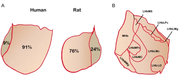
Figure 4 Comparative analysis of human and rat LHb and LHb subnuclear organization
(A). The habenular complex surface is increased in humans compared to rats as well as the relative contribution of the LHb to the total habenular size. In humans the LHb represents ~91% of the total habenular surface vs 9% MHb, while in the rat the LHb to MHb ratio is ~ 76% vs 24% respectively (adapted from Díaz et al., 2011). (B). Subnuclear organization of the LHb. The medial LHb comprises the anterior (LHbMA, not shown), superior (LHbMS), parvocellular (LHbMPc), central (LHbMC) and marginal (LHbMMg) subnuclei. The lateral LHb comprises the parvocellular (LHbLPc), magnocellular (LHbMc), oval (LHbLO), marginal (LHbLMg) and basal (LHbLB, not shown) subnuclei. This distinction has been made based on different cell organization and distinct expression patterns of cellular markers such as the GABAB receptor, the Kir3.2 potassium channel and neurofilament. (adapted from Geisler et al., 2003).
Inputs to the LHb
The habenular complex is positioned at the highway of a major information stream in the brain, connecting the limbic forebrain and basal ganglia regions with midbrain neuromodulatory systems. It receives most of its inputs through a fiber bundle called the stria medullaris and sends its projections to monoaminergic centers through the fasciculus retroflexus (Fig3B), which altogether form the diencephalic conduction system
(Sutherland, 1982). Although the MHb and LHb receive their inputs and send their
outputs using the same fiber tracts, they are differently innervated and target different brain nuclei. A potential connectivity between the MHb and LHb has been debated, but has never been anatomically or functionally proven.
The MHb receives inputs from septal and diagonal band of Broca (DBB) areas and sends in turn its axons to the interpeduncular nucleus of the midbrain (Herkenham and
Nauta, 1979, 1977). In contrast, the LHb receives projections from the output structure
MHb LHbMS LHbLPc LHbLMg LHbMPc LH bM Mg LHbMC LHbLMc LHb LO 91% 9% 76% 24% Human Rat A B
Page | 23 of the basal ganglia - the entopeduncular nucleus (EPN; homologue of the globus pallidus interna in primates and humans) and from limbic forebrain regions including the lateral hypothalamus (LH), lateral preoptic area (LPO), ventral pallidum (VP), BNST, DBB, lateral septum (LS) as well as feedback projections from the laterodorsal tegmentum (LDT), the VTA and the dorsal and median raphe nuclei (DRN and MRN) (Fig5A) (Herkenham & Nauta 1977; Nagy et al. 1978; Parent et al. 1981; Kowski et al. 2008; Li et al. 2011; Tripathi et al. 2013; Lammel et al. 2012; Swanson 1982; Aghajanian
& Wang 1977). A projection from the PFC has also been described (Kim and Lee, 2012;
Li et al., 2011; Warden et al., 2012). Some of the inputs to the LHb represent a
topographical organization potentially important for LHb functions (Fig5B).
3V cc 4V PFC VTA D3V VP LH LDTg LPO BNST sm cc 4V Raphe fr DBB LS EPN - +/-+/- glutamate GABA glutamate&GABA unknown + - +/-LHb MHb EPN terminals VTA terminals LH terminals ++ + ++ A B
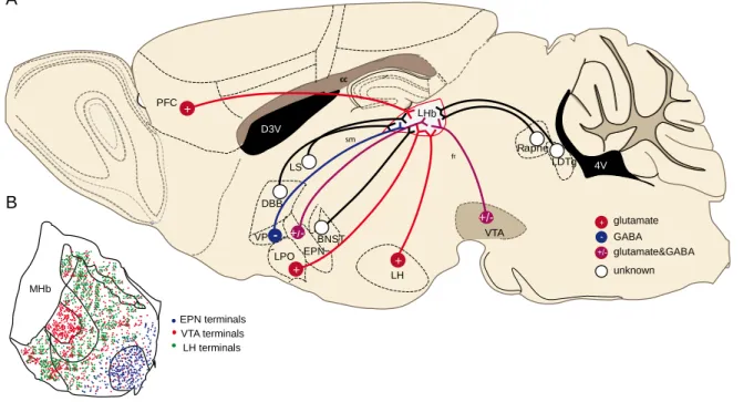
Figure 5Afferents and their territorial distribution within the LHb
(A).The LHb receives glutamatergic (PFC, LPO, LH), GABAergic (VP) and mixed inputs (EPN, VTA and potentially LH). Other inputs and their neurotransmitters are not yet established (DBB, LS, BNST, LDT and raphe). (B). EPN terminals target the lateral LHb (LHbLO), while this subregion is devoid of LH and LPO inputs (Kowski et al., 2008; Poller et al., 2013; Shabel et al., 2012). LH axons are more concentrated in the medial aspect of the LHb (with the exception of the LHbMPc) (Poller et al., 2013). Other input terminals, such as the VTA ones are widely distributed throughout the LHb(Hnasko et al., 2012; Root et al., 2014b). sm: stria medullaris; fr: fasiculust retroflexus, cc: corpus callosum; D3V: dorsal 3th ventricle and 4V: 4th ventricle
Page | 24 Output connectivity of the LHb
LHb neurons are almost exclusively glutamatergic and long-range projecting (Kim and
Chang, 2005; Li et al., 2011; Weiss and Veh, 2011). Although some studies have
suggested the existence of local GABAergic neurons in the medial LHb, functional evidence about a potential LHb microcircuit is still lacking (Li et al., 2011; Zhang et al.,
2016). Anatomical and physiological studies indicate that LHb neurons send their axons
mainly to GABAergic neurons in the midbrain. Indeed, tracing experiments show that neurons located mainly within the lateral LHb project to the GABAergic rostro-medial tegmental nucleus (RMTg; (Balcita-Pedicino et al., 2011; Gonçalves et al., 2012; Jhou et al., 2009b; Meye et al., 2016; Sego et al., 2014), also called tail VTA (Kaufling et al., 2009; Perrotti et al., 2005). In contrast, LHb neurons originating from the medial aspect send their axons preferentially to monoaminergic nuclei (Gonçalves et al., 2012; Sego et
al., 2014), where they form functional synapses with local GABAergic neurons (Lammel
et al., 2012; Weissbourd et al., 2014) as well as with dopamine neurons of the VTA
(Balcita-Pedicino et al., 2011; Lammel et al., 2012) or serotonin neurons in the caudal
dorsal raphe (Dorocic et al., 2014; Sego et al., 2014) (Fig 6B). Further, it has been shown that individual neurons project either to VTA or to raphe nuclei without collateralizing, suggesting that single LHb neurons have distinct output targets (Bernard
and Veh, 2012; Gonçalves et al., 2012; Li et al., 2011; Maroteaux and Mameli, 2012). In
addition, the LHb has been recently shown to send axons to GABA neurons within the LDT and to orexin- and melanin concentrating hormone (MCH)-expressing neurons in the LH (González et al., 2016; Lammel et al., 2012; Yang et al., 2016). While the former has also been functionally proven, it remains to be confirmed whether LHb neurons establish functional synaptic connections in the LH (Fig6A).
Given that LHb neurons are of glutamatergic phenotype and that they project to a vast majority of GABA neurons in the midbrain it is plausible that they may disynaptically inhibit dopamine or serotonin neurons potentially providing aversive signals to the midbrain.
Page | 25 Function of the LHb in reward and aversion encoding
Seminal studies in rhesus monkeys have suggested a role of the LHb in encoding aversive stimuli and in controlling dopaminergic neurons activity, crucially contributing to our understanding of motivational processing. When monkeys were exposed to unexpected aversive air puff, a cue predicting it (after conditioning sessions) or an omission of expected reward, the majority of LHb neurons increased phasically their firing activity. On the contrary, when an unexpected rewarding stimulus or reward-predictive cues were presented to the animals or alternatively an expected punishment was omitted, LHb neurons firing decreased or remained unchanged respectively (Fig7D)
(Matsumoto and Hikosaka, 2009a, 2007). Moreover, there was a linear relationship
between the objective value of the stimulus and the conditioned stimulus (cue) response for negative outcomes, but not for positive ones (Fig7A). This has led to the idea that
3V cc 4V 3V VTA RMTg -DA sm 4V 3V Raphe LH MCH Orx LDTg -5HT LHb + -+ DA 5HT MCH Orx GABA glutamate dopamine serotonin MCH+ Orexin+ RMTg-projecting monoamine-projecting MHb sm D3V A B
Figure 6 LHb efferents and territorial distribution of their cell bodies throughout the LHb
(A).The LHb sends glutamatergic projections mainly to GABA neurons of the midbrain (RMTg, VTA, Raphe and LDT), but also to dopamine and serotonin neurons in the VTA and dorsal raphe. The LHb also projects to MCH and Orexin-expressing neurons in the LH.(B). LHb neurons targeting the RMTg are mainly located in the lateral LHb (Kowski et al., 2008; Poller et al., 2013; Shabel et al., 2012), while those targeting the VTA and dorsal raphe are located in the medial LHb (Poller et al., 2013). sm: stria medullaris; fr: fasiculust retroflexus, cc: corpus callosum; 3V and 4V: 3th and 4th ventricle
Page | 26 LHb neuronal activity discriminates the valence of stimuli and preferentially represents negative-valued events with respect to dopamine neurons, the activation of which preferentially represents reward (Matsumoto and Hikosaka, 2009b; Mirenowicz and
Schultz, 1996). It is important to note that when a reward was fully predictable (100%
reward predictive cue) LHb neurons did not respond to the unconditioned stimulus (reward delivery), whereas if the reward was not fully predictable (50% reward occurrence in previous sessions) LHb neurons were inhibited (Fig7B). The magnitude of the inhibitory response increased with the reward unpredictability. Similarly, in trials where an aversive stimulus was 100% predictable, LHb neurons responded with excitation to the unconditioned stimulus (air puff) and the response was reduced compared to trials where the aversive stimulus was not fully predictable (Fig7C). These data suggest that when an aversive or rewarding stimulus is predictable the response of LHb neurons during the stimulus presentation (excitation or inhibition) is decreased in magnitude or absent compared to when it occurs in an unpredictable manner
(Matsumoto and Hikosaka, 2009a) (Fig7D). Altogether this evidence strongly indicates a
role of LHb neurons in negative-reward prediction error. This behavior of LHb neurons appears opposite to the behavior of dopamine neurons in response to rewarding and aversive stimuli and their predictive cues (Fig7E). As discussed in the previous chapters, unpredicted rewarding stimuli or reward-predictive cues lead to rapid and brief bursts of activity in dopamine neurons. When the reward occurrence becomes predictable dopamine neurons no longer fire, whereas an omission of expected rewards inhibits them (Schultz et al., 1997). In contrast, aversive stimuli such as noxious foot pinch or foot shock mainly decrease dopamine neurons firing rates and bursting activity (Ungless
et al., 2004), although few neurons respond with phasic excitation (Brischoux et al.,
2009). This opposite processing of reward and aversion in the LHb and VTA led to the
idea that LHb neurons provide a negative-reward predictive signal to dopamine neurons
(Bromberg-Martin et al., 2010; Keiflin and Janak, 2015; Schultz, 2007b). Importantly, the activity of LHb neurons is not only opposed, but it also precedes the activity of SNc and VTA dopamine neurons in non-rewarded trials, indicating that LHb neurons exert inhibitory drive onto dopamine neurons (Matsumoto and Hikosaka, 2007). This is in line with an inhibitory effect of LHb electrical stimulation onto dopamine neurons activity
Page | 27
(Christoph et al., 1986; Ji and Shepard, 2007). Moreover, blocking excitatory
transmission within the LHb leads to transient increase of dopamine release in the NAc, dorsal striatum and PFC, suggesting that LHb activity exerts a tonic inhibition on dopaminergic transmission (Lecourtier et al., 2008). However, in a recent study LHb lesions prevented a decrease in dopamine neurons activity only when a reward was omitted but not when an aversive stimulus was delivered, suggesting that LHb neurons are not the only source of negative reward prediction signal to dopamine neurons and that instead the LHb codes rather for disappointment (Tian and Uchida, 2015). A limitation, nonetheless, of this study is the use of electrolytic lesion of the LHb, where circuit adaptations might have occurred as a compensatory mechanism. In this regard, the use of optogenetic or chemogenetic silencing of the LHb activity could be more informative to determine its contribution to negative reward prediction error when controlling midbrain structures.
In humans, functional magnetic resonance imaging (fMRI) showed increased LHb activity when healthy volunteers received negative feedback after failing to perform a task or when they were exposed to aversive stimuli (Hennigan et al., 2015; Ullsperger
and von Cramon, 2003). Furthermore, similarly to monkeys the LHb of humans
responded preferentially to cues signaling negative outcomes (Lawson et al., 2014). Altogether data in human and non-human primates suggest an important role of the LHb in predicting negative outcomes necessary for motivated behaviors.
Page | 28 NO CUE REWARD REWARD PREDICTIVE CUE REWARD
PRE-LEARNING: UNEXPECTED OUTCOME POST-LEARNING: REWARD PREDICTION ERROR
Schultz et al., 1997
NO CUE AIR PUFF AIR PUFF
NO REWARD
NO AIR PUFF
NO CUE REWARD REWARD
REWARD OMISSION PREDICTIVE CUE
NO REWARD
Matsumoto and Hikosaka, 2007; 2009
DA neurons activity LHb neurons
activity
PRE-LEARNING: UNEXPECTED OUTCOME POST-LEARNING: REWARD PREDICTION ERROR
REWARD PREDICTIVE CUE REWARD PREDICTIVE CUE AIR PUFF PREDICTIVE CUE AIR PUFF OMISSION PREDICTIVE CUE D E
Figure 7LHb function in aversion encoding and negative prediction error
(A). Linear relationship between averaged conditioned stimulus response (cue) and the objective value for negative outcomes and non-linear relationship for rewarding outcomes. (B). Relationship between the averaged LHb unconditioned stimulus (reward) response and the probability for reward or reward omission (positive prediction error). (C). Relationship between averaged LHb unconditioned stimulus (air puff) response and the probability for airpuff or airpuff omission (negative prediction error). (From Matsumoto and Hikosaka, 2009a). (D). LHb responses to unpredictable (non-learned) rewarding or aversive stimuli. After learning, LHb neurons responses shift to the cues predicting 100% reward or 100% punishment. However, LHb neurons respond also to the unconditioned stimulus when it is aversive but with smaller magnitude. When a cue predicts omission of reward LHb neurons increase firing and when a cue predicts omission of air puff LHb neurons decrease firing. (E). Dopamine neurons show opposite responses to unpredictable rewards and to cues predicting rewards (increased firing) compared to LHb neurons. When a cue predicts reward dopamine neurons fire, but if the reward is omitted dopamine neurons are phasically inhibited.
Page | 29 Role of LHb output for aversion processing
Following these studies an important question arose: given the excitatory nature of LHb neurons, how does their activity drive inhibition onto dopamine neurons? LHb neurons projections to GABA neurons of the midbrain might mediate LHb inhibitory control onto dopamine neurons. Indeed, an important link between the LHb and dopamine neurons function in reward/aversion encoding is the GABAergic RMTg. Anatomical and ultrastructural studies indicate that the LHb sends a major glutamatergic efferent to the RMTg. RMTg neurons in turn project to dopamine neurons within the VTA and SNc, although some synaptic contacts with non-dopaminergic cells have also been reported
(Balcita-Pedicino et al., 2011; Brinschwitz et al., 2010; Jhou et al., 2009b; Kaufling et al.,
2009). In line with this, retrogradely labeled RMTg neurons projecting to the VTA were
found in close apposition to anterogradely labelled LHb axon terminals (Gonçalves et al.,
2012). Importantly, RMTg neurons encode rewarding or aversive stimuli similarly to the
LHb and opposite to dopamine neurons, by phasically decreasing (reward) or increasing (aversion) their firing (Fig8) (Jhou et al., 2009a; Matsumoto and Hikosaka, 2007).
Functional data in anesthetized rats and in behaving monkeys indicate that RMTg neurons receiving LHb input increase phasically their firing in response to aversive stimuli and inhibit dopamine neurons, further supporting a role of RMTg in relaying LHb signals to dopamine neurons (Hong et al., 2011; Lecca et al., 2012; Matsui and Williams,
2011). In line with a role of LHb output to midbrain GABA neurons in driving negative
teaching signals, light-activation of ChR2-expressing LHb terminals in the RMTg of mice produced real-time and conditioned-place avoidance behaviors (Stamatakis and Stuber,
2012). Moreover stimulation of this pathway produced negative reinforcement since
animals nose-poked to terminate stimulation, while it disrupted positive reinforcement as animals nose-poked less to obtain a reward, suggesting that LHb-to-RMTg pathway activity provides a punishing signal (Fig8) (Stamatakis and Stuber, 2012).
Although LHb neurons strongly project to the RMTg, they also send, direct projections to GABA and dopamine neurons in the VTA. Moreover there is a topographic organization of this connectivity since the VTA projecting neurons are found mainly in the medial division of the LHb and send their axons to the ventral medio-posterior aspects of the
Page | 30
VTA (Gonçalves et al. 2012; Omelchenko et al. 2009; Swanson 1982; Phillipson &
Pycock 1982; Skagerberg et al. 1984). Importantly, light activation of ChR2-expressing
LHb terminals in the VTA of mice evoked responses in half of non-dopaminergic cells (putative GABA neurons), whereas only a very small percentage of dopamine neurons responded to the stimulation (Stamatakis and Stuber, 2012). Altogether these data indicate that the LHb preferentially connects to GABA neurons in the midbrain, likewise exerting an inhibitory control on dopamine neurons (Ji and Shepard, 2007; Matsumoto
and Hikosaka, 2007). This is in line with the results obtained in monkeys showing that
excitation of LHb neurons in response to aversive events is followed by inhibition of dopamine neurons (Matsumoto and Hikosaka, 2007). In agreement with this, light activation of ChR2-expressing GABA neurons in the VTA inhibits dopamine neurons firing, reduces reward-related behaviors (sucrose licking) and induces conditioned place avoidance (Fig2B), similarly to activation of the LHb-RMTg pathway (Fig8) (Stamatakis
and Stuber, 2012; Tan et al., 2012; van Zessen et al., 2012). Moreover, inhibition of
dopamine neurons expressing NpHR led to avoidance behaviors, further supporting that midbrain GABA-mediated inhibition of dopamine neurons is instrumental for the expression of aversive behaviors (Fig2B) (Tan et al., 2012).
The LHb also targets dopamine neurons in the ventromedial and posterior portion of the VTA, which preferentially send axons to the medial PFC and undergo aversive experience-dependent plasticity (Lammel et al., 2012, 2011). Furthermore, optogenetic activation of retrogradely labeled VTA-projecting LHb neurons produces conditioned place avoidance (Fig8) (Lammel et al., 2012).
In humans, together with an increased LHb activity a higher activation of the VTA, SNc and PFC regions were also detected after aversive stimuli and this was associated with increased functional connectivity between the habenula and VTA as well as VTA and
PFC (Hennigan et al., 2015), suggesting that both in rodents and in humans the
habenulo-meso-cortico-limbic circuit is involved in aversion processing.
Collectively, all these anatomical and functional data strongly suggest that LHb output conveys aversive signals via different anatomical connections and ultimately underlies aspects of aversive behaviors. The stimulation of each of these distinct LHb pathways to the midbrain is sufficient, but it remains still unclear whether it is necessary for aversive
Page | 31 behaviors. Other important questions arising following these studies are whether these pathways are simultaneously active or occur at different instances and which particular aspects of aversive behaviors do they encode.
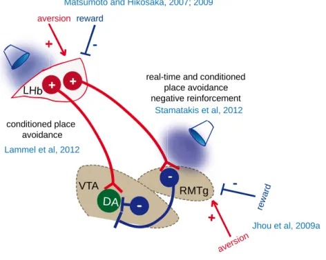
Figure 8 Contribution of LHb output to motivated behaviors
LHb neurons are glutamatergic and are connect predominantly to GABA neurons of the midbrain (Meye et al., 2016). ChR2-driven stimulation of LHb terminals within the RMTg induces real-time and conditioned place avoidance, negatively reinforces behavior and disrupts positive reinforcement. RMTg neurons inhibiting dopamine neurons are excited by aversive stimuli and are inhibited by rewards similarly to LHb neurons. Some VTA-projecting LHb neurons target predominantly dopamine neurons projecting to the PFC. Stimulation of LHb neurons projecting to the VTA induces conditioned place avoidance.
LHb VTA RMTg
-DA + aversion
+
-reward
Matsumoto and Hikosaka, 2007; 2009
+
-rew ard
Jhou et al, 2009a avers
ion real-time and conditioned
place avoidance negative reinforcement Stamatakis et al, 2012 conditioned place avoidance Lammel et al, 2012 +
Page | 32 Role of inputs to the LHb for aversion processing
Data collected so far suggest that the LHb lacks local GABAergic control (Li et al., 2011). Instead its activity is largely shaped by multiple long-range synaptic inputs (Root et al., 2014b; Shabel et al., 2014, 2012; Stamatakis et al., 2016). Indeed, many forebrain and midbrain nuclei send glutamatergic and GABAergic terminals directly to the LHb contributing to its function in reward and aversion. Recent studies have proposed that some inputs to the LHb, including the EPN, VTA and potentially the LH are capable to co-release GABA and glutamate from the same synaptic terminal and to form functional postsynaptic connections with LHb neurons (Root et al., 2014b; Shabel et al., 2014;
Stamatakis et al., 2016). Activating ChR2 specifically in glutamatergic neurons in a
Vglut2-Cre-dependent manner (Vglut2: vesicular glutamate transporter 2) in any of these three inputs led to both AMPA receptor- and GABAA receptor-mediated postsynaptic
currents. Similarly, a Vgat-Cre- or GAD67-Cre-dependent activation of ChR2 (Vgat: vesicular GABA transporter and GAD67: glutamate decarboxylase, the enzyme synthetizing GABA from glutamate) in GABAergic neurons of the VTA or EPN respectively triggered both excitatory and inhibitory postsynaptic currents (EPSCs and IPSCs respectively) in the LHb. A fine ultrastructural analysis in mouse and rat has shown that the majority of VTA-to-LHb neurons co-release glutamate and GABA from the same synaptic terminal forming functional asymmetrical and symmetrical synapses
(Root et al., 2014b). Activation of the EPN and LH inputs drives LHb neurons to fire,
while the predominant effect of VTA-to-LHb pathway stimulation in vivo was inhibitory (Fig9) (Root et al., 2014b; Shabel et al., 2014; Stamatakis et al., 2016). Interestingly, VTA-to-LHb optostimulation resulted in inhibition followed by rebound excitation in some LHb neurons, raising the possibility that the co-release of glutamate and GABA from the same synapse may have a role to temporally control LHb neuronal firing (Root et al.,
2014b). This hypothesis is supported by previous evidence showing that phasic
electrical stimulation of the midbrain decreases the firing rate of a subset of LHb neurons, whereas a tetanic stimulation tends to increase it (Shen et al., 2012).
Importantly, optogenetic activation of specific inputs to the LHb can drive distinct motivational states. Indeed, stimulation of the EPN-to-LHb synapses, which has an
Page | 33 overall excitatory effect on LHb firing, drives real-time place avoidance behavior (Fig9), consistent with the idea that an increased LHb activity is required for the expression of negative states (Shabel et al., 2012). EPN inputs impinge onto neurons of the lateral division of the LHb, which project mainly to GABA neurons of the midbrain, further supporting the role of this pathway in aversion encoding and avoidance behaviors (Fig5B and Fig6B) (Gonçalves et al., 2012; Lecca et al., 2014; Proulx et al., 2014;
Shabel et al., 2012, Meye 2016). Interestingly, although the EPN participates mainly in
the control of body movements (DeLong, 1971), some EPN neurons encode negative-reward prediction error similarly to the LHb, by phasic excitation upon unexpected aversive stimuli or reward omissions and phasic inhibition by unexpected rewards. The response of these negative-reward encoding neurons in the EPN precedes that of LHb neurons, suggesting that they may excite LHb neurons during aversive stimuli or reward omission (Fig9) (Hong and Hikosaka, 2008).
Optostimulation of the VTA-to-LHb pathway also produced avoidance of the light-associated chamber after conditioning, suggesting that this input may be involved in avoidance learning (Fig9) (Root et al., 2014a). Nevertheless, this effect seems puzzling considering that the VTA-to-LHb input inhibits the majority of LHb neurons, which would presumably lead to disinhibition of downstream dopamine neurons and would drive rewarding states. A possible explanation to this issue may reside in that in this study a continuous light activation has been employed when the animal was in the light-associated chamber, which may result in progressive increase of LHb neuronal activity and therefore to increased inhibitory drive onto downstream dopamine targets (Shen et
al., 2012). Whether the balance of excitation and inhibition at co-releasing synapses is
frequency or time-dependent remains however an open question. Alternatively, VTA inputs may target LHb neurons projecting directly onto dopamine neurons, therefore inhibiting them and contributing to avoidance behaviors.
Another important question arising is whether VTA neurons projecting to the LHb release dopamine and what the functional consequences would be on LHb neuronal activity. Indeed, early anatomical and tracing studies have described afferent projection from the ventromedial portion of the VTA to the medial aspect of the LHb (Swanson
Page | 34 positive fibers (TH, the rate limiting enzyme for dopamine synthesis) are found (Aizawa et al., 2012; Geisler et al., 2003; Gruber et al., 2007). Some more recent studies instead report that VTA fibers are widely distributed across the LHb and are predominantly TH negative and Vglut2 positive (Hnasko et al., 2012; Root et al., 2014b). Despite this evidence, characterization of the expression profile of LHb-projecting VTA neurons showed that a non-negligible population expresses TH in addition to glutamate and GABA markers (Root et al., 2014b), suggesting that these neurons are potentially capable to release dopamine. Cre-dependent ChR2 expression in the VTA of TH-Cre mice led to detection of ChR2+ terminals in the LHb, but a weak expression of TH. Optostimulation of these VTATH+/ChR2+ fibers in the LHb failed to release detectable dopamine levels as assessed by fast-scan voltametry. Moreover, these LHb-projecting VTATH+/CHR2+ neurons showed a reduced expression of Vmat2 (vesicular monoamine transporter 2), D2 dopamine receptor (D2R) and DAT (dopamine transporter) and exhibited different electrophysiological properties compared to classical dopamine releasing VTA-to-NAc neurons. Optogenetic activation of VTATH+/ChR2+ axons in the LHb evoked GABAergic postsynaptic responses and decreased firing activity of LHb and RMTg neurons, whereas the same stimulation increased VTA activity. Moreover, stimulation of this pathway led to conditioned place preference as well as to positive reinforcement (Fig9) (Stamatakis et al., 2013). This is in discrepancy with the aversive effect of VTA-to-LHb stimulation reported by Root et al., 2014a. This contradiction may arise from the different genetic approaches employed and the potential targeting of different LHb neuronal subpopulations which may in turn project to distinct downstream targets. Notably, it has also been shown recently that the TH-Cre line can present some ectopic expression in non-TH-expressing neurons, raising the issue of choosing appropriate Cre-driver lines to study complex circuits (Lammel et al., 2015; Stuber et al., 2015).
Other inputs to the LHb have also been implicated in different aversive behaviors. Anatomical studies indicate that predominantly glutamatergic LH inputs target mainly VTA-projecting LHb neurons in the medial division of the LHb (Fig5 and Fig6)
(Gonçalves et al., 2012; Poller et al., 2013). Excitatory, Vglut2-expressing LH neurons
Page | 35 Indeed, optical stimulation of LHVglut2-to-LHb pathway increases LHb neurons activity and induces real-time place avoidance, whereas NpHR-driven inhibition of this pathway leads to real-time preference (Fig9) (Stamatakis et al., 2016). This is in line with the necessity of increased LHb output for the expression of aversive behaviors and a decreased LHb output for reward-related behaviors (Lammel et al., 2012; Stamatakis and Stuber, 2012).
Another glutamatergic projection described to date arises from the mPFC (Kim and Lee, 2012; Li et al., 2011; Warden et al., 2012). Indeed, a study in rats shows that activating glutamatergic mPFC inputs expressing ChR2 in the LHb leads to behavioral despair as shown by decreased mobility in the forced-swim test (FST), a paradigm widely use to screen for depressive-like behaviors (Fig9) (Warden et al., 2012). However, the authors did not further investigate the contribution of this input for LHb neurons activity, leaving it open for further investigation.
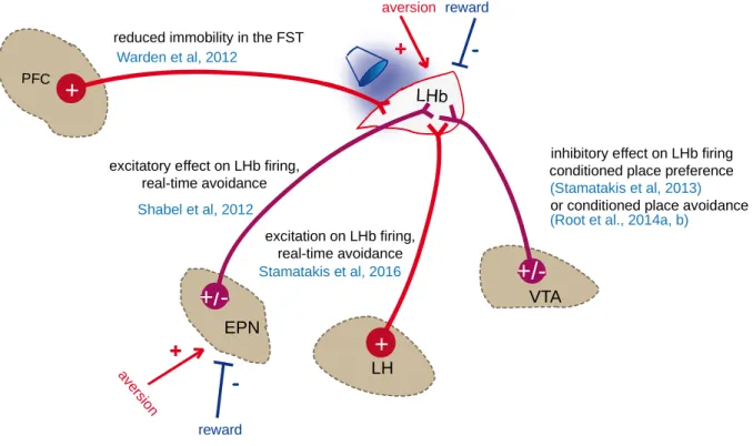
Altogether, these studies demonstrate that distinct excitatory or inhibitory pathways can drive LHb neuronal activity ultimately contributing to the encoding and expression of motivated behaviors. Importantly, excitatory inputs arising from the EPN, LH, VTA and mPFC are capable to drive avoidance behaviors, whereas inhibitory inputs such as the GABAergic VTA-to-LHb component can inhibit neuronal activity and produce preference behaviors (Fig9). A caveat however of these studies is the use of non-specific stimulation patterns to activate exogenously-expressed opsins, which may overcome other physiological mechanisms relevant for these behaviors. It is also interesting to address whether different frequency patterns lead to different glutamate/GABA release ratios from the co-releasing synapses. Another limitation of the so far described approaches is that in the majority of the cases, it has been shown sufficiency of a pathway in inducing specific behaviors, but not its necessity. Nevertheless these studies demonstrate the importance of investigating specific circuits within the LHb to the level of single cell-types for different aspects of motivated behaviors. Furthermore, it is of major interest to closely investigate whether and how synaptic plasticity occurs in the LHb following rewarding or aversive experience and what is its contribution on LHb neuronal output both in physiological and pathological conditions. This is particularly important in light of the role of LHb neurons activity in aversive behaviors (Lecca et al.,
Page | 36
2014; Proulx et al., 2014). A thorough examination of specific plasticity mechanisms
could provide molecular targets to reverse maladaptations in disorders related to LHb dysfunction including addiction and depression.
VTA LH EPN
+/-LHb+
excitatory effect on LHb firing,real-time avoidance
inhibitory effect on LHb firing conditioned place preference or conditioned place avoidance
av ersio n
+
-reward aversion
+
-reward
Hong and Hikosaka, 2008
Matsumoto and Hikosaka, 2007; 2009
Shabel et al, 2012
(Stamatakis et al, 2013) (Root et al., 2014a, b)
PFC
+
excitation on LHb firing, real-time avoidance Stamatakis et al, 2016 reduced immobility in the FST
Warden et al, 2012
Figure 9Contribution of inputs to the LHb to different motivational states
EPN neurons increase their firing in response to aversive stimuli and decrease their firing in response to rewards, similarly to LHb neurons. EPN-to-LHb and LH-to-LHb optogenetic stimulation increases LHb firing and drives real-time avoidance. Stimulation of the PFC-to-LHb input drives behavioral despair. Vglut2-expressing VTA terminals stimulation in the LHb predominantly inhibits LHb neurons firing and drive conditioned place avoidance, while TH-expressing VTA terminals stimulation drives conditioned place preference.