RESEARCH OUTPUTS / RÉSULTATS DE RECHERCHE
Author(s) - Auteur(s) :
Publication date - Date de publication :
Permanent link - Permalien :
Rights / License - Licence de droit d’auteur :
Institutional Repository - Research Portal
Dépôt Institutionnel - Portail de la Recherche
researchportal.unamur.be
University of Namur
Ecarin based coagulation testing
Gosselin, Robert C; Douxfils, Jonathan
Published in:
European Medical Journal Hematology DOI:
10.1002/ajh.25852 Publication date: 2020
Document Version Peer reviewed version
Link to publication
Citation for pulished version (HARVARD):
Gosselin, RC & Douxfils, J 2020, 'Ecarin based coagulation testing', European Medical Journal Hematology, vol. 95, no. 7, pp. 863-869. https://doi.org/10.1002/ajh.25852
Ecarin based coagulation testing
Robert C Gosselin. University of California, Davis Health System, Thrombosis and Hemostasis Center, Sacramento, CA
Jonathan Douxfils. Qualiblood sa, Namur, Belgium Running title: Ecarin testing
Keywords: Ecarin, ecarin clotting time; ecaring chromogenic assay; dabigatran; Direct thrombin inhibitors
Correspondance to: Robert C Gosselin, University of California, Davis Health System, Thrombosis and Hemostasis Center, Sacramento, CA email:[email protected]
Abstract
Ecarin is derived from venom of Echis carinatus and will activate prothrombin into
meizothrombin which will then cleave fibrinogen to result in clot formation. Ecarin based testing has been described for decades, but these assays were typically restricted to reference or speciality coagulation laboratories. This test was initially described for the assessment of direct thrombin inhibitors (e.g bivalirudin lepirudin, or argatroban) and was not affected by heparins or heparinoids. Ecarin based assays were rarely used for anticoagulation monitoring until the emergence of the direct oral thrombin inhibitor dabigatran etexilate in 2010. As this test was mentioned in the prescribing information for dabigatran etexilate, there was increased interest for use by clinical laboratories as the preferred method for assessing the anticoagulant effect of this drug. The purpose of this document is to review the current status of ecarin based assays for assessing dabigatran, with the understanding that these methods can also be
exploited for determining the anticoagulation effect of parenteral direct thrombin inhibitors such as argatroban and bivalirudin.
Introduction
The first published report on the effect of envenomation from the saw-scaled viper, Echis
carinatus demonstrated significant bleeding at wound site, petechiae, conjunctival hemorrhage
and eventual death.1 The early stages of assessing Echis carinatus effect was in experimental observations after toxin exposure in white rats where the thrombin time prolongation was more pronounced than the Quick prothrombin time method, and thromboelastographic changes noted in dogs, 24 hours after toxin exposure. The authors presumed the effect was targeting either “the conversion to fibrinogen or fibrinolysis”, with the author suggesting ecarin primary mechanism being the latter effect.2 Later, it was demonstrated that Echis carinatus venom converts prothrombin to thrombin, but the venom itself lacking thrombin-like activity.3,4 The resultant thrombin produced from Echis carinatus toxin (coined “ecarin”), was thought to
be of different properties than that achieved through normal physiologic processes and
described as ecarin-thrombin or E-thrombin, a metalloprotease.5 In a series of experiments, the authors demonstrated the effect of ecarin was at two cleavage sites: one at the Arg-Ser bond, which is split during the release of the NH2-terminal portion of the molecule, the second being the Arg-Ile bond, which links the A-and B chain thrombin precursors.5 Later it was
demonstrated that there was no Arg-Ser bond cleavage6 but that ecarin creates, rather than thrombin, meizothrombin which is a thrombin analogue consisting of α-thrombin complexed with prothrombin fragment 2, which is generated from cleavage at the Arg323-Ile324 site and has increased esterase activity.7 In contrast, the physiological activation of prothrombin by factor Xa occurs at cleavage site A (Arg273-Thr274, generating prothrombin fragment 1.2) in addition to cleavage site B (Arg322-Ile323).8 Note that the prothrombin fragment 2 generated by
meizothrombin and prothrombin fragment 1.2 generated by factor Xa cleavage are not the same peptides. [Figure 1]
As ecarin is an activator of prothrombin, the potential for clinical use was described in various settings including vitamin K deficiency9 and lupus anticoagulant testing10, using either
chromogenic or clot-based methods. However, the utilization of ecarin based assays for
anticoagulant assessment were fairly limited to research or reference laboratories, but the ECT is commonly used for lupus anticoagulant testing in certain geographical regions.11,12 In 1998, the FDA approved lepirudin (Refludan®, Bayer Healthcare), a recombinant hirudin, for
treatment of heparin induced thrombocytopenia13, representing the first parenteral direct thrombin inhibitor (DTI) inhibiting both thrombin and meizothrombin.14,15 Shortly thereafter, other parenteral DTIs were approved, including bivalirudin and argatroban.14 The use of ecarin based testing now became of more interest as a local based assay, as DTIs became the standard of care for anticoagulating patients with heparin-induced thrombocytopenia.16 Ximelagatran,
an oral direct oral thrombin inhibitor was also reported to be suitably measured by ecarin based assays17, but this drug was removed from the market due to hepatotoxicity.18
The real explosion for ecarin based testing emerged with the approval of dabigatran etexilate (Pradaxa®, Boehringer Ingelheim), a direct oral thrombin inhibitor, approved for use in the US in 2010 as an alternative to warfarin for stroke prevention in patients with nonvalvular atrial fibrillation 19 Since 2010, additional FDA approved indications for dabigatran include treatment of deep venous thrombosis (DVT) and pulmonary embolism (PE) in patients who have been treated with a parenteral anticoagulant for 5-10 days; risk reduction for recurrence of DVT and PE in patients who have been previously treated; and prophylaxis of DVT and PE in patients who have undergone hip replacement surgery20 The timing of approval was different in Europe where the prophylaxis of DVT and PE in patients who have undergone hip replacement surgery was approved in 2008, and in 2014 approval was granted for the treatment of deep venous thrombosis (DVT) and pulmonary embolism (PE) in patients who have been treated with a parenteral anticoagulant for 5-10 days, or for risk reduction for recurrence of DVT and PE in patients who have been previously treated. The current dosing indications are for adult patients and predicated on indication and renal function.18,21 In a study evaluating the prescribing practice in the US, Ziakas and colleagues demonstrated that in 2015, DOACs consisted of ~30% of the oral anticoagulants prescribed for medicare patients.22 Dabigatran constituted approximately 16 – 19% of DOAC prescriptions, mostly by cardiologists and internal medicine physicians.22
While dabigatran is not monitored with the same frequency as warfarin, the need to measure dabigatran may be required for assessing bleeding patients, trauma, or patient requiring immediate intervention (thrombolysis in stroke, emergency surgery, neuraxial
anesthesia).18,21,23 For dabigatran treated patients, ecarin based assays demonstrated a more linear response to drug concentration than the activated partial thromboplastin time (APTT)
with less inter-individual variations.24-28 In contrast, the traditional thrombin time (TT) is too sensitive for assessing dabigatran, with concentrations as low as 25 ng/mL resulting in a 2 to 3-fold increase of the baseline TT, making this test more suitable for excluding drug presence instead of quantifying.26,27 Conversely, the Activated Clotting Time (ACT) has been shown to be less sensitive to dabigatran, with a misprediction as high as 40%.28 As ecarin methods are not affected by heparin, this method would even be suitable for monitoring transition therapy to/from heparins to dabigatran.26
Ecarin tests are based on two different principles, i.e. clot-based and chromogenic-based methods.24 The purpose of this document is to review the testing principles of ecarin based assays as well as their application in the monitoring of anticoagulation. We will only focus on this application as these tests are now primarily used for the assessment of oral or parenteral direct thrombin inhibitors.
The ecarin clot-based assay, also named the ecarin clotting time
Despite its description in the literature for decades, there is to date no standardization of the ecarin clotting time (ECT).29 As previously described, ecarin will activate prothrombin (factor II) creating meizothrombin, which will then convert fibrinogen to fibrin, the principle of all clot-based testing. [Figure 2A] Despite the simple test principle, the variations of testing include: sample dilution (yes or no), degree of dilution (ratio of sample to diluent), diluent type (saline or buffer), whether preincubation of sample or reagents at 37oC, and ecarin concentration. 16,29-32, resulting in different sensitivity to dabigatran exposure and lower limits of
quantitation.[Figure 3] Due to its simplicity, the ECT can be easily automated to modern coagulation analyzers, but necessary performance verification is required, especially to assure absence of pipet/pipetting carryover of ecarin reagent.
Dabigatran etexilate prescribing information indicates that “At recommended therapeutic
doses, dabigatran etexilate prolongs the coagulation markers such as aPTT, ECT, and TT.”20 Additionally, the prescribing information indicates, “In the RE-LY trial, the median (10th to 90th
percentile) trough ECT in patients receiving the 150 mg dose was 63 (44 to 103) seconds.”20 The readers should be informed that the RE-LY trial was assessing the use of dabigatran in
nonvalvular atrial fibrillation (NVAF). The dose for that indication may differ than dose for patients with reduced renal function or for other indications, and thus, the expected dabigatran concentration, and resultant ECT may differ.18,21 A technical procedural note mentioned that the ecarin concentration used for testing in the RE-LY trial was 6 IU/mL.35 However, there was no mention on the dilution ratio of the plasma and thus the final concentration of ecarin. Numerous studies have demonstrated the linearity of the ECT with dabigatran concentration. Most of the publications from Boehringer Ingelheim scientists used ecarin clotting time (expressed as a
ratio)24,36-38,31-33, whereas one publication detailed the comparison between ECT with raw clotting times versus calibrated method to quantify dabigatran levels, with an estimated lower limit of quantitation around 25 ng/mL.39 Another study revealed that the ECT had a zero misprediction, indicating a prolongation of the ECT outside the normal reference interval for concentrations below 30 ng/mL.28 To date, the ECT has been largely used as a research tool with somewhat limited clinical availability in the USA. While development of commercial kits may improve the practicality of this test, these kits have not been standardized or validated with dabigatran. It has also to be noted that low levels of prothrombin or hypofibrinogenemia will cause falsely elevated conventional clotting times unproportionate to dabigatran or DTI concentrations. For these reasons, ECT cannot be recommended for emergency monitoring of anticoagulant effects even if this test is reported in current prescribing information.
The ecarin chromogenic assay
As with ECT, the use of chromogenic ecarin testing has been described for decades. The principle of chromogenic testing is simple (Figure 2B), with differences are noted between sample preparation, addition of a prothrombin buffer, and chromogenic substrate for meizothrombin used.29 There was only one single commercial kit available, the Ecarin Chromogenic Assay (ECA-H/T, “H” and “T” standing for hirudin and DTI, respectively;
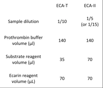
HaemoSys-Ecarin Chromogenic Assay, dist. by Diagnostica Stago) that was designed for use as a means of monitoring DTI anticoagulation.40,41 Differences between the hirudin and the DTI methodology resided in the initial dilution of the sample. Subsequently, that method was replaced by a modified version, the ECA-II (Diagnostica Stago, Parsippany, NJ) in 2015. While both kits were distributed by Diagnostica Stago, there were differences between the two chromogenic ECA methods, as the ECA-H/T kit was designed for microplate techniques, and the ECA-II using an automated instrument platform. (Table 1) Adaptation to an automated analyzer was successfully accomplished using ECA-T method, suggesting a potential comparability between the two chromogenic assays.42
In an unpublished study, samples from dabigatran treated patients were compared at 5 different laboratory sites and tested using both ECA-T and ECA-II methods. For each site, calibration was performed using dabigatran calibrators (Hyphen Biomedical) for the ECA-T and dabigatran calibrators from Diagnostica Stago for the ECA-II method. Precision testing was performed by running two sets of dabigatran controls (Hyphen Biomedical and Diagnostica Stago) in duplicate over 5 days. Accuracy was assessed by analyzing plasma samples from dabigatran treated patients (CliniSys Associates, Ltd, Atlanta, GA) on both ECA methods. Notable differences between the two methods included the lower limit of quantitation (varied between site between 10 – 30 ng/mL) for ECA-T and ECA-II (15 ng/mL) and the raw data from calibration. (Figure 4) Each site was able to demonstrate precision of < 10.2% for control level I
(assigned control range around 50 ng/mL) and < 5.6% for control level II (around 200 ng/mL). Regression was demonstrated to be > 0.961 between ECA-II and assigned values for patient samples, with increasing bias associated with higher dabigatran levels, especially for the site using BCS-XP protocol, which consistently under recovered dabigatran levels when > 200 ng/mL. The ECA-II did improve the accuracy of result recovery for all sites for samples with < 100ng/mL (Figures 5), which has been previously noted.43 The improved accuracy at lower dabigatran concentration is most likely secondary to calibration methods, as ECA-T calibration used 3-4 points (0 [optional], 30, 255, and 468 ng/mL) and ECA-II had 5-point calibration curve (0, 52, 106, 182, and 270 ng/mL). These data, coupled with the protocol changes (Table 1) would suggest that protocol modifications are required to cover the entire testing range, but no change may be necessary if accuracy is more clinically relevant at lower dabigatran
concentrations (<50ng/mL) for cases of emergent interventions.44,45 In addition, the
methodology of Diagnostica Stago for the ECA-II assay uses reflex testing meaning that, when the concentration is above the higher calibration point, an automatic re-dilution of the samples is performed by the analyzer (from 1/5 in the initial analysis to 1/15 in the re-diluted analysis, increasing the potential range of measurement until 750 ng/mL approximatively).
Point of care testing and ecarin
More than 15 years ago, in order to rapidly monitor and evaluate the intensity of
anticoagulation in patients treated with lepirudin, a point-of-care (POC) platform was created based on the activation of the coagulation process by the venom from Echis Carinatus.46 This test was based on the Thrombolytic Assessment System (TAS, Bayer Diagnostics; Rapidpoint Coag Analyzer, Morrisville, North Carolina) and employs the same coagulation principles as other ecarin clotting time assays, but in a 1-step, dry-chemistry platform. This test was commercialized by Pharmanetics but is now longer on the market.
Another microfluidic system based on the principle of lateral flow assays (LFAs), a new paper-based method incorporating both ECA and ECT test platforms, has been reported to be sensitive towards the presence of dabigatran and other DTIs.47 This new system is based on the migration of the plasma on an electronegative environment of nitrocellulose fibers which mimics the exposed phospholipid surface of vascular endothelial cells, which slows down platelet aggregation and subsequent coagulation, suitable for clotting assays. Microfluidic paper analytical devices (micro PAD’s) are more sophisticated and multichannel versions of LFAs are even more advantageous given small volume requirements, low operative costs, and design versatility.47 These paper-based lab-on-a-chip (LOC) platforms have gained increased clinical use as diagnostic tools, especially since these systems can work with smartphone
applications to acquire data thanks to specific applications. However, while this technology may seem promising as a POC application, optimizations and validations are required to improve the sensitivity of this paper-based assay.47
Thromboelastography has also emerged as one possible solution for the monitoring of DOACs. However, reagents triggering either the extrinsic or the intrinsic pathway were not sufficiently sensitive, nor robust. The use of ecarin in thromboelastography permits increasing the
sensitivity, specificity and robustness of thromboelastography for the monitoring of DTIs in whole blood samples.48 Further investigations are still required for the viscoelastic
measurement methods for the assessment of dabigatran. A modified cartridge for the TEG6S (Haemonetics®, Braintree, MA), including an ecarin channel49, is currently under investigation (ClinicalTrials.gov, NCT02798328).
Conclusion
Ecarin is derived from the snake Echis carinatus and converts prothrombin to meizothrombin with the advantage of not being affected by heparin or heparinoids. While this test has been available for decades, it was rarely used by laboratories. However, the widespread approval of
the direct oral thrombin inhibitor, dabigatran etexilate, resulted in an emergence in this method, including citation in the prescribing information of Pradaxa. There are two different methodologies for ecarin based testing: the traditional clot-based assay and the chromogenic assay. It should be appreciated that the clot-based assay can be influenced by prothrombin and fibrinogen levels that may reduce the accuracy of the measurement of the inhibitory effect on thrombin. However, there is a high degree of specificity with ecarin based testing in the assessment of any direct thrombin inhibitor, including the parenteral drugs bivalirudin and argatroban. Dabigatran calibrated ecarin methods have been demonstrated to be equivalent to the gold standard method of mass spectrometry with the advantage of not being influenced by heparin which can be of interest in case of bridging or to ensure better specificity. The arrival of a POC device based on this venom may also provide rapid solutions to estimate the degree of anticoagulation in emergent situations. While this manuscript describes Ecarin-based
anticoagulant monitoring, the interested reader is referred to another current "Test of the Month" article on Ecarin-based lupus anticoagulant testing [Moore G, Am J Hematol].
References
1. Alcock MB. A Case of Venomous Bite by Echis Carinata. Ind Med Gaz. 1888;23(6):175-181.
2. Kornalik F, Pudlak P. Coagulation defect following non-toxic doses of Echis viper venom. Experientia. 1962;18:381-2.
3. Schieck A, Habermann E, Kornalik F. The prothrombin-activating principle from Echis carinatus venom. II. Coagulation studies in vitro and in vivo. Naunyn Schmiedebergs Arch Pharmacol. 1972;274(1):7-17.
4. Dyr JE, Blombäck B, Kornalík F. The action of prothrombin activated by Ecarin on fibrinogen. Thromb Res. 1983;30(3):225-34.
5. Kornalik F, Blombäck B. Prothrombin activation induced by Ecarin - a prothrombin converting enzyme from Echis carinatus venom. Thromb Res. 1975;6(1):57-63.
6. Morita T, Iwanaga S, Suzuki T. The mechanism of activation of bovine prothrombin by an activator isolated from Echis carinatus venon and characterization of the new active intermediates. J Biochem. 1976;79(5):1089-108.
7. Novoa E, Seegers WH. Mechanisms of alpha-thrombin and beta-thrombin-E formation: use of ecarin for isolation of meizothrombin 1. Thromb Res. 1980;18(5):657-68. 8. Rosing J, Tans G. Meizothrombin, a major product of factor Xa-catalyzed prothrombin
activation. Thromb Haemost. 1988;60(3):355-60.
9. Bertina RM, van der Marel-van Nieuwkoop W, Dubbeldam J, Boekhout-Mussert RJ, Veltkamp JJ. New method for the rapid detection of vitamin k deficiency. Clin Chim Acta. 1980;105(1):93-8.
10. Triplett DA, Stocker KF, Unger GA, Barna LK. The Textarin/Ecarin ratio: a confirmatory test for lupus anticoagulants. Thromb Haemost. 1993;70(6):925-31.
11. Moore GW, Smith MP, Savidge GF. The Ecarin time is an improved confirmatory test for the Taipan snake venom time in warfarinized patients with lupus anticoagulants. Blood Coagul Fibrinolysis. 2003;14(3):307-12.
12. Moore GW, Culhane AP, Maloney JC, Archer RA, Breen KA, Hunt BJ. Taipan snake venom time coupled with ecarin time enhances lupus anticoagulant detection in
nonanticoagulated patients. Blood Coagul Fibrinolysis. 2016;27(4):477-80 13. Refludan® prescribing information. Berlex Laboratories, October 2002.
14. Van Cott EM, Roberts AJ, Dager WE. Laboratory Monitoring of Parenteral Direct Thrombin Inhibitors. Semin Thromb Hemost. 2017;43(3):270-276.
15. Hafner G, Roser M, Nauck M. Methods for the monitoring of direct thrombin inhibitors. Semin Thromb Hemost. 2002;28(5):425-30.
16. Gosselin RC, King JH, Janatpour KA, Dager WE, Larkin EC, Owings JT. Comparing direct thrombin inhibitors using aPTT, ecarin clotting times, and thrombin inhibitor
management testing. Ann Pharmacother. 2004;38(9):1383-8.
17. Carlsson SC, Schulman S. A step change in oral anticoagulation: lack of coagulation monitoring with ximelagatran. Semin Vasc Med. 2005;5(3):259-65.
18. Douxfils J, Gosselin RC. Laboratory Assessment of Direct Oral Anticoagulants. Semin Thromb Hemost. 2017;43(3):277-290.
19. United States Food & Drug Administration. Pradaxa (Dabigatran etexilate) drug approval package NDA 22-512 10/19/2010.
https://www.accessdata.fda.gov/drugsatfda_docs/nda/2010/022512Orig1s000Approv.p df. Last accessed 03/10/2020]
20. Pradaxa® prescribing information. Boehringer Ingelheim GmbH. Revised 03/2018
21. Gosselin RC, Adcock DM, Bates SM, Douxfils J, Favaloro EJ, Gouin-Thibault I, Guillermo C, Kawai Y, Lindhoff-Last E, Kitchen S. International Council for Standardization in
Haematology (ICSH) Recommendations for Laboratory Measurement of Direct Oral Anticoagulants. Thromb Haemost. 2018;118(3):437-450.
22. Ziakas PD, Kourbeti IS, Poulou LS, Vlachogeorgos GS, Mylonakis E. Medicare part D prescribing for direct oral anticoagulants in the United States: Cost, use and the "rubber effect". PLoS One. 2018;13(6):e0198674.
23. Douxfils J, Ageno W, Samama CM, Lessire S, Ten Cate H, Verhamme P, Dogné JM, Mullier F. Laboratory testing in patients treated with direct oral anticoagulants: a practical guide for clinicians. J Thromb Haemost. 2018;16(2):209-219.
24. van Ryn J, Stangier J, Haertter S, Liesenfeld KH, Wienen W, Feuring M, Clemens A. Dabigatran etexilate--a novel, reversible, oral direct thrombin inhibitor:interpretation of coagulation assays and reversal of anticoagulant activity. Thromb Haemost.
2010;103(6):1116-27.
25. Douxfils J, Mullier F, Robert S, Chatelain C, Chatelain B, Dogné JM. Impact of dabigatran on a large panel of routine or specific coagulation assays. Laboratory recommendations for monitoring of dabigatran etexilate. Thromb Haemost. 2012;107(5):985-97.
26. Douxfils J, Lessire S, Dincq AS, Hjemdahl P, Rönquist-Nii Y, Pohanka A, Gourdin M, Chatelain B, Dogné JM, Mullier F. Estimation of dabigatran plasma concentrations in the perioperative setting. An ex vivo study using dedicated coagulation assays. Thromb Haemost. 2015;113(4):862-9.
27. Dager WE, Gosselin RC, Kitchen S, Dwyre D. Dabigatran effects on the international normalized ratio, activated partial thromboplastin time, thrombin time, and fibrinogen: a multicenter, in vitro study. Ann Pharmacother. 2012;46(12):1627-36.
28. Hawes EM, Deal AM, Funk-Adcock D, Gosselin R, Jeanneret C, Cook AM, Taylor JM, Whinna HC, Winkler AM, Moll S. Performance of coagulation tests in patients on therapeutic doses of dabigatran: a cross-sectional pharmacodynamic study based on peak and trough plasma levels. J Thromb Haemost. 2013; 11(8):1493-502.
29. Gosselin RC, Douxfils J. Measuring Direct Oral Anticoagulants. Methods Mol Biol. 2017;1646:217-225.
30. Pötzsch B, Hund S, Madlener K, Unkrig C, Müller-Berghaus G Monitoring of recombinant hirudin: assessment of a plasma-based ecarin clotting time assay. Thromb Res 1997; 86:373–383.
31. Nowak G The ecarin clotting time, a universal method to quantify direct thrombin inhibitors. Pathophysiol Haemost Thromb 2003; 33:173–183.
32. Lindhoff-Last E, Piechottka GP, Rabe F, Bauersachs R Hirudin determination in plasma can be strongly influenced by the prothrombin level. Thromb Res 2000; 100:55–60. 33. Pradaxa® prescribing information. Boehringer Ingelheim. Revised 03/2018.
34. Douxfils J, Gosselin RC. Laboratory Assessment of Direct Oral Anticoagulants. Semin Thromb Hemost. 2017;43(3):277-290.
35. Food and Drug Administration Center for Drug Evaluation and Research. Clinical Pharmacology Review, NDA 22-512, Dabigatran. 09-01-2010.
36. Liesenfeld KH, Schäfer HG, Trocóniz IF, Tillmann C, Eriksson BI, Stangier J. Effects of the direct thrombin inhibitor dabigatran on ex vivo coagulation time in orthopaedic surgery patients: a population model analysis. 2006 Br J Clin Pharmacol. 62(5):527-37.
37. Stangier J, Rathgen K, Stähle H, Gansser D, Roth W. The pharmacokinetics,
pharmacodynamics and tolerability of dabigatran etexilate, a new oral direct thrombin inhibitor, in healthy male subjects. Br J Clin Pharmacol. 2007; 64(3):292-303.
38. Stangier J, Stähle H, Rathgen K, Fuhr R. Pharmacokinetics and pharmacodynamics of the direct oral thrombin inhibitor dabigatran in healthy elderly subjects. 2008; Clin
Pharmacokinet. 47(1):47-59.
39. Gosselin R, Hawes E, Moll S, Adcock D. Performance of various laboratory assays in the measurement of dabigatran in patients receiving therapeutic doses: a prospective study based on peak and trough plasma levels. Am J Clin Pathol. 2014;141(2):262-7.
40. Lange U, Nowak G, Bucha E. Ecarin chromogenic assay--a new method for quantitative determination of direct thrombin inhibitors like hirudin. Pathophysiol Haemost Thromb. 2003 Jul-2004;33(4):184-91.
41. Nowak G, Lange U, Bucha E. Drug monitoring of argatroban using the ecarin chromogenic assay. Sem Thromb Hemost 2008;34(Suppl 1): 81-86.
42. Gosselin RC, Dwyre DM, Dager WE. Measuring dabigatran concentrations using a chromogenic ecarin clotting time assay. Ann Pharmacother. 2013;47(12):1635-40. 43. Jaffer IH, Chan N, Roberts R, Fredenburgh JC, Eikelboom JW, Weitz JI. Comparison of the
ecarin chromogenic assay and diluted thrombin time for quantification of dabigatran concentrations. J Thromb Haemost. 2017;15(12):2377-2387.
44. Douketis JD, Spyropoulos AC, Anderson JM, Arnold DM, Bates SM, Blostein M, et al. The Perioperative Anticoagulant Use for Surgery Evaluation (PAUSE) Study for Patients on a Direct Oral Anticoagulant Who Need an Elective Surgery or Procedure: Design and Rationale. Thromb Haemost. 2017;117(12):2415-2424. Erratum in:Thromb Haemost. 2018;118(9):1679-1680.
45. Touzé E, Gruel Y, Gouin-Thibault I, De Maistre E, Susen S, Sie P, Derex L. Intravenous thrombolysis for acute ischaemic stroke in patients on direct oral anticoagulants. Eur J Neurol. 2018;25(5):747-e52.
46. Cho L, Kottke-Marchant K, Lincoff AM, Roffi M, Reginelli JP, Kaldus T, Moliterno DJ. Correlation of point-of-care ecarin clotting time versus activated clotting time with bivalirudin concentrations. Am J Cardiol. 2003;91(9):1110-3.
47. Alouidor B, Sweeney RE, Tat T, Wong RK, Yoon J-Y. Microfluidic Point-of-Care Ecarin-Based Clotting and Chromogenic Assays for Monitoring Direct Thrombin Inhibitor. J Extra Corpor Technol, 2019; 51(1):29-37.
48. Schaden E, Schober A, Hacker S, Kozek-Langenecker S. Ecarin modified rotational thrombelastometry: a point-of-care applicable alternative to monitor the direct thrombin inhibitor argatroban. Wien Klin Wochenschr. 2013;125(5-6):156-9.
49. Bliden KP, Chaudhary R, Mohammed N, Muresan AA, Lopez-Espina CG, Cohen E, Raviv G, Doubleday M, Zaman F, Mathew B, Tantry US, Gurbel PA. Determination of non-Vitamin K oral anticoagulant (NOAC) effects using a new-generation thrombelastography TEG 6s system. J Thromb Thrombolysis. 2017;43(4):437-445.
Summary Table
1. Ecarin based assays are highly sensitive and specific assays to assess the anticoagulant effect of oral and parenteral direct thrombin inhibitors.
2. Ecarin is not affected by heparin or heparin-like anticoagulants, which is an advantage over the thrombin time, aPTT, and ACT, all of which are prolonged by heparin,
interfering in the measurement of DTIs when both anticoagulants are present or when heparin contamination occurs during specimen collection.
3. There are two different ecarin assays: clot-based and chromogenic.
4. Ecarin clot-based assays can be factitiously prolonged, and thus overestimating anticoagulant effect, in patients with reduced prothrombin (factor II) and fibrinogen levels.
5. Dabigatran calibrated chromogenic ecarin assays have been demonstrated to be equivalent to mass spectrometry measurements.
6. Ecarin assays have been demonstrated to be adaptable to current coagulation analyzers or transferred in a point-of-care (POC) device to provide rapid turn-around-time results for any direct thrombin inhibitor drug.
a. The limitations of ecarin methods include the influence of low prothrombin and/or fibrinogen levels, but these do not influence chromogenic ecarin assays b. There is a lack of standardization of ecarin assays, suggesting that ECT raw data
Table 1: Comparison of STA-R Evolution protocol for ECA-T and ECA-II chromogenic ecarin methods.
ECA-T ECA-II Sample dilution 1/10 (or 1/15)1/5
Prothrombin buffer volume (µl) 140 140 Substrate reagent volume (µl) 35 70 Ecarin reagent volume (µL) 70 70
Accepted
Article
The physiologic activation of prothrombin from either activated factor X or ecarin to create thrombin.
Accepted
Testing principles of ecarin clot based (A) and chromogenic based (B) testing18
NOTE: As the ecarin clotting time (A) is dependent on patient prothrombin and fibrinogen levels, the clotting may be prolonged if prothrombin and/or low fibrinogen levels are present. However, with the ecarin chromogenic assay, the sample is diluted in prothrombin buffer, eliminating the effect of low prothrombin levels, and the method is not dependent on fibrinogen conversion, and thus is not affected by low fibrinogen
levels.
Accepted
Performance characteristics of ECT in dabigatran assessment ECT: ecarin clotting time; LOQ: Lower limit of quantitation
Accepted
Differences in calibration raw data for ECA-T (∆) and ECA-II (o) using the BCSXP analyzer and same testing protocols.
Accepted
Comparison of accuracy for samples containing <100ng/mL for ECA-T (A) and ECA-II (B) chromogenic ecarin methods for sample <100ng/mL for ECA-II (Figure A) and ECA-T (Figure B) methods.