HAL Id: tel-02787414
https://hal.inrae.fr/tel-02787414
Submitted on 5 Jun 2020
HAL is a multi-disciplinary open access archive for the deposit and dissemination of sci-entific research documents, whether they are pub-lished or not. The documents may come from teaching and research institutions in France or abroad, or from public or private research centers.
L’archive ouverte pluridisciplinaire HAL, est destinée au dépôt et à la diffusion de documents scientifiques de niveau recherche, publiés ou non, émanant des établissements d’enseignement et de recherche français ou étrangers, des laboratoires publics ou privés.
Involvement of the inflammasome in the response of
host cells during Staphylococcus aureus infection
Elma Lima Leite
To cite this version:
Elma Lima Leite. Involvement of the inflammasome in the response of host cells during Staphylococcus aureus infection. Microbiology and Parasitology. Universidade Federal de Minas Gerais, 2019. English. �tel-02787414�
UNIVERSIDADE FEDERAL DE MINAS
GERAIS INSTITUTO DE CIÊNCIAS
BIOLÓGICAS DEPARTAMENTO DE
BIOLOGIA GERAL PROGRAMA DE
PÓS-GRADUAÇÃO EM GENÉTICA
Tese de Doutorado
Involvement of the inflammasome in the response of
host cells during Staphylococcus aureus infection
Orientada: Elma Lima Leite
Orientadores: Prof./Dr. Vasco Ariston de Carvalho Azevedo Dr. Nadia Nadejda Berkova
Co-supervisor: Dr. Yves Le Loir
Belo Horizonte - Brasil
Rennes - France
Elma Lima Leite
Involvement of the inflammasome in the response of
host cells during Staphylococcus aureus infection
Belo Horizonte - Brasil
Rennes - France
2019
Tese apresentada ao programa de Pós-Graduação em Genética do Departamento de Biologia Geral do Instituto de Ciências Biológicas da Universidade Federal de Minas Gerais como requisito parcial para obtenção do título de Doutora em Genética.
Orientadores:
Prof. Dr. Vasco Ariston de Carvalho Azevedo Dr. Nadia Nadejda Berkova
Ficha elaborada pela Biblioteca do Instituto de Ciências Biológias da UFMG Ficha elaborada pela Biblioteca do Instituto de Ciências Biológias da UFMG Ficha catalográfica elaborada pela Biblioteca do Instituto de Ciências Biológicas da UFMG
043
Leite, Elma Lima.
Involvement of the inflammasome in the response of host cells during Staphylococcus aureus infection [manuscrito] / Elma Lima Leite. – 2019. 199 f. : il. ; 29,5 cm.
Orientadores: Prof. Dr. Vasco Ariston de Carvalho Azevedo, Dr. Nadia Nadejda Berkova. Co-orientador: Dr. Yves Le Loir.
Tese (doutorado) – Universidade Federal de Minas Gerais, Instituto de Ciências Biológicas. Programa de Pós-Graduação em Genética.
1. Genética. 2. Osteoblástos. 3. Staphylococcus aureus. 4. Inflamassomos. 5.
Caspase 1. 6. Interleucina-1. I. Azevedo, Vasco Ariston de Carvalho. II. Berkova, Nadia Nadejda. III. Le Loir, Yves. IV. Universidade Federal de Minas Gerais. nstituto de Ciências Biológicas. V. Título.
“E, naquele dia, sendo já tarde, disse-lhes: Passemos para a outra margem’’.
Acknowledgement
Agradecimentos
-
Ao meu orientador brasileiro, Vasco Azevedo, pela oportunidade, ensinamentos e, principalmente por acreditar em mim;- A minha orientadora francesa, Nadia Berkova;
- A Yves Le Loir, por toda ajuda e pela acolhida em seu Instituto de Pesquisa; - Aos membros da banca pela disponibilidade em avaliarem este trabalho de Tese; - A Martine Deplanche pelos seus ensinamentos e amizade;
- As equipes de pesquisa do INRA de Rennes, pela ajuda e disponibilidade;
- A Pós-graduação em Genética, por todo o suporte na parte administrativa desse doutorado;
- A escola de doutorado do Agrocampus Ouest – Rennes, por todo o suporte na parte administrativa desse doutorado cotutela;
- A agência de fomento, CAPES e ao programa CAPES-COFECUB pelo auxílio financeiro concedido no Brasil e na França durante este período de Tese;
- A todos os membros do laboratório LGCM, especialmente a Fillipe pelo apoio; - Ao meu querido irmão Eldo;
- A Rafael Cabús por todo o apoio nos momentos difíceis e decisivos da minha vida. Você sempre acreditou em mim e me disse que meus sonhos seriam realizados; - A Seu Beto, Dona Suzane, Dona Gegé e Isabelle, obrigada pelo apoio;
- À Houem Rabah, Oumaima, Laurence Fauvel, Mary Bret, muito obrigada!
- Aos meus amigos, Flora, Natália, Angélica, Emmanuele, Amanda, Anne, Renata, Flávia Aburjaile, Rachid, Luisa Osorio, Alberto, Thiago Baiano e Daniel que me apoiaram e me incentivaram de perto ou de longe.
- A Rodolphe por ser sempre forte e me apoiar nestes últimos momentos difíceis. Por sempre me dizer que tudo é possível;
- E especialmente a Deus, minha mãe, Socorro, e ao meu pai, Euclides, que são a minha fortaleza, obrigada pelo apoio e amor incondicional em todos os momentos, eu amo vocês!
II
Remerciements
Je remercie:
- Vasco Azevedo, mon directeur de thèse brésilien, pour l’opportunité qu’il m’a donnée, de son enseignement, et surtout, d’avoir cru en moi ;
- Nadia Berkova, ma directrice de thèse Français;
- Yves Le-Loir, pour toute son aide et pour l’accueil que j’ai reçu dans son institut de recherche ;
- Les membres du jury pour leur disponibilité à évaluer ce travail de thèse ; - Martine Deplanche pour ses enseignements et son amitié, merci beaucoup ;
- Les équipes de recherche de l’INRA de Rennes, pour leur aide et leur disponibilité ; - Le programme de troisième cycle en génétique, pour tout son soutien dans la partie administrative de cette thèse de doctorat en cotutelle ;
- L’École doctorale de l’Agrocampus Ouest – Rennes, pour tout son soutien dans la partie administrative de cette thèse de doctorat en cotutelle ;
- Les organismes de financement CAPES et le Programme CAPES-COFECUB pour toute l’aide financière que j’ai reçue au Brésil et en France pendant la durée de cette thèse ;
- Tous les membres du laboratoire LGCM, en particulier à Fillipe pour le soutien ; - Mon cher frère Eldo;
- Rafael Cabús pour tout le soutien nécessaire dans les moments difficiles et décisifs de ma vie. Tu as toujours cru en moi et m'a dit que mes rêves se réaliseraient;
- Beto, Suzane, Gegé et Isabelle, je vous remercie pour le soutien;
- À Houem Rabah, Oumaima, Laurence Fauvel, Mary Bret, merci beaucoup !
- Mes chers amis Flora, Natália, Angélica, Emmanuele, Amanda, Anne, Renata, Flávia Aburjaile, Rachid, Luisa Osorio, Alberto, Vinícius, Thiago Baiano et Daniel qui m'ont soutenue et encouragée de près ou de loin;
- Rodolphe pour être fort et me soutenir toujours dans ces derniers moments difficiles. Pour me dire toujours que tout est possible ;
- Et surtout à Dieu, ma mère, Socorro, et mon père, Euclides, qui sont ma forteresse, merci pour votre soutien et votre amour inconditionnel à tout moment, je vous aime!
Table of Contents
Acknowledgement ... I Table of Contents ... III List of figures ... VI List of tables ... VIII List of Abbreviations ... IX Resumo ... 1 Abstract ... 2 Résumé ... 3 Collaborations ... 4 Manuscript structure ... 5
General introduction / Introduction générale ... 6
General introduction ... 7
Introduction générale ... 11
Chapter 1: Bibliographic Synthesis ... 15
1. General characteristics of Staphylococcus aureus ... 16
1.1. Definition and morphology ... 16
1.2. General cultural, biochemical characteristics and genome of S. aureus ... 16
1.3. General pathogenesis and clinical diseases ... 17
1.3.1 Pathogenesis ... 17
1.3.2 Virulence factors ... 18
1.3.3 Phenol-soluble modulins (PSMs) ... 22
2. The inflammasome, a primordial complex of innate immunity ... 25
2.1. Mechanisms of the inflammatory response ... 25
2.2. Inducers of the inflammatory response ... 26
2.3. Mediators of the inflammatory response ... 27
2.4. IL-1β ... 28
2.4.1 The maturation of IL-1β ... 28
2.4.2 Excretion of IL-1β ... 29
2.4.3 The regulation of IL-1β ... 30
2.5. Other cytokines ... 31
3. Innate immunity receptors ... 32
3.1. Pattern recognition receptors (PRRs) ... 32
3.2. NOD-like receptors (NLRs) ... 34
4. Molecular platform: The inflammasome ... 35
4.1 The Caspase-1 protein ... 36
4.2 Non-canonical activation of inflammatory caspases ... 37
4.3 The ASC protein ... 37
4.4 The NLRP1 inflammasome ... 38
4.5 The NLRP3 inflammasome ... 40
4.6 The AIM2 inflammasome ... 43
4.7 The NLRC4 inflammasome ... 43
4.8 Others inflammasomes ... 44
4.9 Inflammasome and pathologies... 47
IV
Rationale of the PhD project ... 50
Pertinence du projet de Thèse ... 53
Aim of the PhD project ... 56
Objectif du projet de thèse ... 56
Chapter 3. Literature review. Strain and cell type-specificity of host cell response to Staphylococcus aureus invasion. ... 57
Strain and cell type-specificity of host cell response to Staphylococcus aureus invasion. ... 58
Abstract ... 58
1. Introduction ... 59
2. S. aureus, a versatile opportunistic pathogen ... 59
3. S. aureus adhesion and internalization ... 60
4. Cell response to S. aureus infection is strain-dependent... 61
5. Phenotypic modifications also alter the S. aureus-host cell interaction... 62
6. Strain-specific ability to adhere and internalize ... 63
7. Strain-specific cytotoxicity ... 64
8. Cytotoxicity induced by S. aureus extracellular vesicles... 65
9. Impact on the host cell cycle ... 66
10. Impact on the innate immune response and on the inflammatory response...67
11. Impact on the epigenetic modifications in host cells ... 67
12. Cell types and S. aureus invasion ... 68
13. Conclusion ... 69
Author Contributions ... 70
Funding ... 70
Conflict of Interest Statement ... 70
Acknowledgments ... 70
References ... 70
Figure legend ... 77
Chapter 4. Original article - Involvement of caspase-1 in inflammasomes activation and bacterial clearance in S. aureus-infected osteoblasts... 83
Involvement of Caspase-1 in inflammasomes activation and bacterial clearance in S. aureus-infected osteoblasts. ... 84
Abstract ... 84
1. Introduction ... 85
2. Results ... 87
2.1. Caspase-1 activation and IL-1β release triggered by inflammasomes activators LPS and ATP in human osteoblasts and PMA-treated ThP1 cells ... 87
2.2. Deletion of caspase-1 gene in human osteoblasts MG-63 using the CRISPR/Cas9 gene editing system. ... 87
2.3. Inflammasomes involvement in caspase-1 dependent IL-1β release by S. aureus infected MG-63 osteoblasts... 88
2.4. Pivotal role of S. aureus PSM toxins in stimulation of IL-1β release by infected
osteoblasts. ... 89
2.5. S. aureus clearance by osteoblasts depends on caspase-1 ... 90
3. Discussion ... 91
4. Materials and Methods: ... 93
4.3. Maintenance of eukaryotic cells lines ... 93
4.4. Deletion of the caspase-1 gene in human osteoblast-like MG-63 cells using the CRISPR/Cas9 gene editing system. ... 94
4.5. S. aureus strains description ... 94
4.6. Cell culture infection ... 95
4.7. Western blot analysis ... 95
4.8. Flow cytometry analysis ... 96
4.9. Confocal microscopy ... 96
4.10. IL-1β quantification by ELISA ... 96
4.11. Statistical analysis ... 97
Acknowledgment ... 97
Conflict of Interest ... 98
Figure legend ... 98
References ... 102
Chapter 5. Results and general discussion ... 112
5.1. Caspase-1 activation and IL-1β release triggered by inflammasome activators LPS and ATP in human osteoblasts and PMA-treated ThP1 cells ... 117
5.2. Deletion of caspase-1 gene in human osteoblasts MG-63 using the CRISPR/Cas9 gene editing system... 121
5.3. Inflammasome involvement in caspase-1 dependent IL-1β release by S. aureus infected osteoblasts ... 127
5.4. Pivotal role of S. aureus PSM toxins in stimulation of IL-1β release by infected osteoblasts ... 132
5.5. S. aureus clearance by osteoblasts depends on caspase-1 ... 136
Chapter 6. General conclusion and perspectives of the work performed during PhD project ... 139
General conclusion and perspective ... 140
Conclusion générale et perspectives ... 144
References ... 148
Supplentary Material and methods ... 174
VI
List of figures
Figure 1: Overview of the immune system. ... 8 Figure 2: Accessory gene regulatory system (Agr). ... 22 Figure 3: Overview of phenol-soluble modulin activities in S. aureus. ... 23 Figure 4: Phenol-soluble modulins peptides (PSMs) define the virulence potential of S. aureus. ... 24 Figure 5: Scheme of NLR receptor subfamilies and their structure in humans. ... 35 Figure 6: Scheme of the domain structure of the NLRP3 inflammasome components. ... 36 Figure 7: Human NLRP1 responds to muramyl dipeptide, while Anthrax lethal toxin triggers murine Nlrp1b.. ... 39 Figure 9: AIM2 and NLRC4 inflammasomes are activated by specific PAMPs. ... 44 Figure 8: NLRP3 inflammasome activation mechanism. ... 46 Figure 10: Schematic of the proposed objectives for the first and second part of the thesis. ... 52 Figure 11: Detection of active caspase-1 in MG-63 cell supernatants by Western blot. ... 114 Figure 12: Detection of pro-caspase-1 in lysates of MG-63 and THP1 cells by western blotting. ... 116 Figure 13: Influence of PMA on IL-1β production in culture supernatants of THP1 cells. ... 118 Figure 14: Caspase-1 activation and IL-1β release triggered in human osteoblasts. 120 Figure 15: Detection of pro-caspase-1 in lysates of MG-63-CRISPR/cas9 cells by Western blot. ... 123 Figure 16: Detection of pro-caspase-1 in lysates of CASP1–/– MG-63 cells by Western
blotting. ... 124 Figure 17: Detection of pro-caspase-1 in lysates of CASP1–/– MG-63 cells by Western
blotting. ... 125 Figure 18: Detection of pro-caspase-1 in lysates of CASP1–/– MG-63 cells by Western
blot. ... 125 Figura 19: Deletion of caspase-1 gene in human osteoblasts MG-63 using the CRISPR/Cas9 gene editing system ... 126 Figure 20: NLRP3 protein expression, speck-like aggregate formation and IL-1β secretion by WT MG-63 vs CASP1–/– MG-63 cells exposed to S. aureus. ... 128
Figure 22: Caspase-1 is required for secretion of IL-1β in S. aureus-infected MG-63 cells. ... 132 Figure 23: S. aureus phenol-soluble modulins stimulate IL-1β release from infected osteoblasts. ... 134 Figure 24: kinetics of IL-1β production between 6 h and 9 days post-infection in MG-63 cells by S. aureus clinical strains... 136 Figure 25: Involvement of caspase-1 in bacterial clearance. ... 138
VIII
List of tables
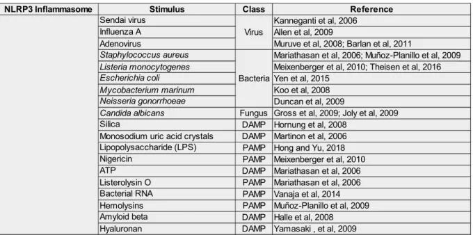
Table 1: Virulence factors of S. aureus and their characteristics... 21 Table 2: List of activators known to activate of the NLRP3 inflammasome. ... 41 Table 3: Different protocols used to detect active caspase-1 in THP1 cells differentiated with (PMA) by western blotting. ... 116
List of Abbreviations
ANOVA: Analysis of variance
ASC: Apoptosis-associated speck-like protein containing a caspase recruitment domain ATP: Adenosine triphosphate
BHI: Brain Heart Infusion
CA-MRSA: Community-Acquired Methicillin-Resistant S. aureus CARD: Caspase recruitment domain
DC: Dendritic cells
DMEM: Dulbecco's Modified Eagle Medium DNA: Deoxyribonucleic acid
ELISA: enzyme-linked immunosorbent assay FACS: Fluorescence-activated cell sorting Fn: Fibronectins
FnBPA and FnBPB: Fibronectins Binding Proteins (A and B) Gent100: Gentamicin 100 μg/mL
Gent25: Gentamicin 25 μg/mL
HA-MRSA: Hospital-Associated Methicillin-Resistant S. aureus ICB: Institudo de Ciências Biológicas
ICE: IL-1-converting enzyme ou enzyme transformant IL-1 IFN-γ: interferon gamma
IL-1β: interleukin-1 beta
INRA: French National Institute for Agricultural Research LDH: Lactate dehydrogenase
LPS: lipopolysaccharide MOI: Multiplicity of infection
MRSA: Methicillin-resistant Staphylococcus aureus MS: Mass spectrometry
MSCRAMMs: Microbial Surface Components Recognizing Adhesive Matrix Molecules NK: Natural killers
NLR: NOD-like receptor
NOD: nucleotide binding oligomerization domain PAMPs: Pathogen associated molecular pattern PBMC: Peripheral blood mononuclear cells PBS: Phosphate Buffered Saline
X
PSM: Phenol-soluble modulins
PVDF: Polyvinylidene fluoride membrane
ROS: reactive oxygen species/espèces réactives de l’oxygène RPMI-1640: Roswell Park Memorial Institute medium
S. aureus: Staphylococcus aureus FCS: Fetal Calf Serum
SCCmec: staphylococcal cassette chromosome mec SCV: Small colony variant
SD: Standard deviation
SDS-PAGE: Sodium dodecyl sulfate polyacrylamide gel electrophoresis SE: Staphylococcal enterotoxins
TBS: Tris-buffered saline Th: T helper cells
TLR: Toll-like receptor
TNF-α: Tumor Necrosis Factor-α
TPA: 12-O-tetradecanoylphorbol 13-acetate UFMG: Universidade Federal de Minas Gerais V: Volts
WB: Western Blotting WT: Wild type
Resumo
Staphylococcus aureus (S. aureus) é uma bactéria Gram-positiva altamente adaptativa e versátil que pode causar uma ampla gama de doenças infecciosas em humanos ou animais. Nos estágios iniciais da infecção, a interação entre S. aureus e as células hospedeiras causa inflamação, cujo processo depende de vários mecanismos celulares complexos e de uma cascata de sinalização coordenada principalmente pelas citocinas e sua ativação. Os principais fatores que determinam o início da inflamação e sua progressão são pathogen-/microbe-associated molecular patterns (PAMPs/ MAMPs), danger-associated molecular patterns (DAMPs), pattern recognition receptors (PRRs), e o sistema imune inato e adaptativo. Inflammassomas, complexos de sinalização multiprotéicos que se agrupam após a detecção de PAMPs/DAMPs, são fatores-chave da resposta imune inata. A maioria dos inflamassomas é formado por um receptor NLR, uma proteína adaptadora ASC e uma Caspase-1 ativa. Uma vez montados, os inflamassomas atuam como plataformas ativadoras que promovem a maturação das citocinas pró-inflamatórias IL-1β e IL-18 em suas formas ativas. Seu papel em diferentes tipos de fagócitos profissionais durante a infecção por S. aureus tem sido extensivamente estudado. Por outro lado, o conhecimento sobre o envolvimento de inflamassomas em fagócitos não profissionais (por exemplo, células epiteliais, células endoteliais ...) é muito fragmentado. O objetivo deste trabalho foi estudar o papel da ativação do inflamassoma durante a infecção por S. aureus em fagócitos não profissionais. Adotamos um modelo de infecção por S. aureus desenvolvido em células osteoblásticas (linhagem MG-63). Análises de Western blot e ELISA foram usadas para controlar a ativação de caspase-1 e a maturação de IL-1β na interação de S. aureus/MG-63. Geramos células MG-63 deletadas do gene da caspase-1 (CASP1–/– MG-63) usando a abordagem CRISPR/Cas9. Isso nos
permitiu demonstrar o envolvimento do inflamassoma na resposta imune inata a S. aureus por fagócitos não profissionais. Inesperadamente, mostramos que S. aureus prolifera nas células CASP1–/– MG-63, sugerindo que Casp1 (ou um fenômeno regulado pela Casp1) está
envolvido na eliminação do S. aureus intracelular. Descobrimos que a formação do inflamassoma nas células MG-63 é mais tardia que nos fagócitos profissionais. Além disso, o uso de cepas de S. aureus (mutantes deletados) expressando ou não Phenol-Soluble Modulins (PSM) nos permitiu determinar o papel das PSMs como desencadeador. Assim, demonstramos que S. aureus é capaz de ativar a formação de inflamassomas em células não fagocíticas (MG-63) e que os PSMs estão envolvidos nesse fenômeno.
2
Abstract
Staphylococcus aureus (S. aureus) is a highly adaptive and versatile Gram-positive bacterium that can cause a wide range of infectious diseases in humans or animals. In the early stages of infection, the interaction between S. aureus and the host cells causes inflammation, the process of which depends on several complex cellular mechanisms and a signaling cascade coordinated mainly by cytokines and their activation. The key factors determining the initiation of inflammation and its progression are pathogen/ microbe-associated molecular patterns (PAMPs/MAMPs), danger-associated molecular patterns (DAMPs), pattern recognition receptors (PRRs), innate and adaptive immunity. Inflammasomes, multi-protein signaling complexes that assemble after sensing PAMPs/ DAMPs, are key players of the innate immune response. Most inflammasomes are formed by an NLR receptor, an ASC adapter protein, and active caspase-1. Once assembled, inflammasomes act as activating platforms that promote the maturation of pro-inflammatory cytokines IL-1β and IL-18 in their active forms. Their role in different types of professional phagocytes during S. aureus infection has been extensively studied. Conversely, knowledge about the involvement of inflammasomes in non-professional phagocytes (e.g. epithelial cells, endothelial cells...) is very fragmented. This work aimed to study the role of inflammasome activation during S. aureus infection in non-occupational phagocytes. We adopted a model of S. aureus infection developed on osteoblastic cells (MG-63 line). Western blot and ELISA analyses were used to control caspase-1 activation and IL-1β maturation in the S. aureus MG-63 interaction. We generated MG-63 cells deleted from the caspase-1 gene (CASP1–/– MG-63 cells) using the CRISPR/Cas9 approach. This allowed us
to demonstrate the involvement of inflammasomes in the innate immune response to S. aureus by non-professional phagocytes. Unexpectedly, we have shown that S. aureus proliferates in CASP1–/– MG-63 cells, suggesting that Casp1 (or Casp1-regulated phenomenon) is involved
in the elimination of intracellular S. aureus. We found that the formation of the inflammasome in MG-63 cells is later than in professional phagocytes. Besides, the use of S. aureus strains (deletion mutants) expressing or not Phenol-Soluble Modulins (PSM) allowed us to determine the role of PSM in this trigger. Thus, we have demonstrated that S. aureus can activate inflammasome formation in non-professional phagocytes (MG-63) cells and that PSM is involved in this phenomenon.
Résumé
Staphylococcus aureus (S. aureus) est une bactérie à Gram positif hautement adaptative et polyvalente qui peut causer un large éventail de maladies infectieuses chez l’homme ou l’animal. Lors des premières étapes de l’infection, l’interaction entre S. aureus et les cellules hôtes entraine une inflammation dont le processus dépend de plusieurs mécanismes cellulaires complexes et une cascade de signalisation coordonnée principalement par des cytokines et leur activation. Les facteurs clés déterminant l’initiation de l’inflammation et sa progression sont les pathogen-/microbe-associated molecular patterns (PAMPs/MAMPs), les danger-associated molecular patterns (DAMPs), les Pattern Recognition Receptors (PRRs), l’immunité innée et adaptative. Les inflammasomes, complexes de signalisation multiprotéines qui s'assemblent après la détection de PAMPs/DAMPs sont des facteurs clés de la réponse immunitaire innée. La plupart des inflammasomes sont composés d’un récepteur NLR, d’une protéine adaptatrice ASC et de la caspase-1 activée. Une fois assemblés, les inflammasomes agissent comme des plateformes activatrices qui favorisent la maturation des cytokines pro-inflammatoires IL-1β et IL-18 sous leurs formes actives. Leur rôle dans les différents types de phagocytes professionnels au cours de l'infection à S. aureus a été largement étudié. A l’inverse, les connaissances sur l'implication des inflammasomes dans les phagocytes non-professionnels (e.g. cellules épithéliales, endothéliales…) sont très parcellaires. L'objectif de ce travail était d'étudier le rôle de l'activation de l'inflammasome lors d’une infection à S. aureus sur des phagocytes non-professionnels. Nous avons adopté pour cela un modèle d'infection à S. aureus mis au point sur des cellules ostéoblastiques (lignée MG-63). Des analyses Western blot et ELISA ont été utilisées pour contrôler l'activation de Caspase-1 et la maturation de l’IL-1β lors de l’interaction S. aureus MG-63. Nous avons généré des cellules MG-63 délétées du gène caspase-1 (CASP1–/– MG-63 cells) en utilisant l'approche CRISPR/Cas9. Ceci nous a
permis de démontrer l’implication des inflammasomes dans la réponse immune innée à S. aureus par des phagocytes non-professionnels. De façon inattendue, nous avons montré que S. aureus prolifère dans les CASP1–/– MG-63 cells, suggérant que Casp1 (ou les phénomènes
régulés par Casp1) est impliquée dans l’élimination de S. aureus intracellulaire. Nous avons constaté que la formation de l'inflammasome dans les cellules MG-63 est cependant plus tardive que dans des phagocytes professionnels. De plus, l'utilisation de souches de S. aureus (mutants de délétion) exprimant ou non des Phenol Soluble Modulins (PSM) nous a permis de déterminer le rôle des PSM dans ce déclenchement. Ainsi, nous avons démontré que S. aureus est capable d’activer la formation d’inflammasome dans des cellules (MG-63), phagocytes non professionnels et que les PSM sont impliqués dans ce phénomène.
4
Collaborations
This PhD thesis was prepared in collaboration between the Federal University of Minas Gerais (UFMG) (Belo Horizonte, Brazil) and the Institute of Agricultural Sciences, Agro-Food, Horticulture and Landscape (Rennes, France). The work presented in this manuscript was performed at the Laboratory Science & Technologie du Lait & de l'Oeuf (STLO) at the Institut National de la Recherche Agronomique (INRA), located in Rennes, France under the supervision of Dr. Nadia Berkova and co-supervision of Dr. Yves Le Loir and at the Laboratory of Cellular and Molecular Genetics in the Institute of Biological Sciences, Federal University of Minas Gerais under the supervision of Prof. Vasco Azevedo. In this context, the objective of my project was to investigate whether Staphylococcus aureus invasion can induce the activation of inflammasomes in non-professional phagocytes, using osteoblast-like cells as a model of cells infections and which S. aureus virulence factors are involved in inflammasomes activation during infection. To address these questions, we generated knock-out osteoblastic-like cells for the caspase-1 gene using CRISPR/Cas9-based genome editing in collaboration with Dr. Arthur Gautron and Dr. David Gilot, IGDR, Rennes, France and Pr. Petr Broz, University of Lausanne, Switzerland. Inflammasomes activation was investigated in infected WT MG-63 vs CASP1–/– MG-63 cells through immunological detection of active caspase-1 and
active IL-1β. Moreover, bacterial clearance in WT MG-63 vs CASP1–/– MG-63 cells was
investigated using gentamicin protection assay and immunofluorescence methodology. To decifer the role of principal S. aureus virulence factors in inflammasomes activation of infected osteoblasts, we used wild type and mutant strains of S. aureus kindly provided by Pr. Michael Otto, NIH, USA and Pr. Friedrich Goetz, Tubiengen University, Germany. Our work brings new knowledge on the pathogenic mechanisms of S. aureus, a major cause of chronic infection in humans and animals.
This work is supported by different funding agencies in Brazil by Coordenação de Aperfeiçoamento de Pessoal de Nível Superior (CAPES, CAPES/COFECUB) – Finance Code 001 and in France by Institut National de la Recherche Agronomique (INRA).
Manuscript structure
This manuscript is subdivided into six major parts:
I. Chapter 1. Bibliographic Synthesis. A general introduction to the following topics: (I) microbiological aspects of Staphylococcus aureus, as well as diseases caused by this pathogen and associated pathogenic processes; phenol-soluble modulins (PSMs) and their role in pathogenesis; (II) the inflammasome, a primordial complex of innate immunity; (III) innate immunity receptors; (IV) molecular platform: the inflammasome.
II. Chapter 2. Problem statement of the project thesis.
III. Chapter 3. Literature review. Strain and cell type-specificity of host cell response to Staphylococcus aureus invasion.
IV. Chapter 4. Original article - Involvement of caspase-1 in inflammasomes activation and bacterial clearance in S. aureus-infected osteoblasts. In this part it will be described (the methodology, the results and the discussion) of the work.
V. Chapter 5. Results and general discussion
VI. Chapter 6. General conclusion and perspectives of the work performed during PhD project.
6
General introduction
Staphylococcus aureus (S. aureus), is a Gram-positive bacterium capable of colonizing both human and animal hosts and have the potential to cause a wide range of diseases including pneumonia, bacteremia, sepsis, endocarditis, osteomyelitis, device-related infections, toxic-shock syndrome, brain abscesses, mastitis among other diseases (FITZGERALD, 2012; FOSTER, 1996; WERTHEIM et al., 2005). S. aureus is a predominant cause of bone and joint infections (BJI), a disease associated with high morbidity and high health costs worldwide (DEL POZO; PATEL, 2009; TUCHSCHERR; GERACI; LÖFFLER, 2017; WRIGHT; NAIR, 2010; ZIMMERLI; TRAMPUZ; OCHSNER, 2004). S. aureus-induced BJI can evolve into chronic infection and become highly refractory to antibiotic treatment (LOWY, 1998). This is probably due to the several strategies that this bacterium has developed to invade host cells and to survive in the intracellular environment (EIFF; PETERS; PROCTOR, 2001; MONTGOMERY; DAVID; DAUM, 2015; TUCHSCHERR; GERACI; LÖFFLER, 2017).
In recent years, considerable progress has been made in our understanding of how pathogens are recognized by the innate immune system that forms the first line of defense against infection (CARRILLO et al., 2017; NICHOLSON, 2016). The invasion of foreign microorganisms induces a spectrum of inflammatory responses in the infected host (Fig. 1). Generally, inflammation is classically described as an essential response of the host immune system that enables survival during infection or injury, and that maintains tissue homeostasis under a variety of noxious conditions (CHEN et al., 2017a; CRUVINEL et al., 2010; MEDZHITOV, 2010a; RANKIN; ARTIS, 2018).
The type of inflammatory response depends on the category of the inflammatory trigger. It was demonstrated that bacterial pathogens are recognized by receptors of the innate immune system, such as Toll-like receptors (TLRs), resulting in the induction of various inflammatory mediators such as inflammatory cytokines and chemokines, as well as prostaglandins, which act on target tissues (CARRILLO et al., 2017; CHEN et al., 2017a; LUIS MUÑOZ-CARRILLO et al., 2019; TURNER et al., 2014). The acute inflammatory response is finished when the triggering agent is eliminated, the infection is cleared, and the damaged tissue is repaired. If this initial inflammatory response does not eliminate the pathogens, as in a case of chronic infections or unrepaired tissue damage, the adaptive immune system comes into play. The cells of the adaptive immune system translocate to the site of infection and begin to eliminate pathogens and damaged cells and tissue (MEDZHITOV, 2008, 2010a). However, in some chronic infections, inflammation can persist without adaptive immunity (CRUVINEL et al., 2010).
8
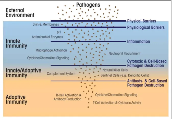
Figure 1: Overview of the immune system.
The innate response is characterized by being the first line of defense against infection in which leukocytes such as (e.g., neutrophils, monocytes, macrophages, etc.), are involved. These defense cells detect and attack other cells carrying pathogen-associated molecular patterns (PAMPs), and small proteins that signal pathogen invasion (i.e., cytokines and chemokines) or short peptides that directly attach to and restrict microbial pathogens. The cells of the adaptive immune system rely on T and B lymphocytes and proteins (i.e., antibodies) that detect and eliminate specific pathogens and also use cytokine/chemokine to signal and recruit other immune cells. Memory T and B lymphocytes have an essential role in immunity against pathogenic invasion. The complement system, along with natural killer cells and dendritic cells, straddles both innate and adaptive immunity. Adopted from (SPIERING, 2015).
Thus, the innate immune response plays a pivotal role in the defense against pathogens and is initiated through the pattern recognition receptors (PRRs). PRRs recognize pathogen/microbe-associated molecular patterns (PAMPs/MAMPs) and endogenous danger-associated molecular pattern (DAMPs), leading to the activation of host defense pathways that result in the clearance of the infection (CARRILLO et al., 2017; JANG et al., 2015; KUMAR; KAWAI; AKIRA, 2011; MOGENSEN, 2009; ROH; SOHN, 2018). Toll-like receptors (TLRs), a group of membrane-bound extracellular and endosomal receptors, sense the presence of infection through recognition of PAMPs. The innate immune response against microbes also involves a major inflammatory pathway known as inflammasomes (MARTINON; BURNS; TSCHOPP, 2002; SCHRODER; TSCHOPP, 2010).
Inflammasomes are multi-protein signaling complex that assembles after recognition of danger signals and/or pathogens by a family of cytosolic receptors called NLRs (nucleotide-binding domain leucine-rich repeat containing receptors) or PYHIN protein family members that consisting of immune sensors of intracellular DNA identified to activate inflammasomes (MARTINON; BURNS; TSCHOPP, 2002). Once assembled, inflammasomes initiate signaling by activation of downstream proteases, most notably 1 and less frequently caspase-11, which then proteolytically mature and promotes the secretion of pro-IL-1β and pro-IL-18, cytokines strongly implicated in the pathology of BJI (BROZ; MONACK, 2013; LATZ, 2010; LATZ; XIAO; STUTZ, 2013a; MARTINON; BURNS; TSCHOPP, 2002; WILSON et al., 1994). Furthermore, inflammasome activation triggers pyroptosis, an inflammatory form of cell death (BERGSBAKEN; FINK; COOKSON, 2009; YANG et al., 2019; YUAN et al., 2018). The role of NLRP1, NLRP3, NLRC4, NLRP6, NLRP7, NLRP12 and the PYHIN proteins AIM2 and IFI16 in assembling inflammasome and controlling the innate immune response against various microbes including different bacteria and the involvement of various bacterial factors have been intensively explored during the past decade (CANEPARO et al., 2018; LIN; ZHANG, 2017; SÁ; FESTA NETO, 2016; VLADIMER et al., 2013). Studies have shown that these inflammasomes form in response to various stimuli and are also subject to negative regulatory mechanisms, which cause the production of inflammatory cytokines to occur in a controlled manner after the onset of the inflammatory response (NAIK; DIXIT, 2010).
The NLRP3 inflammasome is the best characterized NLR so far. NLRP3, together with proteins ASC and caspase-1, form a cytoplasmic oligomeric complex known as the NLRP3 inflammasome, which plays a critical role in initiating innate immune responses, however, the molecular mechanisms of its activation are not fully understood (DAVIS; WEN; TING, 2011; DEIGENDESCH; ZYCHLINSKY; MEISSNER, 2018; MELEHANI et al., 2015). Assembly of the NLRP3 inflammasome has been shown to contribute to several pathologies like autoinflammatory syndromes, chronic inflammation, and metabolic diseases (DEIGENDESCH; ZYCHLINSKY; MEISSNER, 2018).
The role of inflammasomes formed in S. aureus-infected professional phagocytes that ingest and/or kill invasive bacteria has been extensively investigated. However, despite numerous works on the mechanism used by S. aureus to internalize into and survive in non-professional phagocytes, such as epithelial cells (KREMSEROVA; NAUSEEF, 2019; MA et al., 2019), the involvement of inflammasomes and processed IL-1β, which plays a crucial role in bone homeostasis in S. aureus-associated BJI, has not been investigated. This is of particular interest in the case of BJI as S. aureus is shown to internalize and to survive within the osteoblasts, generating chronic infections refractory to antimicrobial treatments. It is therefore
10
essential to develop new approaches to better understand the mechanisms by which staphylococci interact with osteoblasts and, in fine, generates damage to bones.
Introduction générale
Staphylococcus aureus (S. aureus) est une bactérie à Gram positif capable de coloniser l'hôte humain ou animal et de provoquer un large panel d’infections allant de bénignes à potentiellement mortelles telles que pneumonie, septicémie, endocardite, ostéomyélite, infections associées aux dispositifs médicaux internes, syndrome de choc toxique, abcès, ou mammite entre autres maladies (FITZGERALD, 2012; FOSTER, 1996; WERTHEIM et al., 2005). S. aureus est notamment une cause fréquente d'infections des os et des articulations (BJI, pour l’anglais Bone and Joint Infection), maladie associée à une morbidité élevée et à d’importants coûts de santé dans le monde (DEL POZO; PATEL, 2009; TUCHSCHERR; GERACI; LÖFFLER, 2017; WRIGHT; NAIR, 2010; ZIMMERLI; TRAMPUZ; OCHSNER, 2004). Les infections osseuses induites par S. aureus peuvent évoluer en infections chroniques, devenant très réfractaires aux traitements antibiotiques (LOWY, 1998). Cela est probablement dû aux différentes stratégies que cette bactérie à développer pour s’adapter et survivre à l’intérieur des cellules hôtes (EIFF; PETERS; PROCTOR, 2001; MONTGOMERY; DAVID; DAUM, 2015; TUCHSCHERR; GERACI; LÖFFLER, 2017).
Au cours des dernières années, notre compréhension de la manière dont le système immunitaire inné reconnaît les agents pathogènes a considérablement avancé (CARRILLO et al., 2017; NICHOLSON, 2016). Une invasion de microorganismes étrangers induit un spectre de réponses inflammatoires chez l'hôte infecté (Fig. 1). L'inflammation est classiquement décrite comme une réponse essentielle de la réponse immunitaire de l'hôte qui permet la survie pendant une infection ou une blessure et maintient l'homéostasie tissulaire dans diverses conditions nocives (CHEN, L. et al., 2017; CRUVINEL et al., 2010; MEDZHITOV, 2010a; RANKIN; ARTIS, 2018).
Le type de réponse inflammatoire dépend de la catégorie du déclencheur inflammatoire. Il a été démontré que les bactéries pathogènes sont reconnues par les récepteurs du système immunitaire inné, tels que les Toll-like receptors (TLR), entraînant l'induction de divers médiateurs inflammatoires comme les cytokines et les chimiokines inflammatoires, ainsi que les prostaglandines, qui agissent sur les tissus cibles (CARRILLO et al., 2017; CHEN, L. et al., 2017; LUIS MUÑOZ-CARRILLO et al., 2019; TURNER et al., 2014). La réponse inflammatoire aiguë est terminée lorsque l'agent déclencheur de l’infection est éliminé et que les tissus endommagés sont réparés. Si cette réponse inflammatoire initiale n'élimine pas les agents pathogènes, comme dans le cas d'infections chroniques ou de lésions tissulaires non réparées, le système immunitaire adaptatif intervient. Les cellules du système immunitaire adaptatif migrent vers le site de l'infection et commencent à éliminer les agents pathogènes et
12
les cellules et tissus endommagés (MEDZHITOV, 2008, 2010a). Cependant, dans certaines infections chroniques, l'inflammation peut persister sans immunité adaptative (CRUVINEL et al., 2010).
Figure 1: Vue d'ensemble du système immunitaire. La réponse innée est caractérisée par le fait qu'elle constitue la première ligne de défense contre l'infection dans laquelle interviennent des leucocytes tels que des neutrophiles, des monocytes, ou des macrophages. Ces cellules de défense détectent et attaquent d'autres cellules portant des structures pathogen-associated molecular patterns (PAMP) et de petites protéines qui signalent une invasion d'agents pathogènes (cytokines et chimiokines) ou de courts peptides antimicrobiens se liant directement aux agents pathogènes microbiens. Les cellules du système imunitaire adaptatif s'appuient sur des lymphocytes T et B et des protéines (les anticorps) qui détectent et éliminent spécifiquement des agents pathogènes et prodiosent également une cytokine/chimiokine afin de signaler et de recruter d'autres cellules immunitaires. Les lymphocytes T et B jouent un rôle essentiel dans l'immunité contre l'invasion pathogène. Le système du complément, ainsi que les cellules tueuses naturelles (NK) et les cellules dendritiques, interviennent à la fois dans l'immunité innée et adaptative. Reproduit de (SPIERING, 2015).
Ainsi, la réponse immunitaire innée joue un rôle central dans la défense contre les agents pathogènes et est initiée par les pattern recognition receptors (PRR). Les PRR reconnaissent les pathogen/microbe-associated molecular patterns (PAMPs/MAMPs) et les endogenous danger-associated molecular pattern (DAMPs), ce qui conduit à l'activation des voies de défense de l'hôte qui entraînent l'élimination de l'infection (CARRILLO et al., 2017; JANG et
al., 2015; KUMAR, H.; KAWAI; AKIRA, 2011; MOGENSEN, 2009; ROH; SOHN, 2018). Les récepteurs Toll-like (TLR), un groupe de récepteurs extracellulaires et endosomaux liés à la membrane, détectent la présence d'une infection par la reconnaissance des PAMP. La réponse immunitaire innée contre les microbes implique également une voie inflammatoire majeure connue sous le nom d’inflammasomes (MARTINON; BURNS; TSCHOPP, 2002; SCHRODER; TSCHOPP, 2010a).
Les inflammasomes sont des complexes de signalisation multi-protéines qui s'assemblent après la reconnaissance des signaux de danger et/ou des agents pathogènes par une famille de récepteurs cytosoliques appelés NLR (nucleotide-binding domain leucine-rich repeat containing receptors) ou de membres de la famille de protéines PYHIN (MARTINON; BURNS; TSCHOPP, 2002). Une fois assemblés, les inflammasomes initient l’activation de protéases en aval, notamment la caspase-1 et la caspase-11, qui ensuite maturent protéolytiquement la pro-IL-1β et la pro-IL-18 et favorisent leur sécrétion cellulaire (BROZ; MONACK, 2013; LATZ, 2010; LATZ; XIAO; STUTZ, 2013a; MARTINON; BURNS; TSCHOPP, 2002; WILSON et al., 1994).
De plus, l'activation de l'inflammmasome déclenche la pyroptose, une forme inflammatoire de mort cellulaire (BERGSBAKEN; FINK; COOKSON, 2009; YANG, YANG et al., 2019; YUAN et al., 2018). Le rôle de NLRP1, NLRP3, NLRC4, NLRP6, NLRP7, NLRP12 et des protéines AIM2 et IFI16 de PYHIN dans l'assemblage de l'inflammasome et le contrôle de la réponse immunitaire innée contre divers microbes, y compris différentes bactéries, est maintenant bien documenté. De même, l'implication de divers facteurs bactériens dans le déclenchement de l’inflammasome a été intensément explorés au cours des dernières années (CANEPARO et al., 2018; LIN; ZHANG, 2017; SÁ; FESTA NETO, 2016; VLADIMER et al., 2013a). Ces inflammasomes se forment en réponse à divers stimuli et sont également soumis à des mécanismes de régulation négative qui modulent la production de cytokines inflammatoires après le déclenchement de la réponse inflammatoire (NAIK; DIXIT, 2010).
L'inflammasome NLRP3 est le NLR le mieux caractérisé. NLRP3, ainsi que la protéine ASC et la Caspase-1, forment un complexe oligomérique cytoplasmique appelé inflammasome NLRP3, qui joue un rôle essentiel dans l'initiation de la réponse immunitaire innée. Cependant, les mécanismes moléculaires de son activation ne sont pas entièrement compris (DAVIS; WEN; TING, 2011; DEIGENDESCH; ZYCHLINSKY; MEISSNER, 2018; MELEHANI et al., 2015). L'assemblage de l'inflammasome NLRP3 a été associé à plusieurs pathologies telles que des syndromes auto-inflammatoires, l’inflammation chronique et certaines maladies métaboliques (DEIGENDESCH; ZYCHLINSKY; MEISSNER, 2018).
Le rôle des inflammasomes formés dans les phagocytes professionnels infectés par S. aureus qui ingèrent et/ou tuent les bactéries invasives a été étudié de manière approfondie.
14
Cependant, en dépit de nombreux travaux sur le mécanisme utilisé par S. aureus à internaliser dans et survivre dans les phagocytes non professionnels, comme les cellules épithéliales (KREMSEROVA; NAUSEEF, 2019; MA et al., 2019) l'implication des inflammasomes et de l'IL-1β traitée, qui joue un rôle crucial dans l'homéostasie osseuse dans le BJI associée à S. aureus, n'a pas encore été démontrée. Ceci est particulièrement intéressant dans le cas de BJI que S. aureus est représenté à internaliser et à survivre dans les ostéoblastes, générant des infections chroniques réfractaires aux traitements antimicrobiens. Il est donc essentiel de développer de nouvelles approches pour mieux comprendre les mécanismes par lesquels les staphylocoques interagissent avec ostéoblastes et, in fine, génère des dommages aux os.
16
Bibliographic synthesis
1. General characteristics of Staphylococcus aureus
1.1. Definition and morphology
Staphylococcus aureus (S. aureus) was described by Alexander Ogston and Louis Pasteur as cocci with a mean diameter of 0.5 and 1.5 μm, isolated from furuncle and abscesses in 1880 (OGSTON, 1881). The genus Staphylococcus belongs to the family Micrococcaceae, which is composed of Gram-positive bacteria (ROSYPAL; ROSYPALOVÁ; HOREJS, 1966). The name “Staphylococc” derives from the Greek words Staphyle (Grape-like clusters) and aureus (gold) (BROWN; GRILLI, 1998). S. aureus is immobile, non-spore-forming, produces coagulase and is often unencapsulated or has a limited capsule (OGSTON, 1881).
1.2. General cultural, biochemical characteristics and genome of S. aureus
S. aureus is an aerobic and facultative anaerobic bacterium. It readily grows on basal media and forms golden-yellow colonies on nutrient agar. It is positive for catalase, negative for oxidase and capable of fermenting mannitol (LOWY, 1998). S. aureus can grow in a wide range of temperatures. Its optimal growth temperature is 30º- 37ºC, but it can grow between 7º and 48.5ºC. Its optimum pH is 7.2 (but it can grow in a range of 4.2 – 9.3). It also grows in a wide range of sodium chloride concentration (7.5-10% NaCl) (LE LOIR; BARON; GAUTIER, 2003; SCHMITT; SCHULER-SCHMID; SCHMIDT-LORENZ, 1990). In blood agar, S. aureus forms beta-hemolytic colonies due to the production of hemolysins (α-toxin, β-toxin, γ-toxin and δ-toxin) (BLAIR, 1958; DINGES; ORWIN; SCHLIEVERT, 2000).
The first fully sequenced and annotated genomes were published in 2001 by Hiramatsu’s group, comparing the genomes of two methicillin-resistant strains, Mu50 and N315 (KURODA et al., 2001). This opened the way to numerous other sequencing projects, which led to great insights into our understanding of this major pathogen. Genomic analyses of animal and human strains show that the whole genome is about 2.82 Mb in size. The genome of S. aureus contains 2.872 genes encoding 2.767 proteins. The GC content of the genome was found to be 32.7%. The core genome of S. aureus consists of about 80% of conserved genes between strains, encoding essential metabolic and regulatory functions, as well as surface proteins involved in adhesion and surface architecture (BABA et al., 2008; GILLASPY et al., 2006; LINDSAY; HOLDEN, 2004).
The other predicted genes make up an "accessory" genome, comprising a collection of mobile genetic elements (MGEs), including bacteriophages, chromosomal cassette and pathogenicity islands (KURODA et al., 2001; LINDSAY; HOLDEN, 2004, 2006). MGEs encode most of the virulence factors of S. aureus. The exchange of virulence factors that are transported by MGEs from a strain to another is a driving force in S. aureus evolution. The difference in the gene content of S. aureus strains is due to deletions of genes and insertions by MGEs.
Some regions of the central genome show inter-strain variability. The allelic variation of structural and functional genes of S. aureus arises from the random substitution of nucleotides, followed by selection, recombination and horizontal gene transfers (CROSSLEY et al., 2009; FENG et al., 2008; LINDSAY; HOLDEN, 2004). In addition to the chromosome, S. aureus strains can also contain plasmid elements encoding for resistance and virulence determinants (CROSSLEY et al., 2009; FITZGERALD et al., 2001; LINDSAY, 2010; LINDSAY; HOLDEN, 2004, 2006).
1.3. General pathogenesis and clinical diseases
1.3.1 Pathogenesis
The development of S. aureus infections consists of five different stages: bacterial colonization, followed by local infection, dissemination, metastatic infection and toxinosis (GNANAMANI; HARIHARAN; PAUL-SATYASEELA, 2017). S. aureus is often found in healthy carriage in mammals and birds. It is a frequent inhabitant of human retronasal ways, in which it may be part of the human microbiota without causing disease in the host (OTTO, 2010a).
S. aureus is a commensal and opportunistic pathogen that causes a wide range of acute infections (septicemia, bacteremia, pneumonia, meningitis) and chronic diseases of bones, joints, and skin in humans, ranging from mild to life-threatening infections (CHAMBERS, 1997; FITZGERALD, 2012; LOWY, 1998). Osteomyelitis is an inflammatory disease that accompanies bone destruction. It is commonly caused by bacteria. However, infections occurring through fungi or other parasites have also been reported (GOMES; PEREIRA; BETTENCOURT, 2013; URS et al., 2016).
S. aureus is the most common infectious causative organism of osteomyelitis with more than 70% of occurrences. In terms of prevalence, it is followed by Gram-negative bacilli, Streptococcus pneumoniae and Streptococcus pyogenes (CHIAPPINI; MASTRANGELO; LAZZERI, 2016). These bacteria adhere to the bone matrix leading to the formation of biofilms. In these biofilms, pathogens undergo complex metabolic changes that make these bacteria
18
less sensitive, both to the host immune response and to antibiotics. The complex nature of biofilm makes treatment difficult leading to long-term antibiotic therapy, and to high morbidity and elevated cure costs for patients and hospitals (CRAFT et al., 2019).
S. aureus is also a major animal pathogen. In dairy animals, such as cows and small ruminants, mastitis remains a major concern worldwide that affects animal health, and leads to huge economic losses in the dairy industry due to veterinary treatments, premature culling, and the change in milk quality (LESCOURRET; COULON; FAYE, 1995; PETON; LE LOIR, 2014; RAINARD et al., 2018). Although viruses, fungi or protozoa can cause mastitis, the most common causes are bacteria (AMIN; HAMOUDA; ABDEL-ALL, 2011; DEB et al., 2013). Escherichia coli is often associated with acute mastitis in bovine. In contrast, S. aureus most commonly causes subclinical mastitis that tends to become chronic in cows (GÜNTHER et al., 2017; SEARS; MCCARTHY, 2003; YOUNIS; JAVED; BLUMENBERG, 2016).
Subclinical mastitis accounts for about 80% of all economic losses associated with mastitis, related to reduced milk production and quality, as well as treatment and prevention costs (PETROVSKI; TRAJCEV; BUNESKI, 2006). Massive antibiotic use in veterinary medicine is regarded as a major problem in terms of the emergence and spread of antibiotic-resistant strains (FAIR; TOR, 2014; LI; WEBSTER, 2018; VENTOLA, 2015a). Prevention (e.g. anti-mastitis vaccine) is by far the best strategy. However, most vaccines tested to date do not allow the development of an efficient and protective defense against S. aureus mastitis (WALLEMACQ et al., 2012).
In human medicine, S. aureus causes huge problems such as nosocomial infections, notably because some strains are resistant to a wide variety of antimicrobials. Penicillin was discovered by Alexander Fleming in 1928 and was first administered to infected patients as a chemotherapeutic agent in 1941. The resistance to penicillin in S. aureus rapidly appeared in 1942, followed by more general mechanisms of resistance against the family of beta-lactams (CHAIN et al., 1940; FLEMING, 1929a; FLETCHER, 1984; RAMMELKAMP; MAXON, 1942; VENTOLA, 2015a). The resistance of S. aureus to beta-lactams and other types of antibiotics has contributed to a decrease in the number of effective drugs against this microorganism (LEVY; MARSHALL, 2004).
1.3.2 Virulence factors
The importance and success of S. aureus as a pathogen relies on the combination of toxins-mediated virulence, invasiveness and antimicrobial resistance (LE LOIR; BARON; GAUTIER, 2003), as well as cell-cell interaction, immune response evasion, and tissue damage (FENG
et al., 2008; GORDON; LOWY, 2008). The pathogenicity of this bacterium is due to both the diversity of its virulence factors and its ability to express its virulence in various host environments (PEACOCK et al., 2002).
The repertoire of S. aureus virulence factors is extensive and comprises secreted and structural products, including membrane proteins, that direct the process of pathogenesis (GORDON; LOWY, 2008). Due to the impact of this bacterium on human and animal health, the structure and function of its virulence factors, its interaction with the host and the molecular mechanisms involved in resistance to antibiotics have been extensively studied (HECKER et al., 2010; LINDSAY; HOLDEN, 2004).
Based on their mechanism of action and role in pathogenesis, staphylococcal virulence factors are classified as represented in Table 1. Among the virulence factors produced by this microorganism, the Microbial surface components recognizing adhesive matrix molecules (MSCRAMMs) are of particular importance in S. aureus colonization. MSCRAMMs form a group of molecules that bind to components of the extracellular matrix, such as collagen, fibronectin (Fn) and fibrinogen (Fg). Binding to these components facilitates bacterial internalization into the host cells and promotes tissue damage (WANN; GURUSIDDAPPA; HOOK, 2000).
The fibronectin binding proteins FnBPA and FnBPB of S. aureus are responsible for the Fn adhesion process into the host cell surface (FOSTER et al., 2014; FOSTER; HÖÖK, 1998; JOSSE; LAURENT; DIOT, 2017). Binding to Fn plays a major role in bacteria-cell interactions, but S. aureus also produces components able to bind to Fg and collagen (Hienz et al, 1996). The binding to Fg is mainly mediated by Clumping factor A and B (ClfA) and (ClfB) proteins, which allow bacterial adhesion to blood plasma and biomaterials exposed to human or animal blood (FOSTER; HÖÖK, 1998).
All these factors allow S. aureus to bind to the components of the extracellular matrix, facilitating the infection of several cell types, such as epithelial and immunological cells (OTTO, 2010a; PIETROCOLA et al., 2017). S. aureus also produces a variety of other secreted virulence factors including toxins (leucocidins, haemolysins, exfoliative toxins, enterotoxins, (TSST-1) - Toxic shock syndrome toxin -1), enzymes, and a group of factors encompassing the phenol-soluble modulins (PSM) (GNANAMANI; HARIHARAN; PAUL-SATYASEELA, 2017; OTTO, 2014a). PSMs may play an important role in the different stages of pathogenesis, including adhesion, invasion, persistence, evasion, and death of host cells (GORDON; LOWY, 2008; OTTO, 2014b).
The expression of virulence factors is tightly controlled in S. aureus. Following the establishment of the colonization, S. aureus represses the expression of surface proteins
20
associated with adhesion and internalization and produces toxins and other secreted virulence factors responsible for bacterial dispersion and, consequently, the symptoms of infections (NOVICK, 2003; NOVICK; GEISINGER, 2008). The different virulent factors expressed are controlled by a definite locus named the accessory gene regulator (agr) and the staphylococcal accessory regulator (sar) (JARRAUD et al., 2002).
The agr system is formed by a two-component system, which is responsible for the regulation of several virulence factors associated with the infectious processes (NOVICK; GEISINGER, 2008; OTTO, 2004). The agr operon is composed of four genes (agrA, agrB, agrC and agrD) that respectively produce AgrA, AgrB, AgrC and the auto-inducible peptide (AIP) (Fig. 2). Virulence expression is based on a quorum-sensing mechanism. During bacterial growth, AIP accumulates in the surrounding medium and when AIP concentration reaches a given threshold, it induces a shift of expression resulting in a repression of the genes encoding surface proteins and an induction of genes encoding secreted proteins (toxins). AIP, which is processed and secreted by AgrB, binds to the membrane receptor formed by AgrC, which phosphorylates or dephosphorylates AgrA. AgrA functions as a signal transducer of the AIP binding to the ArgC receptor, activating the expression of the agr operon and the RNAIII gene. The latter expresses the RNAIII, a regulator of several genes related to virulence in S. aureus (LE; OTTO, 2015; NOVICK, 2003; NOVICK; GEISINGER, 2008).
Table 1: Virulence factors of S. aureus and their characteristics. Adapted by (GNANAMANI; HARIHARAN; PAUL-SATYASEELA, 2017).
Factors Characteristics References
Helping attachment to host tissues
Breaking/evading the host immunity Polysaccharide microcapsule
Protein A (SpA)
Panton-Valentine leukocidin (PVL) Genestier et al., 2005
Voyich et al., 2006
α-Hemolysin
Chemotaxis-inhibitory protein of S. aureus (CHIPS):
Tissue invasion
Extracellular adherence protein (Eap)
Induces toxinosis
Staphylococcal Enterotoxins (SE) There are more than 25 known SEs. SEs bind MHC class II receptors on T-cells and hyperstimulate the T-cells. S. aureus produces battery of enterotoxins which are potent gastrointestinal exotoxins. The Staphylococcal food poisoning is an intoxication which results from consumption of foods containing sufficient amount of preformed enterotoxins
Argudin et al., 2010
Toxic shock syndrome toxin -1 (TSST-1) TSST-1 and some of enterotoxins are called as pyrogenic toxin super antigens. TSST-1 causes toxic shock
syndrome especially in menstrual women Dinges et al., 2000
Exfoliative toxins A and B Serine proteases which selectively recognize and hydrolyze desmosomal proteins in the skin. ETs cause staphylococca-scalded skin syndrome, a disease predominantly affecting infants
Bukowski et al., 2010 Microbial Surface Components Recognizing
adhesive matrix molecules (MSCRAMM)
Cell surface proteins which interact with host molecules such as collagen, fibronectin and fibrinogen, thus, facilitate the tissue attachment. Staphylococcal protein A, fibronectin-binding proteins A and B, collagen-binding protein and clumping factor A and B belong to this family. They are also involved in host immune evasion
Resist the phagocytosis and killing by polymorphonuclear phagocyte
Vazquez et al., 2011
Nilsson et al., 1997
Proteases, lipases, nucleases, hyaluronatelyase, phospholipase C, metalloproteases (elastase), and
Staphylokinase
These extracellular enzymes cause tissue destruction and,
Hong et al., 2016
Bhakdi, and Tranum-Jensen, 1991
Postma et al., 2004
Edwards et al., 2012 Binding of the Fcγ domain of immunoglobulin (Igs)
PVL is found in most of community-associated MRSA (CA-MRSA). PVL consists of two subunits, LukS and LukF. LukS binds to human complement receptors C5aR and C5L2 and allows docking of LukF. The Luk subunits oligomerizes and form a pore lysing the cell.
Disruption of epithelial or endothelial surfaces
Impediment of neutrophil chemotaxis
22
Cytotoxins and enzymes can be seen as a means to convert host molecules into nutrients for bacterial growth or to cleave antibiotic molecules (OTTO, 2014a). Hemolysins (α, β, γ, and δ), nucleases, proteases, lipases, hyaluronidase, and collagenase can indeed provide nutrients for the bacterium from the host material. Besides, enzymes such as β-lactamases are responsible for inhibiting the activity of antibiotics derived from β-lactam rings. Among the toxins produced by S. aureus, superantigens are potent and non-specific immune response inducers (MAKHLIN et al., 2007; TOKAJIAN et al., 2011). Also referred to as pyrogenic toxins, they include staphylococcal enterotoxins (more than 25 SEs have been described so far) and toxic shock syndrome toxin-1 (TSST-1) (CHOI et al., 1989; DINGES; ORWIN; SCHLIEVERT, 2000; KRAKAUER, 2019; MARRACK; KAPPLER, 1990).
Figure 2: Accessory gene regulatory system (Agr).
Quorum sensing system agr encodes gene products that ultimately promote further transcription of agr locus in a process called autoactivation. According to (NOVICK; GEISINGER, 2008).
1.3.3 Phenol-soluble modulins (PSMs)
In 1999, Seymour Klebanoff described the so-called Phenol-soluble modulins (PSM), a group of molecules contained in the supernatant of Staphylococcus epidermidis, isolated by size exclusion chromatography with the use of a solvent containing phenol (MEHLIN; HEADLEY;
KLEBANOFF, 1999; OTTO, 2004). PSMs are part of a family of short amphipathic, α-helical peptides, which are produced by various staphylococcal strains and which have great importance in the virulence process of S. aureus (Fig. 3) (GONZALEZ et al., 2014; OTTO, 2014b; PESCHEL; OTTO, 2013).
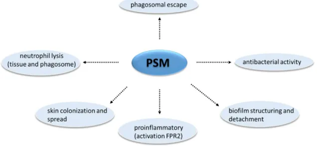
Figure 3: Overview of phenol-soluble modulin activities in S. aureus.
PSMs participate in the formation of biolfims. PSMs may be antimicrobial agents, they also act to induce cell lysis, cytokine production through the FPR2 receptor, and participate in the S. aureus dispersion process.
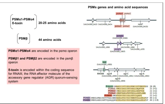
PSMs were described as a proinflammatory "complex" of seven PSMs, including the shortest α type with approximately 20-25 amino acids in length (four PSMα1-PSMα4 peptides), the longest β type with approximately 43- 45 amino acids (PSMβ1 and PSMβ2) and δ-toxin peptides ranging in length from 25 to 26 amino acids (CHEUNG et al., 2014, 2016; MEHLIN; HEADLEY; KLEBANOFF, 1999). The PSM–encoding genes scattered at different locations in the genome. The PSMα1-PSMα4 genes are encoded in the PSMα operon. The PSMβ1 and PSMβ2 peptides encoded in the PSMβ operon and the δ-toxin is encoded within the RNAIII sequence (Fig. 4) (CHATTERJEE et al., 2011; OLIVEIRA; BORGES; SIMÕES, 2018).
Likewise several other major staphylococcal exotoxins, PSMs are positively regulated by agr, the global regulatory quorum sensing system (GOMES-FERNANDES et al., 2017; QUECK et al., 2008; XU et al., 2017). The PSMs are agr-regulated through AgrA, not by RNAIII, which modulates several other virulence genes in S. aureus (MARROQUIN et al., 2019; PESCHEL; OTTO, 2013; TAN et al., 2018).
24
Figure 4: Phenol-soluble modulins peptides (PSMs) define the virulence potential of S. aureus. PSMs have a pronounced ability to lyse human leukocytes and other cell types, to stimulate an inflammatory response and the formation of biofilm (CHEUNG et al., 2014, 2016; PESCHEL; OTTO, 2013; TOWLE et al., 2016).
Their importance in the pathogenesis was reported by Voyich et al. who showed in 2005 that, in the CA-MRSA strains (LAC and MW2), PSMs participate in S. aureus virulence, in a model of skin infections and bacteremia in mice (PESCHEL; OTTO, 2013). Specifically, PSMα group induces a stronger inflammatory response compared to PSM
, whereas the PSMβ group and the δ-toxin did not significantly influence the inflammation process. PSMα group increased the amount of neutrophils and monocyte chemotaxis, a critical step in the establishment of S. aureus infection, in addition to elevating leukocyte death in vitro (WANG et al., 2007). The dissemination capacity of S. aureus and S. epidermidis, associated with biofilm formation, is directly related to the production of PSMs (LE; OTTO, 2015; OTTO, 2017; QUECK et al., 2008; TSOMPANIDOU et al., 2013; WANG et al., 2011).The ability to induce cellular lysis is the most relevant feature of these PSMs (CHEUNG et al., 2014). The action of the PSMs is not specific and is independent of receptors. It affects almost all eukaryotic membranes leading to cell lysis, which is probably due to a disturbance of the plasma membrane (CHEUNG; DUONG; OTTO, 2012; KRETSCHMER et al., 2010). Other S. aureus toxins, such as α-toxin or Panton-Valentine Leukocidin (PVL), specifically lyse polymorphonuclear neutrophils (PMNs) (DIEP et al., 2010; LÖFFLER et al., 2010; OLIVEIRA;
BORGES; SIMÕES, 2018). Although this group of peptides is linked to the cellular lysis process, only a portion of the PSMs is responsible for this activity (CHEUNG et al., 2014). S. aureus PSMα3 and S. epidermidis PSMδ exhibit the most evident cytolytic activities among PSMs, whereas PSMα1 and PSMα2 are moderately cytolytic (CHEUNG; DUONG; OTTO, 2012; CHU et al., 2018; DA et al., 2017; OTTO, 2017).
Among other activities, PSMs induce neutrophil chemotaxis, oxygen radical production, intracellular calcium influx, and release of proinflammatory cytokines such as Interleukin-8 (IL-8), a member of the CXC subfamily of chemoattractant cytokines (HENKELS et al., 2011; PARKER et al., 2016; WANG et al., 2007). The ability of the PSMs to induce an inflammatory response classify them as pathogen-associated molecular patterns (PAMPs), which are a group of microbial molecules that alert the host organism when a pathogen infection occurs (BIANCHI, 2007; CHEUNG et al., 2014). In addition, PSMs have been shown to be capable to decreasie the expression of proinflammatory cytokines such as IL-32, IL-6 and IL-8 (DEPLANCHE et al., 2016).
2. The inflammasome, a primordial complex of innate immunity
2.1. Mechanisms of the inflammatory response
The immune system is formed by a series of effector mechanisms which can destroy a variety of invading pathogenic organisms belonging to four main groups fungi, bacteria, parasites, and viruses (CARRILLO et al., 2017; MESQUITA JÚNIOR et al., 2010; NICHOLSON, 2016). The immune system is composed of two types of responses: antigen-specific adaptive immune response and the innate immune response recognizing microbial associated molecular patterns (MAMP's) (RYU, 2017). Innate immune response described as the organism's first line of defense is of primary importance in the immediate recognition and elimination of invasive microorganisms (ESPINOSA; RIVERA, 2016). The innate immune response consists basically of physical and chemical barriers, reactive oxygen species (ROS), innate immune cells, and soluble mediators such as the complement system, innate antibodies, and associated cytokines and recognizes pathogen-associated molecular patterns (PAMPs) (CARRILLO et al., 2017; CRUVINEL et al., 2010).
PAMPs are recognized by pattern recognition receptors (PRRs), expressed primarily in cells of the innate immune system (KUMAR; KAWAI; AKIRA, 2011; MOGENSEN, 2009). PRRs can also recognize host molecules that contain damage-associated molecular patterns (DAMPs), these molecules are released when a pathogenic organism is able to multiply in the host as a