HAL Id: tel-02917956
https://tel.archives-ouvertes.fr/tel-02917956
Submitted on 20 Aug 2020HAL is a multi-disciplinary open access archive for the deposit and dissemination of sci-entific research documents, whether they are pub-lished or not. The documents may come from teaching and research institutions in France or abroad, or from public or private research centers.
L’archive ouverte pluridisciplinaire HAL, est destinée au dépôt et à la diffusion de documents scientifiques de niveau recherche, publiés ou non, émanant des établissements d’enseignement et de recherche français ou étrangers, des laboratoires publics ou privés.
Imaging of intracellular trafficking of the HIV-1
nucleocapsid protein
Iryna Lysova
To cite this version:
Iryna Lysova. Imaging of intracellular trafficking of the HIV-1 nucleocapsid protein. Virology. Uni-versité de Strasbourg, 2017. English. �NNT : 2017STRAJ092�. �tel-02917956�
UNIVERSITÉ DE STRASBOURG
ÉCOLE DOCTORALE DES SCIENCES DE LA VIE ET DE LA SANTÉ
UMR CNRS 7312 Laboratory of Biophotonics and Pharmacology
THÈSE
présentée par :Iryna LYSOVA
soutenue le : 07 Juliet 2017
pour obtenir le grade de :
Docteur de l’université de Strasbourg
Discipline/ Spécialité: Sciences de la vie/Biophysique
Imagerie quantitative du traffic
intracellulaire de VIH-1
THÈSE dirigée par :
M. MELY Yves Professeur, université de Strasbourg M. DMITRUK Igor Professeur, université nationale Taras Chevtchenko de Kyiv
RAPPORTEURS :
Mme BONNEAU Stéphanie Maitre de Conférences, université Pierre et Marie Curie M. SEKSEK Olivier Chargé de Recherche CNRS, université Paris Sud
AUTRES MEMBRES DU JURY :
M. RUFF Marc Directeur de recherches CNRS, IGBMC Mme ANTON Halina Chargé de Recherche CNRS, université de Strasbourg
2
Table of contents
________________________________________________________________________________
List of abbreviations ... 4
Chapter 1: Introduction ... 6
1.1. AIDS and HIV ... 7
1.2. Subtypes of HIV ... 9
1.3. HIV-1: structure and functions of viral components ... 11
1.3.1. Viral genome ... 12
1.3.2. Structural protein Gag ... 14
1.3.3. Enzymatic proteins ... 16
1.3.4. Envelope proteins ... 17
1.3.5. Regulatory and accessory proteins ... 18
1.4. Viral life cycle ... 19
1.4.1. Early stages of infection ... 20
1.4.2. Late phase of infection ... 25
1.5. NCp7: structure and function ... 27
1.5.1. Structure ... 28
1.5.2. Interaction with nucleic acids ... 28
1.5.3. Chaperone properties ... 29
1.5.4. Interaction with cellular proteins ... 30
1.5.5. Inhibition ... 31
1.6. Role of NCp7 during infection ... 32
1.7. Microscopy approaches to image the HIV-1 virology ... 34
1.7.1. Electron microscopy ... 36
1.7.2. Atomic force microscopy ... 39
1.7.3. Fluorescence microscopy ... 41
1.7.4. High-resolution microscopy ... 45
1.7.5. HIV microscopy by EM and AFM ... 59
1.7.6. HIV-1 imaging by fluorescence microscopy ... 66
Research objectives ... 76
Chapter 2: Materials and methods ... 78
2.1. Cell culture ... 79
2.1.1.Transfection ... 79
2.1.2. Energy depletion ... 80
2.2. Production of pseudoviral particles ... 80
2.2.1. Pseudotyping of the lentiviral vectors ... 80
2.2.2. Vesicular stomatitis virus glycoprotein ... 80
2.2.3. Tetracysteine tag conjugation ... 82
2.2.4. Labeling ... 82
2.3. Imaging ... 82
2.3.1. Confocal ... 83
3 2.3.3. CLEM ... 83 2.4. PALM ... 84 2.4.1. Sample preparation ... 84 2.4.2. PALM ... 85 2.4.3. Data treatment ... 86 2.4.4. Analysis ... 87 Chapter 3: Results ... 89
3.1. Cytoplasmic Release of NCp7 proteins from the HIV-1 viral cores during the early steps of infection ... 90
3.1.1. Introduction ... 90
3.1.2. Publication ... 92
3.2. High resolution imaging of NCp7 in the context of pseudoviral infection ... 109
3.2.1. Introductio ... 109
3.2.2. PALM imaging of the free HIV-1 pseudoviruses ... 110
3.2.3. PALM imaging of the HIV-1 pseudoviruses in the infected cells ... 112
3.2.3.1. Preliminary experiments of PALM imaging of TC-FlAsH pseudoviruses in HeLa cells ... 112
3.2.3.2. Intracellular distribution of Ncp7 during the early stage of infection ... 115
3.2.3.3. Effect of the labeling density ... 116
3.2.3.4. Distribution of the HIV-1 pseudoviruses .. during the time course of the infection ... 119
3.2.3.5. Effect of RT inhibition on the cellular distribution of pseudoviruses ... 123
3.2.4. Discussion and conclusions ... 127
3.3. Mechanism of NCp7 nuclear entry ... 130
3.3.1. Introduction ... 130
3.3.2. Preliminary experiments ... 134
3.3.3. Intracellular distribution of the viral complexes in energy depleted cells ... ... 138
3.3.4. Imaging of NCp7 at the nuclear envelope ... 143
3.3.5. CLEM ... 145
3.3.5.1. Proof of concept ... 145
3.3.5.2. TEM observations of NCp7-TC/ReAsH ... 149
3.3.6. Discussion and conclusions ... 153
Chapter 4: Conclusions and perspectives ... 156
Chapter 5: Another works ... 162
5.1. Tuning luminescent properties of CdSe nanoclusters by phosphine surface passivation ... 163
5.2. Publication ... 166
List of references ... 174
4
List of abbreviations
________________________________________________________________________________
a.a. AFM AIDs ATP AZT CA CCD CLEM CTD DAB EDT EM EMCCD Env ER ESCRT ET FG FIB-SEM FPs Gag GFP Gp gRNA/vRNA GSD GTP HIV HR HTLV IN KPN LTR MA MEA NA NA NCp7 NES NLS NPC NTD Nup PAFPs PAINT PALM PALMIRA PBS PBS amino acidsatomic force microscopy acquired immunodeficiency adenosine triphosphate azidothymidine capsid
camera coupled charged devices correlative light electron microscopy carboxy-terminal domain 3,3’-diaminobenzidine ethandithiol electron microscopy electron multiplier CCD envelope endoplasmic reticulum
endosomal sorting complex required for transport electron tomography
phenylalanine-glycine focused ion beam-sEM fluorescent proteins group specific antigen green fluorescent protein glycoprotein genomic/viral RNA ground-state-depletion microscopy guanosine triphosphate human immunodeficiency high resolution
human T-lymphotropic virus integrase
karyopherin long terminal repeat
matrix cysteamine nucleic acids numerical aperture
nucleocapsid protein 7super-resolution nuclear export signal
nuclear localization signal nuclear pore complex amino-terminal domain nucleoporins
photoactivatable fluorescent protein
point accumulation for imaging in nanoscale topography photoactivation localization microscopy
PALM with independently running acquisition primary binding site
5 PIC Pol PPT PPTc PR PSF QDs R Ran RESOLFT RNP RRE RT RT RTC SEM SIM SL SP SR ssDNA/dsDNA SSIM ssTEM STED STORM SVT TAR TC TEM TIRF U UTR vDNA VLP Vpr VSV ZF βME preintegration complex polymerase
poly purine tract
poly purine tract central protease
point spread function quantum dots
repeat
Ras-related nuclear protein
reversible saturable optical linear fluorescence transitions ribonucleoprotein complex
rev response element reverse transcriptase reverse transcription
reverse transcription complex scanning electron microscopy
structured illumination microscopy stem-loops
spacer to peptide super-resolution
single/double stranded DNA
saturated structured illumination microscopy serial-section TEM
state stimulated emission depletion
stochastic optical reconstruction microscopy single virus tracing
transactivation response element tetracystein
transmission electron microscopy total internal fluorescence illumination unique
untranslated region viral DNA
virus like particle viral protein R
vesicular stomatitis virus zinc finger
6
Chapter 1: Introduction
7
1.1. AIDS and HIV
_____________________________________________________________________________________________
Acquired immune-deficiency syndrome (AIDS), is a disease caused by the Human Immunodeficiency Virus (HIV) types 1 and 2. These viruses attack specifically the cells of the immune system. The infection causes severe immunodeficiency and the infected individual becomes vulnerable to HIV related opportunistic infections. The first period of infection is characterized by acute infection symptoms including fever, inflammation of the throat and of the lymph nodes. Then, follows a latency period that can last from several weeks to several years. At the final stage of infection, the number of immune cells strongly decreases, which leads to secondary infections that lead to death.
HIV belongs to the retroviral family and lentiviral subgroup. Retroviruses are enveloped viruses. A single-stranded positive sense RNA molecule codes their genome. They express a specific viral enzyme called reverse transcriptase, which catalyzes the conversion of viral RNA into DNA.
First cases of AIDS were reported in 1981 with the cases of Pneumocystis carinii and Kaposi sarcoma in the USA (1981). Shortly after, a new virus called lymphadenopathy-associated virus (LAV) was discovered in 1983 in Institute Pasteur, Paris by Barre-Sinoussi (Barre-Sinoussi, Chermann et al. 1983). One year later, Popovic and Gallo studied the development of infected cells and described the Human T-lymphotropic virus type III (HTLV-III) (Gallo, Salahuddin et al. 1984; Popovic, Read-Connole et al. 1984). The International Committee on the Taxonomy of viruses (ICTV) referred these viruses and other viruses from patients with AIDS as HIV in 1986. Almost in the same year, another retrovirus (later assigned as HIV-2) was discovered in patients from West Africa. HIV-1 is found worldwide whereas HIV-2 is restricted to the West African region. Soon after isolation of the HIV viral particles, its genetic information and its specific interactions with CD4 cells were actively studied. The first antiviral treatment based on the azidothymidine (AZT), a specific inhibitor of the reverse transcriptase, was developed in 1987 (Furman, Fyfe et al. 1986; Yarchoan, Klecker et al. 1986; Brook 1987). Figure 1.1.1. represents the crucial points of HIV-1 history from the first detection in 1981 until the development of a combined treatment leading to the decrease of death rates in 2000s. In 2008, the French scientists, F.Barre-Sinoussi and L. Montagnier received the Nobel Prize for HIV-1 discovery.
8
Figure 1.1.1. History of HIV. From the detection of the illness (1981) until first treatment (AZT, 1987) passed just 6 years. Despite extensive research efforts on AIDS/HIV during the past 30 years, more than 30 million people are still infected and no drug is able to eradicate the virus from infected patients.
Progress of HIV infection into AIDS is related to the fact that the virus infects immune cells that do not divide (Fig. 1.1.2.). HIV infected CD4+T cells lose their function and are not replaced by new ones. During the early stages of HIV infection (within 3-12 weeks), the level of CD4+T cells decreases but the hyperactivated immune system maintains the lymphocytic homeostasis by increasing the production of CD8+T cells (Margolick, Munoz et al. 1995). During this period, the anti-HIV-1 antibodies are not yet present in the blood, so that the diagnosis can only be made by detection of viral RNA or p24 (capsid protein of HIV) in the plasma of infected persons. This first acute phase is followed by a latent period that can last up to several years (Pantaleo, Graziosi et al. 1993). Finally, the immune system collapses and the virus mutations activate the latent HIVs. A characteristic of this phase is lymphadenopathy, which coincides with a low viral replication in lymph nodes and accumulation of virions at the surface of follicular dendritic cells (Burton, Keele et al. 2002). The symptomatic phase or AIDS is declared when the number of CD4 + T cells is below 200 cells / L (normal value 600-1200 cells / L). At this stage, the infected person develops opportunistic infections and malignant diseases (lymphoma or non-Hodgkin KS) which cause the patient death (Pantaleo, Graziosi et al. 1993).
9
Figure 1.1.2. Course of HIV infection. A clear dependence between the load of viruses (red line) and number of CD4+ cells (blue line) is observed in non-treated cases (Adapted from (Rowland-Jones and Whittle 2007) ).
Since its discovery almost 30 years ago, nearly 25.3 million people died of AIDS-related illnesses (UNAIDS, 2015: fact sheet: 2014 statistics). In 2014, 0.8% of the global population was living with HIV (UNAIDS, 2015: How AIDS changed everything). This illness caused two millions of deaths per year all around the world. The peak of the epidemic was detected in 1999 (UNAIDS report on the global AIDS epidemic 2010, 2012). Advances in antiretroviral treatment, based on combinations of drugs, cause the decline of the disease rate.
1.2. Subtypes of HIV
________________________________________________________________________________
The genome of HIV was sequenced in 1985. The genetic information was used for the characterization of the origin and evolution of the virus (Sanchez-Pescador, Power et al. 1985). The sequence revealed that HIV-1 and HIV-2 might result from a cross-species transmission of the simian immunodeficiency virus (SIV) (Hirsch, Olmsted et al. 1989; Peeters, Honore et al. 1989).
10
Figure 1.2.1. Origins of HIV. Genetic diversity in HIV-1 M, N, O, and P and HIV-2 with patterns of cross-species transmissions. Indicated years are the estimated times of recent common ancestors of HIV-1 and HIV-2 (Adapted from (Tebit and Arts 2011)).
The diversity of genetically HIV-related groups is the result of the viral phylogeny (evolution) in distinct geographic regions (Fig. 1.2.1.). Nowadays four viral groups (called M, N, O and P) are assigned for HIV-1. The group M (further divided into 9 subtypes) is responsible for 98% of worldwide infection. The origin of the HIV-1 virus is still debated. Several studies revealed that HIV-1 is close to the simian immunodeficiency virus (SIV) found in the captive chimpanzees (Huet, Cheynier et al. 1990). However, only M and N groups were shown to be of the chimpanzees region, whereas no ape virus closely similar to O group was found. Moreover, a similar virus was detected in gorillas (Van Heuverswyn, Li et al. 2006). A smaller viral load (Popper, Sarr et al. 2000) and a frequent arrest of the infection in the latent phase without progress to AIDS (Rowland-Jones and Whittle, 2007) characterize the HIV-2 subtype. Similarly, the SIV strains found in the sooty mangabeys is non-pathogenic in its natural host (Silvestry, 2005)(Silvestri, Fedanov et al. 2005) (Fig. 1.2.1). HIV-2 virus is classified into 8 (A – H) lineages with independent host transfer. Groups A and B are identified to be the most spread among humans, other 6 subtypes are rare and are thought to be ‘dead-end’ transmissions.
11
1.3. HIV-1: structure and functions of viral components
________________________________________________________________________________
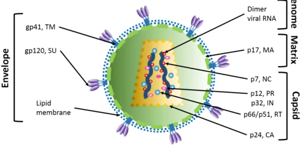
Mature HIV-1 virion is a spherical particle of 90-120 nm. It contains two strands of viral RNA, viral proteins and few host cell proteins. All these components are surrounded by a lipid bilayer (derived from the host cell membrane) covered with two types of glycoproteins (gp120 and gp41). Under the lipid bilayer, the matrix shell is composed of ~2000 matrix (MA) proteins. The mature viral particle contains the conical viral capsid, composed of capsid proteins (CA) organized in a fullerene-like structure. The capsid contains the genomic RNA dimer (9.2 kb), stabilized by the nucleocapsid proteins (NCp7), viral enzymes (reverse transcriptase, integrase and Protease) (Frankel and Young 1998; Turner and Summers 1999) and accessory proteins (Vpr, Vif and Nef) (Figure 1.3.1.). Viral particles contain also cellular components such as tRNA
Lys3
(Kleiman and Cen 2004), actin (Ott, Coren et al. 2003; Chertova, Chertov et al. 2006), ribosomal proteins (Mekdad, Boutant et al. 2016), and cyclophilin A (Braaten, Franke et al. 1996).
Figure 1.3.1. Structure of the mature HIV virion. Main structural departments of the virion are: envelope, matrix and capsid containing viral genome.
1.3.1. Viral genome
Viral RNA
The genome of HIV-1 consists of two copies of positive-sense single stranded RNA molecules (9193 nucleotides) (Wain-Hobson 1998) that form a stable dimer form, involving the 5’ dimer linkage structure (DLS). It is produced after integration of the viral DNA into the host
12
cell genome and after transcription by cellular polymerases. The architecture and secondary structure of the genome were resolved (Watts, Dang et al. 2009).
The viral genome contains both coding and non-coding elements. The three major coding regions correspond to polyproteins gag, pol and env (representing the structural and enzymatic proteins). It codes also for the regulatory proteins (Tat, Rev) and auxiliary proteins (Nef, Vif, Vpr and Vpu). Coding regions are flanked at both ends by noncoding regions and untranslated regions (UTR): 5'-UTR and 3'-UTR (Coffin, Hughes et al. 1997)(Fig. 1.3.2.). During reverse transcription, the vRNA is used as a template for the synthesis of proviral DNA and UTR regions became LTR (Long Terminal Repeat), which are important during the integration and transcription processes.
Figure 1.3.2. HIV-1 genome. The viral genomic RNA contains coding regions, which are flanked by the non-translated regions UTR (UnTranslated Region). During reverse transcription, the viral RNA is reverse transcribed into the viral DNA. The three main central regions code for gag, pol and env, which encode the structural (MA, CA, NC), enzymatic (PR, RT, IN) and envelope (SU, TM) proteins (Suzuki and Suzuki 2011).
The 5'-UTR region is the most conserved part of the HIV-1 genome. It plays an important role during reverse transcription, splicing, packaging and dimerization of the vRNA (Russell, Liang et al. 2004). It is composed of the R (Repeat), U5 (unique 5), PBS (Primary Binding Site), and the encapsidation sequence (called ) (Fig. 1.3.3.).
13
Figure 1.3.3. 5’UTR region of the HIV-1 genome. R region is composed of TAR and PolyA loops, latter is followed by the U5 and PBS regions. Encapsidation sequence comprises 4 stem-loops and psi region (Adapted from Smyth, 2015).
The R sequence, found in two identical copies at the 5’ and 3’termini of the HIV-1 genome, is composed of two stem-loops, the Transactivation Response element (TAR) and the poly (A) hairpin. The TAR sequence, composed of 57 nucleotides is the Tat interaction site and is crucial for the initiation of reverse transcription (Peterlin, Luciw et al. 1986; Harrich, Ulich et al. 1996). The PolyA stem-loop contains the AAUAAA polyadenylation signal (Berkhout 1996) that induces the addition of the poly-A tail at the 3’ terminus of the messenger RNA (Klasens, Thiesen et al. 1999).
The U5 region is a sequence of 85 nucleotides located at the 5ʹ-end of the gRNA and is the first transcribed sequence during reverse transcription.
The PBS (Primer Binding Site) is composed of 18 nucleotides and is complementary to the 18 3’-terminal of tRNALys3
which is the primer of the reverse transcription (Mak and Kleiman 1997; Kleiman 2002).
The encapsidation sequence is composed of 4 stem-loops (SL1-SL4) and is located next to the PBS and includes the starting codon for gag translation (AUG).
SL1 contains the Dimer Initiation Site (DIS), a sequence of six nucleotides (GUGCAC) implicated in the dimerization of the viral RNA (Skripkin, Paillart et al. 1994)
SL2 has a major splice donor site called "SD" where all transcripts of HIV-1 are cleaved generating various viral mRNA.
14
SL3 contains the packaging signal (Ψ) responsible for vRNA encapsidation during the assembly of the newly formed virions (Lever, Gottlinger et al. 1989) SL4 contains the initiation codon of gag translation.
Other non-coding regions of the viral genome are PPT (Poly Purine Tract), PPTc (Poly Purine Tract central) and RRE (Rev Response Element). PPT and PPTc are regions with high purine content. They are resistant to the RNase H activity of the reverse transcriptase and serve as primers for the synthesis of plus-strand DNA (Charneau, Alizon et al. 1992). PPTC is also
responsible for the formation of the DNA flap in the pre-integration complex (PIC), a structure that may play an important role during the nuclear import of the PIC (Arhel, Souquere-Besse et al. 2007; Riviere, Darlix et al. 2010). The RRE segment is a ~ 350-nucleotide sequence located in the env gene. It interacts with the Rev protein, which allows the nuclear export of non-spliced or single-spliced RNAs (Farjot, Sergeant et al. 1999).
Proviral DNA
The HIV-1 proviral DNA in its integrated form is approximately 9.8 kb in length. The sequence is flanked on both ends by the Long Terminal Repeats (LTRs) that are crucial for the integration and the transcription of the vDNA. The coding genes of HIV are located in the central region of the proviral DNA and encode three major polyproteins- Gag (Group specific antigen), Pol (polymerase), and Env (envelope), as well as the regulatory proteins (Tat, Rev) and the accessory proteins (Vpu, Vpr, Vif, Nef) (Frankel and Young 1998; Freed 2001). The structural proteins are further cleaved. The matrix, capsid, nucleocapsid protein, p6, SP1 and SP2 peptide result from the cleavage of the Gag polyprotein. Pol (polymerase) provides the protease (PR), reverse transcriptase (RT) and integrase (IN). Env (envelope), cleaved by a cellular protease, generates the envelope glycoproteins gp120 and gp41.
1.3.2. Structural protein Gag
The
HIV-1 gag gene encodes a 55 kDa Gag polyprotein that is the principal structural unit able to form the viral particles at the cell plasma membrane. This protein is composed of several domains that are cleaved by the viral protease during the maturation of the virus. This cleavage is a complex and highly regulated process (Pettit, Lindquist et al. 2005) . The products of the proteolytic cleavage of Gag are MA, CA, NC and the low molecular products SP1, SP2 and p6 (Fig. 1.3.4.).15
Figure 1.3.4. Map of the Gag precursor polyprotein and major functions of its domains. Due to its myristilated region and basic domain, MA allows stable anchoring of Gag at the plasma membrane. The CA domain is responsible for Gag-Gag interactions and the formation of the viral core. The NC region binds specifically the viral RNA, acts as a chaperone of nucleic acids and protects the viral RNA. P6 is implicated in the release of newly formed viral particles (Adapted from (Freed 2015)).
Matrix
The matrix protein (132 amino acids) presents five α-helices, two short 310 helical
stretches and a three-strand mixed β-sheet (Massiah, Starich et al. 1994; Matthews, Mikhailov et al. 1996). In the mature viral particle, the MA protein lines the inner surface of the viral envelope (Frankel and Young 1998). This multifunctional protein is implicated in early and late stages of the viral life cycle. In the early stages, p17 participates in the preintegration of viral complex in the host cell nucleus (Bukrinskaya, Vorkunova et al. 1992; Bukrinsky, Haggerty et al. 1993) . During the assembly step, the basic residues of the MA domain of Gag interact electrostatically with the inner leaflet of the plasma membrane (Saad, Miller et al. 2006; Saad, Loeliger et al. 2007). Additional contribution to Gag binding att the plasma membrane is ensured by the myristic acid moiety (Myr) that anchors the Gag polyprotein to the phosphatidylinositol rich regions of the inner leaflet of the plasma membrane (Ono, Ablan et al. 2004; Saad, Miller et al. 2006). Furthermore, the Highly Basic Region (HBR) platform allows MA to bind the gRNA in a non-specific manner, suggesting the participation of MA in the recruitment of viral and cellular RNA during assembly (Ott, Coren et al. 2005; Rulli, Hibbert et al. 2007).
Capsid
Figure 1.3.5. HIV-1 CA spatial organization. Crystal structures of hexameric (A) and pentameric (B) CA assemblies. Individual proteins are colored. Each protein contains NTD and CTD regions. (C) 3D representation of the HIV-1 capsid model, NTDs of hexameric and pentameric regions are shown in blue and yellow, respectively and CTD are green (Pornillos, Ganser-Pornillos et al. 2011).
16
CA is a 231 amino acids protein, composed of an amino-terminal domain (NTD) that is connected by a linker with a 310-helix to the carboxy-terminal domain (CTD) (Gitti, Lee et al.
1996; Gamble, Yoo et al. 1997)(Fig. 1.3.5.). CTD is implicated in Gag-Gag multimerization (Briggs and Krausslich 2011), and plays a role in the stabilization of Gag oligomers during the assembly (Tanaka, Robinson et al. 2015). In the mature HIV-1, ~1200 CA molecules are organized in a cone like shell (Pornillos, Ganser-Pornillos et al. 2011; Sundquist and Krausslich 2012; Zhao, Perilla et al. 2013). In the cone, the NTD and CTD regions are positioned outside and inside the capsid, respectively (Meng, Zhao et al. 2011). The broad end contains the high-density region formed by the ribonucleoprotein complex (RNP). The narrow end is in contact with the MA protein (Li, Hill et al. 2000; Mayo, Huseby et al. 2003; Briggs and Krausslich 2011; Zhao, Perilla et al. 2013). By interacting with the Nups, the CA protein is probably involved in the nuclear import of the viral genome (Dismuke and Aiken 2006).
Nucleocapsid
The HIV-1 nucleocapsid proteins (NC) are characterized by two zinc fingers similar to the zinc-finger motifs typical of DNA binding proteins. The NC is a small, 55 amino acid, (7 kDa) protein that exists as a functional domain of Gag and as a free protein in its mature form. NC shows a nucleic acid chaperone activity. Detailed structure and functions of NCp7 will be discussed below.
Protein p6, peptides SP1 and SP2
The p6 protein consists of 52 amino acids and is located at the C-terminus of the Gag polyprotein. P6 interacts with several cellular proteins of the ESCRT (Endosomal Sorting Complex Required for Transport) family and is required for the viral release (Huang, Orenstein et al. 1995).
SP1 and SP2 spacer peptides that separate the CA from NC and the NC from the P6, respectively. SP1 is involved in Gag multimerization (Datta, Temeselew et al. 2011). The function of SP2 is less defined.
1.3.3. Enzymatic proteins
HIV-1 enzymatic proteins are the products of the gag-pol gene translation. This gene encodes for the precursor Pr160Gag-Pol polyprotein, produced as a result of the programmed ribosomal frameshift (5 % frequency) during the translation of Gag/Gag-Pol mRNA. As a result, the Gag to Gag-Pol ratio in the cell is 20 to 1.
17
The first enzymatic HIV-1 protein structurally characterized was the viral protease (PR) (Miller, Schneider et al. 1989). PR is released from the Pr160Gag-Pol by an autocatalytic reaction (Farmerie, Loeb et al. 1987; Jacks, Power et al. 1988). The enzyme is a homodimer, stabilized by an antiparallel β-sheet. The active site of PR is localized at the interface between the two subunits (Navia, Fitzgerald et al. 1989) and bears the catalytic triad, which is responsible for PR cleavage activity. During the maturation of viral particles, PR participates in the proteolytic processing and cleavage of Gag/GagPol (Prabu-Jeyabalan, Nalivaika et al. 2002; Alvizo, Mittal et al. 2012), that results in the formation of mature and ready for infection viruses.
Reverse transcriptase (RT) is a flexible DNA polymerase. The mature form of the protein is a heterodimer, composed of two polypeptide chains p66 and p51. Reverse transcriptase drives the synthesis of the double-stranded DNA (dsDNA) genome from the plus-stranded RNA (Hu and Hughes 2012). In addition to its DNA- and RNA-dependent DNA polymerase activity, the reverse transcriptase exhibits also an endonuclease activity (RNase H) that degrades the RNA template in the RNA/DNA duplex. Reverse transcription takes place in the cytosol of infected cells after viral entry.
HIV-1 integrase (IN) is composed of three structural domains, the N-terminal, central catalytic and N-terminal domain. Inside HIV-1, IN can be found in a tetramer form (Jenkins, Engelman et al. 1996). IN is crucial for the incorporation of the viral DNA into the genome of the host cell trough the IN strand transfer reaction (Wu, Liu et al. 1999; Zhu, Dobard et al. 2004; Tekeste, Wilkinson et al. 2015) by stimulating both the initiation and elongation steps of the (Dobard, Briones et al. 2007). Finally, IN may also play a role in the nuclear import of the PIC by interacting with the nucleoporins (Ao, Jayappa et al. 2012).
1.3.4. Envelope proteins
Env proteins are derived from the polyprotein precursor Pr160Env. Env is organized in
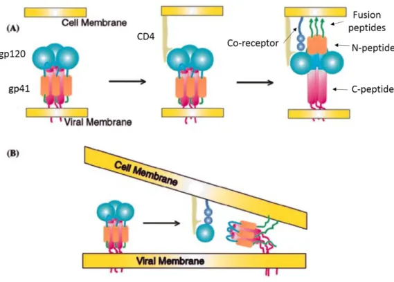
three subunits of non-covalently associated heterodimers of gp120 surface protein (SU) and gp41 transmembrane protein (TM). The Env trimer recognizes CD4 receptors at the surface of T cells and mediates viral entry. gp41 is composed of an N-terminal ectodomain, a transmembrane domain and a C-terminal intraviral segment. During the fusion step, the binding to CD4 and to chemokine co-receptor allows TM to reorient parallel to the membranes, which facilitates the insertion of the gp41 fusogenic peptide into the cellular membrane (Fig. 1.3.6.) (Caffrey, Cai et al. 1998). SU is glycosylated and forms surface-exposed loops (Leonard, Spellman et al. 1990).
18
Figure 1.3.6. CD4 and chemokine receptor induced fusion of the viral and cellular membranes followed by reorientation of the TM protein. (a) Binding of the gp120 to the CD4 receptor is followed by the displacement of the gp41 N-terminal fusogenic peptides toward the cellular membrane triggered by the connection to the chemokine co-receptor; (b) CD4 and chemokine binding causes the loss of the SU proteins enables TM reorientation and membrane fusion (Adapted from (Turner and Summers 1999)).
1.3.5. Regulatory and accessory proteins
The viral DNA codes also for the accessory (Nef, Vif, Vpr, Vpu) and regulatory (Tat, Rev) (Frankel and Young 1998) proteins. The accessory proteins Vpr, Nef, and Vif are encapsidated during assembly, whereas Rev, Tat and Vpu do not appear to be encapsidated.
Nef, the HIV-1 negative factor is a 206 amino acid residues myristilated regulatory factor that localizes in the inner leaflet of the host cell membrane. Nef stimulates the decrease of the number of CD4 receptors at the surface of infected cells (Goldsmith, Warmerdam et al. 1995). Nef also downregulates the co-receptors CXCR4, CCR5 and participates in the prevention of the super-infection of the cells (Michel, Allespach et al. 2005).
Virion Infectivity Factor or Vif is composed of 192 amino acids (its complete 3D structure has not been yet resolved) (Barraud, Paillart et al. 2008). It is integrated in the virion by binding to the 5’ region of RNA or/and to the C-terminal domain of Gag (Huvent, Hong et al. 1998; Henriet, Richer et al. 2005). Vif plays a regulatory function as a nucleic acid chaperone all
19
over the viral life cycle (Batisse, Guerrero et al. 2012). Moreover, it acts as inhibitor of the APOBEC-3G antiviral activity (Mariani, Chen et al. 2003).
Vpr (Viral Protein R) is highly conserved and consists of 96 amino acids. Vpr is bound to the RNA in the viral capsid (Welker, Hohenberg et al. 2000). It participates in numerous stages during the viral life cycle (Guenzel, Herate et al. 2014), especially in the early phase. Vpr is implicated in reverse transcription and during the nuclear import via its interactions with nucleoporins (Le Rouzic, Mousnier et al. 2002). As well, Vpr is responsible for the G2 cell cycle arrest with following apoptosis by the interaction with mitochondrial pores proteins (Basanez and Zimmerberg 2001).
Vpu (Viral protein U) is composed of 81 amino acids (Maldarelli, Chen et al. 1993). It is organized in a transmembrane domain and two regions of alpha helices. Vpu is thought to: 1) decrease the level of CD4 receptors by stimulating their degradation in the endoplasmic reticulum (this degradation stimulates the release of Env (Willey, Maldarelli et al. 1992); 2) favor the budding of viral particles by decreasing the effects of tetherine, known for its antiviral activity (Neil, Zang et al. 2008).
The Transactivator of Transcription (Tat) protein consists of five different regions: N-terminal, cysteine-rich, hydrophobic, arginine rich and glutamine-rich domains. Its size may vary between 86 and 101 amino acids (Jeang and Rauscher 1996). Tat basic domain is crucial for the recognition and binding to the TAR RNA sequence (Calnan, Biancalana et al. 1991). This binding stimulates the expression of the viral genome. Tat is also needed for efficient elongation during the initiation of transcription (Tahirov, Babayeva et al. 2010). Once released in the extracellular medium, Tat enters into surrounding non-infected cells and causes their apoptosis. Finally, Tat has been shown to activate viral transcription in latent cells (Debaisieux, Rayne et al. 2012).
The Regulatory of Viral Expression (Rev) protein consists of 116 amino acids and contains nuclear export and nuclear localization signals (NES and NLS respectively) (Fischer, Meyer et al. 1994; Meyer and Malim 1994). This protein participates in sequence-specific transport of viral mRNA from the nucleus to the cytoplasm.
1.4. Viral life cycle
________________________________________________________________________________
TheHIV-1 viral cycle can be divided in two main phases (Fig. 1.4.1.):
20
early phase : starts from binding of the virus to the CD4 receptors on the cell surface, followed by fusion with host cell membrane and entry of the virus into the cytosol, uncoating of the conical capsid, reverse-transcription of the vRNA to the gDNA, nuclear entry and integration of the vDNA into the host cell genome late phase comprises synthesis of the new viral RNA and expression of viral proteins, assembly of immature virions, budding at the plasma membrane and maturation of the viral particle.
These two phases are separated by a latent period that may last from several days up to several years (Siliciano and Greene 2011).
Figure 1.4.1. Schematic diagram of the HIV-1 life cycle. The viral life cycle can be divided in early and late phases. Early phase comprises binding, fusion and entry, uncoating and reverse transcription, followed by nuclear entry and integration. The late phase begins with the expression of viral genes and continues with the release and maturation of the new viral progeny (adapted from Kate Bishop Lab, Francis Crick institute website).
1.4.1. Early stages of infection
Virus entry
HIV-1 infection starts with the attachment of the viral Env glycoprotein to the CD4 and chemokine receptors (CCR5 or CXCR4) at the surface of the host cell. HIV-1 envelope spike
21
trimers consist of non-covalently linked heterodimers of gp120 and gp41. The interaction of the Env glycoprotein with the cell receptors trigger a sequence of conformational changes leading to the insertion of the fusion peptide at the amino-terminus of gp41, into the host cell membrane. This is then, followed by the formation of a fusion pore permitting the viral entry (Pancera, Zhou et al. 2014). In case of direct cell-to-cell transmission, the virus is transmitted via endocytosis in a cytoskeleton-dependent adhesive junction or so called virological synapse (McDonald, Wu et al. 2003; Jolly, Kashefi et al. 2004; Miyauchi, Kim et al. 2009).
Uncoating and reverse transcription
Cell entry is followed by the uncoating and reverse transcription of the viral genome. These processes seem to be linked however, their order, timing and location are still debated. Three different hypothesis were proposed for the spatial-temporal occurrence of uncoating:
direct uncoating in close proximity to the cell membrane: this hypothesis is based on the detection of small amounts of CA proteins directly after infection (Bukrinsky, Haggerty et al. 1993; Fassati and Goff 2001; Iordanskiy, Berro et al. 2006) and the inability to detect the capsid inside the infected cells by transmission electron microscopy (Grewe, Beck et al. 1990)
progressive uncoating during reverse transcription (Arfi, Lienard et al. 2009; Hulme, Perez et al. 2011): this hypothesis is based on the detection of a broad range of HIV-1 complex morphologies inside the cytoplasm (Nermut, Hockley et al. 1998; Nermut and Fassati 2003; Warrilow, Tachedjian et al. 2009)
loss of capsid near the nucleus, just before nuclear entry (Arhel, Souquere-Besse et al. 2007): this hypothesis is linked to the fact that the intact capsid preserves a high concentration of enzymatic proteins, such as reverse transcriptase needed for completion of reverse transcription
The transport of viral complexes to the perinuclear region is most probably realized by the cellular transport machinery. It was reported that PIC associates with actin cytoskeleton and microtubules (Bukrinskaya, Brichacek et al. 1998; McDonald, Vodicka et al. 2002; Arhel, Munier et al. 2006).
22
Figure 1.4.2. Early stages of infection. Fusion of the virus with cell membrane is followed by uncoating and reverse transcription during the trafficking to the nucleus. Then, the PIC formed after reverse transcription is translocated through the nuclear envelope by an active import mediated by the NPC. Inside the nucleus, the viral DNA is integrated into the genome of the host cell (Suzuki and Craigie 2007).
Reverse transcription
In the cytosol of infected cells, the ribonucleoprotein (RNP) complex (25-125 copies of RT, 12-60 IN, hundreds of Vpr and RNA dimer covered by ~2000 molecules of NCp7 (Thomas and Gorelick 2008), undergoes the reverse transcription process and is transformed to preintegration complex (PIC) (Fig. 1.4.2.).
In the course of RT, genomic single strand genomic RNA (gRNA) is converted into the double stranded viral DNA (Fig. 1.4.3.). This process is orchestrated by the reverse transcription and facilitated by the NCp7. Notably during the beginning of the RT process NCp7 chaperoning properties are valuable for the initiation of the RT and strand transfer, this will be described below. RT requires primer and template, where the primer is the host tRNA and genomic RNA is a template. In the first step, RT is initiated after the annealing of the PBS region of vRNA by tRNA primer. Then, first strand transfer is made possible by the R sequence on the both ends of the template. Further, synthesis of plus-strand DNA is initiated from the purine-rich regions of gRNA. Second-strand transfer is facilitated by the plus- and minus strand DNA copies of PBS. Finally, termination of the upstream plus-strand DNA requires the central termination sequence.
23
Figure 1.4.3. Mechanism of reverse transcription. (A-B) Initiation of reverse transcription by host tRNA and start of (-) ssDNA synthesis; (C-D) start of (+) ssDNA synthesis after minus-strand transfer; (E-F) second (plus-stranded) transfer (Hu and Hughes 2012).
Nuclear import
Biochemical analyses have revealed that the PIC contains viral DNA, IN, Vpr, MA, CA and cellular proteins (Barrier-to-autointegration factor (BAF), LEDGF-75) (Raghavendra (Raghavendra, Shkriabai et al. 2010) (Ciuffi and Bushman 2006) (Llano, Delgado et al. 2004). However, its precise composition and notably, the presence of NCp7 are still debated (Dismuke and Aiken 2006; Iordanskiy, Berro et al. 2006; Thomas and Gorelick 2008).
Because of its size (~tens of nanometers) (Miller, Farnet et al. 1997), the PIC is actively internalized into the nucleus (Mattaj and Englmeier 1998), through the nuclear pore complexes (NPCs) (Fig. 1.4.2) (Konig, Zhou et al. 2008; Di Nunzio, Danckaert et al. 2012). This internalization pathway requires the presence of specific amino acid sequences called Nuclear Localization Signals (NLS). Canonical NLSs were found in MA (Bukrinsky, Haggerty et al. 1993) and IN (Bouyac-Bertoia, Dvorin et al. 2001). Moreover, non-canonical karyophilic signals were identified in Vpr (Fouchier, Meyer et al. 1998). More than ten NUPs were observed to be involved in the nuclear transport of HIV-1 (Fig. 1.4.4.). Among them are Nup98, Nup153, Nup214, Nup358 and Nup62 (Konig, Zhou et al. 2008; Lee, Ambrose et al. 2010; Monette, Pante et al. 2011; Di Nunzio, Danckaert et al. 2012) 2010). HIV-1 infection is clearly decreased by the depletion of Nup358 (Konig, Zhou et al. 2008) or Nup 153 (Lelek, Casartelli et al. 2015). It seems that the Nups influence also the integration process. Nup62, Nup153 and Nup98 can bind
24
to transcriptionally active genes (Di Nunzio, Fricke et al. 2013; Kohl, Ng et al. 2014) in the chromatin and serve as intermediate factors that drive the PIC to its integration site (Jayappa, Ao et al. 2012).
Figure 1.4.4. Interaction of HIV-1 PIC with host cell proteins during translocation through the nuclear envelope (NE). The active import of PIC occurs via Nup358, Nup98, Nup153 and Nup62 proteins (Le Sage and Mouland 2013).
Genome integration
After nuclear import, the proviral DNA is integrated into the genome of the host cell. Initially, the IN specifically binds to the two LTR ends of the viral DNA (Kukolj and Skalka 1995)) and cleaves the last two 3 'nucleotides of each DNA strands releasing a CA dinucleotide and a 3'OH (3 'processing). Subsequently, the tetrameric form of IN cleaves the genomic DNA of the target cell and catalyzes the insertion of the viral genome into the cellular genome (strand transfer). Finally, cellular DNA repair enzymes correct the discontinuities between cellular and viral DNA (Engelman, Mizuuchi et al. 1991; Vink, van Gent et al. 1991; Vink, Yeheskiely et al. 1991). The integrated proviral DNA is replicated in the nucleus of the host cell as any other cellular gene.
25
1.4.2. Late phase of infection
Figure 1.4.5. Late phase of HIV-1 replication. The viral Env is synthesized in the rough endoplasmic reticulum (RER) and transported to the plasma membrane via the secretory pathway. Gag and GagPol precursor polyproteins are synthesized in the cytosol from the full-length vRNA. Gag recruits the viral gRNA, starts to multimerize and reaches the plasma membrane, where it anchors to the membrane lipid rafts. After budding, maturation occurs leading to the formation of the main structural elements and stabilization of genome (Freed 2015).
Late stages of HIV-1 infection start from the transcription of the viral genome, followed by the splicing and the nuclear export of viral RNAs (spliced and unspliced) into the cytoplasm of the host cell (Fig. 1.4.5.). The accessory protein Rev (Freed 2001) that contains a nuclear export signal (NES) and recognizes the RRE sequence on the mRNA mediates the nuclear export. Interestingly, during the Rev-mediated export, the Nup62 is displaced into the cytoplasm (Fig. 1.4.4.) and incorporated into virions (Monette, Pante et al. 2011).
26
In the cell cytoplasm, the mRNAs are translated into viral proteins. Gag is expressed in ribosomes from the unspliced mRNA as a 55kDa precursor polyprotein. Gag-Pol polyprotein precursor is generated by a translational frameshift. The unspliced mRNA can be also encapsidated as genomic RNA into the new viral particles. The mechanisms regulating the equilibrium between translation and encapsidation are not fully understood. Env, Vif, Vpr and Vpu are expressed from single spliced RNA and Tat, Rev and Nef from multi-spliced RNAs.
In the cytosol, Gag molecules bind to the RNA and form (via CA- CA interactions) small oligomers that diffuse to the cell periphery where the myristilated MA domain binds to the inner leaflet of the plasma membrane. Gag binds preferentially to PIP2-enriched domains connected to raft domains, which serve as platforms for viral assembly and may facilitate the incorporation of Env glycoproteins (Ono and Freed 2001). In the majority of cell types, Gag multimerization and virion assembly takes place at the plasma membrane (Raposo, Moore et al. 2002).
Viral RNAs through their encapsidation sequence are specifically recognized by the NC domain of Gag. The RNA molecule serves as a scaffold for further Gag multimerization at the plasma membrane resulting in the formation of round-shaped virions. The NC domain serves as a chaperone of nucleic acids that drives RNA packaging (Levin, Guo et al. 2005). In immature particles, ~2,400 +/-700 Gag molecules (Carlson, Briggs et al. 2008) are packed radially with the C-terminus of Gag directed towards the center and the MA domain directed to the membrane. The Gag lattice presents a hexameric arrangement (Bharat, Davey et al. 2012; Schur, Hagen et al. 2015). The incorporation of Env may possibly occur through a passive process (co-targeting Gag and Env to the plasma membrane) or a direct recruitment of Env by Gag (Checkley, Luttge et al. 2011).
The cellular ESCRT (Endosomal Sorting Complexes Required For Transport) machinery mediates the membrane fission of the nascent viral particles. Interactions between the cellular membrane, ESCRT and the virus are mediated by the p6 domain of Gag (Morita, Sandrin et al. 2007; Carlton and Martin-Serrano 2009; Morita, Sandrin et al. 2011). It was shown that ESCRT elements accumulate at the head or at the neck of the viral bud (Bleck, Itano et al. 2014; Van Engelenburg, Shtengel et al. 2014). Other cellular proteins such as TsG101 and Alix favor the scission of the viruses by recruitment of ESCRT-III complexes (Jouvenet, Zhadina et al. 2011).
New virions are released in immature and non-infectious forms. Viral maturation is triggered by the proteolytic activity of protease that cleaves the Gag and Gag-Pol, generating the mature structural (MA, CA, NC, p6) and viral enzymes (PR, IN, RT). During the process, the
27
viral particle is reorganized; the RNA is condensed and forms, together with the NC and RT the RTC complex. CA molecules form a typical cone shaped capsid.
1.5. NCp7: structure and function
________________________________________________________________________________
Figure 1.5.1. Diagram of Gag polyprotein maturation. Maturation occurs in three steps and produces subsequently p15, p9 and finally the p7 nucleocapsid protein by liberation of the p1 domain (Adapted from (Abd El-Wahab, Smyth et al. 2014)).
The HIV-1 nucleocapsid protein exists in two forms: as a functional NC domain of Gag during the late phase of infection and in mature form (NCp7), after the proteolytic cleavage of the polyprotein.
The mature form of NC (NCp7) is generated within the virion by a series of cleavages catalyzed by the viral protease (Fig. 1.5.1.) (Erickson-Viitanen, Manfredi et al. 1989; Pettit, Moody et al. 1994). The first cleavage occurs between SP1 and the NC region and leads to the liberation of NCp15 composed of NCp7-SP2-p6 (Shehu-Xhilaga, Kraeusslich et al. 2001). This proteolytic intermediate of NCp contains 123 a.a and is subsequently cleaved (during the second step of maturation, which requires binding to gRNA) to the NCp9 (71 a.a.) (Sheng, Pettit et al. 1997). Finally, the third proteolytic cleavage promotes the release of NCp7 and SP2.
The mature viral capsid contains between 1500 to 2000 NCp7 molecules, which cover the RNA dimer. The NCp7 concentration is of the order of millimolar and the nucleotide/NCp7 ratio is about 8-9 (Darlix, Godet et al. 2011)
28
1.5.1. Structure
NCp7 is a small basic protein (pI=9.9) containing 55 aa. It comprises two CCHC zinc fingers composed of Cys-X2-Cys-X4-Cys-His-X4 sequence (X represent variable amino acids) (Fig. 1.5.2.A) connected by a short flexible basic peptide linker and flanked by N- and C- domains (Fig. 1.5.2. B). Zinc fingers and the RAPRKKG linker can fold in a globular conformation (Mely, Jullian et al. 1994; Morellet, de Rocquigny et al. 1994; Lee, De Guzman et al. 1998). The affinity of Zn2+ to the zinc fingers is very high (K
a ~10
14 M-1) (Mely, De Rocquigny
et al. 1996). Despite the similar amino acid composition of the two fingers, their activity and biochemical properties are not equivalent (Fisher, Rein et al. 1998; Fisher, Fivash et al. 2006; Zargarian, Tisne et al. 2014). ZF1 plays an important role in the destabilization of nucleic acid secondary structures while ZF2 increases the rate of annealing of the RNA and/or DNA during the strand transfer, genome dimerization and maturation (Gorelick, Chabot et al. 1993; Heath, Derebail et al. 2003) (Aduri, Briggs et al. 2013; Wu, Mitra et al. 2014). The disruption of zinc fingers folding by mutations leads to a loss of infectivity (Aldovini and Young 1990; Dorfman, Luban et al. 1993; Gorelick, Chabot et al. 1993).
Figure 1.5.2. Structure of NCp7. (A) Amino acid sequence. Zinc fingers are shown in green, the basic linker in yellow, and the two terminals N and C domains are presented in blue and violet respectively. (B) 3D representation of protein structure determined in solution by NMR (Adapted from (Godet, Boudier et al. 2012; Sleiman, Goldschmidt et al. 2012).
1.5.2. Interaction of NCp7 with nucleic acids
NCp7 can bind to almost any nucleic acid sequence of 5-8 nucleotides. Due to its cationic nature, the NC/NA interactions are salt dependent (Urbaneja, Kane et al. 1999; Vuilleumier, Bombarda et al. 1999). The basic residues of NCp7 (notably those of the N-terminal domain and the linker) interact electrostatically with the phosphodiester backbone of any NA (Mely, de Rocquigny et al. 1995).
A number of specific interactions of NCp7 with NA has also been reported (Berglund (Berglund, Charpentier et al. 1997; Wu, Ozarowski et al. 1997; De Guzman, Wu et al. 1998;
29
Bourbigot, Ramalanjaona et al. 2008). NCp7 binds preferentially to guanosine rich regions of single-stranded DNA or RNA. The interaction is strongly dependent on the hydrophobic plateau formed at the top of zinc fingers by the V13, F16, T24, A25 (N-terminal finger) and W37, Q45, M46 (C-terminal finger) residues (Amarasinghe, De Guzman et al. 2000).
1.5.3. Chaperone properties
By NA binding, condensing and annealing as well as by promoting strand transfer reaction, NCp7 remodels the NA structure to its most thermodynamically stable conformation, and thus, performs chaperone activity.
Remodeling of NA structure by NCp7 is based on binding to the NAs, followed by partial destabilization of NA, rapid on/off kinetics of NC-NA interactions and NA aggregation (Darlix, Godet et al. 2011). During this remodeling, the RNA adopts different conformations, in order to lead to the most stable one (Tompa and Csermely 2004). NCp7 chaperone activity depends on the NA occupancy (Fig. 1.5.N). At low occupancy, NCp7 performs limited chaperone activity, while at high occupancy level; NCp7 promotes aggregation and macromolecular crowding effect, followed by annealing (Lapadat-Tapolsky, Gabus et al. 1997). The occupancy level is changing during the viral lifecycle. It is low during the assembly step (1:100 nt) when NCp7 is part of Gag, (Paillart, Shehu-Xhilaga et al. 2004) but increases with Gag multimerization and during the maturation step (1:2-5nt) (Chertova, Chertov et al. 2006). In mature virions, NCp7 coats and protects the viral genome in the nucleoprotein complex and then, promotes reverse transcription in the infected cell. During the integration step, when almost all NC proteins are released, its chaperone activity is questionable (Darlix, Cristofari et al. 2000).
30
Figure 1.5.3. Activity of NCp7 through the viral life cycle. At high occupancy level, the NCp7 protein protects the RNA, whereas at low occupancy, NCp7 is performing nucleic acid chaperoning and specific binding (Adapted from (Darlix, Godet et al. 2011))
1.5.4. Interactions with cellular proteins
Several host proteins have been identified as specific interactions of NCp7 (Mouland (Mouland, Mercier et al. 2000; Zimmerman, Klein et al. 2002; Ueno, Tokunaga et al. 2004; Popov, Popova et al. 2009).
In the course of HIV-1 assembly, the Tsg101 domain of ESCRT is recruited by the NC and p6 domains of Gag and targeted to the viral assembly sites (Chamontin, Rassam et al. 2015). This targeting favors RNA packaging and the budding process. Another ESCRT factor ALIX is involved in virus budding and release. ALIX interacts via its YPXL pattern with the p6 domain of Gag and via its BRO1 domain with the NC-Gag (Popov, Popova et al. 2008; Dussupt, Javid et al. 2009). However, the role of ALIX is still unclear due to the absence of effect of its depletion on viral egress (Fujii 2009).
31
Actin interacts with the N-terminal of NC, both in its mature form or as a Gag domain (Liu, Dai et al. 1999; Wilk, Gowen et al. 1999). This interaction seems to play a role during viral budding where it favors the incorporation of actin into the virus (Ott 2008). However, recent studies have revealed that the NC domain of Gag is dispensable for association of cortical actin to the HIV-1 budding sites (Stauffer, Rahman et al. 2014).
Staufen1 is a host factor that is encapsidated into HIV-1 (Mouland, Mercier et al. 2000; Chatel-Chaix, Clement et al. 2004) and plays a role in the trafficking and metabolism of the mRNA. Staufen1 is recruited by the NC domain of Gag (Chatel-Chaix, Boulay et al. 2008) and participates in the packaging of RNA. Moreover, Staufen1 modulates Gag multimerization during HIV-1 assembly (Chatel-Chaix, Abrahamyan et al. 2007).
ABCE1 (HP68 or RNase L inhibitor) is a 68 kDa cellular protein that is a member of the ATP-binding cassette protein family E. It binds to Gag, directly after its translation via the basic residues of the NC domain (Lingappa, Dooher et al. 2006) and promotes ATP-dependent conformational changes of the capsid assembly intermediates crucial for assembly.
Nucleolin is a typical nucleolar protein. In cells infected with HIV-1, nucleolin is translocated to the plasma membrane (Ueno, Tokunaga et al. 2004). It is thought to play a role in the transport of the intron containing nucleocytoplasmic gRNA (Cochrane, McNally et al. 2006). By interacting with NC-Gag and the Ψ region of the genomic RNA, it could favor the assembly of viral particles (Bacharach, Gonsky et al. 2000).
1.5.5. Inhibition
NCp7 and notably CCHC zinc fingers are highly conserved. Point mutations in these sequences lead to a complete loss of infectivity (Dorfman, Luban et al. 1993; Morellet, de Rocquigny et al. 1994; Berg and Shi 1996; Ramboarina, Morellet et al. 1999; Buckman, Bosche et al. 2003; Thomas and Gorelick 2008). As a result, NCp7 appears as a good candidate for antiviral treatment. Several types of NCp7 inhibitors have been developed.
Zinc ejectors act by an electrophilic attack of the cysteine thiol groups, generating disulfide bridges that lead to ejection of zinc atom. This causes the loss of NCp7 activity and results in a loss of HIV-1 infectivity. 3-nitrobenzamide (NOBA) (Rice, Schaeffer et al. 1993), the first zinc ejector developed initiated the synthesis of many other molecules (de Rocquigny, Shvadchak et al. 2008; Turpin, Schito et al. 2008; Goldschmidt, Jenkins et al. 2010; Garg and Torbett 2014; Mori, Kovalenko et al. 2015). Among them are disulfide benzamide (SBIR) (Rice, Supko et al. 1995), azodicarbonamide (ADA) (Rice, Turpin et al. 1997), pyridinioalkanoyl
32
thioesters (PATEs) (Turpin, Song et al. 1999), s-acyl-2-mercaptobenzamide thioesters (SAMT) (Srivastava, Schito et al. 2004; Jenkins, Byrd et al. 2005), methyl-3-phenyl-2H-[1,2,4]thiazol-5-yideneamine (WDO-217) (Vercruysse, Basta et al. 2012) and N,N′-bis(4-ethoxycarbonyl-1,2,3-thiadiazol-5-yl)benzene-1,2-diamine (NV038) (Pannecouque, Szafarowicz et al. 2010). These inhibitors show an efficient antiviral activity on all HIV-1 strains, leading to the release of noninfectious virus (Turpin, Terpening et al. 1996). The main drawback of these molecules is their cellular toxicity resulting from their non-specific action on cellular proteins containing zinc fingers.
More prominent in terms of specificity might be zinc ejecting inhibitors or non-covalent NC inhibitors (NCIs), targeting the hydrophobic platform of the zinc fingers or nucleic acid partners in the non-covalent mode. ''Gallein'' compound showed activity at nanomolar concentrations on free NCp7 (Stephen, Worthy et al. 2002). In 2009, Shvadchak and colleagues reported the search strategy for NCIs, based on the high-throughput screening (HTC) assay to identify small molecules for inhibition of NCp7 chaperon activity. This assay was used to screen 4800 chemical substances and five compounds were successfully identified as inhibitors. Later, non-toxic 2-amino-4-phenylthiazole (AN3) compound (Mori, Nucci et al. 2014) have been developed on the base of A10 fragment from HTC assay screening (Shvadchak, Sanglier et al. 2009). Further, HTC assay was used to identify an anti-NCp7 of small methylated oligonucleotides (mODNs), rich in GU, mimicking short sequences of LTR. These mODNs affect the chaperone activity of NCp7 and/or the synthesis of viral DNA resulting in an efficient inhibition of viral infection and replication (Grigorov, Bocquin et al. 2011; Avilov, Boudier et al. 2012).
Some aminoglycosides and intercalating peptides can bind to NCp7 preferential sites on viral RNA, such as SL1 stem loop (Chung, Mujeeb et al. 2008; Chung, Ulyanov et al. 2010), SL3 stem loop (Warui and Baranger 2009) or the TAR and PBS sequences (Sosic, Frecentese et al. 2013) which allows a specific inhibition of the interaction of the NCp7 with these sequences.
1.6. Role of NCp7 during infection
_____________________________________________________________________________________________
Functions of NCp7 during the viral infection are mainly related to its nucleic acid chaperone properties. NCp7 plays a crucial role during several steps of the early and late infection stages.
33
During reverse transcription, NCp7 is required for the proper annealing of the tRNA (Lys3), a crucial step of the initiation of reverse transcription (Saadatmand and Kleiman 2012; Sleiman, Goldschmidt et al. 2012). Next, NCp7 promotes the minus-strand transfer by favoring the annealing of the newly synthesized TAR DNA domain (cTAR) to the TAR RNA at the 3’-end of the genomic RNA (Piekna-Przybylska, DiChiacchio et al. 2010). Moreover, NCp7 participates in the removal of the non-PPT vRNA regions during RT translocation along the (-) DNA template (Jacob and DeStefano 2008; Post, Kankia et al. 2009). It was also shown that NCp7 might protect the newly synthesized vDNA ends from nucleases (Buckman, Bosche et al. 2003).
In the cytoplasm of infected cells, the virus undergoes several transformations. Initially, extremely high concentrations of NCp7 (~ 100 mM) in the ribonucleoprotein complex strongly condense the genomic RNA. During reverse transcription, the RTC is remodeled and progressively transformed into the preintegration complex (PIC). Since the affinity of NCp7 for dsDNA is significantly lower as for ssRNA, it is thought to be progressively released from the viral complex (Fig. 1.6.2.). Nevertheless, it is likely that a pool of NCp7 molecules may bind non-specifically to dsDNA in PIC and promote stable binding of IN to LTR ends (Carteau, Batson et al. 1997).
Figure 1.6.2. Model of viral core dismantling controlled by RT. (Adapted from (Mirambeau, Lyonnais et al. 2007).
Role of NCp7 in viral integration
NCp7 was shown to effectively enhance viral DNA integration reactions both in vitro and in in vivo (Carteau, Batson et al. 1997; Buckman, Bosche et al. 2003; Poljak, Batson et al. 2003;
34
Thomas, Gagliardi et al. 2006) et al., 2006). However, the mechanism by which NCp7 promotes integration is not fully understood. It could, as it was demonstrated “in vitro”, directly favor the enzymatic integration steps and assist the formation of a functional integration complex. NCp7 molecules bound to DNA in PIC may mask the non-specific sites for IN binding (Poljak, Batson et al. 2003).
Role of NCp7 in HIV-1 assembly
RNA selection and packaging is a crucial step of the late phase of infection. Two copies of unspliced viral RNA are packed in nascent infectious virions. These molecules are chosen specifically in the pool of spliced viral mRNAs and highly abundant cellular mRNAs. The selection is promoted by the NC domain of Gag (Berkowitz, Fisher et al. 1996) due to the specific recognition and binding of NC to the RNA packaging signals (Ψ-sites). NMR analysis revealed the crucial role of the ZFs in the specific interaction with Ψ-sites (South and Summers 1993). Mutation of NCp7 zinc fingers could significantly reduce the genome packaging (Gorelick, Henderson et al. 1988; Aldovini and Young 1990). The absence of NC domain prevents specific HIV-1 gRNA packaging (Ott, Coren et al. 2005) and results in a severe decrease in the production of virions (Ott, Coren et al. 2003). During the assembly process, the viral genome plays a role of a scaffold for the multimerization of Gag proteins. Deletion of the NC domain or the two ZFs results in impaired Gag oligomerization and delays the production of particles, as compared with the wild Gag (de Rocquigny, El Meshri et al. 2014; El Meshri, Dujardin et al. 2015). The role of NC-Gag in the assembly is likely linked to its ability to bind the RNAs that serves as a scaffold for Gag oligomerization (Cimarelli and Darlix 2002; Alfadhli, Dhenub et al. 2005; Jouvenet, Simon et al. 2009).
NCp7 plays also a crucial role in the dimerization of genomic RNA (Bieth, Gabus et al. 1990; Fu and Rein 1993). This process starts from base pairing between the two RNA molecules, through a “kissing loop structure”. NC then facilitates dimer maturation via its chaperone activity, which promotes the more stable extended duplex.
Finally, in the mature virions NCp7 protects and stabilizes the RNA dimer (Feng, Copeland et al. 1996; Rein, Henderson et al. 1998; Jalalirad and Laughrea 2010).
1.7. Microscopy approaches to image the HIV-1
________________________________________________________________________________
Microscopy techniques represent an important tool for virology research. Visualization of isolated viruses or virions inside host cells can provide a large number information crucial for the
35
understanding of the infectious process and the viral lifecycle. The first attempt to visualize HIV virions was realized in the 80s by transmission electron microscopy (EM). This technique successfully unraveled the morphology and the structure of the viral particles. Since, technical progresses in the microscopes and the development of cryo-tomography enabled to access the detailed structure of mature and immature HIV-1 viral cores (Briggs and Krausslich 2011). Imaging of HIV-1 in infected cells is more challenging due to the difficulty to identify the virus in a dense intracellular environment. However, using immunolabeling techniques, HIV-1 could be imaged during the early stages of infection (Apostolski, McAlarney et al. 1993; Dundr, Leno et al. 1995). Moreover, viral assembly and budding at the plasma membrane were also investigated by EM studies, which enlightened the mechanisms of the generation of the viral particles, their scission and maturation (Larson, Johnson et al. 2005; Arhel, Souquere-Besse et al. 2007; Finzi, Orthwein et al. 2007; Carlson, Briggs et al. 2008; Strauss, Hammonds et al. 2015). Interestingly, during the last years several atomic force microscopy (AFM) imaging studies emerged in the field of virology and brought interesting information about the size distributions and physical properties of the HIV-1virions (Kuznetsov, Victoria et al. 2003; Kol, Shi et al. 2007).
The use of optical microscopy in HIV-1 studies was for a long time limited because of the lack of a proper labeling strategy that preserves the viral infectivity. For instance, insertion of fluorescent proteins in Gag led to generation of non-infectious viruses (Muller, Daecke et al. 2004; Larson, Johnson et al. 2005). A key step was obtained when pseudoviruses containing GFP fused to Vpr were successfully produced and used for tracking viral particles in infected cells in the pioneering work of McDonald (McDonald, Vodicka et al. 2002). Soon after, the fusion of IN to a tetracystein tag permitted to follow the viral core and to quantify its diffusion in the intracellular environment (Arhel, Munier et al. 2006). Several examples of visualization of viral reorganization in the early steps of infection by application of multi-labeled virions have also been shown (Campbell, Perez et al. 2007; Ma, He et al. 2016), 2016). Recently emerged high-resolution (HR) fluorescent microscopy methods found rapidly their place for imaging HIV. HR microscopy has been used for visualizing the recruitment of host cell proteins in the early (Chojnacki, Staudt et al. 2012) and late steps of infection (Malkusch, Muranyi et al. 2013; Van Engelenburg, Shtengel et al. 2014; Prescher, Baumgartel et al. 2015). Moreover, HR microscopy allowed to visualize morphology changes related to RTC to PIC transition of viral particles (Lelek, Di Nunzio et al. 2012) and dynamics of Gag diffusion at the plasma membrane (Manley, Gillette et al. 2008). In the following chapter, we will describe the general principles of microscopy methods and their applications to HIV-1 imaging.
36
1.7.1. Electron microscopy
In electron microscopy, the contrast arises from different types of electron-sample interactions. When electrons hit the sample, elastic and inelastic interactions occur inside the teardrop-shaped volume of the specimen (Fig. 1.7.1). Depending on the energy of the electrons, the size of the volume varies from 100 nm to 5 µm. In case of elastic collisions (which are used after in transmission (TEM) and diffraction electron microscopy methods), no energy is transferred from the electron to the sample, similarly to the case where electrons pass through the sample without any interaction. In this case, the electrons are deflected by Coulomb interaction with the positive potential inside the electron cloud. If energy transfer takes place, the interaction is called inelastic and produces X-ray as well as Auger secondary or backscattered electrons. Among these interactions, transmitted and secondary electrons are used for imaging techniques (secondary electrons are used in scanning electron microscopy (SEM) (Krumeich, Zurich) and the transmitted ones are detected in transmission electron microscopy (TEM), while the other ones provide more specific analytical information about the composition, thickness or the structure of the sample.
Figure 1.7.1. Interaction of the electrons with the sample. Incident electrons interact with the atoms of the sample and produce detectable output with specific information.