HAL Id: hal-02872109
https://hal.archives-ouvertes.fr/hal-02872109
Submitted on 10 Mar 2021
HAL is a multi-disciplinary open access archive for the deposit and dissemination of sci-entific research documents, whether they are pub-lished or not. The documents may come from teaching and research institutions in France or abroad, or from public or private research centers.
L’archive ouverte pluridisciplinaire HAL, est destinée au dépôt et à la diffusion de documents scientifiques de niveau recherche, publiés ou non, émanant des établissements d’enseignement et de recherche français ou étrangers, des laboratoires publics ou privés.
Expression and Activity of Serum Response Factor Is
Required for Expression of the Muscle-determining
Factor MyoD in Both Dividing and Differentiating
Mouse C2C12 Myoblasts
Cécile Gauthier-Rouvière, Marie Vandromme, David Tuil, Nicole Lautredou,
May Morris, Marielle Soulez, Axel Kahn, Anne Fernandez, Ned Lamb
To cite this version:
Cécile Gauthier-Rouvière, Marie Vandromme, David Tuil, Nicole Lautredou, May Morris, et al.. Ex-pression and Activity of Serum Response Factor Is Required for ExEx-pression of the Muscle-determining Factor MyoD in Both Dividing and Differentiating Mouse C2C12 Myoblasts. Molecular Biology of the Cell, American Society for Cell Biology, 1996, 7, pp.719 - 729. �10.1091/mbc.7.5.719�. �hal-02872109�
Vol. 7, 719-729,May1996
Expression
and
Activity of
Serum Response Factor Is
Required
for
Expression
of
the
Muscle-determining
Factor
MyoD
in
Both Dividing and
Differentiating
Mouse
C2C12
Myoblasts
Cecile
Gauthier-Rouviere,* Marie Vandromme,* David Tuil,t
Nicole
Lautredou,*
May
Morris,*
Marielle
Soulez,t
Axel
Kahn,t
Anne
Fernandez,*I
and
Ned Lamb*
*Cell
Biology Unit, Centre de Recherche deMacromoleculaire, CNRS-INSERM, BP 5051, 34033MontpellierCedex, France; and
tInstitut
Cochinde Genetique Moleculaire, INSERM U129, 75014 Paris, FranceSubmitted December 10, 1995; AcceptedFebruary15, 1996 Monitoring Editor:Keith R. Yamamoto
To
understand
the mechanism
by
which the serum response factor (SRF) is involved in
the process
of skeletal
muscle
differentiation,
we
have
assessed the
effect of inhibiting
SRF
activity
orsynthesis
onthe expression
of the
muscle-determining
factor MyoD.
Inhibition
of
SRF
activity
inmouse
myogenic C2C12
cells
through
microinjection
of
either the SRE
oligonucleotide
(which
acts
by
displacing
SRF
proteins
from the
endog-enous
SRE
sequences), purified
SRF-DB
(a
30-kDa portion of SRF containing the
DNA-binding
domain of
SRF,
which acts as a
dominant
negative mutant
invivo),
or
purified
anti-SRF
antibodies
rapidly
prevents
the expression of MyoD. Moreover, the rapid
shutdown
of
MyoD
expression
after
invivo
inhibition of SRF activity is
observed
not
only
inproliferating myoblasts
but also
inmyoblasts
cultured under
differentiating
conditions.
Additionally, by
using
acellular system expressing a
glucocorticoid-induc-ible antisense-SRF
(from
aa 74 to244)
we
have shown that
blocking
SRF
expression
by
dexamethasone induction of
antisense
SRF results
inthe lack
of MyoD expression as
probed by both immunofluorescence
and Northern blot
analysis. Taken together these
data
demonstrate that SRF expression and activity
arerequired for
the expression
of
the
muscle-determining factor MyoD.
INTRODUCTION
Theserumresponsefactor(SRF)belongstothe MADS
(MCM1-agamous-ARG80-deficiens-SRF)-box transcrip-tion factors family (Nurrish and Treisman, 1995) and bindsasequencecalled SRE(serumresponseelement) (PrywesandRoeder, 1987; Treisman, 1987; Normanet
al., 1988). This serum regulatory element is found in
the promoter region of many growth factor-stimu-lated immediate early genes, c-fos being the first de-scribed (Treisman, 1985;Gilman et
al.,
1986;Treisman and Ammerer, 1992). SRE is the prototype ofa large*Correspondingauthor.
family of upstream elements within mammalian
pro-motersthathavea coreconsensussequence ofCC(A/
T)6GG known as CArG box (Minty and Kedes, 1986; Tayloretal., 1988).Inparticular, manymuscle-specific
genes contain this CArG sequence in their promoter region (Minty and Kedes, 1986; Taylor et al., 1988; Klamut et al., 1990; Ernt et al., 1991) and in some of
them, these sequences were shown to be activating elements essential for the expression of
muscle-spe-cific markers (Mohun et al., 1987; Tuilet al., 1993).
SRF binds to CArG motifs present in the promoter regions of the sarcomeric a-actin genes (Boxer et al., 1988)and the chicken cardiac myosinlight-chaingene
(CArG) and serum-responsive (SRE) promoter ele-ments are functionally interchangeable, showing
againthat these two motifs could bind thesamefactor in vivo(Boxer etal., 1988; Taylor etal., 1989;Tuiletal.,
1990). Moreover, Mohun et al. (1991) have reported that SRF is implicated both in muscle-specific gene expression and serum-responsive transcription in Xenopus embryos.
SRF is required for all SRE functions in vivo as
revealed by SRE mutagenesis experimentsthatreduce orblock SRFbinding (Treisman, 1990),microinjection of SRE oligonucleotide or SRF-DB (a portion of SRF protein from amino acids 113-265 containing the DNA-binding and dimerizationdomains),and the
in-hibitionof c-fos induction and cell growth when SRF is depleted from cell nuclei by cytoplasmic microin-jection of SRF-specific antibodies (Gauthier-Rouviere
etal.,1991a,b, 1993). Moreover,throughmicroinjection of anti-SRF antibodies, we have shown that SRF, in
addition to its role in cell proliferation, is also
impli-cated in skeletal muscle differentiation of rat L6 and
mouse C2 cell lines. Inhibition of SRF activity was
found to block myoblast to myotube transition and prevent the expression of two myogenic differentia-tionmarkers, myogenin and troponin T(Vandromme
etal., 1992).
Thisearly requirementfor SRFinmyogenesisledus to investigate whether SRF is implicated in the regu-lation of the muscle-specific regulatory gene MyoD, the expression of which inalarge number of primary cellsand cell linesis sufficientto convertthesecellsto
skeletal myoblasts (Lassar et al., 1986; Tapscott et al.,
1988; Choi et al., 1990). MyoD is a nuclear protein
expressed in skeletal muscle cells and belongs to the
family of muscle-specific basic helix-loop-helix pro-teins that includes myf-5, myogenin, and MRF-4. These proteins, through binding to the E-box
(CANNTG) uponheterodimerization with other basic
helix-loop-helix factors such as the ubiquitously
ex-pressed E12 and E47proteins, act as atranscriptional
activator of genes that encode skeletalmuscle-specific
proteins (Murre et al., 1989; Weintraub et al., 1991; Lassaretal., 1991). MyoDis aconstitutiveproteinboth
in myoblasts and myotubes; we have therefore ques-tionedwhether inhibition of SRF expressionoractivity iscapable ofaffecting MyoD expression. Through
mi-croinjection of either SRE oligonucleotide, purified SRF-DB, or anti-SRF antibodies we have shown that SRFactivity is requiredfor the expression of the
mus-cle-determining factor MyoD. We observed a rapid shutdown of MyoD expression upon inhibition of SRF activity in proliferating myoblasts as wellas in myo-blasts cultured underdifferentiating conditions. Addi-tionally, using a cell line containing an inducible an-tisense expression vector, we have confirmed that induction of antisense SRF led to the progressive ex-tinctionofendogenousSRFand theconcomitant
abo-lition of MyoD expression. Taken together these
com-plementary approaches show that MyoD expression requires the presence and activity of SRF.
MATERIALS AND METHODS Cell Culture
Themyogenic mouseC2C12 cell line(Blauetal., 1983)wasgrown in DMEMsupplementedwith 10%fetalcalfserum.Differentiationwas
induced by plating C2C12 myoblasts at 104 cells/cm2 on plastic dishes in growth medium for 2 days and replacing the growth
medium withdifferentiation medium (DMEM supplemented with
2%fetalcalf serum). Three days after addition of thedifferentiation
medium, 60-80% of thecellshaddifferentiatedintomyotubesand greaterthan99of cellsexpressed MyoD andmyogenin.
Antisense SRFCell Line Establishment and Culture C2CL2is amuscleclone derived from themousemyogenicC2C12 cell linestably transfected with plasmid P501,aplasmid that con-tainsthecDNA encoding the humanglucocorticoid receptor gene
under the controlof theRous sarcoma virus promoter(LeRicousse
etal., in press).
Anti-6 is a clone generated in C2CL2 cells (described above)
stably transfected with plasmid P504, aplasmid bearing the anti-senseSRF cDNArepresenting 517bp (correspondingtoaminoacids 74-244) of the humancDNA sequenceunderthe controlofmouse mammary tumor viruslong terminalrepeat(Soulez,
Gauthier-Rou-viere, Henzen, Vandromme, Lamb, Kahn, and Tuil, unpublished
data).
C2CL2oranti-6cells wereplatedat60,000cells/dishinDMEM
supplementedwith 10% fetal calfserumwithorwithout the glu-cocorticoid dexamethasone (10-6 M). In cells cultured with dexa-methasonea supplementary addition of dexamethasone was
per-formed 36 h after plating. After 3 days, cells were fixed and analyzed for SRF and MyoDexpression(see below).
Northern BlotAnalysis
Poly(A)+ RNAs were isolated from atleast 108 cells with a Fast
TrackKit(Invitrogen,SanDiego,CA). Northern blotanalysiswere
performed as previously described (Concordetetal., 1993).
Mem-branesweresuccessively hybridized with thefollowing randomly labeledprobes: the 1.15-kbhuman SRFCterminalcDNAfragment, the 1.8-kbmouseMyoD cDNAfragment,andtheR45 cDNAprobe (corresponding to a fragment of human 18S rRNA). Hybridized membraneswerescannedwith a Phosphorlmager (Molecular Dy-namics,Sunnyvale, CA).
Microinjection
Formicroinjection studies, cellsgrown onplastic dishes were
mi-croinjected with either SRE oligonucleotide
(5'-AGGATGTC-CATATTAGGACATCTGC-3'), mutated SRE oligonucleotide (5'-AGGATGTCCATATTAACTATTGATG-3') (0.3 mg/ml in the needle), orpurifiedSRF-DB (at 0.3 to 0.5mg/ml in the needle) in a solutioncontainingrabbit marker antibodies(0.5mg/ml).Purified anti-SRFantibodieswereinjectedaloneintothe cytoplasmof cells.
Aftermicroinjection,cellswereeitherkept in the same medium or
transferredtodifferentiationmediumand returned to the incubator. Atdifferenttimescells werefixedandstained for MyoD expression andthepresenceofthe markerantibodies.
Immunofluorescence
Atvarioustimesafter microinjection, cells were fixed for 5 min in 3.7% formalin (in phosphate-buffered saline) followed by a 30-s extractionin-20'Cacetoneandrehydrationinphosphate-buffered
saline containing0.1%bovineserumalbumin. Cellswerestainedfor injected rabbit marker antibody by using fluorescein-conjugated anti-rabbit antibody (1:200; Cappel, West Chester, PA) and for
MyoD expressionbyusing amonoclonalanti-MyoDantibody(1:20;
a generous gift from Jim Hallman and Peter Dias in the Peter Houghtonlaboratory,St.Jude Children's Research Hospital,
Mem-phis, TN)for60 minandbiotinylatedanti-mouse(1:200;Amersham, ArlingtonHeights, IL) for30 minandstreptavidin-Texas Red(1:400, Amersham) for 30 min. For SRFstaining after anti-SRFantibody
microinjection, cellswereprocessed aspreviouslydescribed (Gau-thier-Rouviereetal., 1991).The chromatinwasstained with Hoechst (0.1 jig/ml; Sigma, St. Louis, MO) applied justbefore cells were
mounted and observed by confocal laserscanningmicroscopy.
C2CL2 andanti-6cellswerefixedasdescribed above and stained forSRF expression as previously described (Gauthier-Rouviere et
al., 1991) and MyoDexpression asdescribedabove.
ingfactorMyoD isexpressed in 75-80%of
subconflu-ent proliferating C2C12 myoblasts (as visible in the
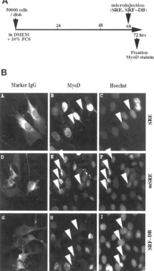
surrounding noninjectedcells in thedifferentpanels of Figure1). Figure 1A details the microinjection protocol and immunofluorescence analyses shown in Figure 1B. InFigure 1B,cellsinjected with SRE (visualized in
panel A by staining for coinjected inert marker anti-body) and fixed 4 h after microinjection show the
complete absence of MyoD expression (Figure
1B,
panel B). Cells were also stained with Hoechst, a
spe-cificstainfor DNA, to allowidentificationof the nuclei inboth microinjected and nonmicroinjected cells
(Fig-Confocal LaserScanningMicroscopy
Dual-channel ConfocalLaserScanning Microscopywasperformed
using the Leica CLSM equippedwith a Krypton-Argon ion laser
using twomajor emissionlines at488 nm for fluorescein
isothio-cyanate excitationand568nmfor rhodamineorTexasRed excita-tion. Planapochromat lenses (40X or63x)wereused andthe
un-treatedimages weredirectly transferred from theVMEbusof the
LeicaMotorola68040to aSilicon GraphicsIRISIndigo workstation (R3000). Images were deconvoluted, gamma mapped, and
con-verted to SGI raster format using "convert" (18). Figures were
assembledcompletely under SGI showcase3.20andprinteddirectly
as postscript files using a Kodak Colorease thermal sublimation
printer(Rochester, NY).
RESULTS
Inhibition of SRF ActivityRapidly Blocks MyoDExpression in C2C12 Myoblasts under
Proliferating Conditions
We have shown previouslythat injection of anti-SRF
antibodies into C2 or L6myoblastsimpairsthe
differ-entiation of these two cell lines by preventing both myogenin expression andthemyoblast-myotube
tran-sition (Vandromme et al., 1992). To investigate this
early requirement of SRF in myogenesis, we ques-tioned the potential implication of SRF inMyoD ex-pression. MyoDthat is akeyregulatorof myogenesis
acts upstreamof myogenin inpromoting cells to dif-ferentiate and its expression isrequiredtopromotethe differentiated phenotype. We chose to inhibit SRF
ac-tivity by microinjection and examine the subsequent
effect of this inhibitionontheexpression ofMyoD. To inhibit SRF activity, subconfluent C2C12 myoblasts growing under conditions that favor proliferation,
were microinjected with either the SRE
oligonucleo-tide (SRE sequence of the c-fos promoter that
corre-sponds to the putative DNA binding site for SRF protein and acts inside cells by squelching SRF
pro-teins) or purified SRF-DB (a 30-kDa portion of SRF containingonlytheDNA-bindingdomain of SRF and acting as a dominant negative mutant in vivo). At differenttimes after injection, cells werefixed and the effect of SRF inhibition on MyoD expression was
as-sessedbyimmunofluorescence. The
muscle-determin-A
50000cells / dish inDMENI +10-c FCS 24 iiiicroinjection (SRE. SRF-DB) 48 68 72 hrs Fixation 'Nl oDstainingB
MarkerIoG MvoD Hoeclist
Figure1. SRF inhibitionthrough microinjectionof SRE oligonu-cleotide or purified SRF-DB blocks MyoD expression in C2C12 myoblastskeptinproliferatingconditions.(A) Schematic represen-tationofthetimingofmicroinjectionandimmunofluorescencedone
in part B. (B) Proliferating C2C12 myoblasts were microinjected
with a solution of rabbit marker antibody containing either SRE
oligonucleotide(A-C),mutated SREoligonucleotide (D-F)or puri-fiedSRF-DB(G-I).Fourhours aftermicroinjection, cellswerefixed
and stained formicroinjected marker rabbit antibodies (A, D,and
ure
1B,
panels C, F, andI). As a control, mutated SREoligonucleotides (inwhichthekeynucleotides for SRF binding have been mutated) were injected (Figure
1B,
panel D) and did not result in any inhibition ofMyoD expression (Figure
1B,
panel E). Similarly,when SRF activity is inhibited after injection of
pu-rifiedSRF-DB (Figure
1B,
panelG)noMyoD expres-sion is detected in injected cells 4 h after injection (Figure1B,
panel H). SRE oligonucleotide or puri-fied SRF-DB were also microinjected into C2myo-blasts, theparental cell line fromwhichC2C12 cells
were derived. As in C2C12, SRF inhibition in C2 cells prevented MyoDexpression (our unpublished observations). However, because C2C12 cells ex-press higher levelsofMyoD, they represent a better
model than the C2 cell line for these experiments
and we continued our studies onlyin C2C12. We next examined whether microinjection of
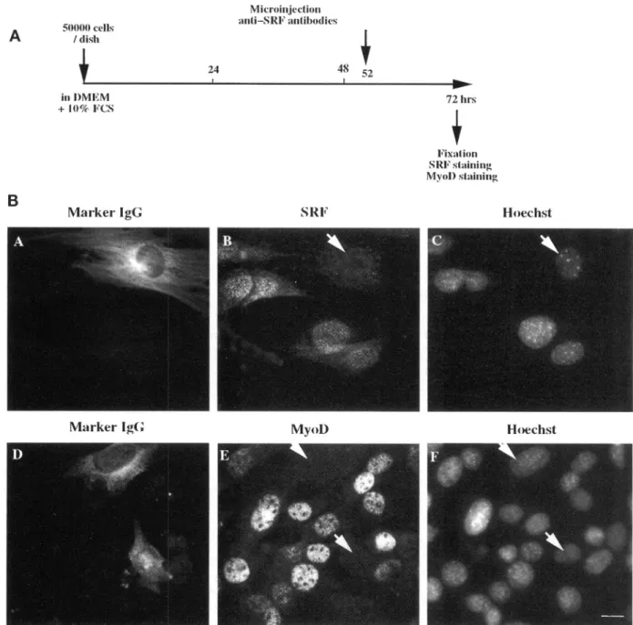
anti-bodies against SRF affected MyoD expression. We haveshowninpreviousstudies that anti-SRF antibod-ies, microinjected in the cytoplasm, bind newly syn-thesized SRF thus preventing it from going into the nucleus (Gauthier-Rouviere et al.,1991a). Assuch, in-hibition ofSRF activity occursonly afterthe
degrada-tion of the preexisting nuclear poolofSRF,which we
have show to take 10-12 h. As shown in Figure 2B,
cytoplasmic injectionof anti-SRF(Figure 2B,panel A) completely abolishes SRF nuclear staining (Figure 2B, panelB)20hafterinjection.Figure 2B,panelsDandE, show that the inhibition ofSRF
by
anti-SRF injection (Figure 2B,panel D) resulted in anabsenceofdetect-able MyoD protein
(Figure
2B,panel E),
whereasmi-croinjection of preimmune antiserum has no effect
either on SRF or MyoD expression (ourunpublished observations). As anadditional controlwe have veri-fied thatcytoplasmic microinjection ofanti-SRFinlate Gl did not affect the distribution of another nuclear protein; cyclinA (ourunpublished observations).
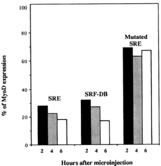
Toestimate moreprecisely thetime atwhich MyoD expression decreases after SRF inhibition, cells were
fixed 2, 4, and 6 h after microinjection of SRE
oligo-nucleotide orpurified SRF-DB. These relativelyshort time points were chosen because of the very short half-life of MyoD protein (MyoD protein turnover is 60-90 min). As summarized in Figure 3, microinjec-tionofSREoligonucleotideorSRF-DB induces arapid
decrease in MyoD expression. Indeed, after2 h, only
28% of cells injected with SRE oligonucleotide and 31% of cells injected with SRF-DBstill show a detect-able level of staining for MyoD. Six hours after SRF inhibition, this had fallen to 18% of cells injected with SRE oligonucleotide and 14% of cells injected with SRF-DB. In contrast, at the different times tested (i.e.,
2, 4, and 6 h), 70% of cells injected with mutated inactive SRE oligonucleotide still expressed MyoD,
showing that microinjection had no significant effect
on MyoD expression (80% of the surrounding
nonin-jected cells express MyoD). Analyzing longer time
periods after microinjection revealed that MyoD ex-pression was repressed for 8 h after microinjection, beginning to resume 10 h after SRE oligonucleotide injection and 12 h afterSRF-DB injection (our unpub-lished observations). This result indicates that the
ef-fectwe observed by microinjection ofSRE or SRF-DB is fully reversible in a time that likely corresponds to the half-life ofSRE and SRF-DBinside living cells.
To confirmthese observations, we used the anti-6/ C2CL2 cell model recently developed (Soulez et al.,
unpublished data). The anti-6 clone is derived from C2C12 cells that have been stably transfected by both a long terminal repeat-glucocorticoid receptor
(LTR-GR) plasmid that encodes the glucocorticoid receptor
(cells called C2CL2) and a plasmid that contains the SRF cDNAinanantisense orientation under the con-trolof the GR element(anti-6). Additionof dexameth-asone to the culture medium activates the expressed glucocorticoidreceptor in turn inanti-6cells, inducing
antisense SRF expression. As observed with injection
of antibodies, the inhibition of endogenous SRF was
assumed tobe completeonly after 12 hof
dexameth-asone induction by which time endogenousSRF was degraded. We investigated the effect of dexametha-sone treatment on the expression of MyoD in both
C2CL2 and anti-6 cells. Both C2CL2 and anti-6 myo-blasts were grown either in proliferating medium
alone orsupplemented with the glucocorticoid dexa-methasone
(10-6
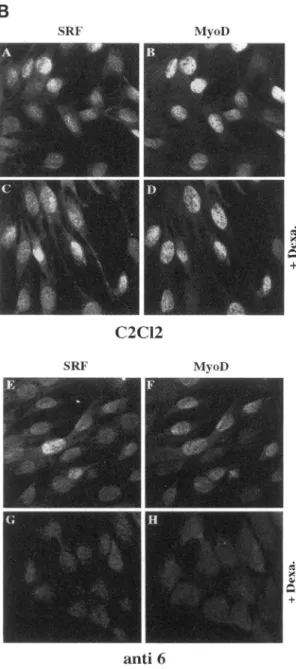
M). After3days,cells werefixed and analyzed forSRF and MyoD expressionby immuno-fluorescence as detailed in Figure 4A. Figure 4B shows SRF expression in parental C2CL2 cells (Figure 4B,panel A). As expected from previous studies SRF is
expressed ubiquitously in the nucleus. Treatment of
C2CL2 cells withdexamethasone(Figure4B, panelC)
resulted in a slight increase in SRF staining. As shown in Figure 4B, panel B, MyoD is expressed in
approxi-mately 80% of the C2CL2 myoblasts, an expression
slightlyincreased after dexamethasone treatment
(be-tween80-85% of MyoD expressing cells; 83% in Fig-ure4B, panel D). Analysis of anti-6 cells (C2CL2 cells containinginducible antisenseSRF) shows that SRF is still expressed in nontreated anti-6 cells (Figure 4B,
panelE). Comparedwiththeparentalcell line aslight
diminution in SRF staining is detectable in the un-treated anti-6 cells (Figure 4B, compare panel A to
panel E). This diminution isalso observableby quan-tification of the mRNAof C2CL2 and anti-6 myoblasts
(Soulez et al.,unpublished data) and has been attrib-uted to an endogenous activation of the LTR-MMTV promoter leading to a slight leak of antisense SRF in these cells. The additionof dexamethasone induces a pronounced diminution of SRF. This is shown in panel G where little or noSRF isdetectable by immunoflu-orescence. MyoD staining in untreated anti-6 myo-blasts is alsoalready diminishedin comparison to the
50000cells
A
/disb in D)MEM + 10%F'CsB
24 Marker IgG MNicroinijection itiSRFantibodies8 5 48 152 SR1FMarkerIgG MyoD) Hoechst
Figure2. SRF inhibitionthrough microinjection of anti-SRF antibodies blocks MyoD expression in subconfluent C2C12 kept inproliferating conditions. (A) Schematic representation of the timing of microinjection and immunofluorescence done in part B. (B) Subconfluent proliferatingC2C12myoblastsweremicroinjectedwithaffinity-purified rabbit anti-SRF antibodies. Twenty hourslater, cellswerefixed and stained forinjectedmarker rabbit antibodies(AandD),SRFexpression (B), MyoD expression (E),and DNA(C and F).
MyoDlevel in C2CL2 myoblasts (Figure 4B, compare
panel F,60%ofMyoD expressing cells,topanelB,80% of MyoD expressing cells). This can most likely be
attributedtothe leak of theantisense SRFinthe anti-6
myoblasts. However, when anti-6 cells are treated
withdexamethasoneto induce antisense SRF,there is
a concomitant decrease inthe level of MyoD (Figure 4B, panel H, 0% of MyoD expressing cells) as SRF expressionfalls(Figure 4B, panel G). Thissuppression
of MyoD expression after dexamethasone treatment cannotbe attributedtoaneffect of thedexamethasone
other than induction of SRF antisense because the parental cell line (which was transfected with the
LTR-GRonly)doesnotshow such inhibitionofMyoD expressionupondexamethasonetreatment(Figure4B, panel D). As soon as SRF level is diminished MyoD expressionfalls(Figure 4B,comparepanelsAandBto
EandF)and becomes undetectable when SRF
expres-72 hrs
Ft'ixat1ion SRF stainilg
MvoI)Staining
o 60
40 SE SRF-DB
20
0
2 4 6 2 4 6 2 4 6
Hoursaftermicroinjection
Figure 3. Inhibitory effectof SRE oligonucleotide orSRF-DB
mi-croinjectioninproliferating C2C12myoblastsonMyoDexpression. Subconfluent proliferating C2C12 myoblasts were injected as de-scribed inFigure 1,fixed2(black),4(dotted),or6 h(white)after injection andstained formicroinjected rabbit antibodies and MyoD expression.The percentage ofinjectedcellsexpressingMyoDwas
measured. The histogramsummarizesthe data fromsix indepen-dentsetsof experiments;40 to50cellswereinjectedineach exper-iment.
sionisfully abolished (Figure4B,panelsG andH).To
confirm that the expression of both SRF and MyoD
wasinhibited inSRFantisense-induced C2CL12 cells,
mRNA levels were analyzed by Northern blot.
Poly(A)+ RNA were isolated fromanti-6cells treated
ornottreatedwithdexamethasoneand
placed
indif-ferentiation medium. In cells treatedwith
dexameth-asone,the twoformsof SRF mRNA weresignificantly reduced,the 2.4 kb form beingundetectable.Asimilar marked reduction of the transcripts for MyoD was observed in SRF antisense-induced cells. Taken
to-gether these results show that inhibition of SRF ex-pression with an antisense SRF, like inhibition of its activitybymicroinjection, results ina complete shut-down of MyoD expression insubconfluent
proliferat-ingmyoblasts.
Inhibition ofSRFActivity in C2C12 Cells Cultured underDifferentiatingConditions Suppresses MyoD Expression
The data reported above clearly show that inhibition of SRFin proliferating C2C12 myoblastsleads to the rapid decrease and abolition of MyoD expression. To investigate whether such effects also take place after C2C12 have ceased to proliferate, we further exam-ined the effect of SRF inhibition on MyoD expression
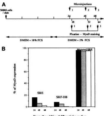
in cells placed in differentiating medium for several hours.C2C12 myoblasts were allowed toproliferatein growth medium until confluence before being in-duced to differentiate by replacing the growth me-dium by differentiation medium. At different times thereafter, differentiating mononucleated C2C12 cells were microinjected with either SRE oligonucleotide,
SRF-DB domain, or mutated SRE oligonucleotide. Cells were fixed 2, 4, or 6 h after microinjection and
processed for detectionofMyoDexpression. Figure 5
shows the results ofexperiments in whichcells were
microinjected 24, 48, or 60 hafter induction of
differ-entiation and fixed 4 h after microinjection. Under these conditions, microinjection of SRE
oligonucleo-tide strongly inhibited MyoD expression. In contrast, in the control experiment in which myoblasts were
injected withmutatedSRE oligonucleotide,no inhibi-tionof MyoDexpression wasobserved.SRF inhibition
through microinjection of SRF-DB also abolished
MyoDexpression. Interestingly, thelaterthe microin-jections wereperformed after addition of
differentia-tionmedium,the morepronouncedwastheinhibition
ofMyoD. In cells placedindifferentiationmediumfor
60 h, no MyoD expression was detected 4 h after
microinjection. Inthese experiments,only
mononucle-atedcellsweremicroinjected. Indeed,36hafter induc-tionofdifferentiationby addition of the differentiation medium,plurinucleated myotubesarealreadypresent
(about 40% of myotubes). Taken together, these data show that inhibition of SRF expression or activity in
differentiating myoblasts blocks expressionof MyoD. Moreover,the efficiencywith whichinhibition ofSRF shutsdown MyoD expression seemsto increase with the time spent in differentiation medium.
DISCUSSION
We have examined thepotential implication of SRF
in the regulation of the expression of the myogenic
regulator MyoD. Through a combination of four
approaches leadingtoeither inhibition of SRF activ-ity (i.e., microinjection of anti-SRF antibodies, SRE
oligonucleotides, or purified SRF-DB) or expression
(inducible antisense SRF-expressing cell line), we
have shown the complete shutdown of MyoD
ex-pression in C2C12 cells. This absence of MyoD ex-pression after inhibition of SRF expression or activ-ity is observed both in subconfluent proliferating
myoblasts and in mononucleated differentiating C2 and C2C12 cells. This regulation of MyoD expres-sion bySRF could explain the early requirement of SRFin myogenesis.
SRFExpression orActivityIs Required for
MyoD Expression
In addition to its well known function in immediate
A
60000 cells / dish 24 hrs 48 hrs 72 hrs~-L)M-DEM
+10%FCS +1-Dexa. Fixation SRF andMyol) stainingC
_ + 4.5Kb_
-;Wi
2.4 Kb -1.bSRF
MyoD
Figure4. SRF inhibitionthrough expression of antisense SRF abol-ishes MyoD expression in myoblasts cultured under proliferating conditions. (A) Schematic representation of the timing of antisense inductionand immunofluorescence doneinpartB. (B) C2CL2 and anti-6cellswereplatedat60,000cells/dishinDMEMsupplemented with10%fetal calfserumwithorwithoutdexamethasone (10-6 M).
Thirty-six hours after plating, dexamethasonewasaddedtothe cells without changingthe culture medium. Cellswerefixed 3 days after
plating and stained for SRF and MyoD expressionasdescribed in
MATERIAL ANDMETHODS. Shownareimmunofluorescent
im-agesofSRF(A, C, E, and G) andMyoD staining (B, D, F, and H).
(A-D) C2CL2 myoblasts; C and D, treated with dexamethasone. (E-H) anti-6 myoblasts; G and H, treated withdexamethasone. (C) Poly(A)+ RNAwereprepared from anti-6 cells cultured in
prolif-erationmedium for 3days andindifferentiation mediumfor2days with or without dexamethasone (10-6 M). Northern blots were
performed usinghuman SRFC-terminalfragment, mouseMyoD,
andR45 human 18SfragmentcDNAasprobes.Shownare
autora-diographs afterhybridation.
involved in the tissue-specific expression of muscle
genes (Miwaand Kedes, 1987; Walsh and Shimmel, 1988). Several studies have demonstrated that SRF binds to CArG box sequences present in the
pro-moter of skeletal as well as cardiac muscle genes (Boxer et al., 1988; Papadopoulos and Crow, 1993).
Moreover, inhibition of SRF activity following
mi-croinjection of anti-SRF antibodies into C2 and L6
myoblasts blocks the cells in a mononucleated
un-differentiated state and prevents expression of two muscle-specific markers myogenin and troponin T
(Vandromme et al., 1992). Although the myogenin
promoter contains one such CArG-box, it was
shown to be nonfunctional both in muscle and in
nonmuscle cells (Santoro and Walsh, 1991). This observation led us to examine the expression of a
upstream effector of myogenin expression, Myod.
Our microinjection experiments show that the ex-pression oractivity of SRF is required continuously
for expression ofMyoDinboth dividingand differ-entiating C2C12 myoblasts. In contrasttomyogenin
and MRF4, which are induced in the course of dif-ferentiation, MyoD expression is constitutive in
bothmyoblasts andmyotubes (Tapscottetal., 1988).
It is required to promote the differentiated
pheno-type and can elicit a muscle determination in vitro
B
SRF MyoDC2CI2
SRF MvoD +4anti
6C.Gauthier-Rouviereetal.
A
50600 cdls /disb Microinjectons tI t2 *3 I I 1 2t 4f 6t a28 M52 t I t2 t3 --- Fbion- MyoDstaining DMEM+10% FCSB
80 -0 .-0 0 60- 40-20 DMEM+2% FCS SRE SRF-DB-
24 48 60 24 48 66 24 48 60 Hours afteradditionofdifferentiationmediumFigure 5. Inhibitory effectof SREoligonucleotideorSRF-DB
mi-croinjection on MyoD expression in C2C12 myoblasts placed in
differentiation medium.(A) Schematic representation of the timing of microinjection and immunofluorescence done in part B. (B)
C2C12 myoblastswereculturedinproliferationmediumfor 3days
and then induced to differentiate by replacing the proliferation
mediumby differentiation medium.After either24(black),48
(dot-ted), or 60h(white)indifferentiation medium, cellswere
microin-jected with either SREoligonucleotide,purified SRF-DB,ormutated SRE oligonucleotide. Four hours after microinjection, cells were
fixed andstained for MyoD expression. The percentageof injected cellsexpressingMyoDwasmeasured.Thehistogramsummarizes
the data fromsixindependentsetsof experiments;40-50cellswere
injectedineach experiment.
program in a number of nonmuscle cell types
(Las-saretal.,1986; Tapscottetal., 1988; Choietal., 1990).
Our findingthat SRF activity is requiredfor MyoD expression provides a target mechanism of the early
requirement forSRF in myogenesis we described
be-fore (Vandromme etal., 1992).
We found that the inhibition of MyoD expression was less marked when SRF inhibition was
per-formed in subconfluent proliferating myoblasts
(with 20-30% of cells still expressing MyoD). In contrast, the inhibition of MyoD expression bySRF was complete in differentiating C2C12 cells (i.e., in
differentiating medium for 48h). Wehave observed that the efficiency with which SRF inhibition led to
an absence of MyoD expression increases with the time spent in differentiating medium. This effect
mightberelated tothe fact that dividing myoblasts
show a heterogeneous expression of MyoD (i.e.,
MyoD is expressed at different levels in
proliferat-ingmyoblasts), a heterogeneity attributed to a cycle-dependent expression of MyoD. Cells are commit-ted todifferentiate and withdraw from the cell cycle at theend ofGi phase (Nadal-Girard, 1978), a time when MyoD expression is maximum. One
explana-tion for the incomplete inhibition of MyoD expres-sion that we observed in proliferative myoblasts
could be that the susceptibility ofMyoD expression to SRF levels is dependent on the cell cycle with a maximum inhibition of MyoD by SRF inhibition when MyoD levels are the highest (end of G1).
In addition to SRF, among the proteins of the MADS-box family are the four MEF2 proteins that
bindthe consensus (C/T)TA(T/A)6 TA(G/A) present in many, if not all, muscle gene regulatory regions
(Cserjesi and Olson, 1991). MEF2A and MEF2C are
specific to differentiated skeletal (and cardiac for
MEF2A) muscle and nerve cells (Yu et al., 1992).
MEF2A expression is concomitant with the early ex-pressionof sarcomeric proteins during myogenesis in vitro, whereas MEF2C appears only later. MEF2D is also restricted tomusclecelllineagesandincontrast to MEF2Aand MEF2C, it is also present inproliferating myoblasts (Breitbartetal., 1993). Consideringthe pos-sible implication of MEF2D in the early commitment eventsleadingtomyogenesis, aswell asthesequence homology between MEF2D and SRF, one may ques-tionwhetherourtoolsinitially designedtospecifically affectSRFactivity, mayalso haveaneffectonMEF2D. However, this could not be the case because of the following: 1)SRFand MEF2bindingsitesareclearly different, and inparticular it was shown that bind-ing of MEF2C to the MEF2 site was notaffected by the presenceof CArG box sequences (McDermottet
al., 1993). 2) MEF2 proteins do not bind the CArG box. 3) Amino acids 138-142 have been reported to be essential for specific binding of SRF to the SRE sequence (Nurrish and Treisman, 1995). These resi-dues are present in SRF-DB (which spans to amino acids 113-265), effectively discounting an effect of
SRF-DB on activity of the MADS-box family mem-bers other than SRF.
How Does SRF Control MyoD Expression?
Our results demonstrate that SRF expression and ac-tivity arerequired for MyoD expression, an effect suf-ficient to explain the early requirement of SRF in myo-genesis. This observation raises the question of how SRF modulates MyoD expression. Different mecha-nisms can be proposed. Simplistically SRF may di-rectly regulate MyoD expression through SRF-bind-ing sites present inthe MyoD promoter. Analysis of the primary nucleotide sequenceofMyoD promoter
(Tapscott etal., 1992) reveals seven putative CArG-like boxes present in both the proximal and the distal regulatory region. Although the CArG-like
boxes diverge variably from the consensus CArG sequence, one of them is fully homologous with a
CArG sequence present in the promoter region of
the mouse myosin light-chain 1A gene, which is capable of binding SRF as efficiently as the wild-type CArG sequence (Catala et al., 1995). The large distance between some of the CArG-like boxes
(es-sentially those of the distal regulatory region) and the transcription initiation site may not be a restric-tion because recently a functional SRE has been identified in the mouse junB gene 2 kb upstream of the site of transcription initiation (Perez-Albuerne et al., 1993), indicating that SREs may exert any influence over long distances in an enhancer-like manner.
Alternatively, the expression of MyoDincultured C2C12 muscle cells may involve the interaction of
one or more muscle-specific factors with a general
transcription factorbindingatthe TATA motif. This kindof mechanism has been proposed for transcrip-tional regulation of Xenopus MyoDa (Leibhametal.,
1994; Wong et al., 1994), which involve interactions between the general transcriptionfactorTFIIDand a
muscle-specific factor MEF2. Interestingly, it has been reported that a related factorTFIIF binds SRF and significantly modulates SRF-activated
tran-scription invitro (Zhuetal., 1994;Jolliotetal., 1995).
Because TFIIDhas been proposed to bind SRF (Zhu
etal., 1991), the possibility remains that SRF bound
to a potential CArG-like box of the MyoD promoter interactswitheither TFIID or TFIIF to promote
tran-scriptional activation. Another possibility wouldbe that SRF, by directly binding to TFIID or TFIIF, induces recruitment of RNA pol II to the initiation
complex or changes the conformation of a large
preexisting complex independently of any binding
to CArG sequence. Alternatively, if the CArG-like boxes in MyoD promoter turn out to be
nonfunc-tional, SRF may be required for the expression of another transcriptionfactor,itself involvedinMyoD transcription. Because MyoD proteins have a very rapid turnover-less than 60 min according to our experiments of microinjection of the protein
(Van-dromme etal., 1994)-such an indirect mechanism is conceivable even though we observed a relatively
rapid shutdown of MyoD expression (within 2 h)
after inhibition of SRF activity.
Thecontrol that SRFexertsonMyoD expressionand the presence of CArG boxes in numerous
muscle-specific genes implicate SRF in at least two levels of
regulationof muscle differentiation:onelevelinwhich SRFexerts acontinuous control ofMyoD expression
in both proliferating and mononucleated differenti-ating musclecells, and a second level inwhich SRF
is involved in the expression of muscle-specific
genes (Mohun et al., 1987) and possibly in
coordi-nation with MyoD protein and/or other myogenic
factors because several muscle promoters contain E and CArG boxes inclose proximity (Sartorelli etal., 1990; Catala et al., 1995).
ACKNOWLEDGMENTS
We aregratefulto J.Demaille and J.C. Cavadore for supportof this work. We thank P. Dias, J. Hallman, and P. Houghton for providing us with anti-MyoD antibodies and R. Treisman for providing us
with SRF cDNA. We thank the Association Francaise contre les
Myopathiesand the Association pour la recherche contre le cancer
for financialsupport.
REFERENCES
Blau,H.M.,Chiu, P., and Webster, C. (1983). Cytoplasmic activation
of human nucleargenes instableheterokaryons.Cell 32, 1171-1180. Boxer, L.M., Miwa, T.,Gustafon,A.,and Kedes, L. (1988).
Identifi-cation and characterization ofa factor that binds to two human
sarcomeric actinpromoters.J.Biol. Chem.264, 1284-1292.
Breitbart,R.E., Liang,C.S., Smoot,L.B.,Laheru,D.A.,Mahdavi, V.,
and Nadal-Ginard,B. (1993).Afourth humanMEF2 transcription
factor, hMEF2D,is anearly markerof themyogenic lineage. Devel-opment118,1095-1106.
Catala, F., Wanner, R., Barton, P., Cohen, A., Wright, W., and
Buckingham,M. (1995).A skeletal muscle-specificenhancer
regu-lated by factors binding to E and CArG boxes is present in the promoterof themouse myosinlight-chain1Agene. Mol. Cell. Biol. 15, 4585-4596.
Choi,J.,Costa,M.L.,Mermelstein, C.S., Chagas,C.,Holtzer,S., and
Holtzer, H. (1990). MyoD converts primary dermal fibroblasts,
chondroblasts, smooth muscle, and retinal pigmented epithelial cells into striated mononucleated myoblasts and multinucleated myotubes.Proc. Natl.Acad. Sci. USA87, 7988-7992.
Concordet, J.P., Salminen, M., Demignon, J., Moch, C.,Maire, P.,
Kahn,A.,andDaegelen,D.(1993).Anopportunistic promoter shar-ingregulatorysequenceswitheitheramuscle-specificor a
ubiqui-tous promoter inthe human AldolaseA gene.Mol. Cell. Biol. 13, 9-17.
Cserjesi,P.,andOlson,E.N.(1991).Myogenininduces the
myocyte-specific enhancer binding factorMEF-2independently of other
mus-cle-specificgeneproducts. Mol. Cell. Biol.11, 4854-4862.
Emt, H.,Walsh, K., Harrison,C.A., andRosenthal, N. (1991).The myosinligh chain enhancerandtheskeletalactin promotershare a
bindingsitefor factorsinvolvedinmuscle-specificgeneexpression.
Mol. Cel. Biol. 11,3735-3744.
Gauthier-Rouviere, C., Basset, M., Blanchard, J.M., Cavadore, J.C., Fernandez,A.,andLamb, N.J.C. (1991a).Casein kinaseII induces
c-fos expression via the serum response element pathway and p67SRF phosphorylation in living fibroblasts. EMBO J. 10, 2921-2930.
Gauthier-Rouviere, C., Cai, Q.Q., Lautredou, N., Fernandez, A., Blanchard,J.M., andLamb, N.J.C. (1993). Expression and
purifica-tion of the DNA-binding domain of SRF: SRF-DB, a part of a
DNA-bindingprotein whichcan act as adominantnegativemutant in vivo. Exp. Cell Res.209,208-215.
Gauthier-Rouviere, C., Cavadore, J.C., Blanchard, J.M., Lamb,
N.J.C., andFernandez, A. (1991b). p67SRFisaconstitutivenuclear proteinimplicatedinthemodulation ofgenesrequiredthroughout
Gilman, M.Z., Wilson, R.N., and Weinberg, R.A. (1986). Multiple protein binding sitesinthe5'-flanking region regulatec-fos expres-sion.Mol. Cell. Biol. 6, 4305-4316.
Joliot, V., Demma, M., and Prywes, R. (1995). Interaction with
RAP74subunitofTFIIF isrequired fortranscriptional activationby
serum responsefactor. Nature373, 632-635.
Klamut, H.J., Gangopadhyay, S.B., Worton, R.G., and Ray, P.N.
(1990). Molecular and functional analysis of the muscle-specific
promoter regionof the Duchenne muscular dystrophy gene.Mol. Cell. Biol. 10,193-205.
Lassar, A.B., Davis, R.L., Wright, W.E., Kadesch, T., Murre, C., Voronova, A.,Baltimore, D., andWeintraub,H. (1991). Functional activityof myogenicHLHproteins requireshetero-oligomerization withE12/E47-like proteinsinvivo.Cell 26,305-315.
Lassar, A.B., Paterson, B.M.,andWeintraub,H.(1986). Transfection ofaDNAlocus thatmediates the conversion of lOT1/2fibroblasts tomyoblasts. Cell 47,649-656.
Leibham, D., Wong, M.W.,Cheng, T.C.,Schroeder, S.,Weil, P.A., Olson, E.N.,and Perry,M.(1994).Binding ofTFIIDandMEF2 tothe
TATA element activates transcription of the XenopusMyoDa pro-moter. Mol.Cell. Biol. 14, 686-699.
McDermott,J.C., Cardoso, M.C., Yu, Y.T., Andres, V., Leifer, D., Krainc, D., Lipton,S.A., andNadal-Ginard,B.(1993). hMEF2C gene encodes skeletal muscle- and brain-specific transcription factors. Mol. Cell. Biol. 13, 2564-2577.
Minty, A., and Kedes, L. (1986). Upstream regions of the human cardiac actin gene thatmodulate itstranscription inmuscle cells: presenceof an evolutionary conserved repeat motif. Mol. Cell. Biol. 6, 2125-2136.
Miwa, T.,andKedes,L.(1987).DuplicatedCArGbox domains have positiveandmutually dependentregulatoryrolesinexpression of thehuman a-cardiacactingene.Mol.Cell. Biol. 7, 2803-2813. Mohun, T., Garrett, N., and Treisman, R.(1987). Xenopus
cytoskel-etalactinand human c-fos gene promoters sharea conserved pro-tein-bindingsite.EMBOJ. 6,667-673.
Mohun, T.J., Chambers, A.E., Towers, N., and Taylor,M.V.(1991).
Expressionof genesencoding the transcription factor SRF during earlydevelopment of Xenopus laevis: identification ofaCArG box-binding activityasSRF. EMBO J. 10,933-940.
Murre,C., McCaw,P.S., Vaessin, H., Caudy, M., Jan, L.Y.,Cabrera, C.V., Buskin, J.N.,Hauschka, S.D., Lassar, A.B. andBaltimore, D.
(1989).Interactionsbetweenheterologoushelix-loop-helix proteins generate complexes that bind specifically to a common DNA
se-quence.Cell 58, 537-544.
Nadal-Ginard,B. (1978). Commitment, fusion and biochemical dif-ferentiation ofamyogeniccellline inthe absenceofDNAsynthesis. Cell 15,855-864.
Norman, C., Runswich, M., Pollock, R., andTreisman, R. (1988). Isolation andproperties ofcDNAclones encoding SRF, a
transcrip-tionfactor thatbinds tothe c-fosserumresponse element.Cell55,
989-1003.
Nurrish, S.J., and Treisman, R. (1995). DNA binding specificity determinantsinMADS-boxtranscriptionfactors. Mol. Cell. Biol. 15,
4076-4085.
Papadopoulos, N., and Crow,M.T.(1993). Transcriptional control of the chicken cardiac myosin light-chain gene is mediated by two AT-rich cis-acting DNA elements andbinding of serumresponse factor. Mol. Cell. Biol. 13,6907-6918.
Perez-Albueme, E.D.,Schatteman, G., Sanders, L.K., and Nathans, D. (1993). Transcriptional regulatory elements downstream of the JunB gene. Proc.Natl.Acad. Sci. USA 90, 11960-11964.
Prywes, R., and Roeder, R.G. (1987). Purification of the c-fos en-hancerbinding protein. Mol.Cell. Biol. 7, 3482-3489.
Santoro, I.M., and Walsh, K. (1991). Natural and syntheticDNA
elements with the CArG motif differ in expression and protein binding properties.Mol. Cell. Biol. 11, 6296-6305.
Sartorelli, V., Webster, K.A., andKedes, L. (1990).Muscle-specific
expressionof the cardiaca-actingene requires MyoDl, CArG-box bindingfactor, and Spl. Genes Dev. 4, 1811-1822.
Tapscott,S.J., Davis, R.L.,Thayer, M.J., Cheng, P.-F., Weintraub, H., and Lassar,A.B.(1988). MyoDl:anuclearphosphoprotein requiring
amychomology regionto convertfibroblaststomyoblasts. Science 242,405-411.
Tapscott,S.J., Lassar, A.B., andWeintraub,A.(1992).Anovel myo-blast enhancer element mediates MyoD transcription. Mol. Cell. Biol. 12,4994-5003.
Taylor, A., Erba, H.P.,Muscat,G.E.O., and Kedes,L.(1988). Nucle-otide sequence and expression of the human skeletal-actin gene:
evolution of functionalregulatory domains. Genomics 3,323-336. Taylor, M., Treisman,R.,Garret,N.,andMohun,T.(1989).
Muscle-specific (CArG)and serum-responsive(SRE)promoterelementsare
functionallyinterchangeableinXenopusembryos andmouse fibro-blasts.Development 106,67-78.
Treisman,R.(1985).Transientaccumulation of c-fosmRNA
follow-ing serumstimulation requiresaconserved 5' element andc-fos 3'
sequence. Cell42,889-902.
Treisman, R.(1987).Identification and purification ofapolypeptide that bindstothe c-fosserumresponseelement. EMBO J. 6, 2711-2717.
Treisman, R. (1990). The SRE:agrowthfactor-responsive
transcrip-tionalregulator. Semin. Cancer Biol. 1, 47-58.
Treisman, R.,and Ammerer, G. (1991). The SRF and MCM1
tran-scription factors. Curr.Opin. Genet. Dev. 2, 221-226.
Tuil,D.,Clergue,N., Montarras,C.,Pinset,C., Kahn,A.,and
Pan-Dinh-Tuy,F.(1990). CCArGG boxes, cis-acting element withadual specificity: muscle-specific transcriptionalactivationandserum
re-sponsiveness.J. Mol. Biol.213,677-686.
Tuil, D., Soulez, M.,Montarras, D.,Pinset, C.,Kahn,A.,and Phan-Dinh-Tuy,F.(1993).Activationofgeneexpression viaCArG boxes during myogenic differentiation. Exp. Cell Res. 205,32-38.
Vandromme, M., Carnac, G., Gauthier-Rouviere, C., Fesquet, D., Lamb, N., and Fernandez, A. (1994). Nuclearimportof the
myo-genicfactorMyoD requirescAMP-dependent protein kinase activ-ity butnot the direct phosphorylation ofMyoD. J. Cell Sci. 107, 613-620.
Vandromme, M., Gauthier-Rouviere, C., Camac, G., Lamb, N.J.C., and Fernandez, A. (1992). Serum response factor p67SRF is ex-pressed and required during myogenic differentiation of both mouseC2 andrat L6musclecelllines. J. Cell. Biol. 118, 1489-1500. Walsh, K., and Schimmel, P. (1988). DNA-binding site for two
skeletalactinpromoterfactorsisimportantforexpression in muscle
cells. Mol. Cell. Biol.8,1800-1802.
Weintraub, H., et al. (1991). ThemyoD gene family: nodal point
Wong, M.W., Pisegna, M., Lu,M.F., Leibham, D., and Perry, M.
(1994).ActivationofXenopusMyoD transcriptionby members of the
MEF2 proteinfamily. Dev.Biol. 166,683-695.
Yu, Y.T., Breitbart, R.E., Smoot, L.B., Lee, Y., Mahdavi, V., and
Nadal-Ginard,B.(1992). Humanmyocyte-specificenhancerfactor2 comprises a groupof tissue-restrictedMADSboxtranscription
fac-tors. Genes Dev.6, 1783-1798.
Zhu, H.,Joliot,V.,and Prywes, R.(1994).Roleof transcription factor TFIIF in serum response factor-activated transcription. J. Biol. Chem.269, 3489-3497.
Zhu, H., Roy, A.L., Roeder, R.G., and Prywes, R. (1991). Serum responsefactoraffectspreinitiationcomplex formationby TFIID in vitro. NewBiol. 3, 455-464.