Biophysical Mechanisms of Lymphocyte Adhesion
to Activated Vascular Endothelium
by MIT LIBRARIES
Gerald C. Koenig B.S. Mechanical Engineering
GMI Engineering and Management Institute (1991)
Submitted to the
Harvard-MI.T. Division of Health Sciences and Technology in partial fulfillment of the requirements for the degree of
Doctor of Philosophy in Medical Engineering at the
MASSACHUSETTS INSTITUTE OF TECHNOLOGY
J
UOL
16 1999
SCHERING
September 1998
© 1998 Massachusetts Institute of Technology 1998. All rights reserved.
Signature of Author: Certified by: -Certified by: Accepted by: Accepted by: MASSACHUSETTS INSTITUTE OF TECHNOLOGY
AUG 2 5 1998
LIBRARIES
Harvard-M.I.T. Division of Health Sciences and Technology July 31, 1998
Rakesh K. Jain Andrew Werk CooWrofessor of Tumor Biology, Harvard Medical School Thesis Supervisor
S a Robert J. Melder
AssistaM Professor of Radiation Oncology, Harvard Medical School Thesis Supervisor
/
. Forbes Dewey, Jr. Professor, Massachu ts Institute of Technology- hairman, Thesis Committee
• .Martha L. Gray
Co-Director, Harvard-M.I.T. Division of Health Sciences and Technology
4A
"
Biophysical Mechanisms of Lymphocyte Adhesion
to Activated Vascular Endothelium
by
Gerald C. Koenig
Submitted to the Harvard-M.I.T. Division of Health Sciences and Technology on July 31, 1998, in partial fulfillment of the requirements for the degree of
Doctor of Philosophy in Medical Engineering
Abstract
The recruitment of lymphocytes to areas of inflammation and angiogenesis involves specific receptor-ligand interactions between cell surface adhesion molecules (CAMs) on lymphocytes and the vascular endothelium. The adhesion process follows a multi-step cascade which has been shown both in vitro and in vivo to involve intercellular adhesion molecule-1 (ICAM-1), vascular cell adhesion molecule-1 (VCAM-1), and endothelial leukocyte adhesion molecule-i (E-selectin) expressed on enidothelial cells. The expression of these CAMs on endothelial cells is modulated by the local concentration of cytokines and growth factors released by resident tissue cells, infiltrating leukocytes, and/or cancer cells. The regulatory mechanisms of adhesion molecule expression by angiogenic factors in
tumor and the resulting leukocyte-endothelial interaction are poorly understood.
The objectives of this thesis were to determine: a) the binding kinetics of each non-activated and IL-2 non-activated lymphocyte subpopulation (CD4+, CD8+, and CD56+ cells), to human umbilical vein endothelial cells (HUVECs) through the use of a parallel-plate flow chamber; b) the expression levels of various cell adhesion molecules on intact HUVEC monolayers treated with tumor interstitial fluid (TIF) and various angiogenic factors, such as VEGF, bFGF, TNFa, and TGFP, using targeted sampling fluorometry (TSF) and flow cytometry; c) the molecular mechanisms used by bFGF in regulating TNFa-mediated cell adhesion molecule (CAM) expression and function; and d) integration of the mechanisms into a framework for lymphocyte-endothelial interaction in disease and health.
These studies demonstrated that both IL-2 activated and non-activated lymphocytes vary in their ability to adhere to activated vascular endothelial cells over a wide-range of physiological flow rates. Also, activated lymphocytes are able to bind with increased levels of efficiency over the non-activated populations, and that variability in lymphocyte subset binding kinetics is dependent on the level of counter-ligand expression and the state of activation. Additional studies demonstrated that IL-2 activated natural killer cells (CD56+) principally utilize an integrin-dependent (P1 and P2), selectin-independent adhesion process in binding to activated vascular endothelium.
The expression studies revealed that fluid extracted from the tumor interstitium upregulates specific CAMs on endothelial cells in vitro, which promotes lymphocyte binding. These results correlated with in vivo findings and validated the use of the tumor fluid as a model for the tumor microenvironment. Analysis of the individual angiogenic factors showed that TNFa and VEGF are able to upregulate specific cell surface CAMs in a dose-dependent manner with similar temporal kinetics. In contrast, bFGF produced a biphasic effect on ICAM-1 regulation, and failed to exhibit an observable effect on the
expression of other CAMs. TGF3 demonstrated no significant differences in CAM expression. Treatment regimens combining bFGF with TNFa or VEGF showed a reduction in the levels of induced CAM expression in a time-dependent process, independent of prior exposure to activating cytokines. This inhibitory effect of bFGF was the result of transcriptional regulation of the inducible CAM genes. Additionally, the early signaling events mediating bFGF action involve the regulation of its receptor tyrosine kinase activity, followed by activation of phospholipase C-y, phospholipase D, and protein kinase C.
The findings from these studies suggest that cytokines and growth factors differentially effect lymphocyte subset binding to activated vascular endothelium by altering cell adhesion molecule expression. Thus, valuable insight is provided into the accumulation of lymphocytes in normal and pathological conditions, such as in tumor and inflammation, and the role of angiogenic factors in regulating the host responsiveness at these sites.
Thesis Supervisor: Rakesh K. Jain
Title: Andrew Werk Cook Professor of Tumor Biology Thesis Supervisor: Robert J. Melder
Title: Assistant Professor of Radiation Oncology Thesis Chairman: C. Forbes Dewey, Jr., Ph.D. Title: Professor of Mechanical Engineering Thesis Reader: Michael A. Gimbrone, Jr., M.D. Title: Elsie T. Friedman Professor of Pathology
Acknowledgments
I would first like to take this opportunity to thank my two thesis advisors, Rakesh Jain and Robert Melder. Their unwavering support and guidance have made this dissertation a reality and a success. I greatly admire them both for their tremendous expertise, enthusiasm, and genuine interest in me, both as a student and as an individual. I could always count on them for much needed advice and encouragement.
I would also like to thank my other two committee members, C. Forbes Dewey, Jr. and Michael A. Gimbrone, Jr. Professor Dewey, who served as my committee chairman, provided beneficial engineering guidance to my thesis and many helpful suggestions and comments. Dr. Gimbrone was an invaluable source of support and provided tremendously insightful advice and comments.
An additional thanks also goes to Roger Mark, former co-director of HST, from whom I received a great deal of inspiration and direction. Dr. Mark was instrumental in my decision to join HST and served as a compassionate and thought-provoking mentor.
Other faculty members in the Steele Laboratory also contributed to this research. Lance Munn introduced me to many of the lab culture and experimental techniques, and continued to provide endless support and expertise throughout my studies. Fan Yuan, Larry Baxter, and Yves Boucher provided encouragement and advice which significantly enhanced my experience.
The Steele Laboratory technicians, Yi Chen and Sylvie Roberge, also provided valuable assistance. Yi contributed significant time and expertise to facilitate the molecular biology experiments, and Sylvie dedicated time and energy to implanting tumors and extracting the fluid for experimental analysis.
My other colleagues and associates in the lab have been a constant source of encouragement, assistance, and friendship. Hera Lichtenbeld, Gabriel Helmlinger, and Paolo Netti were especially so with their genuine good-nature and expertise. Yong, Jin, Nils, Sybill, Dai, Claus, and Christian deserve special mention for their added helpfulness and viewpoints. My fellow HST classmates also provided a stimulating and enriching environment, out of which came many endearing friendships.
The administrative staff also made graduate life a little more enjoyable and easier to handle. From the Steele Laboratory, Carol Lyons and Phyllis McNally were extremely helpful and provided a warm and pleasant environment to work. I especially thank Carol for all her extra support and compassion. From the HST organization, Patty Cunningham, Keiko Oh, Ron Smith, and Carol Campbell all provided significant contributions.
Despite all which the aforementioned have provided, I especially thank my parents, Roland and Gerlinde Koenig, and my sister, Michaela, for their tremendous love and support in all my pursuits in life. Their hard work, dedication, and unending encouragement have instilled in me a passion for life and exploring the unknown. Also, thanks to my loving friends and relatives, for their fresh viewpoints and good-natured spirit.
Finally, I would like to thank my wife Marie, who provided me with unconditional support, understanding, and patience throughout my graduate studies. I am truly blessed to have her as my soul mate for life, and for giving me the joy of a son, Alexander. With an endless love, I cherish thee both.
This work has been partly supported by a fellowship from the MGH Biomedical Engineering Discovery Fund.
To Marie, Alexander, and my parents
Table of Contents
Chapter 1 1.1 1.2 1.3 1.4 Introduction ... ... Motivation and Background ...Hypotheses and Specific Aims ... Significance ... Organization of Thesis ... Chapter 2 Background ...
2.1 Introduction ... 2.2 Cell Adhesion Molecules ...
2.1.1 Selectins ... 2.1.2 Integrins and Immunoglobulin Superfamily ... 2.3 Leukocyte-Endothelial Interaction ... 2.4 Cell Signaling ... 2.4.1 Basic FGF Signaling ... 2.4.2 VEGF Signaling ... 2.4.3 TNFa Signaling ... 2.4.4 TGF3 Signaling ... 2.5 Fluid Dynamics ... Chapter 3 Materials and Methods ...
3.1 Introduction ... ... 3.2 Lymphocyte Preparation ... 3.2.1 Lymphocyte Isolation ... 3.2.2 Lymphocyte Labeling ... 3.2.3 Lymphocyte CAM Receptors... ... 3.3 Endothelial Cell Preparation ...
3.3.1 Endothelial Cell Culture... 3.3.2 Endothelial Cell Monolayer Preparation ...
17 17 19 20 21 23 23 25 25 25 26 28 31 34 35 37 39 44 44 45 45 46 47 47 47 48
3.4 Reagents... ... 49
3.4.1 Cytokines and Growth Factors ... 49
3.4.2 TIF Collection ... 49
3.4.3 Monoclonal Antibodies ... 49
3.4.4 Signaling Inhibitors ... 50
3.5 Functional Adhesion Assay ... 50
3.6 Protein Expression Assays ... 53
3.6.1 Targeted Sampling Fluorometry ... 53
3.6.2 Flow Cytometry (FACS) ... 55
3.6.3 Fluorescence Immunoassay (FIA) ... ... 56
3.6.4 Enzyme-Linked Immunosorbent Assay (ELISA) ... 56
3.7 Northern Blot Analysis ... 57
3.7.1 Blot Preparation and Hybridization ... 57
3.7.2 cDNA Probe Preparation ... 58
3.8 Signal Transduction Analysis ... 59
3.9 Statistical Analysis ... ... ... 59
Chapter 4 Lymphocyte Binding Kinetics ... 60
4.1 Introduction ... 60
4.2 Non-Activated Lymphocyte Binding ... 62
4.3 IL-2 Activated Lymphocyte Binding ... 72
4.4 IL-2 Activated Natural Killer Cell Binding Mechanisms ... 82
4.5 Conclusions... 90
Chapter 5 CAM Modulation by Angiogenic Factors ... 93
5.1 Introduction ... 93
5.2 Surface Expression and Activation Kinetics ... 94
5.2.1 Tumor Interstitial'Fluid (TIF) ... 95
5.2.2 TNFa ... 98
5.2.3 VEGF ... ... 100
5.2.4 bFG F ... 100
5.2.5 T G F O ... . 103
5.2.6 Combination of Angiogenic Factors ... 105
5.3 Functional Correlation ... 108
5 .3 .1 T IF ... . 108
5.3.2 Individual Angiogenic Factors ... 109
5.4 Conclusions ... 118
Chapter 6 Molecular Mechanisms of bFGF in CAM Modulation... 120
6.1 Introduction ... 120
6.2 Characterization of bFGF-Mediated Inhibition ...121
6.2.1 Protein and Functional Level ... 121
6.2.2 mRNA Level ... 124
6.3 mRNA Regulation ... ... 128
6.4 Signaling Pathways ... 132
6.4.1 Surface Protein Expression ... 132
6.4.2 Functional Correlation ... 143 6.5 Generalization of Findings ... 149 6.6 C onclusions ... . 151 Chapter 7 7.1 7.2 7.3 Appendix A. 1 A.2 A.3 Appendix B.1 B.2 B.3 Appendix C.1 C.2 C.3 Conclusions... ... 154 Sum m ary ... .. 154 Future Work ... ... 155
Clinical Significance and Implications ... 158
A Binding Kinetics Data...161
Non-Activated Lymphocyte Binding Data ... 162
IL-2 Activated Lymphocyte Binding Data ... 166
A-NK Receptor Expression Data ... 170
B CAM Expression Data... ... 174
TSF Algorithm for NIH Image ... 174
CAM Expression Kinetics by TIF and Angiogenic Factors...180
Functional Kinetics of Angiogenic Factors ... 189
C Northern Blot and Signal Transduction Analyses ... 92
Kinetics of CAM mRNA Expression ... 193
Level of mRNA Regulation ... 200
Signaling Inhibition Data ... 205
C.3.1 Dose Response Curves ... 205
C.3.2 Adhesion Assay ... 207
C.3.3 FIA Analysis ... 216
List of Figures
1-1 Schematic outline of the thesis ... 22
2-4-1 Schematic of the potential signaling pathways used by bFGF ... 33
2-4-2 Schematic of the signaling pathways used by VEGF... ... 35
2-4-3 Schematic of the signaling pathways used by TNFa... 37
2-4-4 Schematic of the potential pathways used by TGFP ... ... 39
2-5-1 Wall shear stress as a function of the transverse coordinate in the parallel-plate flow chamber ... 42
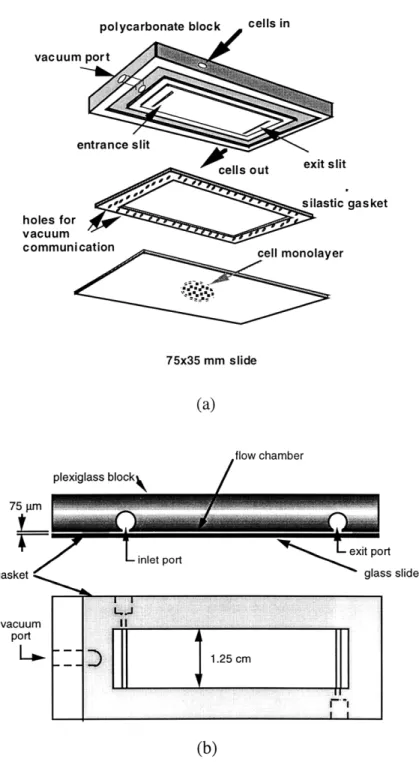
3-1 Slide preparation for monolayers to be used in the adhesion assay ... 48
3-2 Schematic of the Gullino chamber used for the collection of tumor interstitial fluid ... ... 49
3-3 Parallel-plate flow chamber ... 51
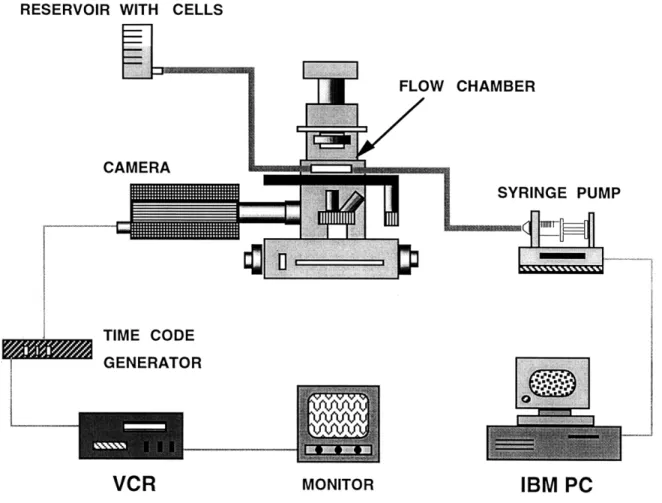
3-4 Schematic of the in vitro adhesion assay set-up ... 52
3-5 Targeted sampling fluorometry ... 54
4-2-1 Cumulative binding curves of non-activated CD4+, CD8+, and CD56+ cells on non-activated HUVEC monolayers ... 63
4-2-2 Cumulative binding curves of non-activated CD4+, CD8+, and CD56+ cells on TNFa-activated HUVEC monolayers ... 64
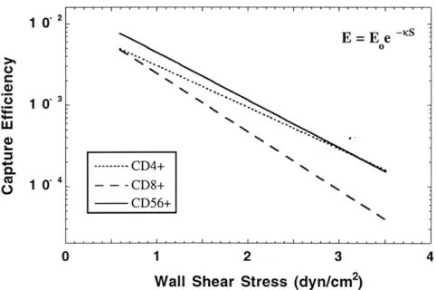
4-2-3 Comparison of binding efficiencies between non-activated CD4+, CD8+, and CD56+ cells on TNFa-activated HUVEC monolayers... 66
4-2-4 Comparison of the fitted parameters: a) , capture coefficient, and b) Eo, efficiency at zero shear generated from the capture efficiency plots of
non-activated lymphocytes on TNFa-non-activated monolayers ... 66 4-2-5 Cumulative binding curves of non-activated CD4+, CD8+, and CD56+
cells on bFGF activated HUVEC monolayers ... 67 4-2-6 Cumulative binding curves of non-activated CD4+, CD8+, and CD56+
cells on TNFa + bFGF activated HUVEC monolayers ... 69 4-2-7 Comparison of binding efficiencies between non-activated CD4+, CD8+,
and CD56+ cells on TNFa + bFGF activated HUVEC monolayers ... 71 4-2-8 Comparison of the fitted parameters: a) K, capture coefficient, and b) Eo,
efficiency at zero shear generated from the capture efficiency plots of
non-activated lymphocytes on TNFa + bFGF activated monolayers ... 71 4-3-1 Cumulative binding curves of IL-2-activated CD4+, CD8+, and CD56+
cells on non-activated HUVEC monolayers ... 72 4-3-2 Cumulative binding curves of IL-2-activated CD4+, CD8+, and CD56+
cells on TNFa-activated HUVEC monolayers ... 74 4-3-3 Comparison of binding efficiencies between IL-2-activated CD4+, CD8+,
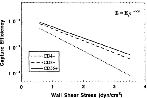
and CD56+ cells on TNFa-activated HUVEC monolayers... 76 4-3-4 Comparison of the fitted parameters: a) K, capture coefficient, and b) Eo,
efficiency at zero shear generated fromi the capture efficiency plots of
activated lymphocytes on TNFa-activated monolayers ... 76 4-3-5 Cumulative binding curves of IL-2-activated CD4+, CD8+, and CD56+
cells on bFGF activated HUVEC monolayers ... 77 4-3-6 Cumulative binding curves of IL-2-activated CD4+, CD8+, and CD56+
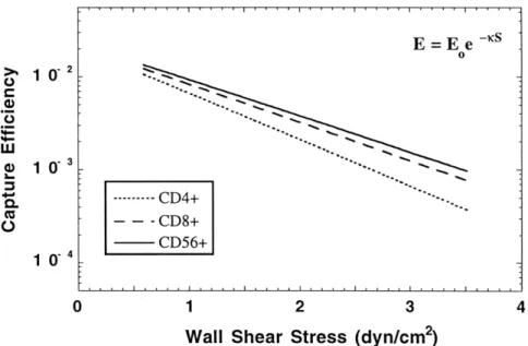
cells on TNFa + bFGF activated HUVEC nionolayers... 79 4-3-7 Comparison of binding efficiencies between IL-2-activated CD4+, CD8+,
and CD56+ cells on TNFa + bFGF activated HUVEC monolayers ... 81 4-3-8 Comparison of the fitted parameters: a) K, capture coefficient, and b) Eo,
efficiency at zero shear generated from the capture efficiency plots of
activated lymphocytes on TNFa + bFGF activated monolayers ... 82 4-4-1 Cumulative binding curves of A-NK cells on TIF-activated and
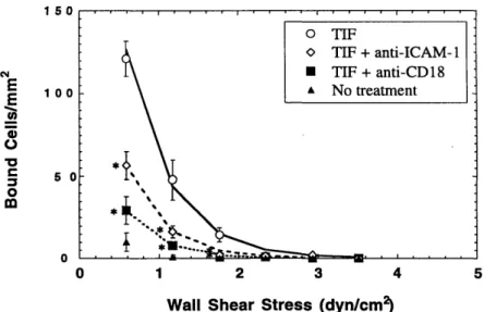
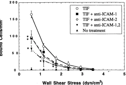
non-activated endothelial cell monolayers ... 83 4-4-2 A-NK cell adhesion to TIF-activated HUVEC monolayers showing the role
of CD 18:ICA M -1 ... 84 4-4-3 A-NK cell adhesion to TIF-activated HUVEC monolayers showing the role
of ICAM -1:ICAM -2 ... 85 4-4-4 A-NK cell adhesion to TIF-activated HUVEC monolayers showing the
4-4-5 A-NK cell adhesion to TIF-activated HUVEC monolayers showing the role
of E-selectin and sialyl-Lewis X ... . 86 4-4-6 A-NK cell adhesion to TIF-activated HUVEC monolayers showing the role
of P-selectin ... 87 4-4-7 A-NK cell adhesion to TIF-activated HUVEC monolayers showing the
combined effects of blocking a) CD 18 and VLA-4 on A-NK cells, and b)
ICAM-1 and VCAM-1 on endothelial cells ... 88 4-4-8 A-NK cell adhesion to TNFa-activated HUVEC monolayers showing the
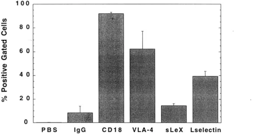
roles of a) 1I and P2 integrins, and b) E- and P-selectin... 89 4-4-9 A-NK expression levels of CD 18, VLA-4, L-selectin, and sialyl Lewis X ... 90 5-2-1 Kinetics of CAM expression on HUVEC monolayers treated with LS 174T
TIF for 24 hrs using targeted sampling fluorometry ... 96 5-2-2 Concentration of a) human VEGF, b) human TNFa, and c) human bFGF
in the tumor interstitial fluid of LS 174T xenotransplants ... 97 5-2-3 Kinetics of CAM expression on HUVEC monolayers treated with TNFa
for 6 and 24 hrs using flow cytometry ... 99 5-2-4 Kinetics of CAM expression on HUVEC monolayers treated with VEGF
for 6 and 24 hrs using TSF ... 101 5-2-5 Kinetics of CAM expression on HUVEC monolayers treated with bFGF
for 6 and 24 hrs using flow cytometry ... 102 5-2-6 Kinetics of CAM expression on HUVEC monolayers treated with TGFP
for 6 and 24 hrs using flow cytometry ... 104 5-2-7 Kinetics of CAM expression on HUVEC monolayers treated with a
combination of TNFa (50 ng/ml) and bFGF (10 ng/ml) for 6 hrs using
T SF ... ... 106 5-2-8 Kinetics of CAM expression on HUVEC monolayers treated with a
combination of TNFo (50 ng/ml) and bFGF (10 ng/ml) for 24 hrs using
T SF ... 107 5-3-1 Cumulative binding curves of A-NK cells on TIF-activated (24 hr) and
non-activated endothelial cell monolayers ... 109 5-3-2 TNFa and TGFP dose response curves of activated NK cell binding to
HUVEC monolayers treated with the cytokines for 24 hr ... 110 5-3-3 VEGF and bFGF dose response curves of activated NK cell binding to
HUVEC monolayers treated with the angiogenic factors for 24 hr ... 110 5-3-4 Temporal kinetics of 50 ng/ml TNFC and 10 ng/ml bFGF on A-NK cell
5-3-5 Temporal kinetics of 35 ng/ml VEGF and 10 ng/ml TGFP on A-NK cell
binding to activated HUVEC monolayers ... 12 5-3-6 Dose response curve of activated NK cell binding to HUVEC monolayers
treated simultaneously with TNFa and bFGF for 24 hr ... 113 5-3-7 Dose response curve of activated NK cell binding to HUVEC monolayers
treated simultaneously with VEGF (35 ng/ml) and bFGF for 24 hr...14 5-3-8 Dose response curve of activated NK cell binding to HUVEC monolayers
treated simultaneously with TNFa (50 ng/ml) and TGFp for 24 hr ... 114 5-3-9 Inhibition kinetics of A-NK cell binding to HUVEC monolayers treated
simultaneously with 50 ng/ml TNFa and 10 ng/ml bFGF ... 1...15 5-3-10 Antibody blocking experiments demonstrating a direct correlation between
ICAM-1 and VCAM-1 expression and A-NK cell binding to HUVEC monolayers treated simultaneously with 50 ng/ml TNFa and 10 ng/ml
bFG F ... 116 5-3-11 Inhibition kinetics of A-NK cell binding to activated HUVEC monolayers
pretreated with either a) 50 ng/ml TNFa (24 hr total exposure time) and then 10 ng/ml bFGF for the time indicated, or b) 10 ng/ml bFGF for the
time indicated and then 50 ng/ml TNFa for 5 hrs...117 6-2-1 Kinetics of A-NK cell binding to HUVEC monolayers treated with
bFGF-conditioned medium and 50 ng/ml TNFa ... ... 123 6-2-2 Kinetics of A-NK cell binding to HUVEC monolayers initially treated with
50 ng/ml TNFa for 6 hrs (t = 0) and then subsequently washed and treated
with + 10 ng/ml bFGF ... 125 6-2-3 Northern blot analysis showing the kinetics of bFGF (10 ng/ml)-induced
inhibition of ICAM-1, VCAM-1, and E-selectin mRNA levels in HUVECs
simultaneously activated by TNFa ... ... 126 6-2-4 Graphic representation of the kinetics of bFGF (10 ng/ml)-induced
inhibition of (a) ICAM-1, (b) VCAM-1, (c) E-selectin, and (d) ICAM-2
mRNA levels in HUVECs simultaneously activated by TNFa ... ...127 6-3-1 Graphic representation of the effects of bFGF (10 ng/ml) on the stability of
(a) ICAM-1, (b) VCAM-1, (c) E-selectin, and (d) ICAM-2 mRNA in
HUVECs initially activated with TNFa ... ... 130 6-3-2 Graphic representation of the effects of bFGF (10 ng/ml) on (a) ICAM-1,
(b) VCAM-1, (c) E-selectin, and (d) ICAM-2 mRNA in HUVECs initially
activated with TNFa ... 131 6-4-1 Simplified schematic of the TNFa and bFGF signaling pathways ... 133 6-4-2 Effect of bFGF neutralizing antibody on TNFa and bFGF induced
6-4-3 Effect of supplemental heparin on TNFa and bFGF induced HUVEC
protein expression ... ... 135
6-4-4 Effect of the receptor tyrosine kinase inhibitor MDHC on TNFa and bFGF induced HUVEC protein expression ... 136
6-4-5 Effect of the protein tyrosine phosphatase inhibitor sodium orthovanadate on TNFa and bFGF induced HUVEC protein expression ...138
6-4-6 Effect of the phospholipase C inhibitor NCDC on TNFa and bFGF induced HUVEC protein expression ... 139
6-4-7 Effect of the phospholipase D inhibitor propranolol on TNFa and bFGF induced HUVEC protein expression ... 140
6-4-8 Effects of the protein kinase C inhibitors calphostin C and bisindolylmal-eimide (BIM) on TNFa and bFGF induced HUVEC protein expression... 141
6-4-9 Effects of the protein kinase C inhibitor calphostin C on PMA and bFGF induced HUVEC protein expression ... 142
6-4-10 Effect of neutralizing bFGF Ab (10 mg) on A-NK cell binding to HUVEC monolayers ... 143
6-4-11 Effect of exogenous heparin (10 mg/ml) on A-NK cell binding to HUVEC m onolayers ... 144
6-4-12 Effect of MDHC (5 mM) on A-NK cell binding to HUVEC monolayers... 144
6-4-13 Effect of sodium orthovanadate (30 mM) on A-NK cell binding to HUVEC monolayers ... ... 145
6-4-14 Effect of NCDC (50mM) on A-NK cell binding to HUVEC monolayers... 146
6-4-15 Effect of propranolol (50 mM) on A-NK cell binding to HUVEC monolayers ... 147
6-4-16 Effect of calphostin C (50 nM) on A-NK cell binding to HUVEC monolayers ... 147
6-4-17 Effect of calphostin C (50 nM) on A-NK cell binding to HUVEC monolayers treated with bFGF (10 ng/ml) and PMA (100 nM)...148
6-4-18 Effect of A-NK cell binding by long term, high dose PMA treatment of HUVEC monolayers ... 149
6-5-1 Broader ramifications of the studies presented ... 150
A-1 Non-activated lymphocyte binding data ... 162
A-2 IL-2-activated lymphocyte binding data ... 166
B-2 CAM expression kinetics by angiogenic factors ... 180
B-3 Functional kinetics of angiogenic factors ... ... 189
C-1 Kinetics of CAM mRNA expression ... 193
C-2 Level of mRNA regulation ... 200
List of Tables
2-1 Regulation of adhesion molecules by cytokines and angiogenic factors ... 24 2-2 Known ap3 integrin complexes ... 26 4-2-1 Comparison of cell binding densities of non-activated CD4+, CD8+, and
CD56+ cells on non-activated HUVEC monolayers... 63 4-2-2 Comparison of cell binding densities of non-activated CD4+, CD8+, and
CD56+ cells on HUVECs treated with TNFa for 24 hrs ... ... 65 4-2-3 Comparison of cell binding densities of non-activated CD4+, CD8+, and
CD56+ cells on HUVECs treated with bFGF for 24 hrs ... 68 4-2-4 Comparison of cell binding densities of non-activated CD4+, CD8+, and
CD56+ cells on HUVECs treated with TNFa+bFGF for 24 hrs ... 69 4-3-1 Comparison of cell binding densities of IL-2 activated CD4+, CD8+, and
CD56+ cells on non-activated HUVEC monolayers... 73 4-3-2 Comparison of cell binding densities of IL-2 activated CD4+, CD8+, and
CD56+ cells on HUVECs treated with TNFa for 24 hrs ... 74 4-3-3 Comparison of cell binding densities of IL-2 activated CD4+, CD8+, and
CD56+ cells on HUVECs treated with bFGF for 24 hrs ... 78 4-3-4 Comparison of cell binding densities of IL-2 activated CD4+, CD8+, and
CD56+ cells on HUVECs treated with TNFa+bFGF for 24 hrs ... 79 6-1 Specific signal transduction inhibitors and their modes of action ... 133 C -3 FIA analysis ... .. 216
Chapter 1
Introduction
1.1 Motivation and Background
Each year in the United States, over 1.2 million individuals are diagnosed with cancer for the first time. According to the American Cancer Society, cancer took the lives of over 560,000 Americans in 1997, representing 23% of all mortality and the second leading cause of death, as defined by 1994 statistics [Vital Statistics of the United States, 1994]. More than 85% of these cancers are solid tumors, and approximately one-half of the patients with these tumors die of their disease. A most sobering and troublesome reality.
Solid tumors, derived from normal host cells which have gone awry, have a fundamental characteristic in being dependent on blood supply for oxygen, nutrients and waste removal beyond a size of 1-2 mm in diameter [Folkman, 1990]. The development of new blood vessels from pre-existing vessels is termed angiogenesis, and occurs in a range of pathologies in addition to cancer (tumor angiogenesis), including atherosclerosis (hyperproliferation of the vasa vasorum within atherosclerotic plaques), endometriosis, and diabetic retinopathy.
Cancer cells, as well as surrounding and infiltrating host cells, produce multiple polypeptide cytokines that are essential for promoting the process of angiogenesis, and are appropriately termed angiogenic growth factors. Among the most well-documented are vascular endothelial growth factor (VEGF), basic fibroblast growth factor (bFGF), transforming growth factor-P (TGFP), and tumor necrosis factor-a (TNFa). These factors, in addition to other cellular products produced by the neoplastic and non-neoplastic
cells, influence the tumor microenvironment by establishing temporal and spatial heterogeneities in blood flow, altering expression of cell adhesion molecules on parenchymal, stromal, and vascular cells, and modifying physiological and metabolic properties. The changes in the microenvironment produce effective barriers against the delivery of cells and molecular therapeutics to tumors.
The mounting and delivery of an effective immunogenic response to tumors by the host immune system involves a progressive sequence of steps, each of which is potentially modified by the tumor microenvironment. The steps include: a) entering the tumor vasculature from host sites, b) interacting with the vascular compartment and forming stable cell:cell adhesion to the vessel wall, c) transporting across the microvascular wall into the interstitium, d) transporting through the interstitial compartment to reach the cancer cells, and then e) actively attacking or supporting the killing of the cancer cells.
The mechanisms of lymphocyte interaction with the vascular endothelial cells serves as a particular point of interest for its importance in the initial steps of the immune response and potentially subserving specific cell populations. The capture and adhesive interactions are influenced by the local hemodynamic forces, determined by the vessel diameter, viscosity, and fluid velocity, and by the adhesive forces, determined by the spatial arrangement and level, affinity, and kinetics of bond formation between receptor-ligand pairs. A working hypothesis is that the specific microenvironment present in regions of angiogenesis, specifically the biological influence of growth factors on the vascular endothelium, is necessary for the interactive process.
The same or closely related adhesion molecules used in angiogenesis to promote cell migration and proliferation also mediate immune cell adhesion to the vasculature. This seemingly paradoxical influence is further complicated by the fact that immune cells themselves are able to produce significant levels of angiogenic factors to either facilitate additional immune cell infiltration or promote tumor vessel formation. An increased understanding of the temporal regulation of the adhesion molecules and angiogenic factors expressed in tumors, as well as the particular molecular mechanisms used by lymphocyte subpopulations, is needed to further elucidate this interactive process.
The influence of angiogenesis and the growth factors and cytokines involved in the interaction of blood leukocytes with the vascular endothelium extends beyond the pathophysiology of tumors. Other diseases with fundamentally similar angiogenic processes involved include atherosclerosis, vasculitis, allograft rejection, graft vs. host disease, and chronic inflammation. It is also vital to several normal physiological processes as well, such as acute inflammation, tissue repair, and embryogenesis.
1.2 Hypotheses and Specific Aims
In formulating the thesis, a set of hypotheses were initially postulated which provided a framework on which the specific objectives were derived. The original hypotheses were as follows:
Tumor interstitial fluid and purified angiogenic factors can modulate cell adhesion molecules (CAMs) on the endothelium. Diversity among angiogenic factors can result in the differential regulation of CAMs.
Endothelial CAMs induced by angiogenic factors can be recognized differentially by lymphocyte sub-populations, namely T lymphocytes and natural killer cells.
Differences in the intracellular signaling pathways used by the angiogenic factors can result in differential CAM modulation.
The specific goals of this thesis were to test these hypotheses by performing the following experimental studies:
Measure the binding kinetics of each specific non-activated and IL-2 activated lymphocyte subpopulation, namely CD4+, CD8+, and natural killer cells (CD56+), through the use of parallel-plate flow chamber studies.
Quantify the expression of various cell adhesion molecules on intact HUVEC monolayers treated with tumor interstitial fluid and various angiogenic factors, such as VEGF, bFGF, TNFa, and TGF, using targeted sampling fluorometry (TSF) and flow cytometry.
Determine the molecular mechanisms used by bFGF in regulating TNFao-mediated cell adhesion molecule (CAM) expression and function.
Integrate the mechanisms into a framework for lymphocyte-endothelial interaction in disease and health
1.3
Significance
Current therapies, which include chemotherapy, radiation, and surgery, can cure about half of the diagnosed cancers. While encouraging, it still leaves a large population of individuals that cannot be treated successfully. New therapies are continuously being explored, including combined treatment protocols. Of particular interest are gene-based therapy [Goldspiel, et al., 1993], and adoptive immunotherapy, whereby TIL (tumor-infiltrating lymphocyte) or LAK (lymphokine-activated killer) cells are delivered to cancer patients in the hope that the body's natural immune cells will resolve the tumor [Rosenberg, 1990]. However, both have had only obtained limited success, but have helped raise questions about the fundamental mechanisms by which the immune system acts.
The studies proposed in this thesis can provide increased understanding to several physiological and pathophysiological processes. The binding kinetics and molecular mechanisms used by the individual lymphocyte subpopulations in interacting with the vasculature, both in tumor, inflammation, and wound healing, are essential for determining mechanisms used by the immune system in localizing to areas requiring active immunological response. In addition, improvements can be made to current modeling schemes which attempt to predict the biodistribution of effector cell populations in the human.
The effects of angiogenic factors on cell adhesion can provide necessary insight into the regulatory and protective mechanisms potentially found in tumors, as well as in inflammation and wound healing. It may also contribute to a better understanding of metastasis and the role of cytokines and growth factors in the process.
Finally, defining the signaling mechanisms used by specific growth factors, may contribute to a better understanding of the molecular mechanisms used by the growth factors to modify cell adhesion molecule expression, and its relation to the other physiological functions which it produces. Also, it gives insight into the angiogenic process and the kinetic responses elicited by specific growth factors in the microenvironment.
Clearly, a better understanding of these processes may provide new insights into the mechanisms of tumor evasion, immune response and neovascular growth, which may be used for developing alternative or modified modalities of therapies against cancer and other diseases.
1.4
Organization of Thesis
The thesis is organized into the following chapters, and is outlined in Figure 1-1: Chapter 1 gives the motivation and significance of the work, as well as the hypotheses and specific aims addressed in the thesis.
Chapter 2 provides a summary of the background essential for the studies conducted in this investigation on lymphocyte-endothelial interactions and cell signaling.
Chapter 3 discusses the materials and methods used in this research. Included is a description of the lymphocyte and endothelial preparations, as well as the protein, mRNA, and functional adhesion assays performed.
Chapter 4 provides results and discussion on the experiments performed examining the lymphocyte binding kinetics for both non-activated and activated lymphocyte
subpopulations.
Chapter 5 presents results and discussion on the analysis of cell surface adhesion molecule expression induced by the angiogenic factors TNFoa, VEGF, bFGF, and TGF3, as well as the tumor interstitial fluid.
Chapter 6 describes the results and provides discussion on the studies performed analyzing the molecular mechanisms used by bFGF in modulating CAM expression.
Chapter 7 provides a summary of the studies conducted in the thesis, and discusses future directions for the research, along with its clinical significance.
The work presented in this thesis has at least partially contributed to four published manuscripts [Munn, et al., 1995; Melder, et al., 1996; Melder, et al., 1996; Jain, et al., 1996], and two in preparation involving the findings presented in Chapters 4 and 6. In addition, a patent is pending with the U.S. Patent Office, based on the inhibitory mechanisms of basic fibroblast growth factor (bFGF) and includes as co-inventors: R.K. Jain, R.J. Melder, and L.L. Munn.
IVEGF
CAM Modulation *bFGF
b *TGF S
Chapter 4 Chapter 6
Lymphocyte Binding olecular Mechanis
Kinetics of bFGF Signaling
on- activated CD4+,CD8+,CD6+ * functional effects n- activated CD4+,CD8+,CD56+ Chapter 3 * mRNA effects .-2 activated CD4+,CD8+,CD56+
Methods,.,
Chapter 7
Summary Conclusions
Figure 1-1. Schematic outline of the thesis. * n
*IL
Chapter 2
Background
2.1
Introduction
The active recruitment of circulating lymphocytes to sites of inflammation, wound repair, and tumor is an integral event in mounting an effective immune response. Adhesive forces resulting from the spatial arrangement, quantity, affinity and kinetics of bond formation between receptor-ligand pairs of cell adhesion molecules (CAMs) must overcome the hemodynamic forces present in the blood vessels. The expression of these CAMs on the endothelium is modulated by the local concentration of inflammatory cytokines and angiogenic factors, see Table 2-1 [Osborn, 1990; Jain, et al., 1996]. The majority of studies involving the growth factors have focused on the specific angiogenic responses. Only recently has there been increased interest in defining the role of these factors in cell adhesion molecule induction and modulation. Growing interest has been placed on the specific signaling transduction pathways used by these cytokines and growth factors in regulating cellular responses in the hopes of developing new therapies for several pathological conditions, such as cancer and heart diseases.
This chapter is designed to serve as a basic reference of the literature related to the understanding of lymphocyte capture mechanisms, the function and signaling pathways used by several cytokine and growth factors, and the fluid mechanics of the in vitro vessel model used in this thesis. With this background, it is hoped that the reader will have a better fundamental understanding of the studies conducted in this thesis, and be able to follow the general findings and appreciate the significance of this work.
Table 2-1. Regulation of adhesion molecules by cytokines and angiogenic factors. Adapted from [Jain, et al., 1996].
Cytokine Cell Type ICAM-1 VCAM-1 E-Selectin P-Selectin
bFGF HUVEC -, L -, , - -HDMEC 1 _ _ NM IFNY HUVEC <> <> NM HDMEC NM <) NM NM MME. NM NM RME NM T NM NM IL-1 HUVEC 1 1 NM HDMEC - T NM MME 1" NM NM IL-4 HUVEC 1, _ _ ", I NM HDMEC NM - NM NM MME NM + NM NM BDEC , T NM TGFP HUVEC <4 <_ , NM MME +, + NM NM TNFa HUVEC T T T T HDMEC NM 1T NM NM MME - NM NM BDEC " T NM BCE NM NM T NM VEGF HUVEC T T T
IL-1 + IL-4 HUVEC , I NM
IL-1 + bFGF HUVEC 1 <.> NM
IL-1 +IFN HUVEC T NM T NM
IL-1 + TGF3 HUVEC NM NM ,I NM
TNFa + IL-4 HUVEC 4 " NM
HDMEC NM < NM NM
BDEC + (. < -, NM
TNFa + IFNy HUVEC t NM 1 NM
TNFa + bFGF HUVEC - , NM
TNFa + TGFp HUVEC _ 4 , NM
TGFP + IL-8 HUVEC NM NM NM
bFGF + VEGF HUVEC _ NM NM NM
IFNy + IL-4 HUVEC 1 .. NM NM
IL-4 + TGF3 HUVEC NM 'NM I - NM
1 = upregulation, <-4 = no effect, 1 = downregulation, NM = not measured. Effect of combined treatments are relative to the individual treatments, whereby: 1TT = synergistic upregulation, 1 = additive effect, <-* = no difference from responsive individual treatment, 1 = negative combined effect. HUVEC - Human Umbilical Vein Endothelial Cell (EC), HDMEC = Human Dermal Microvascular EC, MME = Murine Microvascular Endothelium, RME = Rat Microvascular Endothelium, BDEC = Baboon Dermal EC (in vivo), BCE = Bovine Capillary EC.
2.2 Cell Adhesion Molecules
There are basically two families of CAMs involved in producing the adhesive forces between leukocytes and endothelial cells: 1) the selectins (e.g. E-, L-, and P-selectin) and 2) the immunoglobulin (Ig) superfamily members (ICAM-1 and VCAM-1) on endothelial cells and the corresponding integrin receptors on the leukocytes (02 and
1
3) [Springer,1995; Collins, 1995].
2.1.1
Selectins
Selectins consist of an N-terminal lectin domain, one epidermal growth factor-like module, and from two to nine short consensus repeats [Lawrence and Springer, 1991]. To date, they have only been shown to exist on circulating cells and the endothelium, unlike integrins and Ig superfamily members. The three members of this group of CAMs are: a) E-selectin (ELAM-1, CD62E), b) P-selectin (PADGEM, CD62P), and c) L-selectin (LECAM-1, CD62L). E-selectin is synthesized by endothelial cells in response to inflammatory agents and promotes attachment of monocytes, neutrophils, and certain lymphocytes by combining with the ligand sialyl-Lewis X [Bevilacqua, et al., 1989]. P-selectin is expressed in Weibel-Palade bodies of endothelial cells and in a granules of platelets, becoming mobilized to the cell membrane upon inflammatory activation and promoting adhesion to monocytes and neutrophils [Larsen, et al., 1989]. L-selectin is expressed on leukocytes, promoting lymphocyte adhesion in peripheral lymph nodes and in neutrophils, monocytes, and lymphocyte emigration at inflammatory locales [Spertini, et al., 1991]. The structure of the selectins and their corresponding carbohydrate ligands allow for a high on-rate to complex formation resulting in efficient interactions of rapidly flowing cells with the endothelium, and along with a high off-rate, permit active rolling to occur. Thereby, the selectins are considered to primarily mediate leukocyte capture and rolling.
2.1.2
Integrins and Immunoglobulin Superfamily
Integrins consist of a oap heterodimer, with the a subunits (120-180 kD) covalently associated with a p subunit (90-110 kD) [Hynes, 1992]. Most integrins are expressed on a wide variety of cells and are major receptors for cell attachment to extracellular matrices and in mediating cell-cell adhesion events. There consists a large, yet restrictive, diversity of integrins with currently 8 known 0 units and 15 known a subunits (see Table 2-2) [Hynes, 1992; Rouslahti, 1991]. The a and P subunit combination determines the ligand specificity. While the majority of these integrins are recognized by extracellular matrix
proteins involved in cell-substratum adhesion, there are some which recognize integral membrane proteins of the immunoglobulin superfamily and mediate direct firm cell-cell adhesion.
The Ig superfamily consists of a group of proteins with one or more Ig domains, regions of 70 to 110 amino residues homologous to either Ig variable (V) or constant (C) domains. Most members of this superfamily are integral plasma membrane proteins with Ig domains in the extracellular portions, transmembrane domains composed of hydrophobic amino acids, and widely divergent cytoplasmic tails with no homology to one another. The most common of these which are associated with leukocyte adhesion to vascular endothelium are ICAM-1 (intercellular adhesion molecule-i), ICAM-2, and VCAM-1 (vascular cell adhesion molecule-1) [Rothlein, et al., 1986; Stauton, et al., 1989; Osborn, et al., 1989]. ICAM-1 (CD54) and ICAM-2 (CD102), a constitutively expressed adhesion molecule, on the endothelium bind to LFA-1 (P2 integrin) on the leukocytes, and VCAM-1 (CD106) on the endothelium binds to VLA-4 (P1 integrin) on the leukocytes [Marlin and Springer, 1987; Elices, et al., 1990; Hemler, 1990].
Table 2-2. Known ap integrin complexes. 01 [CD29] - al (CD49a), a2 (CD49b), a3 (CD49c),
a4 (CD49d), a5 (CD49e), a6 (CD49f),
a7, a8, a9, Cv (CD51)
P2 [CD18] - caL (CDla), xM (CDllb), aX (CDllc) P3 [CD61] --> alib , xv 04 [CD104] cx6 06 -4 av 37 --> x4, aIEL P8 av
2.3
Leukocyte-Endothelial Interaction
As for functional studies involving cell adhesion, many investigators have analyzed leukocyte adhesion to cytokine activated endothelial cell monolayers in vitro [Bevilacqua, et al., 1985; Schleimer and Rutledge, 1987; Cavender, et al., 1987; Cotran and Pober, 1990]. Early studies were performed using static conditions and showed that basal adhesion of blood monocytes was significantly greater than that of neutrophils and lymphocytes [Bevilacqua, et al., 1985; Luscinskas and Lawler, 1994]. On activated endothelium, i.e. TNFa, IL-1, or LPS treatment, increases of 2- to 5-fold were seen in monocyte and lymphocyte adhesion and a 10- to 15-fold increase in neutrophil adhesion [Bevilacqua, et
al., 1985; Lo, et al., 1989; Pober, et al., 1987]. Multiple adhesion molecules and leukocyte chemoattractants, e.g. ICAM-1, VCAM-1, E-selectin, P-selectin, L-selectin, MCP-1 and PAF, were proven to be essential in the process and were suggestive of redundant or overlapping function [Springer, 1994]. More recent studies using defined laminar flow conditions have revealed a more complex process in which sequential and overlapping functions exist for the multiple receptor-ligand pairs [Lawrence and Springer, 1991; Springer, 1994; Luscinskas, et al., 1994; Butcher, 1991]. The general model commonly accepted now involves a multi-step cascade consisting of four events (phases 1-4): (i) leukocyte capture and rolling mediated by L-selectin, E-selectin, and P-selectin, (ii) leukocyte arrest dependent on 1 integrins/VCAM-1 with (iii) leukocyte firm adhesion and spreading mediated by P2 integrins/ICAM-1, and (iv) in conjunction with CD31, P2 mediated leukocyte diapedesis, with some overlap in the functions of the adhesion molecules.
Studies have been carried out defining the specific adhesive interactions between T lymphocytes and vascular endothelium. Previous in vitro studies have demonstrated the roles of LFA-1/ICAM-1, VLA-4/VCAM-1, and sialyl-Lewis X/E-selectin in resting CD4+ cell adhesion to activated endothelial cells under static conditions [Shimizu, et al., 1991; Dustin and Springer, 1988; Bierer and Burakoff, 1988]. More recent in vitro studies have determined the cellular processes and molecular events involved in resting CD4+ and CD3+ T cells to TNFa-activated HUVEC monolayers and murine-transfected cell lines [Luscinskas, et al., 1995; Melder, et al., 1995]. Results using resting CD4+ T cells showed that a) P-selectin, but not E- or L-selectin, mediated the initial capture and rolling, b) VLA-4 (13 integrin) participated in rolling and was predominant in stable arrest, and c) LFA-1 (P2 integrin) contributed to stable arrest and predominated cell spreading and transmigration [Luscinskas, et al., 1995]. Resting CD3+ T cells displayed similar characteristics in terms of VLA-4/VCAM-1 and LFA-1/ICAM-1 contributions to rolling, stable arrest, and cell spreading [Melder, et al., 1995]. Interestingly, the contributions of E- and L-selectin were shown to be essential in mediating the initial interaction process, being limited to the range of 1-3 dyn/cm2 [Melder, et al., 1995; Yago, et al., 1995]. It has also been observed that the number of rolling CD45RA- T cells on E-selectin-transfected cells was much higher than those of CD45RA+ T cells, emphasizing a potential molecular difference between naive and memory T cells [Yago, et al., 1995].
When compared to other leukocytes, T cells exhibit some inherent differences in their adhesive characteristics. Rolling velocities for CD4+ T cells were reported to be approximately half that of neutrophils under similar flow conditions [Abassi, et al., 1993], preferential cell attachment downstream of adhered cells was shown to be characteristic of
monocytes and not of CD4+ T cells, and that P-selectin mediated a significant role in CD4+ T cell initial contact and not L-selectin, as was observed in monocyte adhesion [Luscinskas, et al., 1994].
Recent studies have shown the localization of lymphocytes in the microvasculature of growing tumors [Sasaki, et al., 1991; Melder, et al., 1995], as well as along the invasive margin [Suzuki, et al., 1995]. Evidence suggests that the localization of these lymphocytes is due primarily to the adhesive characteristics of the endothelium [Butcher, 1991; Basse, et al., 1991], whereby a variable set of cell adhesion molecules are expressed [Luscinskas and Lawler, 1994], and the effective level of receptor expression and affinity for the particular expressed ligand. The alteration in endothelial cell phenotype is likely a result of the tumor microenvironment, particularly the angiogenic factors present [Folkman, 1995]. The effects of the individual factors alone, or in combination with each other, in terms of CAM modulation have only been explored to a limited extent, and are summarized in Table 2-1 [Melder, et al., 1996; Kitayama, et al., 1994; Gamble and Khew-Goodall,
1993].
2.4
Cell Signaling
The ability of the cell to sense and respond to its environment is essential for its survival and proper function. Extracellular molecules, such as cytokines, polypeptide hormones, neurotransmitters, and antigens bind to cell surface receptors and generate intracellular signals that are transmitted to different systems of the cell. These signaling systems involve interactions between various components of a single pathway, as well as complex interactions between mediators of other pathways and common factors involved in different processes.
A growing interest has accumulated towards determining the specific signal transduction pathways used by the various cytokines and growth factors in elucidating the mechanisms of cell proliferation and differentiation, angiogenesis, apoptosis, cell adhesion molecule regulation, as well as to enhance the use of novel chemotherapy, immunotherapy, and gene therapy [Levitzki, 1994].
Several pathways are shared among the different cytokines and growth factors, and provide a means of cross-talk between different signaling pathways. Among the intracellular signaling molecules commonly utilized are the second messengers cyclic adenosine monophosphate (cAMP), inositol-1,4,5-triphosphate (Ins 1,4,5P3), and calcium
ions (Ca2+). Elevated levels of these compounds can directly activate some metabolic pathways, as well as stimulate the activity of certain protein kinases. The protein kinases
can consequently activate other enzymes, eventually leading to the modulation of gene expression.
Receptors for several activating agents are linked to the enzyme, adenylyl cyclase, through a G-protein. Activation of adenylyl cyclase results in the conversion of adenosine trisphosphate (ATP) into adenosine 3',5'-monophosphate, or cyclic AMP. A variety of enzymes within the cell, including a number of protein kinases (PKA), interact with cAMP to modulate specific metabolic pathways.
Another set of specific receptors on endothelial cells regulates the second-messenger system involving the stimulation of phosphoinositide hydrolysis. Often through a G-protein, but not always the case, the receptors stimulate the activity of a phospholipase C (PLC) that specifically hydrolyzes phosphatidylinositol-4,5-bisphosphate (PIP2) and
phosphatidylcholine, two of four major phospholipids found in the plasma membrane. PtdInsP2 activates two second messengers, diacylglycerol (I)AG) and
inositol-1,4,5-trisphosphate (IP3),which lead to further downstream release of cytoplasmic Ca2+ stores
and the activation of protein kinase C (PKC), which has multiple functions within cells. The receptors for most of the growth factors are transmembrane tyrosine-specific protein kinases (PTK) [Carpenter, 1987]. PTK can be classified as (a) receptor tyrosine kinases (RTK), which directly receive signals through the growth factor-bound extracellular domain, and (b) cellular tyrosine kinases (CTK), which are signal transducers. The family of receptor tyrosine kinases can be divided into a number of structural subfamilies, in which all signal cells through tyrosine phosphorylation reactions. The signaling begins with the dimerization of the receptor upon ligand binding and the subsequent trans-autophosphorylation, or cross-phosphorylation of the dimer [Yu, et al., 1985; Sternberg and Gullick, 1990; Ullrich and Schlessinger, 1990]. The activated receptor then phosphorylates exogenous substrates and recruits adapter molecules and enzymes through its autophosphorylated domains via SH2 domains [Sierke and Koland, 1993]. These events lead to the signal propagating through a number of pathways within the cytoplasm, with the specific pathway dependent on the given cell type. Ultimately, the signals lead to nuclear activation of gene regulatory proteins and stimulation of specific gene transcription (early- and delayed-response).
RTK signaling can be inhibited for study by blocking ligand binding, receptor dimerization, RTK activity or recruitment of signaling molecules. Inhibition of growth factor ligand binding has been performed using growth factor antagonists (i.e., suramin), growth factor toxins (i.e., chimeras between the growth factors and a bacterial or plant toxin), antibodies and antibody-toxin chimeras. Inhibition of the tyrosine-kinase activity has been carried out using: (a) tyrphostins, a family of synthetic benzyldiaminonitrile
compounds, and (b) genistein, methyl 2,5-dihydroxycinnamate, lavendustin A, and
2,5-dihydroxybenzyl-aminobenzoic acid [Levitzki, 1992; Hawker and Granger, 1994]. The inhibition of PLC-y and phospholipase D (PLD) has been done using 1-O-octadecyl-2-O-methyl-lac-glycerol-3-phosphocholine or 2-nitro-4-carboxyphenyl N,N-diphenylcarbamate (NCDC), and propranolol, respectively. The blocking of signaling transducers further downstream has involved inhibition of RAS activation by interrupting the Sos exchanger, Raf-1 Ser/Thr kinase, and post-translational modification by farnesylation. Blocking of Ca2+ activity has been demonstrated using carboxamide amino-imidazole (CAI), which had no effect on cAMP production or inositol phosphate. Lastly, high doses of phorbol esters and moderate doses of bisindolylmaleimide, calphostin C, and sphingosine have been used to inhibit the protein kinase C isozymes [Levitzki, 1994].
The protein kinase C family has been a particular source of interest for its diverse physicochemical and regulatory properties in mammalian cells, as well as its differential tissue expression with specific intracellular localization. Ten isoforms or subspecies of PKC have been identified which are all dependent on phosphatidylserine, but exhibit different requirements of phospholipid metabolites and Ca2+ [Nishizuka, 1992; Asaoka, et
al., 1992]. The isozymes may be tentatively divided into three groups based on their dependent processes: cPKC (ca,I/P3I,y), nPKC (8,e,r,O), and aPKC (S,X). Those PKC isozymes which have been shown to be specific for human endothelial cells are:
cPKC-xa,3, nPKC-8,rj,0, and aPKC- , with nPKC-c being specifically absent [Kent, et al.,
1995]. Differences have also been found in the cellular distribution of PKC-X,E with the number of endothelial cell passages (1 vs. 3) [Haller, et al., 1996].
Most of the individual signaling pathways which have been elucidated are derived from studies involving epidermal growth factor (EGF) and platelet-derived growth factor (PDGF) [Cadena and Gill, 1992; Jaye, et al., 1992]. A limited number of studies have defined the pathways explicitly relevant to bFGF, VEGF, TNFa, and TGF3 [Hawker and Granger, 1994; Jaye, et al., 1992]. While the receptors for bFGF and VEGF have been proven to be protein tyrosine kinases [Jaye, et al., 1992; DeVries, et al., 1992], the receptors for the TGFP superfamily have been identified as serine/threonine protein kinases [Segarini, 1993]. TNFa has been shown to influence the behavior of vascular endothelial cells by inducing granulocyte-macrophage colony-stimulating factor [Broudy, et al., 1986], with its receptor being a protein tyrosine kinase, transient expression of surface antigen correlated with enhanced leukocyte binding to the endothelium [Pober, et al., 1986], and increasing the sensitivity of cells to undergo apoptosis through the sphingomyelin signal transduction pathway [Kolesnick, et al., 1994].
Transcription of cell adhesion molecules and the role of individual cytokines and angiogenic growth factors in the process have provided a direct linkage to the integration of angiogenesis, adhesion, and metastasis, three areas previously considered to be distinct [Jain, et al., 1996; Friesel and Maciag, 1995]. Positive regulatory domains required for cytokine induction (TNFa, IL-1I) have been determined in the promoter regions of E-selectin, ICAM-1, and VCAM-1 [Collins, et al., 1995]. The endothelial cell NF-KB/IKB system and a limited set of other transcriptional activators (e.g. ATF-2, IRF, and HMG I(Y)) are essential in the assembly of unique transcriptional factor complexes that activate multiple endothelial genes.
The following section provides a more detailed description of the signaling pathways used -by bFGF, VEGF, TNFa, and TGFp, and, where possible, emphasis on signaling within endothelial cells and the regulation of cell adhesion molecule expression by these factors.
2.4.1
Basic FGF Signaling
Basic fibroblast growth factor (bFGF or FGF-2) is the prototypical member of the FGF gene family, which is comprised of a total of seventeen members (FGF-1 to 17) which share a partial amino acid sequence homology [Friesel and Maciag, 1995; Hoshikawa, et al., 1998]. FGFs are potent mitogens for mesenchymal and neuroectoderm-derived cells, including vascular endothelial cells, and elicit diverse biological activities on a large number of different cell types. Basic FGF, along with the other FGFs, have been implicated in several physiological and pathological processes, including embryonic development, angiogenesis, wound healing, tissue repair, differentiation, neuronal outgrowth and function, migration, and cell survival [Christofori, 1997]. Additionally, bFGF is known to be the only cytokine that can trigger all the phenomena associated with angiogenesis, including EC proliferation, migration, increase plasminogen activities, collagenase production, and decrease plasminogen activator inhibitors [Kumar, et al., 1998].
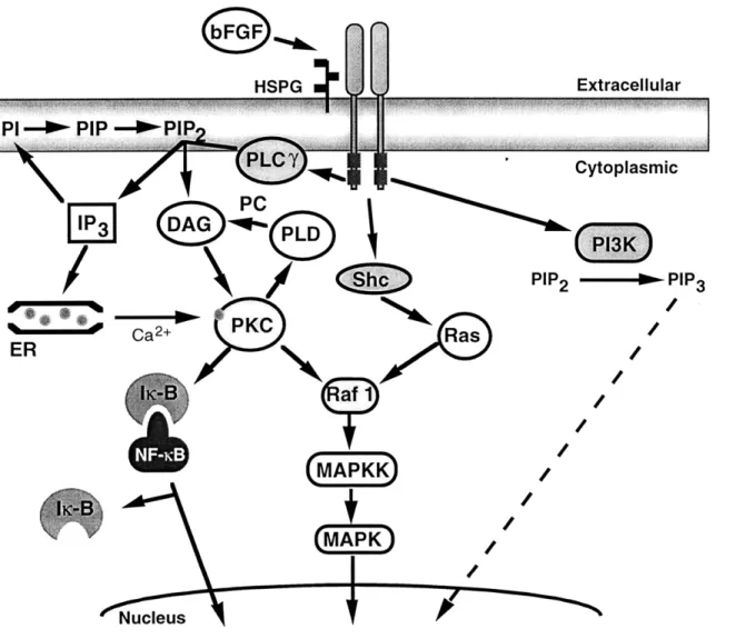
The biological effects of bFGF on endothelial cells, as well as numerous other tissues and cell types, are mediated through four high-affinity, transmembrane tyrosine kinase receptors: FGFR-1 (flg, cek 1), FGFR-2 (bek, cek 3), FGFR-3 (cek 2), and FGFR-4. These receptors belong to the superfamily of RTKs and possess high diversity with variable ligand specificity and affinity. Basic FGF also binds to heparan sulfate polysaccharides (low-affinity receptors) and heparin, which act to form a ternary complex
with bFGF and its high-affinity receptor to potentially increase signaling efficiency and bioactivity [Jaye, et al., 1992; Yayon, et al., 1991].
Binding of bFGF induces conformational changes to its high-affinity receptors, followed by dimerization and transphosphorylation of its tyrosine residues [Ullrich and Schlessinger, 1990]. The autophosphorylated intracellular tyrosine kinase domains become targets for SH2, SH3, and phosphotyrosine-binding domain-containing molecules. Intracellular mediators interacting via such a process are phospholipase C-yl (PLC-yl) [Roth, 1994], cortactin [Zhan, et al., 1994], Shc [Spivak-Kroizman, et al., 1994] and phosphatidyl-inositol 3' kinase [Varticovski, et al., 1994]. Activated PLC-yl catalyzes the breakdown of the membrane phospholipid, phosphatidylinositol bisphosphate, to generate inositol 1,4,5-triphosphate and diacylglycerol [Ullrich and Schlessinger, 1990]. Inostitol triphosphate subsequently mobilizes Ca2+ from intracellular stores, which combines with
DAG to activate protein kinase C. PKC can, in turn, activate phospholipase D to catalyze the hydrolysis of phosphatidylcholine (PC) to form phosphatidic acid (PA), a precursor of DAG [Billah and Anthes, 1990; Exton, 1990]. PC-PLD may also be activated via an independent pathway from PKC activation, and thus may serve as a regulator of long-term activation of PKC [Ahmed, et al., 1994]. The secondary messengers generated in this cascade may activate Raf kinase, mitogen-activated protein (MAP) kinase, extracellular signal-regulated kinase (ERK), and NF-icB, and have been implicated in controlling many of the physiological responses produced by bFGF, namely cell proliferation,
differentiation, migration and angiogenesis [Friesel and Maciag, 1995]. However, the specific role of each messenger in mediating the cellular functions of bFGF are strongly cell-type and function-dependent [Jaye, et al., 1992; Peters, et al., 1992; Mohammadi, et al., 1992]. A schematic of the bFGF-mediated signaling pathways is shown in Figure
2-4-1 for reference.
Studies involving the elucidation of the second messengers activated by bFGF using several of the aforementioned inhibitors have been controversial. In fibroblast cell lines, it has been shown that bFGF activates the enzyme phospholipase Cyl, yet it has not been shown to correlate with increased FGF mitogenicity [Peters, et al., 1992; Coughlin, et al., 1988]. Additionally, neither Ca2+ nor phosphotidylinositol hydrolysis appears to play
a role in FGF mitogenic activity [Peters, et al., 1992; Mohammadi, et al., 1992]. In endothelial cells, there have also been conflicting results with emphasis on the role of PKC in the mitogenic signaling mechanism. PKC has been shown to inhibit bFGF-dependent DNA synthesis in capillary endothelial cells [Doctrow and Folkman, 1987], yet be essential
for bFGF mitogenic activity in normal and transformed fetal bovine aortic endothelial cells [Presta, et al., 1989]. Additionally, studies have demonstrated increased bFGF-activated
PKC-xo, 8, F, 0, rI, and immunoreactivity in human umbilical vein endothelial cells [Kent, et al., 1995; Haller, et al., 1996], and that endothelial proliferation may be mediated by a Ca2+-independent PKC isozyme activated by bFGF [Kent, et al., 1995].
bFG F HSPG Extracellular Cytoplasmic
/
Ca2+ ER PIP2 PIP 32.4.2
VEGF Signaling
Vascular endothelial growth factor (VEGF), also known as vascular permeability factor (VPF), is a homodimeric, heparin-binding glycoprotein with potent angiogenic, mitogenic, chemotactic, and vascular permeability-promoting activities specific for endothelial cells. The VEGF gene encodes four different proteins (VEGF12 1, VEGF1 65,
VEGF1 89, and VEGF206) as a result of alternative splicing [Tisher, et al., 1991].
VEGF12 1 and VEGF1 65 are diffusible proteins that are readily secreted, while VEGF1 89
and VEGF20 6 have high affinity for heparin and principally remain bound to heparin-containing proteoglycans in the extracellular matrix [Ferrara, et al., 1992]. VEGF has been implicated in inflammation and in normal and pathological angiogenesis associated with wound healing, embryonic development, growth and metastasis of solid tumors.
The biological effects of VEGF on endothelial cells are mediated through two receptor tyrosine kinases, Flt-1 (fins-like tyrosine kinase-1) and KDR (kinase-insert-domain-containing receptor; or the mouse homologue, Flk-1, fetal liver kinase-1). Binding of VEGF induces conformational changes in KDR and Fit-1, followed by dimerization and autophosphorylation on tyrosine residues [Heldin, 1995]. Activation of the low-affinity, high-capacity KDR mediates actin reorganization, membrane ruffling, chemotaxis, and mitogenicity [Waltenberger, et al., 1994]. The functions and mechanisms of action of the high-affinity, low-capacity Flt-i are less clearly understood, but appear to mediate chemotactic activity in monocytes and stimulate tissue factor expression in monocytes and endothelial cells [Clauss, et al., 1996].
In molecular terms, VEGF is able to act through KDR and at least partly through Fit-i to increase the tyrosine phosphorylation of phospholipase Cy, phosphatidylinositol 3-kinase, and GTPase-activating protein in endothelial cells [Waltenberger, et al., 1994; Guo, et al., 1995]. The activation of these early signal transduction events by VEGF, as for bFGF, leads to the phosphorylation of protein kinase C via phospholipase Cy and the hydrolysis of phosphatidylinositol-4,5 bisphosphate, and MAP kinases via the Ras pathway. PKC isoforms a and
PII
have been found to be translocated in VEGF-stimulated endothelial cells, while PKC-5 and -E are not, and that the stimulated cell growth is largely mediated by the PKC-P isoform [Xia, et al., 1996]. Additionally, VEGF-activated PKC has been shown to stimulate PLD in HUVECs, which is unlikely to be directly involved in the control of DNA synthesis, but rather in regulating cytoskeleton-dependent effects such as cell migration [Seymour, et al., 1996]. A schematic of VEGF cell signaling is shown in Figure 2-4-2.HSPG Fit-1 Extracellular Cytoplasmic PIP2 PP 3 Ca2+ ER
28
-Figure 2-4-2. Schematic of the signaling pathways used by VEGF.
2.4.3 TNFa Signaling
Tumor necrosis factor-a (TNF(), or cachectin, is a pleiotropic polypeptide that is member of a broader, TNF ligand superfamily consisting of nine different proteins. It is capable of producing a wide variety of effects on a large number of cells by activating multiple signal transduction pathways, inducing or suppressing a vast number of genes, and using ubiquitous receptors. As a consequence of this diversity, TNFa has been recognized in many physiological and pathological processes, including normal host resistance to infection, angiogenesis, cachexia, septic shock, autoimmune disorders, and meningococcal septicemia.
H