Publisher’s version / Version de l'éditeur:
BMC Systems Biology, 3, 21, 2009-02-18
READ THESE TERMS AND CONDITIONS CAREFULLY BEFORE USING THIS WEBSITE. https://nrc-publications.canada.ca/eng/copyright
Vous avez des questions? Nous pouvons vous aider. Pour communiquer directement avec un auteur, consultez la première page de la revue dans laquelle son article a été publié afin de trouver ses coordonnées. Si vous n’arrivez pas à les repérer, communiquez avec nous à [email protected].
Questions? Contact the NRC Publications Archive team at
[email protected]. If you wish to email the authors directly, please see the first page of the publication for their contact information.
NRC Publications Archive
Archives des publications du CNRC
This publication could be one of several versions: author’s original, accepted manuscript or the publisher’s version. / La version de cette publication peut être l’une des suivantes : la version prépublication de l’auteur, la version acceptée du manuscrit ou la version de l’éditeur.
For the publisher’s version, please access the DOI link below./ Pour consulter la version de l’éditeur, utilisez le lien DOI ci-dessous.
https://doi.org/10.1186/1752-0509-3-21
Access and use of this website and the material on it are subject to the Terms and Conditions set forth at
Protein evolution on a human signaling network
Cui, Qinghua; Purisima, Enrico O.; Wang, Edwin
https://publications-cnrc.canada.ca/fra/droits
L’accès à ce site Web et l’utilisation de son contenu sont assujettis aux conditions présentées dans le site LISEZ CES CONDITIONS ATTENTIVEMENT AVANT D’UTILISER CE SITE WEB.
NRC Publications Record / Notice d'Archives des publications de CNRC:
https://nrc-publications.canada.ca/eng/view/object/?id=ca0a391d-620e-4822-b8aa-a6e8cbdfc1e2 https://publications-cnrc.canada.ca/fra/voir/objet/?id=ca0a391d-620e-4822-b8aa-a6e8cbdfc1e2
Open Access
Research article
Protein evolution on a human signaling network
Qinghua Cui
1,3, Enrico O Purisima
1and Edwin Wang*
1,2Address: 1Biotechnology Research Institute, National Research Council Canada, Montreal, Quebec, Canada, 2Center for Bioinformatics, McGill
University, Montreal, Quebec, Canada and 3Department of Medical Informatics, Peking University Health Science Center, Beijing, PR China
Email: Qinghua Cui - [email protected]; Enrico O Purisima - [email protected]; Edwin Wang* - [email protected]
* Corresponding author
Abstract
Background: The architectural structure of cellular networks provides a framework for innovations as well as constraints for protein evolution. This issue has previously been studied extensively by analyzing protein interaction networks. However, it is unclear how signaling networks influence and constrain protein evolution and conversely, how protein evolution modifies and shapes the functional consequences of signaling networks. In this study, we constructed a human signaling network containing more than 1,600 nodes and 5,000 links through manual curation of signaling pathways, and analyzed the dN/dS values of human-mouse orthologues on the
network.
Results: We revealed that the protein dN/dS value decreases along the signal information flow from
the extracellular space to nucleus. In the network, neighbor proteins tend to have similar dN/dS ratios, indicating neighbor proteins have similar evolutionary rates: co-fast or co-slow. However, different types of relationships (activating, inhibitory and neutral) between proteins have different effects on protein evolutionary rates, i.e., physically interacting protein pairs have the closest evolutionary rates. Furthermore, for directed shortest paths, the more distant two proteins are, the less chance they share similar evolutionary rates. However, such behavior was not observed for neutral shortest paths. Fast evolving signaling proteins have two modes of evolution: immunological proteins evolve more independently, while apoptotic proteins tend to form network components with other signaling proteins and share more similar evolutionary rates, possibly enhancing rapid information exchange between apoptotic and other signaling pathways. Conclusion: Major network constraints on protein evolution in protein interaction networks previously described have been found for signaling networks. We further uncovered how network characteristics affect the evolutionary and co-evolutionary behavior of proteins and how protein evolution can modify the existing functionalities of signaling networks. These new insights provide some general principles for understanding protein evolution in the context of signaling networks.
Background
Proteins in cells tend to form a complex cellular signaling network that responds to various signals, ranging from
environmental conditions, hormones or neurotransmit-ters to ions, and perform a series of tasks such as cell growth, maintenance of cell survival, proliferation, differ-Published: 18 February 2009
BMC Systems Biology 2009, 3:21 doi:10.1186/1752-0509-3-21
Received: 18 July 2008 Accepted: 18 February 2009 This article is available from: http://www.biomedcentral.com/1752-0509/3/21
© 2009 Cui et al; licensee BioMed Central Ltd.
This is an Open Access article distributed under the terms of the Creative Commons Attribution License (http://creativecommons.org/licenses/by/2.0), which permits unrestricted use, distribution, and reproduction in any medium, provided the original work is properly cited.
BMC Systems Biology 2009, 3:21 http://www.biomedcentral.com/1752-0509/3/21
entiation, development and apoptosis [1-4]. Cellular sig-naling networks are ubiquitous in various prokaryotes and eukaryotes and play pivotal roles in fundamental processes. Most studies on signaling have so far focused on certain particular signaling pathways or cascades, which represent a family of genes or specific biological processes. However, signaling pathways normally cross talk, branch out, form loops and are linked together to form a complex network. Therefore, it is necessary to study biological questions in a broader network context [5-7]. At present, one of the obstacles to performing large-scale analysis of signaling networks is the lack of a com-prehensive signaling network dataset, because cellular sig-naling information is scattered in literature. So far only a few studies have been conducted for understanding topo-logical organization, cancer signaling and microRNA reg-ulation on literature-mined signaling networks [2,8-10]. At the molecular level, the architectural structure of cellu-lar networks could provide constraints and functional innovations for protein evolution. Using protein interac-tion networks, previous studies addressing this quesinterac-tion analyzed the conservation of network motifs [11,12], link numbers, interacting partners and functional modules of the network proteins [13-15] and regions of network topology [16]. Although cellular signaling is one of the most important biological processes, how signaling net-works provide constraints on protein evolution and what functional consequences of signaling networks are caused by protein evolution have not been studied. To address these questions, we used our previously literature-mined human cellular signaling network which contains more than 1,600 nodes and 5,000 interactions [8,10] to system-atically analyze the dN/dS of human-mouse orthologues on the human signaling network.
Results
To understand how the architectural structure of signaling networks provides constraints for protein evolution, we first constructed a human signal transduction network by manually curating signaling pathways [8,10]. We merged the curated data with other literature-mined human cellu-lar signaling pathways such as a small signaling network containing ~500 genes [2]. As a result, the signaling net-work contains ~1,600 nodes and ~5,000 interactions [10]. In the network, nodes represent proteins/genes, while
neutral and directed links represent physical interactions and activating/inhibitory relations between proteins, respectively. Directed links have two types: positive links (an upstream protein activates a downstream protein) and negative links (an upstream protein inhibits a down-stream protein). The network contains 2,403, 741, 1,915 and 30 links with positive, negative, neutral and unknown type, respectively. To study the evolutionary rate of the proteins in the network, we mapped the dN/dS values of
human and mouse orthologues onto the network pro-teins. The value of dN/dS is the ratio of the rate of DNA sub-stitutions affecting the amino-acid composition of the gene product (dN) to the rate of DNA substitutions that are silent at the amino-acid level (dS). The value of dN/dS can be used to measure the rate of protein evolution after con-trolling for mutation rate [17]. Therefore, in this study, we used dN/dS as a metric to measure the rate of protein
evo-lution. The dN/dS values were calculated based on the dN and dS values which have been deposited in the database H-InvDB (see Methods).
Protein evolutionary rates differ along the signaling information flow
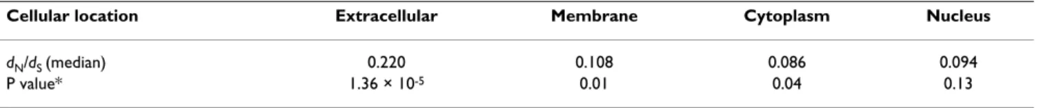
Normally, cellular signaling information flow propagates from the extracellular space to the nucleus. Therefore, we asked how protein evolutionary rates vary along the sign-aling information flow. To answer this question, we first sorted the network proteins into four groups: extracellular space, membrane, intracellular space, and nucleus, based on their cellular locations in the signaling information flow. We then calculated the average dN/dS in each group. We found that the average dN/dS are different for each group along the signal information flow (Table 1). These results suggest that proteins in different stages of the sign-aling information flow (different cellular locations) evolve at different rates, and further indicate that different cellular compartments have different protein evolution-ary rate. Proteins in extracellular space and cellular mem-brane account for the fastest evolving proteins, while proteins in intracellular space and nucleus account for the slowest evolving proteins. Proteins in these two groups show significantly different evolutionary rates (median dN/dS: 0.124 vs. 0.088, 2.5% and 97.5% percentage quan-tiles, [0.007, 0.668] and [0.000, 0.463], respectively, P = 3.36 × 10⁻⁷).
Table 1: Protein evolutionary rates distribution along the signaling information flow
Cellular location Extracellular Membrane Cytoplasm Nucleus
dN/dS (median) 0.220 0.108 0.086 0.094
P value* 1.36 × 10⁻⁵ 0.01 0.04 0.13
*The P values were calculated by Wilcoxon test by comparing the protein evolutionary rates in each group with the evolutionary rates of the whole network proteins.
Protein evolutionary rates are associated with network features
We performed a detailed analysis of the protein evolu-tionary rate on the network using several network fea-tures. Let Δij(dN/dS) represent the difference in dN/dS for a pair of genes. We calculated Δij(dN/dS) for all the pairs of genes (network nodes), which are connected by either a directed or neutral link. Directed links are signaling inter-actions that activate or inhibit while neutral links are just physical interactions. We also did the same for an equal number of random gene pairs in the network. We found that Δij(dN/dS) is significantly smaller for connected pairs of genes than that of random pairs (median value 0.072 vs. 0.092, 2.5% and 97.5% percentage quantiles, [0.002, 0.506] and [0.004, 0.536], respectively, P = 5.66 × 10⁻¹³,
Wilcoxon Test). This result indicates that interacting pro-teins in the network tend to evolve together: fast or co-slow. In signaling networks, proteins have different rela-tionships. We asked whether different types of interac-tions have different constraints on protein evolution in the network. To address this question, we classified the links into three groups according to their link types: neu-tral link, positive (activating) link, and negative (inhibit-ing) link groups. We calculated the average Δij(dN/dS) for protein pairs in each group, respectively. We found that these three types of links act differently on protein co-evo-lution. The median values for neutral, positive and nega-tive link groups are: 0.067, 0.072, and 0.088, respecnega-tively. The 2.5% and 97.5% percentage quantiles are [0.003, 0.359], [0.002, 0.601] and [0.003, 0.507], respectively (P = 0.02, Kruskal-Wallis test). Negative links account for the highest median Δij(dN/dS), followed by the positive links and neutral links (P = 0.02, P = 0.003, respectively). It is also clear that the signaling link group (combining the positive and negative link groups) has a higher median Δij(dN/dS) value than the neutral link group (median value
0.075 vs. 0.067, 2.5% and 97.5% percentage quantiles, [0.002, 0.601] and [0.003, 0.359], respectively, P = 0.04, Wilcoxon test). These results hint that the types of interac-tions between proteins in the network can have different effects on the co-evolution of the proteins. Neutral links representing protein-protein interactions within protein complexes in the signaling network tend to have more similar evolutionary rates and might be more co-evolved. Physically interacting proteins in signaling networks often form protein complexes that are used for isolating certain signaling cascades from other reactions, or for signaling protein translocations. Therefore, co-evolution of the physically interacting proteins could enhance the coordi-nation of these processes. Positive and negative links rep-resent the reactions exerted by signaling enzymes, i.e., kinases and phosphatases. Unlike neutral links, these appear to co-evolve less and might have different evolu-tionary mechanisms (see more in Discussion).
To further differentiate the evolutionary behavior of directed and neutral links, we investigated the association of the distance between two proteins in the network with their evolution rate. In the network, signals can be trans-duced from one node to another through many different cascades, one of which contains the least number of links and is called the shortest path. We defined the network distance to be the shortest path between two nodes. We first sorted the shortest paths between any two nodes in the network using Dijkstra's algorithm. The shortest paths consisting of directed and neutral links were examined independently. We calculated the Δij(dN/dS) between all pairs of nodes having either a directed or neutral shortest path. For either path type, we grouped the pairs of pro-teins according to the length of their shortest paths (net-work distances) and calculated the average Δij(dN/dS) in each group. As shown in Figure 1, Δij(dN/dS) increases as
the network distance increases when the directed shortest path was examined (Spearman's correlation R = 0.83, P = 0.005). These results indicate that for the directed shortest paths, the more distant two proteins are, the less chance they share similar evolutionary rates. The same could not be said with statistical confidence for the neutral shortest path (Spearman's correlation R = 0.61, P = 0.063). How-ever, as shown in Figure 1, when the length of their short-est paths is greater than 7, the average Δij(dN/dS) starts to decrease. To understand this observation, we defined the types of node pairs and calculated the fractions of node-pair-types for each group. We defined node-node-pair-types based on the cellular locations (i.e., extracellular space, membrane, intracellular space, and nucleus) of the nodes in each pair [see Additional file 1], i.e., for a node pair, if
Correlation between network distance and Δij(dN/dS)
Figure 1
Correlation between network distance and Δij(dN/dS). Δij(dN/dS), the absolute difference of dN/dS was calculated for all pairs of genes, and plotted against the network distance, defined by the shortest directed path between them.
1.15E-01 1.20E-01 1.25E-01 1.30E-01 1.35E-01 1.40E-01 0 2 4 6 8 10 12 Networ k Distance ij ( dN/ dS)
BMC Systems Biology 2009, 3:21 http://www.biomedcentral.com/1752-0509/3/21
one node is located in intracellular space and the other is located in nucleus, we defined this pair as a Cy (intracel-lular space)-Nu (nucleus) type. As shown in Additional file 1, the group average Δij(dN/dS) is affected by the frac-tions of the node-pair-types. In particular, two types of the node pairs in each group, Cy-Cy and Ex-Cy might play important roles for the group average Δij(dN/dS) [see Addi-tional file 1].
As we uncovered above, interacting proteins in the net-work show similar evolutionary rates. To understand this phenomenon in more detail quantitatively, we extracted the network components in which the proteins have sim-ilar dN/dS. A network component is a connected sub-net-work. Two nodes are in a same network component if there is a path between them. We identified network com-ponents formed by proteins with dN/dS values in the top
10% (dN/dS > 0.316) and the bottom 10% (dN/dS < 0.016), respectively. We found that both groups of proteins tend to form bigger network components than a randomly selected of proteins consisting of 10% of the network nodes (P = 0.004 and 0.0002, respectively, randomization tests).
To understand the functional consequences of the low and fast evolving network proteins, we analyzed the enrichment of biological functions of the proteins in the highest and lowest 10% of dN/dS proteins, respectively, using FatiGO software tool [18]. The analysis revealed that high dN/dS proteins are significantly enriched with apoptotic signaling (P = 8.9 × 10⁻⁷) and immunological
signaling (P = 9.6 × 10⁻⁶), while low d
N/dS proteins (dN/dS
< 0.016) are significantly enriched with GTP binding (P = 1.0 × 10⁻⁷) and hydrolase activity (P = 3.3 × 10⁻⁶). Because
a higher dN/dS value represents fast evolution of a protein, these results suggest that the proteins of apoptotic signal-ing and immunological signalsignal-ing are highly divergent. Although both apoptotic and immunological signaling are intensively involved in host defense responses, they evolve in different ways. More specifically, among pro-teins in the highest 10% dN/dS, apoptotic signaling
pro-teins preferentially form network components with other proteins, i.e., 18 out of 28 proteins in the largest network component (which we called it 28-cluster) are signaling proteins. In contrast, immunological signaling proteins (antigens) in the same top 10%dN/dS group, were isolated and were not part of large network components. Inde-pendently fast-evolving antigens will increase the diverse
responses of the host cells. On the other hand, interde-pendently fast-evolving apoptotic signaling proteins (i.e., the 28-cluster) might enhance coordinated responses from the host cells and the rapid information transfer needed for survival of the organisms.
We further catalogued the orthologues of the 28-cluster proteins across several model organisms such as Escherichia coli, yeast (Saccharomyces cerevisiae), worm (Caenorhabditis elegans), fly (Drosophila melanogaster) and zebrafish (Danio rerio). A similar analysis was also extended to whole network genes. Not surprisingly, the 28-cluster proteins have much fewer orthologues in the model organisms than the network proteins (Table 2). These results indicate that high dN/dS apoptotic signaling proteins (dN/dS > 0.316) lead to multiple and more flexi-ble and adaptive cell death signaling pathways in human. Indeed, only one primitive dedicated apoptotic signaling pathway is known in C. elegans [19], while several cell death signaling pathways have evolved in human and mouse genomes. Extensive expansion of apoptotic signal-ing proteins in human leads to the integration of a signif-icant portion of apoptotic proteins into the signaling processes that are used in normal physiological condi-tions. For example, apoptotic proteins such as caspases are involved in many non-apototic signaling processes in human and mouse, i.e., cell proliferation and differentia-tion [20,21]. In mice, caspase-9 is involved in both apop-tosis and inner ear epithelium development [22], while caspase-8 is involved in critical signaling for cardiac and neural development during early embryogenesis [23]. Conversely, multiple normal signaling mechanisms have been recruited to cell death either as backups or parallel mechanisms of apoptosis. For example, cytochrome c is a key electron carrier of mitochondrial complex III for res-piration. However, in mammals cytochrome c is involved in apoptosis when mitochondria are damaged [24]. As a result, the mammalian cell death machinery is inter-twined with multiple cellular signaling processes that are part of normal cellular physiological signaling processes, providing backups and flexible signaling mechanisms to cell death signaling. We found that ten out of the 28-clus-ter proteins are not apoptotic proteins. Fast co-evolution of apoptotic proteins with other proteins would enhance the rapid information transfer between apoptotic signal-ing pathways and other pathways. These diverse and flex-ible apoptotic signaling makes possflex-ible a rapid response to a variety of complex internal and external stress signals.
Table 2: Percentage of orthologues of the human signaling network proteins across species
E. coli S. cerevisiae C. elagans D. melanogaster D. rerio
28-cluster protein 0 0 0 25% 64%
Finally, the co-evolution of network components signifi-cantly promotes new functionalities arising from the inte-gration of diverse signaling cascades in signaling networks.
Sensitivity analysis
The human signaling network is incomplete and contains errors. In order to investigate the potential effects of data incompleteness and possible errors, we performed a sen-sitivity analysis by randomly removing 10% of the links and adding the same number of random links into the network. By doing so, we have artificially introduced approximately 10% false negatives and 10% false posi-tives into the network. We examined the effect on the main results described in the previous sections. For pro-tein co-evolution, we found that Δij(dN/dS) is still signifi-cantly less than for a random pair (median value 0.074 vs. 0.081, 2.5% and 97.5% percentage quantiles, [0.002, 0.507] and [0.004, 0.580], respectively, P = 2.5 × 10⁻⁹,
Wil-coxon Test). We found that the three types of links still contribute differently to protein co-evolution (median: 0.069, 0.076, 0.084, 2.5% and 97.5% percentage quan-tiles, [0.003, 0.384], [0.002, 0.586] and [0.004, 0.495], respectively, P = 0.09, Kruskal-Wallis test). As before, neg-ative links account for the highest Δij(dN/dS), followed by
the positive links and neutral links (P = 0.06, P = 0.01, respectively). It is also clear that the signal link group (combining the positive link and negative link groups) has higher Δij(dN/dS) than the neutral link group (median value 0.078 vs. 0.069, 2.5% and 97.5% percentage quan-tiles, [0.002, 0.573] and [0.003, 0.384], respectively, P = 0.06, Wilcoxon test). The Δij(dN/dS) increases as the net-work distance increases (Spearman's correlation R = 0.636, P = 0.05 for the directed path). We found that pro-teins belonging to network components with the highest and lowest 10% dN/dS values still tend to form bigger net-work components than a randomly selected set of 10% of the proteins in the network (P = 0.004 and 0.0002, respec-tively, randomization tests). These results indicate that most of the major conclusions in this study remain unchanged by the addition of a moderate amount of false positives and false negatives. Therefore, the results we obtained are fairly robust.
Discussion
Previous studies in protein interaction network evolution have made several major conclusions: (a) hub proteins or proteins having more interacting links tend to be more conserved [25]; (b) proteins in the network periphery undergo positive selection while those in the network center are more conserved [16]; (c) network proteins appear to be co-evolved with their neighbors [25]; (d) interacting proteins with high local clustering tend to be more conserved [26].
In this study, we constructed a human signaling network and analyzed the protein evolutionary rate on the net-work. Consistent with the studies of protein interaction networks, we find that proteins appear to be co-evolved with their neighbors in the signaling network. However, in our analysis, we further found that in signaling net-works different types of interactions have different strength of constraints on protein co-evolution, in which proteins linked by physical interactions tend to be more co-evolved. Furthermore, for directed shortest paths, the more distant two proteins have, the less chance they share similar evolutionary rates. However, such a correlation was not observed with respect to the neutral shortest path. Positive and negative links in signaling networks include the major signaling regulatory mechanism: protein phos-phorylation and dephosphos-phorylation, which are exerted by kinases and phosphatases. Both types of signaling enzymes are multiple domain proteins which often con-tain, in addition to their core catalytic function, multiple independently folding domains or motifs that mediate connectivity by interacting with other signaling elements [27]. Therefore, signaling enzymes are known to have high modular strategies for controlling their input and output connectivities: the core catalytic activity of a sign-aling protein is physically and functionally separable from molecular domains or motifs that determine its link-age to both inputs and outputs. These features of signaling enzymes suggest that they have distinct evolutionary mechanisms from other proteins, i.e., insertion and recombination of modules are suggested to be a common mechanism of the evolution of new proteins and connec-tions [27,28]. Collectively, these features of signaling enzymes might explain the evolutionary rates differences between the signaling enzymes and their connecting part-ners. Furthermore, negative regulators such as phos-phatases are more promiscuous in their selectivity for their targets/substrates. This fact might explain why phos-phatases (forming negative links in the network) have even weaker co-evolution rates with their connecting part-ners. On the other hand, neutral links represent physical protein interactions in the signaling network. Physically interacting proteins in signaling networks often form pro-tein complexes that are used for isolating certain signaling cascades from other reactions, or signaling protein trans-locations. Therefore, co-evolution of the physically inter-acting proteins will enhance the coordination of the processes mentioned above.
In this study we showed that extracellular proteins are evolving faster, which is in agreement with several previ-ous studies [16,29]. Signaling proteins in the extracellular space are the stimuli of intra- and inter-cell signaling. Fast evolving proteins in the extracellular space allow cells to explore various responses to new stimuli and might estab-lish novel communications between cells. This would
BMC Systems Biology 2009, 3:21 http://www.biomedcentral.com/1752-0509/3/21
promote the cell's capability to respond and adapt to envi-ronmental changes and explore new envienvi-ronmental niches. Recently, Kim et al. showed that proteins in the peripheral regions (i.e., extracellular and membrane pro-teins) of protein interaction networks undergo positive selection, while proteins in the center of the protein inter-action networks are conserved [16]. Protein interinter-action networks collect the global protein interactions in the cell while signaling networks represent a part of cell activities (i.e., cell signaling) [3]. The extracellular components of the signaling network are similar to the peripheral regions of protein interaction and gene regulatory networks, which count for many adaptive properties of the organism [16,30]. Consistently, both Kim et al. [16] and our studies show that proteins in this region are fast evolving. In this study, we further showed that evolutionary rates of pro-teins decrease along the signaling information flow from extracellular space (input layer), intracellular space to nucleus (output layer). The downstream portion of the signaling network evolves more conservatively. This is understandable given that the downstream segment of the signaling network ultimately governs cellular behavior and activities. It is therefore not surprising to find that tumor driver mutating genes, even highly mutated ones, are enriched in the downstream portion of human signal-ing network [8,10]. The existence of fast and slowly evolv-ing proteins in the signalevolv-ing network upstream and downstream portions, respectively, suggests that proteins in the upstream portion of the signaling flow are more adaptable and could be more easily rewired to generate different combinatory regulation mechanisms for the downstream portion of the signaling flows. Thus, it would be more critical to regulate the genes in the downstream portion of the network. Indeed, we do find that the genes in the downstream portion of the signaling network are more significantly regulated by microRNAs than the upper portion of the signaling information flow [9]. It is known that apoptotic and immunological signaling proteins are fast evolving. However, using a network approach, we found that both signaling processes have different modes of evolution: fast evolving immunologi-cal signaling proteins are more independent, while fast evolving apoptotic signaling proteins tend to form net-work components and co-evolve with other signaling pro-teins. Apoptotic signaling proteins are extensively expanded in mammalian genomes in comparison to other genomes such as those of yeast and fly. The diverse and flexible apoptotic signaling makes it possible for mammals to rapidly respond to a variety of complex inter-nal and exterinter-nal stress siginter-nals. Fiinter-nally, the functiointer-nal con-sequences of co-evolution of the apoptotic proteins by forming network components significantly enhance the integration of diverse signaling cascades to cell death sig-naling and make the information transfer more efficient
between apoptotic signaling and other signaling path-ways. Our findings will improve our understanding of sig-naling protein evolution and the mechanism of signal integration in signaling networks caused by protein evolu-tion.
Conclusion
Several major conclusions on protein evolution in protein interaction networks have been previously described. In this work, we further uncovered how network characteris-tics affect the evolutionary and co-evolutionary behavior of proteins. For example, we showed that in signaling net-works different types of interactions have different strength of constraints on protein co-evolution, in which proteins linked by physical interactions tend to be more co-evolved. Furthermore, for directed shortest paths, the more distant two proteins have, the less chance they share similar evolutionary rates. However, such a correlation was not observed with respect to the neutral shortest path. We further showed that evolutionary rates of proteins decrease along the signaling information flow from extra-cellular space (input layer), intraextra-cellular space to nucleus (output layer). The downstream portion of the signaling network evolves more conservatively.
Our analysis further suggested how protein evolution could modify the existing functionalities of signaling net-works. For example, we showed that fast evolving apop-totic signaling proteins tend to form network components and co-evolve with other signaling proteins. The diverse and flexible apoptotic signaling makes it possible for mammals to rapidly respond to a variety of complex inter-nal and exterinter-nal stress siginter-nals. Fiinter-nally, the functiointer-nal con-sequences of co-evolution of the apoptotic proteins by forming network components significantly enhance the integration of diverse signaling cascades to cell death sig-naling and make the information transfer more efficient between apoptotic signaling and other signaling path-ways. These new insights provide some general principles for understanding protein evolution in the context of sig-naling networks.
Methods
Datasets
The human-mouse protein dN and dS data were down-loaded from H-InvDB http://jbirc.jbic.or.jp/hinv/dataset/ download.cgi. We calculated the dN/dS value for each pro-tein [see Additional file 2]. We extracted human-mouse orthologues from a database, Inparanoid (hsamus_ortholog.txt, http://inparanoid.sbc.su.se/).
Signaling network construction
To construct the human cellular signaling network, we manually curated signaling pathways from the BioCarta database http://www.biocarta.com/genes/allpath
ways.asp, which so far is the most comprehensive data-base for cellular signaling pathways. The curated pathway dataset recorded gene names and functions, cellular loca-tions of each gene and relaloca-tionships between the genes. We merged these genes and their interactions with another literature-mined signaling network that contains ~500 proteins [2]. To ensure the accuracy and the consist-ency of the data, each referenced pathway was cross-checked by different researchers and finally all the docu-mented pathways were checked by one researcher. As a result, the merged signaling network contains more than 1,600 nodes and 5, 000 links [8,10]. The human signaling network data are accessible from Cui et al [10].
Gene Ontology analysis
To examine the enrichment of biological processes for a set of genes, we used FatiGO tool [18] and the default parameters. The whole network genes were used as a back-ground gene set.
Statistical analysis
We performed Wilcoxon tests, Kruskal-Wallis tests, and Spearman's correlation using R, a software environment for statistical computing http://www.r-project.org/. Details for randomization tests of cellular networks have been described previously [31]. Briefly, randomization tests of the network components formed by a set of genes were conducted by taking the same number of genes ran-domly from the network for 5,000 times and calculating its network components each time.
Authors' contributions
QC carried out the analysis, EW conceived of the study, and participated in its design and coordination. QC, EW and EOP wrote the manuscript. All authors read and approved the final manuscript.
Additional material
Acknowledgements
This work is partially supported by Genomics and Health Initiative. We thank Dr. Z. Yu for reading the manuscript and comments.
References
1. Balazsi G, Barabasi AL, Oltvai ZN: Topological units of
environ-mental signal processing in the transcriptional regulatory network of Escherichia coli. Proc Natl Acad Sci USA 2005, 102:7841-7846.
2. Ma'ayan A, Jenkins SL, Neves S, Hasseldine A, Grace E, Dubin-Thaler B, Eungdamrong NJ, Weng G, Ram PT, Rice JJ, et al.: Formation of
regulatory patterns during signal propagation in a Mamma-lian cellular network. Science 2005, 309:1078-1083.
3. Wang E, Lenferink A, O'connor-McCourt M: Cancer systems
biol-ogy: exploring cancer-associated genes on cellular networks.
Cell Mol Life Sci 2007, 64:1752-1762.
4. Yarden Y, Sliwkowski MX: Untangling the ErbB signalling
net-work. Nat Rev Mol Cell Biol 2001, 2:127-137.
5. Vidal M: Interactome modeling. FEBS Lett 2005, 579:1834-1838. 6. Letunic I, Yamada T, Kanehisa M, Bork P: iPath: interactive
explo-ration of biochemical pathways and networks. Trends Biochem
Sci 2008, 33:101-103.
7. Karimpour-Fard A, Leach SM, Hunter LE, Gill RT: The topology of
the bacterial co-conserved protein network and its implica-tions for predicting protein function. BMC Genomics 2008, 9:313.
8. Awan A, Bari H, Yan F, Mokin S, Yang S, Chowdhury Q, Yu Z, Puri-sima EO, Wang E: Regulatory network motifs and hotspots of
cancer genes in a mammalian cellular signaling network. IET
Syst Biol 2007, 1:292-297.
9. Cui Q, Yu Z, Purisima EO, Wang E: Principles of microRNA
reg-ulation of a human cellular signaling network. Mol Syst Biol
2006, 2:46.
10. Cui Q, Ma Y, Jaramillo M, Bari H, Awan A, Yang S, Zhang S, Liu L, Lu M, O'connor-McCourt M, et al.: A map of human cancer
signal-ing. Mol Syst Biol 2007, 3:152.
11. Wuchty S, Oltvai ZN, Barabasi AL: Evolutionary conservation of
motif constituents in the yeast protein interaction network.
Nat Genet 2003, 35:176-179.
12. Mazurie A, Bottani S, Vergassola M: An evolutionary and
func-tional assessment of regulatory network motifs. Genome Biol
2005, 6:R35.
13. Amoutzias GD, Pichler EE, Mian N, De GD, Imsiridou A, Robinson-Rechavi M, Bornberg-Bauer E, Robertson DL, Oliver SG: A protein
interaction atlas for the nuclear receptors: properties and quality of a hub-based dimerisation network. BMC Syst Biol
2007, 1:34.
14. Beltrao P, Serrano L: Specificity and evolvability in eukaryotic
protein interaction networks. PLoS Comput Biol 2007, 3:e25.
15. Berg J, Lassig M, Wagner A: Structure and evolution of protein
interaction networks: a statistical model for link dynamics and gene duplications. BMC Evol Biol 2004, 4:51.
16. Kim PM, Korbel JO, Gerstein MB: Positive selection at the
pro-tein network periphery: evaluation in terms of structural constraints and cellular context. Proc Natl Acad Sci USA 2007, 104:20274-20279.
17. Swanson WJ, Vacquier VD: The rapid evolution of reproductive
proteins. Nat Rev Genet 2002, 3:137-144.
18. Al-Shahrour F, Minguez P, Tarraga J, Medina I, Alloza E, Montaner D, Dopazo J: FatiGO +: a functional profiling tool for genomic
data. Integration of functional annotation, regulatory motifs and interaction data with microarray experiments. Nucleic
Acids Res 2007, 35:W91-W96.
19. Metzstein MM, Stanfield GM, Horvitz HR: Genetics of
pro-grammed cell death in C. elegans: past, present and future.
Trends Genet 1998, 14:410-416.
20. Kuida K, Haydar TF, Kuan CY, Gu Y, Taya C, Karasuyama H, Su MS, Rakic P, Flavell RA: Reduced apoptosis and cytochrome
c-medi-ated caspase activation in mice lacking caspase 9. Cell 1998, 94:325-337.
21. Yoshida H, Kong YY, Yoshida R, Elia AJ, Hakem A, Hakem R, Pennin-ger JM, Mak TW: Apaf1 is required for mitochondrial pathways
of apoptosis and brain development. Cell 1998, 94:739-750.
22. Cecconi F, Roth KA, Dolgov O, Munarriz E, Anokhin K, Gruss P, Salminen M: Apaf1-dependent programmed cell death is
Additional file 1
Fractions of node-pair-types in each shortest path group. The data
pro-vided represent the analysis of fractions of the node-pair-types in each shortest path group.
Click here for file
[http://www.biomedcentral.com/content/supplementary/1752-0509-3-21-S1.xls]
Additional file 2
The values of dN/dS of the human-mouse orthologues on the human
signaling network. The file provides the dN/dS data for the human-mouse
orthologues on the human signaling network.
Click here for file
[http://www.biomedcentral.com/content/supplementary/1752-0509-3-21-S2.txt]
Publish with BioMed Central and every scientist can read your work free of charge "BioMed Central will be the most significant development for disseminating the results of biomedical researc h in our lifetime."
Sir Paul Nurse, Cancer Research UK Your research papers will be:
available free of charge to the entire biomedical community peer reviewed and published immediately upon acceptance cited in PubMed and archived on PubMed Central yours — you keep the copyright
Submit your manuscript here:
http://www.biomedcentral.com/info/publishing_adv.asp
BioMedcentral
BMC Systems Biology 2009, 3:21 http://www.biomedcentral.com/1752-0509/3/21
required for inner ear morphogenesis and growth.
Develop-ment 2004, 131:2125-2135.
23. Sakamaki K, Inoue T, Asano M, Sudo K, Kazama H, Sakagami J, Sakata S, Ozaki M, Nakamura S, Toyokuni S, et al.: Ex vivo whole-embryo
culture of caspase-8-deficient embryos normalize their aber-rant phenotypes in the developing neural tube and heart. Cell
Death Differ 2002, 9:1196-1206.
24. Liu X, Kim CN, Yang J, Jemmerson R, Wang X: Induction of
apop-totic program in cell-free extracts: requirement for dATP and cytochrome c. Cell 1996, 86:147-157.
25. Stumpf MP, Kelly WP, Thorne T, Wiuf C: Evolution at the system
level: the natural history of protein interaction networks.
Trends Ecol Evol 2007, 22:366-373.
26. Wuchty S, Barabasi AL, Ferdig MT: Stable evolutionary signal in
a yeast protein interaction network. BMC Evol Biol 2006, 6:8.
27. Bhattacharyya RP, Remenyi A, Yeh BJ, Lim WA: Domains, motifs,
and scaffolds: the role of modular interactions in the evolu-tion and wiring of cell signaling circuits. Annu Rev Biochem 2006, 75:655-680.
28. Lander ES, Linton LM, Birren B, Nusbaum C, Zody MC, Baldwin J, Devon K, Dewar K, Doyle M, FitzHugh W, et al.: Initial sequencing
and analysis of the human genome. Nature 2001, 409:860-921.
29. Waterhouse RM, Kriventseva EV, Meister S, Xi Z, Alvarez KS, Bar-tholomay LC, Barillas-Mury C, Bian G, Blandin S, Christensen BM, et
al.: Evolutionary dynamics of immune-related genes and
pathways in disease-vector mosquitoes. Science 2007, 316:1738-1743.
30. Davidson EH, Erwin DH: Gene regulatory networks and the
evolution of animal body plans. Science 2006, 311:796-800.
31. Wang E, Purisima E: Network motifs are enriched with
tran-scription factors whose transcripts have short half-lives.