Artificial Gravity:
Neurovestibular Adaptation to Incremental Exposure to Centrifugation.
by
Sylvain Bruni
French Engineering Degree, Supélec (Ecole Supérieure dʹElectricité, France, 2004)SUBMITTED TO THE DEPARTMENT OF AERONAUTICS AND ASTRONAUTICS IN PARTIAL FULFILLMENT OF THE REQUIREMENTS FOR THE DEGREE OF MASTER OF SCIENCE IN AERONAUTICS AND ASTRONAUTICS AT THE MASSACHUSETTS INSTITUTE OF TECHNOLOGY SEPTEMBER 2004
© Sylvain Bruni, 2004. All rights reserved.
Signature of Author: ___________________________________________
Sylvain Bruni Department of Aeronautics and Astronautics August 20, 2004Certified by: _________________________________________________
Laurence R. Young Apollo Program Professor of Astronautics and Professor of Health Sciences and Technology Thesis SupervisorAccepted by: _________________________________________________
Jaime Peraire Professor of Aeronautics and Astronautics Chair, Committee on Graduate StudentsThe author hereby grants to MIT permission to reproduce and to distribute pub‐
licly paper and electronic copies of this thesis document in whole or in part.
___________________________________
Sylvain Bruni, August 20
th, 2004.
The author hereby grants to Supélec permission to reproduce and to distribute
publicly paper and electronic copies of this thesis document in whole or in part.
___________________________________
Sylvain Bruni, August 20
th, 2004.
Artificial Gravity:
Neurovestibular Adaptation to Incremental Exposure to Centrifugation.
by
Sylvain Bruni
Submitted to the Department of Aeronautics and Astronautics on August 20 2004, in Partial Fulfillment of the Requirements for the Degree of Master of Science in Aeronautics and Astronautics
Abstract
In order to counteract the debilitating effects of the space environment on the human body, short‐radius intermittent centrifugation is investigated as a possible means to expose astro‐ nauts to artificial gravity. Whereas AG is efficient in providing stimuli for muscles, bones and cardiovascular system, short‐radius centrifugation elicits discomfort and illusory sensa‐ tions of motion if particular head movements are made while spinning. Past research has shown that human beings can adapt to these sensations and undergo various stimuli with‐ out the disturbing effects of motion sickness, sensations of tumbling and inappropriate eye movements. However, current protocols for adaptation basically consist in repeated expo‐ sure to the discomfort. This solution is not satisfactory because the drop‐out rate oscillates between 30 and 50%. Since it is not acceptable to spend days of training on astronauts who, in the end, because of this training, could become unsuitable for flight, it is of primary im‐ portance to find a training protocol that achieves adaptation without going through perma‐ nent discomfort.
Incremental exposure to centrifugation is expected to be a compromised protocol to bring trainees to adaptive level without exposing them to maximum discomfort. Seven subjects were exposed to centrifugation during a five‐day protocol, over which the speed of rotation was progressively increased. As in previous protocols of adaptation, subjects performed provocative head movements at all speeds. A control experiment had ten subjects exposed to centrifugation without making head turns, in order to verify to what extent the experimental conditions of measurement impact the subjectsʹ behavior and reactions. While subjects in the control experiment did not build up adaptation, all subjects in the ex‐ perimental group who completed the protocol showed signs of adaptation to the stimulus. Only one subject did not complete the five sessions, setting the drop‐out rate at about 14%. If this conclusion holds true with more subjects, then a better protocol of adaptation has been unveiled. Thesis supervisor: Laurence R. Young
Title: Apollo Program Professor of Astronautics and Professor of Health Sciences and Technology
Acknowledgements
Thanks! - Merci!
I am deeply indebted to my thesis supervisor, Dr. Laurence Young, for offer‐ ing me the opportunity to come to MIT and carry out research in the area that interested me most. Thank you for your constant guidance and mentor‐ ship.
My appreciation also goes to Dr. Thomas Jarchow for all his help, support and ideas on the project for this past year. Thanks a lot for introducing me to real hands‐on experiments! I would like to thank Dr. Alan Natapoff for re‐ freshing my memories on statistics and providing me with the right methods to analyze all the data sets I accumulated over the months. Thanks a lot to Andy Liu for solving my never‐ending printer problems. Thank you to Liz Zotos for all her help in administrative matters. Many thanks to all MVL faculty members: Dava Newman, Chuck Oman, Jeff Hoffman. You enriched me so much with the diversity of your personal ex‐ periences. I would like to acknowledge the support of NSBRI, for equipment and mate‐ rials.
I owe a lot to all graduate students in MVL. Infinite thanks to Jessica Ed‐ monds for teaching me how to really speak American, to Ian Garrick‐Bethell for teaching me ʺcentrifuge 101ʺ, to Sophie Adenot for sharing with me the joys of completing a Master of Science in one year, to Erika Wagner for all her past work in AG and being such a formidable resource in the domain. Thanks also to my officemates Kristen Bethke, Phil Ferguson and Chris Carr for their insights and inspiration! Thanks also to the VR lab students Jessica Marquez, Kevin Duda and David Benveniste for contributing to the legen‐ dary friendly atmosphere in MVL.
My sincere appreciation also goes to the summer UROPs Tom Walker and Ben Feinberg for running experiments and helping out so much.
Many thanks to all Supélec staff, in particular to Marie‐Dominique de Swarte who constantly took care of me before and during my studies in the US. Last, but not least, all my love and thanks to my family, my parents and my brother for believing in me and supporting me quoi quʹil arrive! Thanks so much.
Table of Contents
1. Introduction. 15 1.1 The New Quest for the Next Frontier. 15 1.2 Considering Humanʹs Finitude. 17 1.3 Motivation and Rationale. 17 1.4 Hypothesis. 19 1.5 Thesis Organization. 20 2. Background. 21 2.1 Step 1: The Need to Send Humans to Space. 21 2.2 Step 2: Review of the Effects of Space Environment. 22 2.2.1 External Factors. 22 2.2.2 Psychological Isolation and Social Issues. 23 2.2.3 Physiological Effects. 24 2.3 Step 3: Past and Current Countermeasures. 28 2.4 Step 4: Artificial Gravity as a Global Countermeasure. 28 2.5 Step 5: Why Do Humans Experience Discomfort on a Centrifuge? 33 2.5.1 Vestibular Physiology. 33 2.5.2 The Physics of Head Turns in a Rotating Environment. 36 2.6 Step 6: Adaptation. 44 2.6.1 Adaptation to Low Speed Centrifugation. 44 2.6.2 Adaptation to High Speed Centrifugation. 45 2.6.3 Retention of Adaptation. 45 2.6.4 Transfer of Adaptation. 45 2.6.5 Other Factors of Adaptation. 46 2.6.6 Habituation vs. Adaptation. 48 2.7 Step 7: Incremental Adaptation. 49 2.8 Theory of Adaptation. 51 2.8.1 Definitions. 51 2.8.2 Goals. 52 2.9 Psychophysics. 54 3. Methods. 59 3.1 Experimental Design. 59 3.1.1 Experimental Group. 59 3.1.2 Control Experiment. 62 3.2 Equipment. 63 3.2.1 The Centrifuge. 63 3.2.2 Eye Movements Recording Goggles. 68 3.3 Subjects. 69 3.4 Measurements. 70 3.4.1 Physiological Measurements: Eye Data. 70
3.4.2 Subjective Assessments. 71 3.5 Protocol. 73 3.5.1 Pre‐Experiment. 73 3.5.2 Experiment. 74 4. Data Analysis. 77 4.1 Eye Data Analysis. 77 4.1.1 Basic Goal of Eye Data Analysis. 77 4.1.2 Data Processing. 77 4.2 Subjective Assessments Analysis. 82 4.3 Missing Values. 82 5. Results. 83 5.1 Overview. 83 5.2 Comments on Drop‐Out. 84 5.3 Experimental Group. 84 5.3.1 Eye Data. 84 5.3.2 Subjective Assessments. 91 5.4 Control Experiment. 98 5.4.1 Eye Data. 98 5.4.2 Subjective Assessments. 101 6. Discussion. 103 6.1 Main Findings. 103 6.2 Incremental Adaptation. 104 6.3 Motion Sickness. 106 6.4 Direction Asymmetry. 107 6.5 Control Experiment. 108 6.6 Limitations. 109 6.7 Recommendations for Future Work. 111 7. Conclusions. 113 References. 115 Appendix A: Medical Form. 123 Appendix B: Consent Form. 125 Appendix C: Pre‐Experiment Briefing. 129 Appendix D: Raw Experimental Data. 133
List of Figures and Tables
Figure 2.1. Changes in the cardio‐vascular system in response to changes of gravity load. 25 Figure 2.2. Werner Von Braun’s concept of rotating station. 29 Figure 2.3. 2001 A Space Odyssey, movie poster. 29 Figure 2.4. Concept of tethered spacecraft. 30 Figure 2.5. View of the tethered Agena (Gemini 11 Mission). 30 Figure 2.6. View of the tethered Agena (Gemini 11 Mission). 30 Figure 2.7. STS‐42 Mission Specialist Hilmers in IML‐1’s MVI rotator chair. 31 Figure 2.8. STS‐42 Payload Specialist Bondar in IML‐1’s MVI rotator chair. 32 Figure 2.9. Location of the vestibular system in the inner ear. 33 Figure 2.10. Orientation of the semi‐circular canals. 35 Figure 2.11. Mechanism of detection of self‐motion. 35 Figure 2.12. Subject lying supine NU on a centrifuge. 36 Figure 2.13. Cupula deflection in the roll canal. 41 Figure 2.14. Cupula deflection in the pitch canal. 42 Figure 2.15. Cupula deflection in the yaw canal. 42 Figure 2.17. Psychological magnitude vs. stimulus magnitude. 55 Figure 3.1. Logarithmic evolution of perception to stimulus. 61 Figure 3.2. General view of the centrifuge. 64 Figure 3.3. Close view of the new footplate. 65 Figure 3.4. Close view of the restraining helmet. 65 Figure 3.5. Subject in NU head position. 66 Figure 3.6. Subject in RED head position. 66 Figure 3.7. Archive view of the centrifuge equipped with the opaque canopy. 67 Figure 3.8. Inside view of the centrifuge equipped with the opaque canopy. 67 Figure 3.9. View of the new stabilizing tetrapod. 68 Figure 3.10. Subject wearing the new eye‐movements measurement device. 68 Figure 3.11. IT report explanation. 72 Figure 4.1. Typical SPV recording. 78 Figure 4.2. Typical SPV decay. 79 Figure 4.3. Typical gaze shift, residuals of blink activity and exponential fit. 79 Figure 4.4. Working plot of data elimination routine. 80 Figure 4.5. Working plot of data elimination routine, after data removal. 80 Figure 4.6. Fitted curve. 80 Figure 4.7. Exponential fit on SPV recording, without data elimination. 81 Figure 4.8. Exponential fit on SPV recording, after data elimination. 81 Figure 5.1. Distribution of A by subject. 85 Figure 5.2. Amplitude vs. direction. 86 Figure 5.3. Distribution of tau for each subject. 87
Figure 5.4. Decrease of tau over phases. 87 Figure 5.5. Tau over rep for each direction. 88 Figure 5.6. Decrease of tau over days. 89 Figure 5.7. Tau over days for each phase. 89 Figure 5.8. Tau vs. direction. 90 Figure 5.9. Decrease of MS over days (phase 1, to‐NU). 92 Figure 5.10. Decrease of MS over days (phase 3, to‐NU). 92 Figure 5.11. Increase of MS over days (phase 2, to‐RED). 93 Figure 5.12. Increase of MS over days (phase 2, to‐NU). 93 Figure 5.13. Decrease of IS over days (phase 1, to‐RED). 94 Figure 5.14. Decrease of IS over days (phase 1, to‐NU). 94 Figure 5.15. Decrease of IS over days (phase 3, to‐RED). 95 Figure 5.16. Decrease of IS over days (phase 3, to‐NU). 95 Figure 5.17. Increase of IS over days (phase 2, to‐RED). 96 Figure 5.18. Increase of IS over days (phase 2, to‐NU). 96 Figure 5.19. Decrease of IT over days (phase 1, to‐RED). 97 Figure 5.20. Decrease of A over phases. 98 Figure 5.21. Amplitude vs. direction. 99 Figure 5.22. Amplitude over replication for each phase (day 1). 99 Figure 5.23. Amplitude over replication for each phase (day 2). 99 Figure 5.24. Amplitude over replication for each direction (phase 1). 100 Figure 5.25. Amplitude over replication for each direction (phase 3). 100 Figure 5.26. Decrease of MS over days. 101 Table 2.1. Representative exponents of the power functions relating subjective mag‐ nitude to stimulus magnitude. 56 Table 3.1. Summary of the experimental design. 59 Table 3.2. Velocity profile. 60 Table 5.1. Summary of statistical methods used. 83 Table 5.2. Summary of the results of a mixed regression on amplitudes. 85 Table 5.3. Summary of the results of a GLM ANOVA on tau. 86 Table 5.4. Summary of the results of a mixed regression on time constants. 88 Table 5.5. Summary of the results of Page tests on subjective assessments. 91 Table 5.6. Summary of the results of a GLM ANOVA on amplitudes. 98 Table 5.7. Summary of the results of Page tests on subjective assessments. 102
O Investigator, do not flatter yourself that you know the things nature performs for herself, but rejoice in knowing the purpose of those things designed by your own mind. Leonardo Da Vinci.
Gravitation cannot be held responsible for people falling in love. Albert Einstein.
Gravity Of Love ‐ Digital picture by Swedish artist Rasmus Wängelin. [http://artofrasmus.cjb.net]
Chapter 1
Introduction
1.1 The New Quest for the Next Frontier.
On January 14th, 2004, American President Georges W. Bush announced the
new Vision for Space Exploration Program that will drive the National Aero‐ nautics and Space Administration (NASA) for the decades to come. After completion of the International Space Station and retirement of the 30‐year‐ old Shuttle fleet, both by 2010, the agency will draw its efforts to the return of human beings on the Moon, with the aim of building there a permanent re‐ search base, that could later become a virtual launch pad for missions to Mars and beyond. To travel to the Moon, and then to Mars, a new transporta‐ tion system, the Crew Exploration Vehicle (CEV), will be designed. With the successes of the Mars Exploration Rovers missions (Spirit and Opportunity) led by NASA, and of the Mars Express orbiter sent by the European Space Agency (ESA), public attention has recently been more and more focused on the possibilities of a manned mission to the red planet. Debate is still vivid about the need for such an expensive mission whereas money could be spent in enhancing life down here, on Earth. But for the scientific community, there is no doubt space exploration must be accomplished, in order to answer cru‐ cial questions about ourselves: how life has emerged, how did our planet evolve, is there life somewhere else in the universe…? (Zubrin, 2003). Send‐ ing robots is just not enough to perform accurate and proficient science where clues can be found about our common past and future in the Universe. Human beings are creative, flexible and full of intuition, at the execution of tasks, far better than current robots are. Real time research is only possible with humans on the field: robotic exploration always suffers from time lags. Sending humans out there, in space, is also a formidable dream and stimu‐
lant for any science apprentice, and for national or international pride and cooperation. In June 2004, for the first time in the history of astronautics, a privately built spacecraft has been launched into space. SpaceShipOne, developed by Scaled Composites and funded by Paul Allen’s Vulcan Inc., rocketed to 62 miles of altitude (100 kilometers), into sub‐orbital space. This flight opened the space frontier to private enterprise, and to the possibilities of affordable missions for launches in near‐Earth space. With private companies expanding their capabilities in this area, government‐funded space projects may be dedicated to further destinations.
Following the American successes and new impulses for space exploration, international enthusiasm has arisen. In 2003 already, China has become the third country to gain the capabilities to launch a manned vessel to space. Now, the Chinese are already planning to settle a base on the Moon and sending Mars missions for the next decades. The ESA, through its Aurora Program, is also envisaging a manned mission to Mars. Scientific and techno‐ logical efforts are in progress all around the world, in order to achieve one of the most magnificent goals mankind could dream of: reaching another fron‐ tier. May this quest be peaceful and cooperative, and may it bring humanity as one. ʺMy wish is that we would allow this planet to be the beautiful oasis that she is, and allow ourselves to live more in the peace that she generates.ʺ Ronald E. McNair ‐ NASA Astronaut. Russian space pioneer Konstantin E. Tsiolkovsky famously said: ʺEarth is the cradle of humanity but one cannot live in the cradle foreverʺ. Earth as the cradle of life and mankind is the most beautiful jewel men and women all around the
world should preserve, protect, respect and cherish. In a few billion years, the Sun will disappear in a cataclysmic explosion, absorbing in its core every entity of the solar system. The Earth and its inhabitants, if mankind has not disappeared before that, will be exterminated. In a human lifetime, billion years are eternity, and any person reading this will not experience this event. But our descent will. Therefore, may it be a question of exploration or of sur‐ vival, time will come in humansʹ history to find a way to leave the cradle.
1.2 Considering Humansʹ Finitude.
Humans are not perfect. They are finite, and conditioned to live in a specific environment. One might wonder if the environment is responsible for what the human being is, or if the human being modifies its environment to its convenience. A combination of both is obviously the safe answer. The former is concordant with Darwinʹs evolution theory over a large time scale. The lat‐ ter goes in favor of the humans’ will and power over their environment, in a lifetime scale: conscious of its finitude, human beings modify their way of living and their close environment in order to adapt it to them, and enhance their own life. But it remains the case that some environments are, so far, not understood or reachable enough to be modified. It is potentially the case for space and extra‐terrestrial bodies. The point is that a combination of human adaptation to environment and environmental adaptation to humans will be necessary in space exploration. The trade‐off is to be investigated carefully, in order both to preserve human life when swapping between environments, and to preserve the environment from potential human harmfulness.1.3 Motivation and Rationale.
The motivation for the present research can be summed up in a 7‐step ration‐ ale. (1) As emphasized in this introduction and by the space community in general, space exploration not only by robots and probes, but also by human beings themselves is both needed and unavoidable in the years, decades, cen‐
turies to come. (2) But extensive exposure to space environment is dangerous for humans. Past research and current data collection aboard the Interna‐ tional Space Station show that a general deconditioning of the human body occurs due to the absence of gravity, in addition to all the external threats, such as vacuum or radiations. (3) Past and current research also proves that countermeasures exist to fight against the deconditioning: negative body pressure suits, fluid loading, medication, and so on. But none of them targets all physiological effects at once. Artificial gravity is investigated as a general countermeasure targeting the very cause of the deconditioning. (4) Two basic implementations of artificial gravity can be considered: large or short radius centrifugation. The former would either be a gigantic rotating spacecraft or two smaller tethered spacecrafts rotating around a common center. The lat‐ ter, preferred in current studies, would feature short‐radius centrifuges in‐ side the spacecraft. Astronauts would then spend a few hours every day on them to be exposed to an artificial gravity field. (5) But the major drawback to short‐radius centrifugation is that head movements performed while spin‐ ning elicit sensations of tumbling and general discomfort. (6) Among other, research in the Man‐Vehicle Lab at MIT has proven that adaptation to these sensations is possible, and that discomfort can be considerably reduced to tolerable levels. (7) Now the question is: what is the best way to adapt? Every step of this rationale is explained in details in Chapter 2.
Current protocols of adaptation to the discomfort resulting from head movements on a centrifuge include prolonged exposure to this discomfort so that the body naturally reacts in order not to suffer indefinitely. The advan‐ tage is that if astronauts get adapted before flying, then they wonʹt suffer up in space. The major disadvantage of this solution is the drop‐out rate: about 30 to 50% of the subjects cannot stand prolonged exposure to this discomfort (Sienko, 2000; Lyne, 2000; Brown, 2002; Newby, 2002). Thus, it is fair to won‐ der if adaptation could be reached without going through all this discomfort.
For example, progressive exposure to centrifugation, and therefore to the discomfort, may still achieve adaptation while reducing the drop‐out rate.
1.4 Hypothesis.
Subjects exposed to centrifugation at incrementally growing rotational speeds will adapt to motion sickness and illusory sensations with less dis‐ comfort than in previous full‐speed exposure protocols (Sienko, 2000; Brown, 2002). Adaptation will be measured by the reduction in motion sickness scores, in intensity of sensation scores, in illusory body tilt perception and in inappropriate compensatory eye movements, between pre‐ and post‐ adaptation phases. The experimental group will be exposed daily to centrifu‐ gation at constant speed, but the speed will increase over the days to finally reach the maximum desired speed of 23 rpm. During these exposures, sub‐ jects will perform head movements to build up adaptation.
In addition, a control experiment will be implemented: subjects will be ex‐ posed to the maximum speed of rotation, but will not perform head move‐ ments, so as to verify to what extent the experimental conditions of meas‐ urement impact the subjectsʹ behavior and reactions.
It is expected that subjects in the experimental group will build up adapta‐ tion without suffering too much from the discomfort: we expect the drop‐out rate to be lower than 30%. It is also expected that subjects in the control ex‐ periment will build up no or significantly less adaptation than subjects in the experimental group, showing both that head movements are the source of adaptation and that our measurement protocol does not distort the experi‐ ment.
1.5 Thesis Organization.
Chapter 1 ‐ Introduction. This chapter was aimed at giving a broad view of the context in which this research was performed.
Chapter 2 ‐ Background. This chapter will provide general background in‐ formation on all the elements discussed in the Rationale (Section 1.3).
Chapter 3 ‐ Methods. This chapter will explain in details the experimental design of this study, and describe the equipment used, the subject selection process, the measurements made and the technical protocol of experimenta‐ tion. Chapter 4 ‐ Data Analysis. This chapter will describe how the data was ana‐ lyzed. Chapter 5 ‐ Results. This chapter will expose the results of this study. Chapter 6 ‐ Discussion. This chapter will discuss the results of Chapter 5, and their significance with regard to other research. Implications of the results will be considered, as well as the incumbent limitations of this work. Rec‐ ommendations for future work will also be addressed.
Chapter 7 ‐ Conclusions. This chapter will summarize the key findings of this study, and present concluding remarks for this research.
Chapter 2
Background
2.1 Step 1: The Need to Send Humans to Space.
As stated in the Introduction, space exploration will not happen without human beings sent to extraterrestrial destinations.
First of all, and most important, men and women are endowed with flexibil‐ ity and adaptability qualities virtually unachievable by robots or robotic mis‐ sions as we know them (Larson and Giffen, 2004). Humans are able to imag‐ ine solutions and behaviors in a manner hardly implementable to machines. In some areas, human capabilities extend far beyond the technology cur‐ rently achievable. For example, pattern recognition of geological activity, in situ experiments and sample findings are areas of performance where hu‐ mans excel. Humans are also able to make unanticipated decisions, whereas robots have to be programmed for decision‐making processes. The rapidity of action of humans and their possibility of self decision‐making would be invaluable when time lags separate the crew from the ground‐based opera‐ tive center. Finally, sending humans to deep zones of our knowledge is also a formidable source of motivation for the actual science community, but also for scientists‐to‐be. Zubrin (2003) explained before the American Congress that ʺnations, like people, thrive on challenges and decay without itʺ. Such a human challenge would be a major, powerful message to the youth, to regain attention to sciences and cooperative endeavors. International partnerships would be ineluctable, contributing to a better, cordial friendship between participating countries, as it happened after the cold war between the United States and Russia.
But this dream has its drawbacks. Wertz and Larson (2004) explained that human exploration impacts mission designʹs complexity on four major is‐ sues: 1) safety and reliability (survivability of the crew becomes the first con‐ cern), 2) pressurized structures and dedicated subsystems (to allow for life support and environmental protection), 3) human factors (sociology, psy‐ chology, physiology, comfort and productivity issues must be addressed with more emphasis), 4) logistics (life support systems may need ʺre‐supplyʺ of vital needs, and more maintenance and repair operation may be foreseen). The following paragraphs expose the dangers of the space environment, re‐ lated to these four categories.
2.2 Step 2: Review of the Effects of Space Environment.
2.2.1 External factors. 2.2.1.1 Vacuum.Because of space vacuum, spacecrafts and spacesuits may have to be oper‐ ated at reduced pressure, in order to diminish the overall structural loading. The major risk of such an implementation is decompression sickness and air embolism, phenomena very similar to that of scuba diving (Parker and West, 1973). Current research on spacesuits is aimed at balancing the need for ma‐ neuverable devices, to limit the fatigue while using it, with the requirements of stiffness linked to the inside air pressure (Jones and Schmitt, 1992; New‐ man and Barratt, 1997; Pitts, 2003).
2.2.1.2 Radiations.
Radiation threats include high energy particles, photons and short‐wave elec‐ tromagnetic radiations. These particles can infiltrate humans and the space‐ craftʹs material and structure, leading to a decrease in performance and strength. Looking at all components of the mission, the lowest radiation damage threshold is for living tissues (humans and animals), in the order on
0.1 to 1 Gy (Gy = gray = J/kg). In order of comparison, glasses and ceramic are damaged between 104 and 106 Gy, and metals between 107 and 109 Gy (Trib‐
ble, 2004). Exposition to such sources may result in acute radiation syn‐ drome, or even cancers. Therefore, spacecraft design must include specific shielding against radiations, more resistant than for the structure itself. High energy particles protection has been demonstrated to be effective for most of them, even though some solar flares might still get through (Ohnishi et al., 2001).
2.2.2 Psychological Isolation and Social Issues.
Psychological issues in an extreme environment can be classified into three types: cognitive, affective and behavioral responses to long‐duration space missions (Shaw and Hackney, 1989).
The main effect of long‐duration spaceflights on cognitive responses is re‐ lated to modifications of spatial perception. Illusory sensations (like over‐ turning, inversion of the body, disorientation, movements of objects in the visual field), and time compression phenomena (altered sense of time) occur, and are probably due to high mental workload and interfering cognitive processes (Christenson and Talbot, 1986).
During Russian missions aboard MIR, the affective response was character‐ ized by increased levels of anxiety, of boredom, of irritability, of hostility, and of anger (Santy, 1983). This appeared to be closely linked to the length of mission, since frequency and intensity of psychologically negative events in‐ creased with mission duration. Such affective issues can compromise the mission and astronautsʹ safety in case of interpersonal conflicts both within the crew and between the crew and ground control.
Behavioral responses include increased fatigue and states of lethargy, di‐ rectly correlated with the affective states (Shaw and Hackney, 1989), de‐ creased motivation, inappropriate psychosocial interactions (Santy, 1983).
Sleep / wake cycle disturbances and sleeping disorders are the major conse‐ quences of the modification of global behavioral responses of astronauts in space.
These last two psychological issues can be grouped as psychological isolation problems, and have been well documented in the past decade. Psychological isolation and inter‐crew social problems have been noted in the Shuttle/Mir missions (Kanas et al., 2002), although astronauts’ selection include the screening of about thirty specific psychological traits aimed at reducing these risks (Santy, 1994). Whereas very few experiments have been planned so far (Tomi et al., 2001), training in space analogs (Brubakk, 2000; Stuster, 2000; Dator, 2003) may help astronauts in areas such as interpersonal tension man‐ agement, crew cohesion, leadership roles, or enhanced connection with the outer world (Kanas et al., 2001).
2.2.3 Physiological Effects.
2.2.3.1 The Immune System.
Immunological modifications during spaceflight are mainly cell‐mediated immune responses: leukocyte proliferation, cytokine production, and leuko‐ cyte subset distribution have been noted. But both the causes and conse‐ quences of these modifications still have to be established (Sonnenfeld, 2002; Sonnenfeld and Shearer, 2003). Sonnenfeld proposed that a combination of exposure to microgravity, to stress, and to radiations might intervene and in‐ teract in the biomedical mechanism that affects the immune system.
2.2.3.2 The Cardio‐Vascular System.
Figure 2.1 shows a diagram of the postulated mechanism of cardio‐vascular deconditioning.
Figure 2.1. Changes in the cardio‐vascular system in response to changes of gravity load (Howard, 1982).
In Earth gravity (‐1‐), most of the activity and blood presence is located at heart level and in lower limbs. When in weightlessness (‐2‐), the absence of gravity elicits a fluid shift toward the upper body, increasing the cardio‐ vascular activity at heart level, in the chest and in the head (hence the facial ʺpuffinessʺ of astronauts), and decreasing the activity and quantity of blood in the legs and lower body. After less than a week in a zero gravity environ‐ ment, the body adapts to this condition by eliminating the surplus of blood and body fluids in the upper body (‐3‐): the renal system activates the proc‐ esses of diuresis and fluid shedding, in order to get rid of the apparent excess of fluid in the upper body, so as to return to homeostasis. This also leads to the destruction of blood cells. So far, this adaptation is perfectly understand‐ able and logical with regard to the novel environment. This adaptation is not a threat in zero gravity. It becomes one upon re‐entry in a gravity field. The effect of gravity is to pull down body fluid to the lower body and the legs, thus decreasing the quantity available at heart level and in the upper body, to a level below what it was before launch (‐4‐). This situation is dangerous be‐ cause the lack of a fully capable cardio‐vascular system can lead to heart dis‐ ease and strokes.
2.2.3.3 The Musculo‐Skeletal System.
Muscles.
The influence of microgravity on the musculo‐skeletal system is asymmetric. As the astronaut is floating in the air, he/she will use his/her upper limbs to navigate in weightlessness, and not the lower limbs. This leads to a maintain‐ ing of the upper body muscle activity, but an atrophy in the lower body. There, oxidative slow twitch fibers disappear to the profit of glycolytic fast twitch fibers. The former provide endurance and long‐duration posture con‐ trol; the latter, precision control and short bursts of strength. Muscle fibers basically maintain balance between oxidative, glycolytic, and contractile en‐ zyme activities (Lieber, 1992). Since the body is no longer subject to hydro‐ static gradients, a down‐regulation of baroreceptors occurs, leading to an at‐ rophy of cardiac muscles. It has been estimated that the reduction in muscle strength of astronauts upon return after one week missions lies between 10% and 40%, and between 20% and 30% for muscle atrophy (Newman, 2003). Bones. The absence of load on the skeletal structure leads to a decrease in bone min‐ eral density (BMD), via the imbalance between osteoblasts and osteoclasts ac‐ tivities: bone growth is slowed down because of the reduction in prolifera‐ tion and activity of osteoblasts. As a result of demineralization, calcium ex‐ cretion increases in weightlessness, worsening the negative balance. On MIR station, bone loss was quantified as a reduction of 1% to 2% in BMD in a month (Newman, 2003). This is equivalent to ten years of normal aging on Earth, and implies a reduction of bone strength, hence an increase in the risk of fracture, which would be catastrophic if happening on Mars, for example.
2.2.3.4 The Neuro‐Vestibular System.
Space motion sickness is the most visible effect of the impact of the absence of gravity on the neuro‐vestibular system (Berry, 1970). The otolith organs (see Section 2.5.1.1) in charge of the detection of the difference (
f
r
=
g
r
−
a
r
) of gravity (g
r
) and linear accelerations (a
r
) are perturbed. It is assumed that the vestibular system infersa
r
by subtractingf
r
fromg
r
(Merfeld et al., 2004). In weightlessness, there is nog
r
, therefore, the vestibular system mis‐estimates linear accelerations by a constant component. In the central nervous system, this elicits a conflict between the expected linear acceleration (based on vi‐ sion, knowledge of conditions…) and the detected linear acceleration (Oman, 1988; Oman, 2003). This is called the Sensory Conflict Theory, and is at the origin of the motion sickness 75% of astronauts suffer from. Because of this lack of cues to maintain balance and motor control, vision becomes a pre‐ ponderant signal, and otoliths’ signals are interpreted as pure linear accelera‐ tion. Space adaptation of the neuro‐vestibular system is basically the result of its desensitization to certain signals (Lathan and Clément, 1997; Young and Sinha, 1998). 2.2.3.5 Implications. All these physiological changes are aimed at appropriately adjusting the hu‐ man body to its novel environment. But, while being bearable and useful in weightlessness, they will induce series of problems upon return in a gravita‐ tional field, because of their being inconsistent with the environment. In the case of a Martian landing, immediate medical assistance and facilities will not be available to cope with these problems. Furthermore, after long‐ duration exposure to weightlessness, the possibilities and conditions of re‐ adaptation are not well known.Therefore, any long‐duration space mission will have to provide astronauts with countermeasure devices and processes, to maintain their physiological capabilities.
2.3 Step 3: Past and Current Countermeasures.
Current countermeasures to fluid redistribution, cardiac and skeletal decon‐ ditioning, and loss of BMD include 1 or 2 hours of daily aerobic and resis‐ tance exercise, with treadmills and cycling machines. However, such exer‐ cises provoke hull vibrational stress on the spacecraft/station structure, and contaminate the cabin with sweat droplets (Czarnik, 1999). These kinds of exercises are efficient for counteracting muscle atrophy and, to a certain point, bone loss, but remains non‐sufficient for cardio‐vascular decondition‐ ing. To solve this problem, the use of lower body negative pressure has been combined with classic exercising, but leading to mixed results on Earth‐based tests (Zhang, 2001).
Exercising is also envisioned as a countermeasure for psychological prob‐ lems: it has been shown to enhance affective and behavioral states (post exer‐ cise Profile of Mood States showed better scores). The sensation of well‐being that occurs after an important exercising activity may be used as a tool to in‐ duce positive affective states to crewmembers (Shaw and Hackney, 1989).
2.4 Step 4: Artificial Gravity as a Global Countermeasure.
Despite their effectiveness, existing countermeasures only result in partial re‐ conditioning or maintaining, in the sense that each solution targets a specific problem, but none is able to address all the debilitating effects at the same time.An optimal countermeasure would address the very cause of the decondi‐ tioning, that is the absence of gravity. Re‐creating gravity in a spacecraft may be enabled by the use of centripetal acceleration of a rotating environment.
Artificial gravity would then respond to all the physiological problems, as an integrated countermeasure (Young, 1999). In 1911, Tsiolkovsky first imagined the potential that artificial gravity could bring; in 1927, Hermann Noordung studied the engineering feasibility of an ʺartificial‐gravity stationʺ; and in 1952, Werner Von Braun started his rotating station project (Figure 2.2). The concept of a large rotating spacecraft as a rotating torus was often pre‐ sented in the domain of science‐fiction (Figure 2.3). However the cost of launch of all elements, and the cost and planning of in‐space assembly are still insurmountable obstacles, preventing this solution from being yet dem‐ onstrated. Another solution would be the tethered spacecraft: a ʺropeʺ holding together two separate elements rotating with respect to each other, around a common center of gravity (Figure 2.4). This solution reduces the mass (hence the cost Figure 2.2. Werner Von Braunʹs concept of rotating station. (Source: http://grin.hq.nasa.gov) Figure 2.3. 2001 A Space Odyssey, movie poster. (Source: http://en.wikipedia.org/wi‐ ki/Image:2001_movie_poster.jpg)
of launch) and allows for very long radii (thus slow rotational rates, minimiz‐ ing Coriolis forces and gravity gradients). Major problems would occur in case of rupture of the tether, like in the 1996 TSS‐1R and 1994 SEDS‐2 mis‐ sions: in addition to the lost of artificial gravity, the mission can be compro‐ mised because modules would be ejected outward. The first test of tethered spacecrafts was done in 1966 when Richard F. Gordon and Peter Conrad suc‐ cessfully created the first artificial microgravity system by linking a tethered Agena module to their Gemini capsule (Figures 2.5 and 2.6). The tethered spacecraft was moved into slow rotation in space. Figure 2.4. Concept of tethered spacecrafts (Czarnik, 1999). Figures 2.5 and 2.6. Views of the tethered Agena (Gemini 11 Mission). (source: http://www.astronautix.com/flights/gemini11.htm) Either consisting of a torus or a tethered vehicle, the spacecraft would be ro‐ tating at a low angular velocity in order to produce the required gravity at its rim. In this case, the gradient of gravity (difference of gravity between the head and the feet of the astronaut) would affect human performance: in or‐ der to minimize the discomfort of the gravity gradient, the radius should
stand between 15.2 and 16.8 meters, in order to provide speeds of rotation under 6 rpm (Stone, 1970; Stone, 1973).
The main advantage for large rotating spacecraft is that no exercise is needed, thus saving precious time for astronauts; and no hazardous material would be floating inside the cabin (Czarnik, 1999). So far, NASA has not ruled out the hypothesis of a rotating spacecraft for the Crew Exploration Vehicle (CEV), even though priority is given to short‐radius centrifuges (SRC). SRC would be used on a regular basis as an intermittent countermeasure to weightlessness, just like any other exercise device (Figures 2.7 and 2.8). Figure 2.7. STS‐42 Mission Specialist Hilmers in IML‐1ʹs MVI rotator chair. (Source: http://images.jsc.nasa.gov)
Figure 2.8. STS‐42 Payload Specialist Bondar in IML‐1ʹs MVI rotator chair. (Source: http://images.jsc.nasa.gov)
Short‐radius centrifugation stimulates efficiently the baroreceptors, which can help reduce orthostatic intolerance upon return on the ground (Burton and Mecker, 1992). Onboard centrifugation of animals on Kosmos‐782 in 1975 proved that 1g for 24 hours is sufficient to stop the overall deconditioning (Shipov, 1975). But SRC still suffers from the problem of gravity gradients. The gravity gradient is inversely proportional to the radius of centrifugation, hence short radius means huge gravity gradients, which might affect the cardio‐vascular system. However, further studies are needed to better char‐ acterize the influence of gravity gradients on physiological systems. In any case, it would be probably bearable since SRC is not a permanent exposure. A second major problem is the impact of SRC on the neurovestibular system. Due to the nature of the vestibular system in the inner ear and to the stimuli given by centrifugation, illusory sensations and motion sickness are elicited when the vestibular system is moved with respect to the planes of rotation. Vestibular functions can disturb an astronautʹs ability to perform assigned tasks, and decrease efficiency (Young, 2000).
2.5 Step 5: Why Do Humans Experience Discomfort on a Centri‐
fuge?
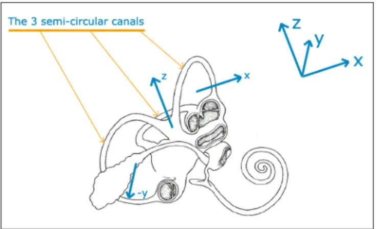
Because of busy schedules, it is then not conceivable to force the astronauts to stay motionless on a bed during short radius centrifugation. These periods would better be used by astronauts to do emails, to read, to talk to their fami‐ lies via videophone, to exercise, even maybe to sleep. Such activities require certain degrees of freedom of the body and the head, which will probably not be totally restrained. Subsequent paragraphs investigate the problems en‐ gendered by head movements in these conditions. In order to understand the phenomena, the related background on vestibular physiology is first ex‐ posed. 2.5.1 Vestibular Physiology. [note: references for this section are Young, 1983; Young, 2003a and 2003b; Oman, 1988; Oman, 2003; Guedry and Benson, 1978] The vestibular system is a physiological structure that allows for self‐motion perception: it sends information to the brain about the relative movement of its organs so that the brain interprets this information and translates it into motion data. The vestibular system is located in the inner ear (one set of it for each ear). Figure 2.9 shows the relative location of the vestibular organs in the inner ear.
Figure 2.9. Location of the vestibular system in the inner ear. Illustration taken from McGill Faculty of Medicine website.
The vestibular system consists of two types of motion sensors: the otolith or‐ gans (saccule and utricle) which are sensitive to linear accelerations and gravity; and three semi‐circular canals (anterior, posterior and lateral canals) which are sensitive to angular acceleration. 2.5.1.1 The Otolith Organs. The otolith organs are sensitive to linear acceleration and gravity. Each organ (saccule or utricle) consists of a macular plane composed of hair cells project‐ ing into a gelatinous membrane. In its top layer are small calcium carbonate aggregates or otoconia (similar to little stones, hence the name otolith: oto = ear, lith = stone). In the presence of linear acceleration and/or gravity, the oto‐ conial membrane moves in the endolymph, thus moving the otoconia, acti‐ vating the hair cells in the direction of the sum of these forces.
In the case of centrifugation, with head at the center of rotation, otoliths should not be stimulated, apart from the natural gravity if centrifugation is operated on Earth.
The accepted mathematical model for the otoliths organs is to consider the otoconia as seismic masses, with linear spring restoring forces due to the macula, and inertial drag due to the surrounding endolymph (see Wilson and Melvill‐Jones, 1979, for a complete description of the underlying mathe‐ matical model).
2.5.1.2 The Semi‐Circular Canals.
The semi‐circular canals can be considered as angular accelerometers: they provide both the magnitude and direction of the rotation(s) they undergo, in a three dimensional space. They are almost perpendicular to each other: each semi‐circular canal is dedicated to a specific axis of rotation. The canal is ly‐ ing in the plane which is orthogonal to the axis considered. A canal is “acti‐ vated” when a rotation around its corresponding axis occurs. See Figure 2.10
for a representation of the axis and the frame of reference (x forward, y to the left and z up). Figure 2.10. Orientation of the semi‐circular canals. Illustration adapted from a drawing from Queen Mary University of London. The activation of the canals comes from the relative motion between the ca‐ nal itself and the viscous liquid (called endolymph) it contains. When the ca‐ nal is rotating around its corresponding axis, the endolymph does not move immediately with respect to the outside frame, because of its inertia. Thus, it implies a relative movement of the endolymph with respect to the canal, but in the other sense of rotation, as if the fluid were rotating inside the canal. Each canal is interrupted by an ampula that contains a thin membrane (the cupula) which supports the pressure of the moving endolymph, and pre‐ vents it from circulating freely inside the canal. The vestibular hair cells in‐ side the cupula react to the pressure of the fluid on the cupula and send the information to the brain. See Figure 2.11 for a representation of the mecha‐ nism.
Illustration adapted from a drawing from Queen Mary University of London.
The deflection of the cupula by the endolymph results in the transmission of afferent signals successively to the vestibular nuclei, the cerebellum, the ocu‐ lar‐motor nuclei, and finally the cerebral cortex.
If the rotation lasts more than twenty seconds, the effect of inertia stops, and the endolymph starts rotating with the canal. The hair cells are not stimu‐ lated anymore and their firing rate goes back to normal. If the constant veloc‐ ity rotation stops, the same initial phenomenon due to the inertia of the endo‐ lymph happens: the hair cells detect an illusory rotation in the reverse direc‐ tion. 2.5.2 The Physics of Head Turns in a Rotating Environment. 2.5.2.1 Cross‐Coupled (Coriolis) Acceleration. Let suppose that a human being is on a centrifuge, rotating about an axis Ac, with an angular velocity
ω
cr
. In addition, his/her head is rotating about an‐ other axis Ah, with an angular velocityω
r
h. This situation generates, for thetime of the head rotation (usually about one second), an additional angular acceleration about an axis Acc which is orthogonal to both Ac and Ah. The cor‐
responding angular velocity emerges from the cross‐product of
ω
cr
andω
hr
: h c ccω
ω
ω
r
=
−
r
×
r
(eq. 0) Figure 2.12. Subject lying supine NU on a centrifuge.Letʹs define the two frames of reference (see Figure 2.12):
(
S
;
x
r
,
y
r
,
z
r
)
is attached to Earth.(
S
′
;
x
r
′
,
y
r
′
,
z
r
′
)
is attached to the centrifuge. Suppose the centrifuge is rotating clockwise. Therefore,ω
cr
is pointing ʺdownʺ:z
c c=
−
′
r
r
ω
.
ω
Letʹs use the rotation operator that transforms a vector from one frame of ref‐ erence (S) into another frame of reference (Sʹ):(
.
/
) (
=
.
/
)
′+
′×
.
S S S Sd
dt
dt
d
ω
r
(eq. 1)Letʹs apply this to a vector (
position
) describing the position in the rotating frame of reference Sʹ. We obtain:(
d
position
dt
) (
d
position
dt
)
position
S S S S
=
′+
ω
′×
r
/
/
which is equivalent to:position
velocity
velocity
S=
S′+
ω
r
c×
(eq. 2) Now, letʹs apply the left side of eq. 1 to the left side of eq. 2 and the right side of eq. 1 to the right side of eq. 2. We obtain:(
)
[
d
position
dt
]
(
position
)
velocity
on
accelerati
on
accelerati
c c S c S c S S×
×
+
×
+
×
+
=
′ ′ ′ω
ω
ω
ω
r
r
r
r
/
(eq. 3) Which is also:(
position
)
velocity
velocity
on
accelerati
on
accelerati
c c S c S c S S×
×
+
×
+
×
+
=
′ ′ ′ω
ω
ω
ω
r
r
r
r
(eq. 4)Also written:
(
position
)
velocity
on
accelerati
on
accelerati
S′=
S−
ω
c×
S′−
ω
c×
ω
c×
r
r
r
.
2
(eq. 5) Equation 5 is the general expression for the Theorem of Coriolis. It describes linear acceleration in a rotating environment. The term−
ω
c×
velocity
S′r
.
2
isthe Coriolis acceleration and the term
−
ω
c×
(
ω
c×
position
)
r
r
is the centrifu‐gal force.
French and Ebison (1986) extended this expression to angular accelerations. The general expression for angular accelerations in a rotating environment is:
(
c h)
c h c h hα
ω
ω
ω
ω
ω
α
r
′
=
r
−
2
.
r
×
r
′
−
r
×
r
×
r
(eq. 6) whereα
hr
represents an angular acceleration of the head in S,′
hα
r
represents the same angular acceleration of the head in Sʹ,ω
hr
represents the head veloc‐ ity in S andω
h′
r
represents the head velocity in Sʹ. The term−
ω
c×
ω
h′
r
r
.
2
is the cross‐coupling acceleration term, and is domi‐ nant in the expression.Lyne (2000) summed up the consequences on the canals. In the case of a head turn from RED (Right‐Ear‐Down) position to NU (Nose‐Up) position, the pitch canal is being stimulated from rest by the cross‐coupled acceleration. The stimulation can be modeled as a sinusoidal function (because the orien‐ tation of the canal changes with regard to the axis of rotation), with a maxi‐ mum amplitude of
ω
c.
ω
h. Thus, the acceleration acting on the pitch canalduring a yaw head turn is:
ω
c.
ω
h.
sin
( )
ω
h.
t
(whereω
h depends on time t).eration modeled as
ω
c.
ω
h.
cos
( )
ω
h.
t
. The yaw canal stays at rest during a yawhead turn on the centrifuge.
A hypothesis for the use of the sine and cosine functions is their periodicity. At the beginning of the head turn (t = 0), the pitch canal gets no stimulation from the centrifuge rotation, but the roll canal gets a stimulation. At the end of a 90° head turn, the pitch canal is stimulated by the centrifuge rotation, whereas the roll canal is not anymore. The sine and cosine functions assure continuity of the stimuli. 2.5.2.2 Canals In/Out of Plane Effect. This effect emerges from the loss of inertia of the endolymph. When a rota‐ tion is sustained in time, after a period of about 20 seconds, the endolymph starts rotating with the canals. Therefore, subject to its elastic restoring force, the cupula returns to its neutral position. If the result of a head movement is a change in the orientation of the plane of the canal with respect to the initial rotation, then the endolymph in the canal will undergo an angular accelera‐ tion corresponding to the initial angular acceleration (that of the centrifuge). Whereas in a stable situation, head turns cause the canals to follow the pro‐ file of angular velocity (acceleration and then deceleration, characteristics of a normal head turn), in a rotating environment, the canals do not. The cupula decay (about 6 seconds) leads to a misrepresentation of
ω
r
, and thus a percep‐ tion of self‐motion. Letʹs go back to the example of the subject lying supine with head at the cen‐ ter of rotation, in the NU position (see Figure 2.12). In this position, the roll canal is stimulated during the rotation, which gives the normal perception of rotation (because the subject knows he/she is lying on his/her back: proprio‐ ceptive channel). After a certain period of time (around 20 seconds), the roll canal response declines to zero: the subject feels no motion (which is a falseperception). If a head turn RED is made, then two canals are involved: the roll canal goes out of the plane of rotation and the pitch canal goes in the plane of rotation.
The roll canal experiences the change in angular velocity because of the ʺcounter‐reactionʺ of the endolymph stopping its rotation in phase with the canal, and decelerating in the clockwise direction, giving the sensation of a roll to the right. The sensation perceived is strong because only the decelera‐ tion phase is present here, not the acceleration that would occur in a station‐ ary environment. The pitch canal experiences the reverse effect: an acceleration because of the stimulation of the centrifuge rotation. This leads to the perception of pitching backwards (forwards in the case of a Left‐Ear‐Down head turn). These effects can be modeled with a block diagram. For example, a modified Young‐Oman transfer function model (Young and Oman, 1969) can help visualize the dynamics of the cupula (and thus of the endolymph) and the in‐ fluence of adaptation of the system (refer to Young and Oman, 1969, for more details).
The first block models the dynamics of the canals (e.g. simple gain K = 1, time constant Tc = 6 seconds); the second block models the neural adaptation (time constant Ta >> Tc, e.g. Ta = 80 seconds). The following graphs were plotted for these values (Lyne, 2000).
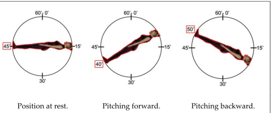
Figure 2.13 shows the velocity profile of the roll canal. Initially in the plane of rotation, it goes out of it, thus undergoing a velocity change of 23 rpm in one
second (thin line). The response of the cupula (its deflection) is immediate (endolymph motion implies cupula deflection), and then goes back to rest at the neutral position (in about 10 seconds): it does not follow the velocity pro‐ file.
Figure 2.14 shows the velocity profile of the pitch canal. Initially out the plane of rotation, it goes in it, thus undergoing a velocity change of 23 rpm in one second (thin line), in the other direction compared to the roll canal. The deflection of the cupula is immediate and then goes back to rest at the neutral position (in about 20 seconds): it does not follow the velocity profile.
Figure 2.15 shows the velocity profile of the yaw canal. Its velocity profile (thin line) corresponds to the normal head turn that would occur in a station‐ ary environment (acceleration and deceleration in one second). The response of the cupula follows the velocity profile and then goes back to rest at the neutral position (in about 6 seconds).
Figure 2.13. Cupula deflection in the roll canal (for a velocity change of 23rpm in 1 second) represented by the tick line. The thin line shows the velocity profile.
Figure 2.14. Cupula deflection in the pitch canal (for a velocity change of 23rpm in 1 sec‐ ond) represented by the tick line. The thin line shows the velocity profile. (note that the saturation is an artefact of simulation) Figure 2.15. Cupula deflection in the yaw canal (for a velocity change of 23rpm in 1 sec‐ ond) represented by the tick line. The thin line shows the velocity profile. (note that the saturation is an artefact of simulation)