Publisher’s version / Version de l'éditeur:
Vous avez des questions? Nous pouvons vous aider. Pour communiquer directement avec un auteur, consultez la première page de la revue dans laquelle son article a été publié afin de trouver ses coordonnées. Si vous n’arrivez Questions? Contact the NRC Publications Archive team at
PublicationsArchive-ArchivesPublications@nrc-cnrc.gc.ca. If you wish to email the authors directly, please see the first page of the publication for their contact information.
https://publications-cnrc.canada.ca/fra/droits
L’accès à ce site Web et l’utilisation de son contenu sont assujettis aux conditions présentées dans le site LISEZ CES CONDITIONS ATTENTIVEMENT AVANT D’UTILISER CE SITE WEB.
Combustion Institute Canadian Section, 2007 Spring Technical Meeting [Proceedings], 2007
READ THESE TERMS AND CONDITIONS CAREFULLY BEFORE USING THIS WEBSITE.
https://nrc-publications.canada.ca/eng/copyright
NRC Publications Archive Record / Notice des Archives des publications du CNRC :
https://nrc-publications.canada.ca/eng/view/object/?id=19f0b7f4-04e3-4859-ad5e-5ee386f0b31e https://publications-cnrc.canada.ca/fra/voir/objet/?id=19f0b7f4-04e3-4859-ad5e-5ee386f0b31e
Archives des publications du CNRC
This publication could be one of several versions: author’s original, accepted manuscript or the publisher’s version. / La version de cette publication peut être l’une des suivantes : la version prépublication de l’auteur, la version acceptée du manuscrit ou la version de l’éditeur.
Access and use of this website and the material on it are subject to the Terms and Conditions set forth at
Laser desorption/laser ionization/time-of-flight mass spectrometer for the detection of polycyclic aromatic hydrocarbons desorbed from soot
Thomson, Kevin; Ziskind, M.; Faccinetto, A.; Therssen, E.; Desgroux, P.; Focsa, C.
LASER DESORPTION/LASER
IONIZATION/TIME-OF-FLIGHT MASS SPECTROMETER FOR THE
DETECTION OF POLYCYCLIC AROMATIC
HYDROCARBONS DESORBED FROM SOOT
K. Thomson
1,2,3, M. Ziskind
1, A. Faccinetto
1,2, E. Therssen
2,
P. Desgroux
2and C. Focsa
11
Laboratoire de Physique des Lasers, Atomes et Molécules,
2
Laboratoire de Physico-chimie des Processus de Combustion et de l’Atmosphère, Centre d'Etudes et de Recherches Lasers et Applications
Université des Sciences et Technologies de Lille
3
Combustion Group, Institute for Chemical Process & Environmental Tech., NRC
INTRODUCTION
Recent advances in the field of laser desorption/laser ionization mass spectrometry (LD/LI/MS) have renewed interest in these separation methods for fast analysis of chemical species adsorbed on soot particles [1-7]. These techniques provide mass-separation of the desorbed phase with high selectivity and sensitivity and require very small amounts of soot to be collected. In particular the techniques provide a means to measure adsorbed polycyclic aromatic hydrocarbons (PAHs). PAHs are important precursors of carbonaceous soot particles, thus influence the quantity and morphology of particulate emission from various combustion processes [8,9]. Furthermore, PAHs absorbed on the surface of the soot particles contribute to the carcinogenicity of the particles. Therefore, there is scientific interest in characterizing and quantifying these adsorbed PAHs in order to provide key information about the mechanism of the soot formation for various fuels and combustors and to understand their health impact. This has motivated various studies [1-7]; however, an extensive characterization of the PAHs absorbed onto soot particles remains a challenge.
An experimental set-up based on the coupling of laser desorption / photoionization / time-of-flight mass spectrometry (LD/LI/TOF-MS) techniques has been developed at the Université des Sciences et Technologies de Lille (USTL) and is dedicated to the analysis of PAH desorbed from soot with particular focus on the dependence of the adsorbed PAHs on the stage of combustion and the nature of the fuel.
In previous papers [10-12], we have explored the influence of various experiment parameters (e.g. fluence and wavelength of desorption and ionization lasers) on the measured mass spectra. In particular, in [12], we explored a change to the ionization laser, increasing the ionization volume through the use of a cylindrical lens (to create a laser sheet) in place of a spherical lens (to create a laser spot). It was concluded that enlarging the ionization volume greatly enhanced the sensitivity of the diagnostic and thus allows the use of significantly lower fluence. This eliminated fragmentation in the spectra from pure PAH and synthetic soot sample spectra. A significant challenge of LD/LI/TOF-MS which remains is to measure mass spectra which are truly representative of the species absorbed on the surface of the soot particles and not contaminated by artifacts of the soot sampling, laser desorption, or ionization steps (e.g., condensed gas phase PAH from the flame, sublimated soot, and gas species fragmentation).
Though the cylindrical lens improved system performance, it is not ideal because laser fluence in the ionization volume is not homogeneous and therefore is difficult to define or control. The same problem exists with the desorption laser. In this paper we explore aperture imaging of the two lasers to create a well defined desorption surface and ionization volume with objective of establishing appropriate operating fluences for the LD/LI steps of LD/LI/TOF-MS of PAH adsorbed on soot.
EXPERIMENTAL DETAILS
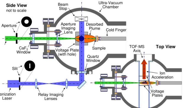
The experimental apparatus is shown schematically in Figure 1 and is described in the context of the experimental method which can be divided into four key steps; sample collection and preparation, laser desorption, laser ionization, and TOF-MS. The steps are described briefly below while more detail is available from previous papers [7,10-12].
Figure 1 – Schematic of Laser Desorption / Laser Ionization / Time-of Flight Mass Spectrometer
Sample collection and preparation
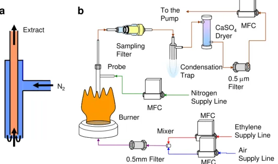
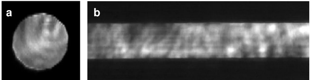
The LD/LI/TOF-MS diagnostic is an ex-situ technique. Sample preparation methods vary with the sample type. Synthetic soot is created by mixing carbon black with PAH dissolved in dimethyl ether (DME) solution. The carbon adsorbs the PAH from solution and the solution is then removed by drawing it through a paper filter. Visible/UV spectrometry of the residual solution is used to confirm that all PAH was adsorbed on the soot. To create a solid sample for use in the TOF-MS, the carbon black powder can be pressed directly in a sample holder to create a sintered disk (Figure 2a). Flame soot is collected from a McKenna premixed burner (1.28 l min-1 ethylene, 8.74 l min-1 air) by an extractive vacuum probe (1.10 l min-1 extraction flow, 0.51 l min-1 dilution nitrogen) onto a borosilicate filter (Figure 2b). A schematic of the soot vacuum probe is included in Figure 3. Sample acquisition times are 15 minutes. Samples enter the TOF-MS via a sample preparation chamber separated from the TOF-TOF-MS via a gate valve where they are cooled on a liquid nitrogen cooled cold finger to a temperature of about -170oC (to avoid
Side View not to scale Top View Aperture Slit Ionization
Laser Relay ImagingLenses
Ultra-Vacuum Chamber CaF2 Window Quartz Window Aperture Imaging Lens Beam Stop Sample Cold Finger Desorbed Plume Voltage Plate (with hole) TOF-MS Axis Ion Acceleration Voltage Plates
PAH sublimation) while simultaneously being pumped to an ultra-high vacuum (residual pressure: 10-7 Torr). Once these conditions are met, the gate valve is opened and the sample is moved into the TOF-MS where the residual pressure is 10-9 Torr.
a
b
Figure 2 - a.) PAH adsorbed on carbon black pressed into holder, b.) soot vacuum extracted from flame onto borosilicate filter
Nitrogen Supply Line Ethylene Supply Line Air Supply Line To the Pump 0.5mm Filter 0.5 µm Filter MFC MFC MFC MFC CaSO4 Dryer Condensation Trap Probe Burner Sampling Filter Mixer N2 Extract
a
b
Figure 3 - a.) stainless steel extractive vacuum probe, b.) complete burner and extraction system
Laser desorption
The samples are irradiated at normal incidence by the beam of a 7 ns doubled Nd:YAG laser (λ=532 nm, Quantel Brillant) which is aperture imaged using a 10 cm focal length plano-convex CaF2 lens to a near top-hat spot (fluence variation of 30%, 95% confidence interval) of 1 mm
diameter (Figure 4a). Laser fluences of 0.064 to 0.255 J/cm2 are typically used and induce desorption of neutral species from the sample, forming a desorbed plume which propagates norm to the sample surface and between the extraction voltage plate.
Laser ionization
The desorbed plume is irradiated by a 7 ns quadrupled Nd:YAG laser (λ=266 nm, Continuum Powerlit) apertured using a 0.5 x 10 mm slit and then relay imaged to the center of the extraction voltage plates using 50 and 30 cm plano-convex quartz lenses forming a laser sheet with a cross-section of ~0.3 x 6 mm. These dimensions are approximate because at the time of publication, a beam profile was only available for the reverse lens configuration (i.e. 30 then 50 cm lens). This magnifies rather than demagnifies the slit as shown in Figure 4b. Fluence calculations are therefore only approximate and the full characterization of the beam is a priority. This stated, the fluences used ranged from 0.009 to 0.9 J/cm2. Timing of the desorption and ionization pulse was
controlled using a digital four channel delay generator and set for a delay of 50 µs between the two lasers.
a
b
Figure 4 - a.) beam profile of 532 nm desorption laser at sample surface (dia. 1 mm), b.) section of beam profile of 266 nm ionization laser in ionization zone for 30 followed by 50 cm lens configuration (total profile is 0.86 x 17 mm).
Laser ionization
The desorbed plume is irradiated by a 7 ns quadrupled Nd:YAG laser (λ=266 nm, Continuum Powerlit) apertured using a 0.5 x 10 mm slit and then relay imaged to the center of the extraction voltage plates using 50 and 30 cm plano-convex quartz lenses forming a laser sheet with a cross-section of ~0.3 x 6 mm. These dimensions are approximate because at the time of publication, a beam profile was only available for the reverse lens configuration (i.e. 30 then 50 cm lens). This magnifies rather than demagnifies the slit as shown in Figure 4b. Fluence calculations are therefore only approximate and the full characterization of the beam is a priority. This stated, the fluences used ranged from 0.009 to 0.9 J/cm2. Timing of the desorption and ionization pulse was controlled using a digital four channel delay generator and set for a delay of 50 µs between the two lasers.
Time of Flight Mass Spectrometer
The ions produced in this way are mass-analyzed in a 1-m reflectron TOF-MS (RM Jordan, VA1
= 1022 V, VA2 = 709 V, VA3 = ground, VXY = 65 V, VR1 = 682 V, VR2 = 1172 V, and VD1 = 2500
V). A mass resolution of m/∆m ~ 1000 is achieved throughout a typical spectrum. Ion detector signals are recorded using a digital oscilloscope at a time resolution of 4 ns/point. Mass calibration of the spectra was achieved by marking peaks for compounds of known atomic mass.
RESULTS AND DISCUSSION
The LD/LI/TOF-MS technique has been applied to synthetic and flame collected soot. Figure 5 includes spectra collected from synthetic soot samples (pyrene adsorbed on carbon black). The influence of ionization fluence on PAH fragmentation is clearly demonstrated. For an ionization fluence of 8 mJ/cm2 the only peaks observed are the parent peak at 202 a.m.u. along with the secondary C13 peak at 203 a.m.u. and dehydrogentated pyrene peaks at 200 and 201 a.m.u.
Otherwise, the spectrum is clean, indicating that pyrene is the only material desorbed from the synthetic soot sample and that there is no fragmentation of the PAH. Conversely, when an ionization fluence of 108 mJ/cm2 is used, a spectrum is recorded which is rich in peaks from 12 to 100 a.m.u. which is indicative of fragmentation of the pyrene during ionization. Through more extensive testing it has been determined that the ionization laser fluence should be kept below 18 mJ/cm2 in order to maximize PAH signals while minimizing fragmentation. Similar
testing shows that the desorption laser fluence should be kept below 150 mJ/cm2. It is noted that the desorption and ionization processes are coupled and in the regime of fragment free desorption, higher desorption fluence necessitates lower ionization fluence to avoid fragmentation due to ionization. The capability in the technique to track mass signal with laser shot number is demonstrated in Figure 5c.). It is observed that the PAH is desorbed over about 40 laser shots and that the yield of pyrene in the spectra is depressed during the first several laser shots for the high ionization fluence condition. Further testing is needed to confirm the repeatability of the decay curves; however, there is hope that the decay trends will give some insight into the nature of the PAH – carbon bonds.
Shot number 0 20 40 60 80 100 120 Integ ra ted si g nal ( V ns) 0 50 100 150 200 250 Eion = 8 mJ/cm 2 Eion = 108 mJ/cm 2 Mass (a.m.u.) 0 50 100 150 200 250 Si g nal ( V ) 0.0 0.2 0.4 0.6 0.8 1.0 1.2 1.4 Mass (a.m.u.) 0 50 100 150 200 250 Si g nal ( V ) 0 1 2 3 4 5 6 7 200 202 204 0 2 4 6 8 70 72 74 76 78 0.0 0.2 0.4 0.6 a b c
Figure 5 – LD/LI/TOF-MS of pyrene adsorbed on carbon black, 10 shot averages, Edes = 0.127
J/cm2, Eion = a.) 0.008,b.) 0.108 J/cm2 and c.) decay of 202 a.m.u. peak with shot number
Shot number 0 5 10 15 20 25 30 35 40 Integ rated si g nal (V ns) 0 50 100 150 200 250 Eion = 10 mJ/cm 2 Eion = 110 mJ/cm 2 Mass (a.m.u.) 0 50 100 150 200 250 300 350 400 Si g n al (V) 0.0 0.2 0.4 0.6 0.8 1.0 1.2 1.4 1.6 1.8 Mass (a.m.u.) 0 50 100 150 200 250 300 350 400 Si g n al (V) 0 1 2 3 4 5 a b c
Figure 6 – LD/LI/TOF-MS of soot collected from McKenna burner, 10 shot averages, Edes =
0.127 J/cm2, Eion = a.) 0.010,b.) 0.110 J/cm2 and c.) decay of 202 a.m.u. peak with shot number
Spectra from flame collected soot are included in Figure 6. For the recommended low ionization fluence, peaks are resolved between 152 and 278 a.m.u. with the stongest peaks at 174, 190, and 202 amu. Very similar peak voltages are recorded as compared to the synthetic soot. When ionization fluence is raised above the recommendation of 18 mJ/cm2, mass peaks appear in the 12 through 100 a.m.u. range which are quite consistent with the fragment peaks observed in the synthetic soot samples. It is therefore likely that these additional peaks are products of fragmentation rather than peaks which can only be accessed through high ionization fluence. The decay of the 202 a.m.u. peak is somewhat different for the flame collected soot, showing a faster decay, more noise, and a late strong peak around the 20th laser shot for both ionization
conditions. Further testing is need to resolve differences in the decay patterns from synthetic and flame soot.The strong similarity in the response of the synthetic and flame soot to ionization laser fluence is very encouraging and suggests that the recommended laser fluences of < 150 mJ/cm2 for the desorption laser and < 18 mJ/cm2 for the ionization laser are correct to avoid fragmentation of the desorbed PAH during the measurement of flame soot samples.
CONCLUSIONS
In this work, we have reported improvements made to a laser desorption/laser ionization/time-of-flight mass spectrometer used to desorb and mass analyze polycyclic aromatic hydrocarbons adsorbed on soot. Through improvements to the optics used to shape the desorption and ionization laser beams, more accurate quantification of the laser fluences is possible, which in turn makes determination of appropriate fluences for the measurement of unfragmented PAH possible. This work remains preliminary and further testing of synthetic and flame collected soot is needed.
ACKNOWLEDGEMENTS
The Centre d’Etudes et de Recherches Lasers et Applications is supported by the Ministere chargé de la Recherche, the Région Nord-Pas de Calais and the Fonds Européen de Développement Economique des Régions. We acknowledge the Natural Science and Engineering Research Council of Canada for Postdoctoral Funding for Kevin Thomson.
REFERENCES
1. R.A. Dobbins, R.A. Fletcher and W. Lu, Combust.Flame 100:301 (1995) 2. R.A. Dobbins, R.A. Fletcher and H.C. Chang, Combust.Flame 115:285 (1998) 3. S.M. Hankin and P. John, Anal. Chem. 71 :1100 (1999)
4. R. Zimmermann, L. Van-Vaeck, M. Davidovic, M. Beckmann and F. Adams, Environ. Sci. Technol. 34:4780 (2000)
5. V. Carré, L. Vernex-Loset, G. Krier, P. Manuelli and JF. Muller, Anal. Chem. 76:3879 (2004)
6. B. Öktem, M.P.Tolocka, B. Zhao, H. Wang and M.V. Johnston, Combust.Flame 142:364 (2005)
7. Y. Bouvier, C. Mihesan, M. Ziskind, E. Therssen, C. Focsa, J.F. Pauwels, P. Desgroux, Proceedings Combust. Institute 31:841 (2007)
8. J.A. Miller, J.V. Volponi, and J.F. Pauwels, Combust.Flame 105, 454 (1996) 9. H. Richter and J.B. Howard, Prog. Energy Combust. Sci. 26, 565 (2000)
10. C. Mihesan, M. Ziskind, E. Therssen, P. Desgroux and C. Focsa, J. Phys. Conf. Ser.,
submitted
11. C. Mihesan, M. Ziskind, B. Chazallon, E. Therssen, P. Desgroux, S. Gurlui and C. Focsa, Appl. Surf. Sci., 253:1090 (2006)
12. K. Thomson, M. Ziskind, C. Mihesan, E. Therssen, P. Desgroux, C. Focsa, Appl. Surf. Sci., in press (2007)