HAL Id: hal-02088274
https://hal.univ-lorraine.fr/hal-02088274
Submitted on 2 Apr 2019
HAL is a multi-disciplinary open access
archive for the deposit and dissemination of
sci-entific research documents, whether they are
pub-lished or not. The documents may come from
teaching and research institutions in France or
abroad, or from public or private research centers.
L’archive ouverte pluridisciplinaire HAL, est
destinée au dépôt et à la diffusion de documents
scientifiques de niveau recherche, publiés ou non,
émanant des établissements d’enseignement et de
recherche français ou étrangers, des laboratoires
publics ou privés.
Proteomic Consequences of a Human Mitochondrial
tRNA Mutation beyond the Frame of Mitochondrial
Translation
Petra Tryoen-Toth, Sophie Richert, Bénédicte Sohm, Manuele Miné, Cécile
Marsac, Alain van Dorsselaer, Emmanuelle Leize, Catherine Florentz
To cite this version:
Petra Tryoen-Toth, Sophie Richert, Bénédicte Sohm, Manuele Miné, Cécile Marsac, et al.. Proteomic
Consequences of a Human Mitochondrial tRNA Mutation beyond the Frame of Mitochondrial
Trans-lation. Journal of Biological Chemistry, American Society for Biochemistry and Molecular Biology,
2003, 278 (27), pp.24314-24323. �10.1074/jbc.M301530200�. �hal-02088274�
Proteomic Consequences of a Human Mitochondrial tRNA Mutation
beyond the Frame of Mitochondrial Translation*
Received for publication, February 12, 2003, and in revised form, April 16, 2003 Published, JBC Papers in Press, April 24, 2003, DOI 10.1074/jbc.M301530200
Petra Tryoen-To´ th‡, Sophie Richert§, Be´ne´dicte Sohm‡, Manuele Mine¶, Ce´cile Marsac¶, Alain Van Dorsselaer§, Emmanuelle Leize§, and Catherine Florentz‡储
From the ‡UPR 9002, Institut de Biologie Mole´culaire et Cellulaire du CNRS, 15 Rue Rene´ Descartes
67084 Strasbourg Cedex, §Laboratoire de Spectrome´trie de Masse Bio-Organique, Ecole de Chimie des Polyme`res, 25 Rue Becquerel, 67087 Strasbourg Cedex 2, and¶Laboratoire CERTO, Faculte´ Necker, 156 Rue de Vaugirard, 75015 Paris, France
Numerous severe neurodegenerative and neuromus-cular disorders, characterized biochemically by strong perturbations in energy metabolism, are correlated with single point mutations in mitochondrial genes cod-ing for transfer RNAs. Initial comparative proteomics performed on wild-type and Myoclonic Epilepsy and Ragged Red Fibers (MERRF) mitochondria from sibling human cybrid cell lines revealed the potential of this approach. Here a quantitative analysis of several hun-dred silver-stained spots separated by two-dimensional gel electrophoresis was performed in the specific case of a couple of mitochondria, containing or not mutation A8344G in the gene for mitochondrial tRNALys,
corre-lated with MERRF syndrome. Computer-assisted analy-sis allowed us to detect 38 spots with significant quan-titative variations, of which 20 could be assigned by mass spectrometry. These include nuclear encoded pro-teins located in mitochondria such as respiratory chain subunits, metabolic enzymes, a protein of the mitochon-drial translation machinery, and cytosolic contami-nants. Furthermore, Western blotting combined with mass spectrometry revealed the occurrence of numer-ous isoforms of pyruvate dehydrogenase subunits, with subtle changes in post-translational modifications. This comparative proteomic approach gives the first insight for nuclear encoded proteins that undergo the largest quantitative changes, and pinpoints new potential mo-lecular partners involved in the cascade of events that connect genotype to phenotype.
Mitochondria are the center of numerous metabolic functions of primary importance to the cell. They are responsible for the major ATP synthesis pathway and thus deliver energy through the activity of respiratory chain complexes and ATP synthase, all located in the inner mitochondrial (mt)1membrane.
Mito-chondria also contain major catabolic pathways including the
citric acid cycle, fatty acid oxidation, part of the urea cycle, and a number of biosynthetic pathways such as those for heme, ubiquinol, and cardiolipin (1). To fulfill these functions, mito-chondria produce a subset of 13 proteins by translation of their own genes and import a large number of proteins encoded by the nuclear genome (estimated 400 –2000), translated within the cytosol. Among these are numerous proteins allowing maintenance and expression of the mt genome (replicase, tran-scription factors, and translation proteins). Thus, mt functions depend on well coordinated cross-talks between mt and nuclear genomes, allowing for a controlled balance between mitochon-dria encoded and nuclear encoded proteins and associated en-zymatic activities.
Mt genomes are very sensitive to mutations (2– 4). In the case of human, as many as 150 point mutations have been correlated already with various neuromuscular and neurode-generative disorders (5– 8). tRNA genes, which correspond only to about 10% of the mt genome, are particularly sensitive because they host about 80 of the known pathogenesis-associ-ated mutations. Genotype/phenotype relationships for these disorders are complex because one mutation can be related to several disorders, and conversely a same disorder can arise from several different mutations. Due to the central role of tRNAs in protein synthesis (9), defects in structure/function relationships of mutant tRNAs have been sought (e.g. reviewed in Refs. 5, 6, 8, 10 –13). The rate of synthesis of the 13 mito-chondria-encoded proteins is decreased, and tRNA maturation, stability, post-transcriptional modification, structure, amino-acylation, and/or codon reading properties can be affected. However, depending on the specific case studied, distinct re-sults were observed, confirming a complex relationship be-tween genotypes and phenotypes and suggesting the contribu-tion of addicontribu-tional factors to the expression of the disease (14). Furthermore, in some instances no defect in protein synthesis was observed, a discovery in support of mediators outside mt translation (15).
Comparative proteomics is a powerful approach to investi-gate quantitative variations in several hundred proteins at once (16, 17). Its application to human mitochondria would enlarge insight into unexpected consequences of a single point mutation in a tRNA gene on the global mt protein content. Such an approach particularly enables the analysis of the fate of nuclear encoded proteins located in mitochondria. Investiga-tion of human mt tRNA gene mutaInvestiga-tions suffers so far from the absence of animal model systems (e.g. Refs. 18 –20). An initial proteomic study performed on mitochondria from sibling cybrid cell lines, in the presence or absence of a single point mutation in one mt tRNA gene, has already proven the feasibility and the potential impact of such an approach for the investigation of mt
* This work was supported by CNRS, Universite´ Louis Pasteur Stras-bourg, European Community Grant QLG-CT-1999-00660, Program Physique-Chimie du Vivant from CNRS, and by the Association Fran-c¸aise pour les Myopathies. The costs of publication of this article were defrayed in part by the payment of page charges. This article must therefore be hereby marked “advertisement” in accordance with 18 U.S.C. Section 1734 solely to indicate this fact.
储To whom correspondence should be addressed. Tel.: 33-3-88-41-70-59; Fax: 33-3-88-60-22-18; E-mail: [email protected].
1The abbreviations used are: mt, mitochondrial; MERRF, Myoclonic
Epilepsy and Ragged Red Fibers; MALDI-TOF, matrix-assisted laser desorption ionization time-of-flight; MS, mass spectrometry; PDH, pyruvate dehydrogenase; DTT, dithiothreitol; CHAPS, 3-[(3-cholamido-propyl)dimethylammonio]-1-propanesulfonic acid; COX, cytochrome c oxidase.
THEJOURNAL OFBIOLOGICALCHEMISTRY Vol. 278, No. 27, Issue of July 4, pp. 24314 –24323, 2003 © 2003 by The American Society for Biochemistry and Molecular Biology, Inc. Printed in U.S.A.
This paper is available on line at http://www.jbc.org
24314
by guest, on March 9, 2011
www.jbc.org
tRNA disorders (21). In mitochondria carrying tRNALysgene
mutation A8344G, associated with Myoclonic Epilepsy and Ragged Red Fibers (MERRF syndrome (22)), a large decrease in the steady-state levels of two nuclear encoded subunits of cytochrome c oxidase, a respiratory chain complex, was identi-fied upon simple visual comparison of two-dimensional PAGE (isoelectrofocusing/SDS-PAGE) followed by mass spectrometric assignment. Here the cybrid model system was further ex-plored by a detailed computer-assisted quantitative compari-son of over 800 spots obtained from wild-type and mutation A8344G-carrying mitochondria. This first large scale quantita-tive analysis of the protein profile of mitochondria representa-tive of a tRNA disorder, using cybrid cell lines as a model system, revealed that in addition to the 13 mitochondria coded proteins, all known to be affected, several nuclear en-coded proteins of mt location undergo quantitative and/or qual-itative changes.
The proteomic approach enlarges the view of the status of a disease-carrying cell, highlighting new molecular species rele-vant to perturbations within the whole cell, beyond the conse-quences of the primary molecular impact of a mutation on the tRNA structure/function relationship in translation. Under-standing how each of these species affects the cell will shed new light on the genotype/phenotype relationships of mt disorders. Although some of the affected proteins may contribute to the expression of the disease, others may be compensating.
EXPERIMENTAL PROCEDURES
Cell Cultures
Human osteosarcoma-derived cybrid cell lines R2-1A and R1C3 were a kind gift of A. Chomyn and G. Attardi (California Institute of Tech-nology, Pasadena). Cell line R2-1A carried the wild-type mt tRNALys
gene in homoplasmic form, and cell line R1C3, associated with MERRF disease, carries an A to G transition at position 8344 in this gene in homoplasmic form (23). Cell lines were grown in Dulbecco’s modified Eagle’s medium with 10% fetal calf serum, 100 units/ml penicillin, 100 g/ml streptomycin, and 100 g/ml bromodeoxyuridine at 37 °C in the presence of 5% CO2and 85% humidity. R2-1A cells were seeded at a
density of 2⫻ 105cells/10 ml of medium/10-cm diameter Petri dish, and
R1C3 cells were seeded at a density of 5 ⫻ 105cells/10 ml/10 cm
diameter. Upon reaching confluence, cells were passed (not more than 4 or 5 times), and 50 –100 dishes, containing ⬃4 ⫻ 106cells each
(confluent culture), were harvested for mitochondria isolation. Stability of the cell lines was verified in regard to the tRNALys
and tRNALeu(UUR)
gene sequences, which were found to be unchanged over the period of experimentation.
Isolation of Mitochondria
Mitochondria were purified according to established procedures (21) with slight modifications. Briefly, cybrid cells were detached by trypsinization, centrifuged, and washed twice with cell dissociation buffer (25 mM Tris-HCl, pH 7.4, 135 mM NaCl, 5 mM KCl, 0.5 mM
NaH2PO4). Cell pellets were resuspended in 210 mMmannitol, 70 mM
sucrose, 5 mMHepes, pH 7.2, and 2 mMEDTA (isolation buffer).
Cyto-solic membranes were permeabilized by a short digitonin treatment (1 min on ice, 0.04 to 0.1 mg/100 mg of total protein as measured by the Bradford technique). Permeabilization efficiency was monitored under the microscope by trypan blue staining. Cells were centrifuged, sus-pended in the same buffer without digitonin, and broken in a Potter-Elvehjem homogenizer with a rotating pestle up to⬃80% cell breakage. The homogenate was centrifuged at 900⫻ g for 5 min at 4 °C to remove nuclei and large debris. The supernatants were recovered and centri-fuged again at 900⫻ g for 5 min at 4 °C. This centrifugation step was repeated until the pellet became invisible (about 7– 8 times). The final supernatant was centrifuged for 20 min at 10,000⫻ g to collect mito-chondria. Mitochondrial pellets were washed once, then resuspended in a small volume of isolation buffer, and stored at⫺80 °C. Protein content was estimated by the Bradford method used directly with mitochondria. Mitochondria per cell, estimated by citrate synthase activity (units per mg of total cellular protein content), was found to be similar in both cell lines (⬃125 milliunits/mg).
Two-dimensional PAGE
For analytical gels, samples consisted of 50 –150g of mt protein in 100l of isolation buffer. For preparative gels, samples consisted of 500 g of protein. Mitochondria were solubilized by addition of 400 l of “lysis buffer” containing 9Murea, 2.5Mthiourea, 5% CHAPS, 12.5 mM
dithiothreitol (DTT), and 0.5% carrier ampholytes (pH 3–10) to the 100-l mt suspension. The mixture was sonicated for 3 min before application to the strip.
Samples were deposited on linearly immobilized 17 cm long, pH 5– 8 IPG strips (Bio-Rad), followed by passive overnight rehydration at 22 °C. Strips were prevented from dehydration and oxidation by cover-ing with mineral oil (Bio-Rad). Isoelectrofocuscover-ing was performed for a total of 53,600 V-h at 22 °C (Bio-Rad, Protean IEF Cell) by application of 300 V for 2 h (to remove salt), 1500 V for 2 h (fast increasing slope), 3000 V for 16 h (focusing), and 500 V for 4 h (stabilization). Strips were equilibrated 2 times for 15 min in 0.125MTris-HCl, pH 7.7, 6Murea,
30% glycerol, and 2.5% SDS, first with 65 mMDTT (reduction step), and finally with 216 mMiodoacetamide (alkylation step).
The second dimension was run after fixing each strip with 1% low melting point bromphenol blue containing agarose solution, on top of a 20⫻ 20-cm 10% acrylamide gel, cast in 0.2MTris-HCl, pH 8.1. Protein samples were electrophoresed using Tris taurine (0.05MTris, 0.2M
taurine, 0.1% SDS) as upper buffer (cathode), and Tris glycine (0.05M
Tris, 0.4Mglycine, 0.1% SDS) as lower buffer (anode) (21). Samples
were electrophoresed at 40 mA/gel until the bromphenol blue reached the bottom of the gel (about 5 h). This allowed resolution of low molec-ular mass proteins down to 7 kDa. Proteins from wild-type and MERRF mitochondria were always run together in the same electrophoresis tank.
Protein Staining
Silver Staining—Gels loaded with 100 –150 g of proteins were
stained according to Ref. 21. Gels were fixed overnight with 5% acetic acid, 30% ethanol, sensitized with 0.02% sodium thiosulfate for 1 min, stained with 0.2% AgNO3in ultrapure water for 90 min, developed in
4% potassium carbonate, 0.25 ml/liter formaldehyde, 0.025% sodium thiosulfate. Staining reaction was stopped by 4% Tris base (m/v), 2% acetic acid treatment. Gels were rinsed, scanned (GS-710 Calibrated Imaging Densitometer, Bio-Rad), and dried. This staining method is sensitive enough to detect about 800 spots on gels loaded with 100g of protein. For gels loaded with 50 –70g of proteins, a more sensitive silver staining method (PlusOne Kit, Amersham Biosciences) was ap-plied according to manufacturer’s instructions.
Colloidal Blue (G-250) Staining—Preparative gels loaded with 500
g of proteins were fixed 3 times for 30 min with 30% ethanol, 2% phosphoric acid. Gels were rinsed 3 times for 20 min with 2% phoric acid and then equilibrated with a solution containing 2% phos-phoric acid, 18% ethanol, 15% ammonium sulfate for 30 min. Colloidal Blue G (Sigma) was added to the equilibration solution at a final concentration of 0.02%. Gels were stained for 24 –36 h, washed with water to decrease background, and scanned. Finally spots of interest were excised with caution to avoid keratin contamination and stored at ⫺20 °C until mass spectrometry analysis.
Gel Imaging and Spot Quantification
For quantitative analysis, gels were silver-stained. This method has a more restricted linear range compared with colloidal blue staining but requires about 10 times less protein for detection, an important factor when preparing mitochondria from adherent cell cultures. Comparison of spot intensities in two independent experiments revealed by either silver or colloidal blue staining lead to the same quantitative results except in the case of fairly dark (still not saturated) spots. In this latest case, intensity differences for a same spot in wild-type and mutant mitochondria were greater in blue gels. Thus, silver staining may tend to underestimate some of the spot intensity increases in two-dimen-sional maps.
Quantitative, computer-assisted gel analysis was carried out with the help of PD-QUEST software (Bio-Rad). Each scanned image was converted to gray scale pixel values by the program. All spot values were normalized according to total density in the gel image, which allows the precise comparison of gels even with different (high and low) backgrounds. Eleven gel couples (wild-type and MERRF mitochondria), run in parallel, were analyzed. After automatic detection, spots result-ing from non-protein sources (e.g. dust, silver particles, and scratches) were filtered out. Operator intervention was required to set landmarks on each gel for accurate cross-gel image matching. This matching pro-cedure resulted in a “master gel” containing all spot information of both
by guest, on March 9, 2011
www.jbc.org
gels (spot size, density, quantity in volume (pixel⫽ OD ⫻ spot surface), etc.). Spot volumes were compared across 11 “master” gels for each matched spot, and data were analyzed under Excel. Ratios of protein quantities (spot volumes) were established for corresponding spots (wild-type and mutation carrying sample), and mean values as well as standard errors were calculated.
Spot Analysis by Mass Spectrometry
Up-regulated spots were analyzed after excision from gels containing mutated mitochondria, and down-regulated spots were analyzed from wild-type mitochondria gels.
In-gel Digestion—Reduction was achieved by a 1-h treatment of the
gel spots in 10 mMDTT at 57 °C. Alkylation reaction was performed using 25 mMiodoacetamide for 45 min at room temperature and pro-tected from light. Tryptic digestion was performed in 12.5 ng/l trypsin (Promega, V5111) in 25 mMNH4HCO3(freshly diluted) at 35 °C
over-night. Gel pieces were centrifuged, and 5l of 25% H2O, 70%
acetoni-trile, 5% HCOOH were added to extract peptides. For MALDI-MS analysis, the supernatant volume was reduced under nitrogen flow to 4 l; 1 l of H2O, 5% HCOOH were added, and 0.5l of the mix were used
for analysis. For nano-LC-MS-MS, the supernatant solvent was com-pletely evaporated in order to remove all acetonitrile from the sample. Then 10l of H2O, 5% HCOOH were added and injected in the high
pressure liquid chromatography system.
MALDI-MS—For MALDI mass spectrometry, mass measurements
were carried out on a Bruker BIFLEX IIITM
MALDI-TOF equipped with the SCOUTTMHigh Resolution Optics with X-Y multisample probe and
gridless reflector. Internal calibration was performed with tryptic pep-tides coming from autodigestion of trypsin, with monoisotopic masses at
m/z⫽ 842.51, 1045.564, and 2211.105. Monoisotopic peptide masses
were assigned and used for data base searches. These files were then fed into the search engine MASCOT (Matrix Science, London, UK). The data were searched against the NCBI non-redundant protein sequence data base with trypsin plus potentially two missed cleavages. All pro-teins present in NCBI were taken into account without any pI and molecular weight restrictions. Some variable modifications were taken into account, like methionine oxidation, cysteine carbamidomethyla-tion, and serine, threonine, or tyrosine phosphorylations. Detection of phosphorylated peptides resulted in matches at 80 Da higher masses/ phosphate group, compared with non-phosphorylated peptides. The peptide mass error was limited to 50 ppm.
Nano-LC-MS-MS—Nanoscale capillary liquid
chromatography-tan-dem mass spectrometric (LC-MS-MS) analysis of the digested proteins were performed using a CapLC capillary LC system (Micromass, Manchester, UK) coupled to a hybrid quadrupole orthogonal accelera-tion time-of-flight tandem mass spectrometer (Q-TOF II, Micromass). The LC-MS union was made with a PicoTip (New Objective, Woburn, MA) fitted on a ZSPRAY (Micromass, Manchester, UK) interface. Chro-matographic separations were conducted on a reversed phase capillary column (Pepmap C18, 75m inner diameter, 15 cm length, LC Pack-ings) with a 200 nl/min flow. The gradient profile used consisted of a linear gradient from 95% A (H2O, 0.05% HCOOH) to 45% B
(acetoni-trile, 0.05% HCOOH) over 35 min followed by a linear gradient to 95% B for 1 min. Mass data acquisitions were piloted by MassLynx software (Micromass, Manchester, UK) using automatic switching between MS and MS-MS modes. Mass data collected during a LC-MS/MS analysis were processed and converted into a .PKL file to be submitted to the search software MASCOT (Matrix Science, London, UK). Searches were done with a tolerance on mass measurement of 0.25 Da in MS mode and 0.5 Da in MS/MS mode.
Western Blotting
Mitochondrial proteins derived from four independent prepara-tions (10g) were run on one-dimensional 10% SDS-polyacrylamide gels (10⫻ 8 cm) and transferred to Immobilon P membrane (Milli-pore). After saturation with 5% milk powder in 20 mMTris-HCl, pH 7.5, 0.5MNaCl, reaction with specific antibodies (dilution 1:1000) was performed. The three different antibodies tested were anti-bo-vine pyruvate dehydrogenase E1 (gift of Dr. G. Lindsay), anti-boanti-bo-vine mt elongation factor (gift of Dr. L. Spremulli), and anti-human lysyl-tRNA synthetase (gift of Dr. K. Shiba). Each antibody was used at 1:1000 dilution. Secondary antibody was horseradish peroxidase cou-pled anti-rabbit IgG (dilution 1:5000), and reaction was revealed by the ECL method (Amersham Biosciences). After exposure, quantita-tive comparison of wild-type and mutant samples was performed with Quantity One software (Bio-Rad). Western blotting was also per-formed on two-dimensional gels loaded with 50 –250g of mt proteins
and probed with either of the three antibodies. Treatment of Immo-bilon membrane was as described above.
Pyruvate Dehydrogenase Activity
Total and basal activities were assayed by the release of14CO 2from
[1-14C]pyruvic acid, as described (24) with minor modifications. Cell
lines were grown on a 6-ml dish until confluent. Each dish was har-vested in 1 ml of lysis buffer (100 mMKH2PO4, pH 7.4, 2 mMEDTA, 1
mMDTT), frozen in liquid nitrogen, and stored at⫺80 °C. For the assay, cells were thawed and gently sonicated (4 pulses, microtip 20, 50%). Three dishes of each cell type were pooled, and the assay was performed in triplicate. Cells were preincubated for 10 min at 37 °C with 0.04 mM
MgCl2and 0.04 mMCaCl2(basal activity) or with 16 mMMgCl2and 0.4
mMCaCl2(total activity). They were incubated for 15 min at 37 °C as
described (24). The reaction was stopped with 0.1 ml of 50% trichloro-acetic acid and left at room temperature for 90 min to allow capture of CO2.
RESULTS
Model System—The protein content of mitochondria isolated from sibling cybrid cell lines, homoplasmic either for wild-type or for the MERRF mutation A8344G in the tRNALysgene, has
been compared. The origin of both cybrid cell lines is an osteo-sarcoma cell line devoid of mitochondria, fused with enucleated myoblasts of a MERRF patient carrying mutation A8344G in a heteroplasmic form (25). Emerging cybrid cells were selected for the presence (R1C3 cells) or the absence (R2-1A cells) of mutation A8344G in the tRNALysgene in homoplasmic form. In
consequence, these cell lines have the same nuclear back-ground and are distinguished only by the presence or absence of mutation A8344G in their mtDNA. This model system has already been genetically and biochemically characterized and used to study MERRF mutation-related biochemical and mo-lecular events (10, 21, 23, 26, 27). In the absence of transgenic mice and/or access to significant patient biopsy samples, cybrid cell lines represent the only valuable model system to evaluate direct or indirect effects of a human mt tRNA gene mutation at the cellular level and are thus used here for a comparative proteomic approach.
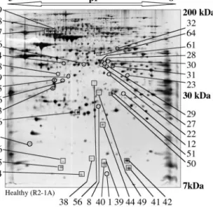
A major limitation in establishing mt proteomes resides in the difficulty of organelle purification and thus in the possible presence of lysosomal, peroxisomal, and cytosolic proteins in mt preparations. This is indeed a drawback when searching for the exact exhaustive protein content within a given organelle, a situation different from the aim of the present study. Here the comparative proteomic strategy was applied to highlight proteins undergoing quantitative changes when comparing wild-type and MERRF mitochondria. Due to the large amounts of protein required per analysis and to the low yield of mito-chondria from adherent cybrid cell growth, purification of mi-tochondria was limited to differential centrifugations, so that contaminants on two-dimensional maps were not unexpected. Two-dimensional PAGE of Total Mitochondrial Proteins and Quantitative Aspects—Well resolved silver-stained two-dimen-sional maps of mt proteins were obtained in a reproducible manner according to procedures established previously (21) and allowed detection of about 800 spots (Fig. 1). Quantifica-tion was performed on 11 gel couples to allow for a significant analysis by comparison of spot intensities, measured as spot volumes. Normalization of gels (loaded with the same amount of mt proteins), done either according to the total quantity in valid spots or to the total density in gel, leads to the same results and allowed differences linked to experimental proce-dures (protein loading and staining) to be eliminated so that only significant variations related to the type of sample (wild-type or mutant) were considered. For the same spot, intensity varied on average by 20 – 40% over the different gels of the same type (i.e. within all wild-type or all mutant gels). Accord-ingly, spot intensity variations greater than these values were
Proteomics on MERRF Cybrids
24316
by guest, on March 9, 2011
www.jbc.org
sought when comparing normalized wild-type and MERRF-related two-dimensional maps. Change by a factor 2 was cho-sen as the basis for systematic comparison.
The majority of the spots (about 760) did not show significant intensity variations between wild-type and mutant gels. Alto-gether, 38 spots (4.75% of detectable spots) were found to be affected in intensity, 19 of which decreased and 19 increased. They are located all over the two-dimensional gels, covering small as well as large molecular weight domains and all the pI ranges considered (Fig. 1). Fig. 2, A and B, summarizes quan-titative variations of these individual spots as an outcome of the 11 independent experiments. The range of differential spot intensities between wild-type and MERRF mitochondria varied from 2.1- to 9-fold, with most spots affected 2–5-fold. The ma-jority of affected spots had low expression levels, with 27 spots out of 38 with intensities under 700 pixels, the highest inten-sity reaching 3700 pixels in both types of samples.
Spot Assignment—Differentially expressed protein spots as well as a number of non-affected spots were assigned by mass spectrometric (MS) techniques. Identification (in 2–3 independ-ent experimindepend-ents) was mainly done by MALDI-TOF on colloidal blue-stained gels, based on a minimum of 3 or 4 matching peptides with a peptide mass tolerance of 50 ppm. A typical case is shown in Fig. 3. Nano-LC-MS-MS, an approach leading to the amino acid sequence of a given peptide, was used as a complementary technique in some instances (Fig. 4). For some spots, identification was not successful (lack of signal for spots of low density, recovery of only a few peptides for proteins of low molecular weight, proteins poor in trypsin cleavage sites, low efficiency of peptide extraction from gel, etc.).
Table I lists identified spots. Most proteins correspond to nuclear encoded mt proteins, but some were cytosolic contam-inants. All proteins of expected mt location appeared at gel coordinates in the range of molecular weight and pI values of their mature forms, and no peptides issued from the targeting
signals were detected by MALDI analyses. Differences between theoretical and experimental pI were accounted for either by distortions in the pH gradient or by post-translational modifi-cations of the corresponding protein (see below).
Spots with decreased intensities in MERRF mitochondria as compared with wild-type mitochondria were assigned princi-pally as nuclear encoded subunits of respiratory chain com-plexes, especially from complex IV or cytochrome c oxidase (COX VIa (spot 41), COX Vb (spot 42), and COX VIb (spot 34)) and complex I or NADH ubiquinone oxidoreductase (subunits 13 (spot 35) and 24 kDa (spot 46) as well as three isoforms (same molecular weight but slightly different pI values) of the 75-kDa subunit (spots 67, 68, and 69)). Furthermore, mitochon-drial ornithine aminotransferase (spot 61), a protein involved in arginine metabolism, underwent a 2.8-fold decrease in in-tensity, and 2 isoforms of peroxisomal enoyl-CoA hydratase (spots 50, 51) underwent a 2.1–3.1-fold decrease.
Within spots of increased intensity, 4 have been assigned as proteins of mt location. They correspond to subunits of the metabolic enzyme pyruvate dehydrogenase (PDH) subunit E1␣ (spot 28), an isoform of this protein (spot 31) and subunit E1 (spot 16). Several non-mitochondrial proteins such as cytosolic translation elongation-factor 1␥ (spot 30), septin 2 (spot 27), -actin (spots 13 and 15), guanine nucleotide-binding protein subunit3 (spot 13), and lysosomal tripeptidyl peptidase (spot 24) were also detected.
A series of spots showing no quantitative changes have also been assigned. They include a 75-kDa heat shock protein, sub-units of succinate dehydrogenase (complex II of the respiratory chain), isocitrate dehydrogenase, succinyl-CoA synthetase, pyr-roline-5 carboxylate reductase, prohibitin, and metaxin 2 (re-sults not shown).
Perturbations within Pyruvate Dehydrogenase E1—Spots of increased intensity in MERRF mitochondria are of particular interest because they reveal active molecular events taking place. This is mainly the case for PDH E1 subunits. The pyru-vate dehydrogenase complex plays a central and strategic role in the control and use of carbohydrates as source of oxidative energy or as precursors of fatty acid biosynthesis (28). It is formed of three enzymes, namely pyruvate dehydrogenase (PDH E1), dihydrolipoyl trans-acetylase (PDH E2), and dihy-drolipoyl dehydrogenase (PDH E3). PDH E1 is a heterotet-ramer (␣22).
To understand differences in gel coordinates of spots 28 (⬃43 kDa, pI 6.8) and 31 (⬃42 kDa, pI 6.9), assigned as the same protein PDH E1␣, the presence for post-translational modifi-cations was investigated with particular attention to possible phosphorylation. Indeed, negative charges of phosphates are the best candidates to influence the pI of proteins. New data bank searches performed with peptide masses, so far not in-cluded in the assignments, combined with screening for phos-phorylation on serine, threonine, or tyrosines fitted with the possibility that PDH E1␣ extracted from spot 28 was phospho-rylated at least at 4 positions (Ser-312, Ser-314, and two posi-tions out of Ser-275, Thr-286, and Tyr-287) and that PDH E1␣ isoform extracted from spot 31 was phosphorylated at least at 5 different positions (Thr-35, Thr-124, Thr-126, Ser-130, and Thr-139) (Table II). However, because phosphorylation lowers the pI of a protein by about 0.2 units/phosphate group, protein in spot 28, migrating at lower pI coordinates on the two-dimen-sional gel than spot 31, is likely phosphorylated to a higher extent than the protein in spot 31 (see location of spots in Fig. 1 and Fig. 5A). The definite degree of phosphorylation of each subunit escapes MS analysis because of intrinsic limitations of this technique (incomplete extraction of hydrophobic
pep-FIG. 1. Typical silver-stained two-dimensional gel
electro-pherogram of wild-type mitochondria from cybrid cells (R2-1A) and location of protein spots that undergo quantitative changes when compared with A8344G mutation-carrying mitochondria.
Mitochondria were purified from adherent cell lines and solubilized by high concentration of denaturing agents and non-ionic detergents to recover a large proportion of hydrophobic proteins. Proteins were sep-arated by isoelectrofocusing (pH 5– 8) followed by electrophoresis on a 10% polyacrylamide-SDS gel. Silver-stained gels were scanned and quantified using PD-Quest software. Spots that display a greater than 2-fold decrease are indicated by open squares, and those displaying a greater than 2-fold increase are shown by open circles. Spot numbers at the sides of the gel are also relevant for later figures.
by guest, on March 9, 2011
www.jbc.org
tides from gels, non-detectable peptides) and remains so far unknown.
Although in wild-type mitochondria intensities of spot 16 (subunit) and spots 28 ⫹ 31 (subunits ␣) fit with a 1 to 1 stoichiometry, this is not the case in MERRF mitochondria (Fig. 2). Thus, additional spots corresponding to alternative
isoforms of PDH E1 subunits were suspected to be present in two-dimensional gels. To investigate this possibility further, two-dimensional gels with healthy and MERRF mt proteins were submitted to Western analysis in the presence of antibod-ies directed against the PHD E1 (␣22) (Fig. 5, B and C). This approach, which is much more sensitive than silver staining,
FIG. 2. Detailed quantitative view
of variations in spot intensity be-tween wild-type and MERRF mu-tation-carrying mitochondria. Only
spots exhibiting greater than 2-fold changes in their quantity are reported. A, histogram of the 19 spots with decreased intensities in mutant mitochondria. Ra-tios of intensities in wild-type/MERRF mitochondria (wt/mt) are indicated. The range of decrease in spot intensities var-ies between 2.1- and 9.0-fold. B, histo-gram of the 19 spots of increased intensi-ties in MERRF mitochondria as compared with wild-type mitochondria. Here ratios of spot intensities in mutant over wild type are given. The range of spot intensity increment varies between 2.4- and 8.3-fold. Bars for wild-type mitochondria are indicated in white, and bars for mutant samples are indicated in black. Mean val-ues of 11 independent experiments with corresponding standard errors are given.
FIG. 3. Representative result of MALDI-TOF mass spectrometric assignment. A, MALDI-TOF mass spectrum of in-gel tryptic peptides, extracted from spot 31, identified as pyruvate dehydrogenase E1␣ subunit. Masses of the matching peptides allowing for identification are indicated, and masses of phosphopeptides are underlined. B, linear representation of the polypeptide chain with matching peptides indicated in
gray. The protein sequence coverage was 42%. Amino acids forming the mt signal peptide are indicated at the N terminus of the sequence by a zig-zag line. No matching peptides were found in this domain, in support to the absence of the targeting signal of the protein analyzed.
Proteomics on MERRF Cybrids
24318
by guest, on March 9, 2011
www.jbc.org
indeed revealed the presence of additional spots aligned either with spots 28 –31 or with spot 16. MALDI-TOF analysis of one spot in each of the two domains of interest in a corresponding colloidal blue-stained gel (the only spots seen on blue gels, namely spots 9 and 20), confirmed an additional subunit␣ (spot 20, with 39% sequence coverage) and subunit (spot 9, with 52% sequence coverage), respectively, both with phosphoryla-tion sites (not shown). Thus, the distribuphosphoryla-tion of PDH E1
sub-units into different isoforms is strongly perturbed in MERRF mitochondria.
Comparison of Western blots showed not only large qualita-tive but also significant quantitaqualita-tive differences (Fig. 5, B and C). Complementary analysis by Western blotting on a one-dimensional 10% polyacrylamide/SDS gel revealed that␣ and  subunits are present in equimolar amounts in both wild-type and mutated samples (not shown) and that the total quantity of
FIG. 4. Representative result of a nano-LC-MS-MS analysis. A, combined spectrum (retention time from 15 to 50 min) of in-gel tryptic digested peptides of spot 22, identified as the mt 28 S ribosomal protein MRP-S22. Masses of peptides allowing identification are indicated. B, linear representation of the polypeptide chain with sequences of matching peptides. Total sequence coverage was 16%. No match for the mt target signal (zigzag line) was found.
TABLE I
Assignment of proteins in spots undergoing quantitative changes
Spots are listed according to numbers shown in Fig. 1. Two classes of spots are distinguished:2, spots with decreased intensities in MERRF samples as compared with wild-type mitochondria;1, spots with increased intensities in mutant samples. Protein assignment was done by mass spectrometric approaches. Variations in spot intensities are expressed as ratios of either wild-type/mutant (wt/mt) or mt/wt. Names of protein, SwissProt data bank accession numbers, subcellular localization, theoretical molecular weights or isoelectric points (calculated for the mature forms of the proteins), and % of mass coverage for peptides allowing for assignment, are indicated. Loc., subcellular localization; Mt, mitochondria; C, cytosol; L, lysosome; Px, peroxisome.
Experimental Data bank exploration Spot
Number Ratio Molecularmass/pI Protein name Accessionno. Loc. Molecularmass pI coverageMass 2 Decrease in intensity
kDa Da
35 ⫺2.7 12/6.05 NADH/ubiquinone oxidoreductase, 3-kDa subunit Q16718 Mt 13.327 5.77 68 46 ⫺2.7 25/6.2 NADH/ubiquinone oxidoreductase, 24-kDa subunit P19404 Mt 23.760 5.71 45 67 ⫺3.6 78/5.6 NADH/ubiquinone oxidoreductase, 75-kDa subunit P28331 Mt 77.053 5.33 37 68 ⫺2.6 78/5.7 NADH/ubiquinone oxidoreductase, 75-kDa subunit P28331 Mt 77.053 5.33 28 69 ⫺3.3 78/5.8 NADH/ubiquinone oxidoreductase, 75-kDa subunit P28331 Mt 77.053 5.33 52 42 ⫺6.1 16/7.15 Cytochrome c oxidoreductase, subunit Vb P10606 Mt 10.613 6.74 34 41 ⫺9.0 13/7.2 Cytochrome c oxidoreductase, subunit VIa Q02221 Mt 9.750 6.97 62 34 ⫺7.8 10/6.4 Cytochrome c oxidoreductase, subunit VIb P14854 Mt 10.061 6.79 65 61 ⫺2.8 44/6.7 Ornithine aminotransferase P04181 Mt 44.808 5.72 28 50 ⫺3.1 33/6.8 Enoyl CoA hydratase Q08426 Px 35.816 8.07 22 51 ⫺2.1 34/6.9 Enoyl CoA hydratase Q08426 Px 35.816 8.07 21 1 Increase in intensity
28 ⫹8.3 43/6.8 Pyruvate dehydrogenase subunit E1␣ P08559 Mt 40.229 6.51 28 31 ⫹3.4 41/6.9 Pyruvate dehydrogenase subunit E1␣ P08559 Mt 40.229 6.51 42 16 ⫹2.4 35/6.0 Pyruvate dehydrogenase subunit E1 P11177 Mt 35.890 5.38 31 22 ⫹5.8 38/6.8 Mt ribosomal protein 28 S subunit S22 P82650 Mt 41.280 7.70 31 30 ⫹7.5 47/6.8 Elongation factor-1␥ P26641 C 50.119 6.25 28 15 ⫹4.8 38/5.85 -Actin P02570 C 41.053 5.56 44 13 ⫹4.0 35/5.8 -Actin⫹ guanine nucleotide-binding protein
subunit3 P02570⫹ C 41.053 5.56 44 P16520 C 37.221 5.39 37 27 ⫹3.9 43/6.7 Septin 2 Q15019 C 41.487 6.15 57 24 ⫹4.8 43/6.05 Lysosomal tripeptidyl peptidase O14773 L 39.790 5.61 35
by guest, on March 9, 2011
www.jbc.org
PDH E1 subunits increased about 2-fold in MERRF samples compared with wild type.
PDH E1 activity is under control of phosphorylation/dephos-phorylation at well defined sites of E1␣ (28). Both forms (phos-phorylated and non-phos(phos-phorylated) co-exist in a given cell and allow for sensitive regulation by shifting their balance. It is possible to measure basal activity, representative of the non-phosphorylated population of enzyme, and total activity subse-quent to dephosphorylation by calcium and magnesium activa-tion of phosphatases (24). Neither basal nor total activities are
significantly different in wild-type and MERRF mitochondria in the presence of pyruvate as substrate (Table III).
DISCUSSION
Comparative proteomics, combining high resolution of pro-teins with mass spectrometry techniques for rapid assignment, have presently been recognized as a powerful tool for the in-vestigation of a number of human diseases (e.g. Refs 17, 29, and 30) including mt disorders (31–33). Further, proteomic maps of mammalian mitochondria become progressively presented in the case of human placenta (34) and rat liver (35).
Couples of cybrid cell lines form a valuable model system to investigate biochemical and molecular consequences of a mu-tation in the mt genome because they possess the same nuclear background and differ by a single point mutation (36). Osteo-sarcoma-derived cybrids were used previously to study the physiological, biochemical, and molecular effects of the A8344G mutations in the mt tRNALysgene which is correlated to the
maternally inherited MERRF syndrome (22). MERRF cells have a decreased growth rate, lower oxygen consumption, and reduced cytochrome c oxidase activity and ATP synthesis, so that the cell undergoes a severe energy deficit (25, 27). At the molecular level, mt protein synthesis is severely impaired, and the steady-state level of the 13 mitochondria encoded proteins is decreased according to their lysine content (26). Initial com-parative proteomics on these cybrid cell lines revealed that at least two nuclear encoded proteins of mt location (COX sub-units Va and VIb) have severely decreased steady-state levels (21). The fate of the 13 mitochondria-encoded proteins cannot be explored by two-dimensional gel analysis due to their hy-drophobicity and/or their extreme pI (34). The present work, exploring silver-stained gels on a computer-assisted basis, en-larges the knowledge of the fate of about 800 proteins contained in MERRF mitochondria compared with wild type.
Variations in Steady-state Levels of Non-mitochondrial Pro-teins—Assignment of regulated spots as non-mitochondrial proteins was not unexpected because mitochondria were not purified to completion. Several of these proteins are eliminated when mitochondria are further purified on a metrizamide gra-dient before their analysis on two-dimensional gels (results not shown). However, the very low yield of this purification proce-dure prevented its systematic use in the present work. The fact that certain contaminants systematically co-purify with
al-TABLE III
Activity of pyruvate dehydrogenase in wild-type and MERRF mutation containing mitochondria (pmol/min/mg
of total cellular proteins)
Each activity was measured in triplicate on cell extracts pooled from three cell cultures.
Wild type MERRF Basal activity 187 141 Total activity 641 673
FIG. 5. Detection of pyruvate dehydrogenase E1 subunit
iso-forms by Western blotting. A, detail of a silver-stained
two-dimen-sional gel of MERRF mutation-carrying mitochondria with spots as-signed by MALDI-TOF as PDH E1 subunits␣ (spots 28 and 31) and subunit (spot 16) indicated. B and C, Western blots of the same domain of non-stained two-dimensional gels performed with antibodies directed against PDH E1 holoenzyme. B corresponds to a wild-type mt sample, and C corresponds to a MERRF mitochondria. Immunoreactiv-ity for subunit resulted mainly in one major spot in wt (spot 9) and four spots in mutant samples. Immunoreactivity for␣ subunits resulted in a major spot (20) in wt, and to a large row of spots of the same molecular weight (about 40 kDa) in the mutant sample. Within all these newly detected spots, two were detectable on colloidal blue-stained two-dimensional gels (9 and 20) and have been further analyzed. MALDI-TOF measurements support the assignment of phosphorylated isoforms.
TABLE II
Phosphorylated peptides in isoforms of pyruvate dehydrogenase E1 subunits
Masses of phosphorylated peptides are shown according to experimental data by MALDI-TOF (Mrobserved), to data bank derived values (Mr
expected), and to “normalized” data (Mrcalculated). Phosphorylation sites are underlined in peptide sequences, and the number of
phosphoryla-tions is indicated. The peptides reported here probably correspond only to a subset of phosphorylation sites. Tryptic peptides corresponding to regulatory sites already reported for the enzyme are not detected by MALDI-TOF MS, probably due to their high hydrophobicity and acidic pI.
Mrobserved Mrexpected Mrcalculated Amino acids Peptide sequence No. of phosphates
Spot 28 (E1␣) 1307.63 1306.62 1306.56 312-321 SKSDPIMLLK 2 1768.77 1767.76 1767.77 275-288 SGKGPILMELQTYR 2 Spot 31 (E1␣) 1477.82 1476.81 1476.66 29-40 NFANDATFEIKK 1 1673.97 1672.96 1672.79 128-141 GLSVREILAELTGR 2 1688.73 1687.72 1687.67 120-132 AHGFTFTRGLSVR 3
Proteomics on MERRF Cybrids
24320
by guest, on March 9, 2011
www.jbc.org
tered proportions in MERRF mt samples suggests that their detection is significant and may have a link with the patholog-ical status of the whole cell. They may be linked to biochempatholog-ical perturbations (e.g. acidification of the cytosol, change in ionic concentrations, and variation in mt membrane potential (37– 39)) leading to alterations in physicochemical properties of these proteins. Some cytosolic proteins may become bound more or less tightly to the outer mt membrane. Alternatively, detection of quantitative changes in cytosolic proteins may reflect true molecular long range effects of the mt tRNA gene mutation such as overexpression or down-regulation of cytoso-lic proteins. This awaits further experimentation.
Decrease in Nuclear Encoded Subunits of Respiratory Chain Complexes—Among the 38 protein spots found to be quantita-tively affected, 6 were assigned as nuclear encoded subunits of the respiratory chain complexes, i.e. subunits of complex I and of complex IV. Interestingly, those affected subunits from the same complex were decreased by the same factor, i.e. about 7.5-fold for complex IV subunits and about 3-fold for complex I subunits.
Complexes of the mt respiratory chain (with the exception of complex II) are composed of both mt and nuclear encoded subunits assembled into functional entities in the inner mt membrane. The stoichiometric assembly of these complexes necessarily depends on a well organized nucleo-mitochondrial communication allowing for fine-tuning of the expression levels of the corresponding genes in both genomes (40, 41). Although a strong decrease in steady-state levels of any nuclear encoded mt protein could result from different molecular regulatory events taking place either at the gene expression level (tran-scription and translation) or at the mt import level, it is likely, as suggested earlier (21), that the decreases observed here for nuclear respiratory chain subunits are the direct consequence of their degradation. Indeed, because mtDNA encoded subunits are synthesized at a lower rate, as a direct consequence of the tRNA mutation (26), their nuclear partners likely remain non-assembled and are therefore degraded. Such a situation has been demonstrated for COX nuclear encoded subunits in cell lines deprived of mtDNA or where mt translation has been inhibited (42– 46). Interestingly, subunits from complex II (suc-cinate dehydrogenase), which are all nuclear encoded and are thus not linked to mt translation, were not found to be regu-lated in the present work.
The five mt respiratory chain complexes are formed from 83 different subunits, of which only 13 are of mt origin. Only 6 of the 70 nuclear encoded subunits were found to be down-regu-lated here. The fate of the 64 remaining subunits is thus uncertain. Calculation of theoretical molecular weight and pI values for each subunit revealed that at least 22 of them should have coordinates within the two-dimensional gels studied in the present work. Their absence could reflect either their mem-brane location and thus difficulty to be extracted (in the x-ray structure of complex IV (47), the three subunits detected here are located on the matrix and intermembrane surfaces of the enzyme rather than within the membrane), their presence within spots which could not be assigned, or that they are not regulated or are regulated below a 2-fold factor.
Variation in Steady-state Levels of Proteins Involved in Mi-tochondrial Translation—Due to impairment of mt translation by mutation A8344G in the tRNALysgene, effects on proteins
involved in the translational machinery may be affected. Only one ribosomal protein, MRP-S22, was found in increased quan-tity in MERRF mitochondria (about 6-fold). The regulation of the steady-state level of only one of the⬃80 proteins which form the mt ribosome is surprising. Protein MRP-S22 belongs to a new class of ribosomal proteins, specific to mammalian
ribosomes (48, 49). This protein may well have an additional alternative function, so far unknown, as observed for other newly discovered mt proteins. MRP-S29 is a death-associated protein involved in apoptosis (50), and MRP-S31 is associated with type 1 diabetes (51). To explore further possible variations in proteins belonging to the mt translational machinery, West-ern blotting with antibodies directed against human lysyl-tRNA synthetase (the enzyme which catalyzes aminoacylation of tRNALys, the tRNA bearing the MERRF mutation
investi-gated here) and against mt translation elongation factor Tu was performed. No significant changes in the steady-state lev-els of these two particular proteins were found (not shown).
Variations in Steady-state Amounts of Metabolic Enzymes— Two metabolic enzymes were found to be affected in MERRF cells. Mitochondrial ornithine aminotransferase decreased, and pyruvate dehydrogenase subunits E1␣ and E1 increased. Or-nithine aminotransferase participates in amino acid metabo-lism, catalyzing inter-conversion of ornithine and glutamate. The contribution of the decrease of ornithine aminotransferase to the pathological status of the cybrid cells remains unclear. However, reduced activity of the respiratory chain correlates directly with reduced activity of the Krebs cycle, which in turn is linked via ␣-ketoglutarate to glutamate. Interestingly, a reduced activity of this enzyme was observed previously in the brains of Huntington’s disease-affected patients. Huntington’s disease is associated with the deficiency of the neurotransmit-ter glutamate and of mt oxidative phosphorylation (52).
The pyruvate dehydrogenase complex converts pyruvate into acetyl-CoA and has thus a central role in energy metabolism. Comparative proteomics associated with Western analysis re-vealed that (i) the total amount of PDH E1 subunits increased about 2-fold in MERRF mitochondria, and (ii) both subunits␣ and of this enzyme are distributed over a series of isoforms in the wild-type sample, but this distribution is wider in the mutated sample. MALDI-TOF assignments are in favor of a variation in the number and distribution of phosphorylation sites as post-translational modifications in the different iso-forms. The detected phosphorylation sites differ from those involved in regulation of PDH E1 activity (53) in agreement with maintenance of the same enzymatic PDH activity in both wild-type and MERRF cell lines. Whereas full understanding of biochemical and molecular perturbations of the PDH complex and their relationships with the presence of the MERRF mu-tation in the mt genome awaits further experiments, it is important to notice that the results of the present investigation do not preclude on the activity of the PDH complex in vivo. Indeed, in addition to phosphorylation/dephosphorylation at three specific sites, the activity of the complex can be alloster-ically regulated by NADH and acetyl-CoA (28). Both com-pounds accumulate in mitochondria as a consequence of respi-ratory deficiency. As an outcome of the present data, it is tempting to speculate that accumulation of the enzyme may represent a compensatory mechanism by the cell to overcome the energetic deficiency but that parallel activation of kinase(s) hinders effective changes in activity. Interestingly, a case of up-regulation of the amounts of PDH E1 proteins has been reported recently for butyrate-treated human colon cancer cells (54).
Expectation for Further Variations—Comparative proteom-ics allowed for an enlarged view of quantitative and qualitative changes in about 800 protein spots observed on two-dimen-sional gels from wild-type mitochondria and mitochondria con-taining a single point mutation in a tRNA gene. The present analysis revealed 38 affected spots, of which 20 could be as-signed, and about 760 unaffected spots. As a major outcome, mitochondria with mutation A8344G in the tRNALysgene show
by guest, on March 9, 2011
www.jbc.org
significant perturbations in the steady-state amounts of pro-teins involved in translation (MRP-S22), respiration (respira-tory chain subunits), and carbohydrate and amino acid metab-olism (PDH, ornithine aminotransferase). Also a series of proteins of cytosolic location were reproducibly found to be affected (either increased or decreased). These results demon-strate that the presence of a single point mutation in an mt tRNA gene not only affects mt translation but also several categories of nuclear encoded proteins, both of mt and cytosolic location. Possible links between these proteins and the muta-tion in an mt tRNA gene are schematized in Fig. 6.
However, it must also be stated that the number of changes observed in the MERRF cell line is rather limited in regard of the severity of the physiological and energetic perturbations taking place. The observed quantitative variations reveal only the strongest intensity changes taking place (variations of 2.1– 9-fold) and certainly correspond only to a subset of molecules that undergo perturbations in the disease-related cells. Sensi-tive quantitaSensi-tive variations within a 2-fold range may be sig-nificant biologically and relevant. They escape the present analysis, however, due to limited accuracy and reproducibility. As discussed above, the fate of many respiratory chain complex subunits escaped analysis. Furthermore, changes in enzymes involved in the glycolytic pathway or those involved in other energy-related pathways would be expected to be affected. Thus, it is likely that only the “tip of the iceberg,” in regard to regulatory events taking place in mitochondria, was detected so far. Many proteins may well have escaped the present anal-ysis due to technical limitations including limited sensitivity of silver-stained gels for poorly represented proteins, analysis of basic and hydrophobic proteins, and difficulty in assignment of some proteins (especially those of low molecular weight). Re-cent developments in proteomic technology (e.g. Refs. 55–59) should allow some of these difficulties to be overcome and lead
to more precise comparative analyses. Also comparison of the same quantity of proteins in either wild-type or mutation-carrying mitochondria, as done here, does not exclude the pos-sibility of larger cellular changes such as regulation in the biogenesis of mitochondria per cell, for example. The two typ-ical mt enzymatic activities measured for both cell lines (citrate synthase and pyruvate dehydrogenase) argue, however, against this possibility. Finally, one should keep in mind that regulation of enzymatic activities does not necessarily involve quantitative variations of their steady-state levels but can be controlled by other biochemical and biophysical means.
Perspectives—The present work used osteosarcoma cybrid cell lines as a model system to explore long range effects of a single mt tRNA gene mutation on the global mt proteome, outside the frame of mt translation. The initial results reported here clearly show the occurrence of such events and open new questions on the molecular mechanisms linking all events. Indeed, it remains to be explored if the observed effects are specifically related to the A8344G mutation or if this is general for other tRNA mutations. Initial studies on cybrid cell lines carrying mutation A3243G in the tRNALeu(UUR) gene
repre-senting a model system of the Mitochondrial Epilepsy, Lactic Acidosis, and Stroke-like episode syndrome already revealed a decrease in two COX subunits, as is the case with the MERRF mutation (21). Furthermore, systematic analysis of cell lines with different nuclear backgrounds for the same tRNA muta-tion (e.g. mutamuta-tion 8344 in myeloblasts or fibroblasts) should allow one to discriminate between mutation-specific and non-specific perturbations and to decipher the contribution of the nuclear background. The present proteomic investigation, which so far uncovered several unsuspected new players that participate in the disease status of the cell, opens new territory for molecular and biochemical investigation. Their exploration will shed light on the intricate mechanistic pathways linking a
FIG. 6. Schematic representation of the most dramatic perturbations observed by comparative proteomics on two-dimensional
silver-stained gels in mitochondria harboring tRNALys
gene mutation A3243G. Mt and nuclear gene expression machinery are considered,
and their convergence at the level of respiratory chain complexes is illustrated. Major relevant energetic metabolic pathways and their connections to the respiratory chain are indicated. Decreases and increases in protein spots as seen on two-dimensional gels are indicated by arrows. Decreases in the 13 mt encoded subunits of the respiratory chain complexes as described (e.g. Ref. 26) were not observed on two-dimensional gels due to their hydrophobicity.
Proteomics on MERRF Cybrids
24322
by guest, on March 9, 2011
www.jbc.org
single point mutation in the mt tRNA gene to phenotypic ex-pression of mitochondrial diseases.
Acknowledgments—We are grateful to T. Rabilloud and J. Lunardi
for their constructive suggestions on the two-dimensional gel technol-ogy and on mitochondrial dysfunction. A. Chomyn and G. Attardi are acknowledged for providing human cybrid cell lines, and G. Lindsay, L. Spremulli, and K. Shiba are acknowledged for the generous gifts of antibodies. We thank C. Prip-Buus for fruitful discussions, M. Sissler for critical comments on the manuscript, L. Levinger for language improvements, and Caroline Paulus for excellent technical assistance.
REFERENCES
1. Scheffler, I. (1999) Mitochondria, John Wiley & Sons, Inc., New York 2. Kogelnik, A. M., Lott, M. T., Brown, M. D., Navathe, S. B., and Wallace, D. C.
(1998) Nucleic Acids Res. 26, 112–115
3. Ingman, M., Kaessmann, H., Pa¨a¨bo, S., and Gyllenstein, U. (2000) Nature 408, 708 –713
4. Servidei, S. (2001) Neuromuscul. Disord. 11, 508 –513
5. Schon, E. A., Bonilla, E., and DiMauro, S. (1997) J. Bioenerg. Biomembr. 29, 131–149
6. Wallace, D. C. (1999) Science 283, 1482–1488
7. Chinnery, P. F., and Turnbull, D. M. (2000) Mol. Med. Today 6, 425– 432 8. DiMauro, S., and Andreu, A. (2000) Brain Pathol. 10, 431– 441
9. So¨ll, D., and RajBhandary, U. L. (eds) (1995) tRNA: Structure, Biosynthesis,
and Function., American Society for Microbiology, Washington, D. C.
10. Chomyn, A. (1998) Am. J. Hum. Genet. 62, 745–751
11. Chomyn, A., Enriquez, J. A., Micol, V., Fernandez-Silva, P., and Attardi, G. (2000) J. Biol. Chem. 275, 19198 –19209
12. Florentz, C. (2002) Biosci. Rep. 22, 81–98
13. Florentz, C., and Sissler, M. (2003) in Translation Mechanisms (Lapointe, J., and Brakier-Gingras, L., eds), Landes Biosciences, Georgetown, TX, in press
14. Jacobs, H. T., and Holt, I. J. (2000) Hum. Mol. Genet. 9, 463– 465
15. Janssen, G., Maassen, J., and van den Ouweland, J. (1999) J. Biol. Chem. 274, 29744 –29748
16. Blackstock, W. P., and Weir, M. P. (1999) Trends Biotechnol. 17, 121–127 17. Celis, J., Kruhoffer, M., Gromova, I., Frederiksen, C., Ostergaard, M.,
Thykjaer, T., Gromov, P., Yu, J., Palsdottir, H., Magnusson, N., and Orntoft, T. (2000) FEBS Lett. 480, 2–16
18. Jacobs, H. T. (2001) Trends Genet. 17, 653– 660
19. Larsson, N., and Rustin, P. (2001) Trends Mol. Med. 7, 578 –581 20. Silva, J., and Larsson, N. (2002) Biochim. Biophys. Acta 1555, 106 –110 21. Rabilloud, T., Strub, J. M., Carte, N., Luche, S., Van Dorsselaer, A., Lunardi,
J., Giege´, R., and Florentz, C. (2002) Biochemistry 41, 144 –150 22. Shoffner, J., Lott, M., Lezza, A. M. S., Seibel, P., Ballinger, S. W., and Wallace,
D. C. (1990) Cell 61, 931–937
23. Helm, M., Florentz, C., Chomyn, A., and Attardi, G. (1999) Nucleic Acids Res.
27, 756 –763
24. Clot, J., Benelli, C., Fouque, F., Rozenn, B., Durand, D., and Postel-Vinay, M. (1992) J. Clin. Endocrinol. Metab. 74, 1258 –1262
25. Chomyn, A., Meola, G., Bresolin, N., Lai, S. T., Scarlato, G., and Attardi, G. (1991) Mol. Cell. Biol. 11, 2236 –2244
26. Enriquez, J. A., Chomyn, A., and Attardi, G. (1995) Nat. Genet. 10, 47–55 27. Villani, G., and Attardi, G. (1997) Proc. Natl. Acad. Sci. U. S. A. 94, 1166 –1171 28. Patel, M., and Korotchkina, L. (2001) Exp. Mol. Med. 33, 191–197 29. Banks, R. E., Dunn, M. J., Hochstrasser, D. F., Sanchez, J. C., Blackstock, W.,
Pappin, D. J., and Selby, P. J. (2000) Lancet 56, 356 –1749
30. Chambers, G., Lawrie, L., Cash, P., and Murray, G. I. (2000) J. Pathol. 8, 192–280
31. Scheffler, N., Miller, S., Carroll, A., Anderson, C., Davis, R., Ghosh, S., and Gibson, B. (2001) Mitochondrion 1, 161–179
32. Mitsumoto, A., Takeuchi, A., Okawa, K., and Nakagawa, Y. (2002) Free Radic.
Biol. Med. 32, 22–37
33. Lopez, M. F., and Melov, S. (2002) Circ. Res. 90, 380 –389
34. Rabilloud, T., Kieffer, S., Procaccio, V., Louwagie, M., Courchesne, P., Patterson, P., Martinez, P., Garin, J., and Lunardi, J. (1998) Electrophoresis
19, 1006 –1014
35. Fountoulakis, M., Berndt, P., Langen, H., and Suter, L. (2002) Electrophoresis
23, 311–328
36. King, M. P., and Attardi, G. (1989) Science 246, 500 –503
37. Asoh, S., Mori, T., Hayashi, J.-I., and Ohta, S. (1996) J. Biol. Chem. 120, 600 – 607
38. Antonicka, H., Floryk, D., Klement, P., Stratilova, L., Hermanska, J., Houstkova, H., Kalous, M., Drahota, Z., Zeman, J., and Houstek, J. (1999)
Biochem. J. 342, 537–544
39. Mirabella, M., Di Giovanni, S., Silvestri, G., Tonali, P., and Servidei, S. (2000)
Brain 123, 93–104
40. Coenen, M., van den Heuvel, L., and Smeitink, J. A. (2001) Curr. Opin. Neurol.
14, 777–781
41. Garesse, R., and Vallejo, C. (2001) Gene (Amst.) 263, 1–16
42. Nijtmans, L., Spelbrink, J. N., Van Galen, M. J. M., Zwaan, M., Klement, P., and Van den Bogert, C. (1995) Biochim. Biophys. Acta 1265, 117–126 43. Nijtmans, L., Taanman, J.-W., Muijsers, A. O., Speijer, D., and Van den
Bogert, C. (1998) Eur. J. Biochem. 254, 389 –394
44. Bentlage, H. A., Janssen, A. J., Chomyn, A., Attardi, G., Walker, J. E., Schagger, H., Sengers, R. C., and Trijbels, F. J. (1995) Biochim. Biophys.
Acta 1234, 63–73
45. Bai, Y., Shakeley, R., and Attardi, G. (2000) Mol. Cell. Biol. 20, 805– 815 46. Chomyn, A. (2001) J. Bioenerg. Biomembr. 33, 251–257
47. Tsukihara, T., Aoyama, H., Yamashita, E., Tomisaki, T., Yamaguchi, H., Shinzawa-Itoh, K., Nakashima, R., Yaono, R., and Yoshikawa, S. (1996)
Science 272, 1136 –1144
48. Cavdar Koc, E., Burkhart, W., Blackburn, K., Moseley, A., Koc, H., and Spremulli, L. L. (2001) J. Biol. Chem. 276, 19363–19374
49. Cavdar Koc, E., Burkhart, W., Blackburn, K., Moyer, M. B., Schlatzer, D. M., Moseley, A., and Spremulli, L. L. (2001) J. Biol. Chem. 276, 43958 – 43969 50. Kissil, J., Cohen, O., Raveh, T., and Kimchi, A. (1999) EMBO J. 18, 353–362 51. Arden, S., Roep, B., Neophytou, P., Usa, E., Duinkerken, G., and de Vries, R.
(1996) J. Clin. Invest. 97, 551–561
52. Wong, P. T., McGeer, P. L., Rossor, M., and McGeer, E. G. (1982) Brain Res.
231, 466 – 471
53. Kolobova, E., Tuganova, A., Boulatnikov, I., and Popov, K. M. (2001) Biochem.
J. 358, 69 –77
54. Tan, S., Seow, T., Liang, R., Koh, S., Lee, C., Chung, M., and Hooi, S. (2002)
Int. J. Cancer 98, 523–531
55. Gygi, S., Rist, B., Gerber, S., Turecek, F., Gelb, M., and Aebersold, R. (1999)
Nat. Biotechnol. 17, 994 –999
56. Gygi, S., Rist, B., and Aebersold, R. (2000) Curr. Opin. Biotechnol. 11, 396 – 401 57. Smolka, M., Zhou, H., and Aebersold, R. (2002) Mol. Cell. Proteomics 1, 19 –29 58. Lin, T.-K., Hughes, G., Muratovska, A., Blaikie, F. H., Brookes, P. S., Darley-Usmar, V., Smith, R. A. J., and Murphy, M. P. (2002) J. Biol. Chem. 277, 17048 –17056
59. Pflieger, D., Le Caer, J., Lemaire, C., Bernard, B.-A., Dujardin, G., and Rossier, J. (2002) Anal. Chem. 74, 2400 –2406
by guest, on March 9, 2011
www.jbc.org