HAL Id: inserm-00290139
https://www.hal.inserm.fr/inserm-00290139
Submitted on 24 Jun 2008HAL is a multi-disciplinary open access archive for the deposit and dissemination of sci-entific research documents, whether they are pub-lished or not. The documents may come from teaching and research institutions in France or abroad, or from public or private research centers.
L’archive ouverte pluridisciplinaire HAL, est destinée au dépôt et à la diffusion de documents scientifiques de niveau recherche, publiés ou non, émanant des établissements d’enseignement et de recherche français ou étrangers, des laboratoires publics ou privés.
deficient mice
Anika Vaarmann, Dominique Fortin, Vladimir Veksler, Iman Momken, Renée
Ventura-Clapier, Anne Garnier
To cite this version:
Anika Vaarmann, Dominique Fortin, Vladimir Veksler, Iman Momken, Renée Ventura-Clapier, et al.. Mitochondrial biogenesis in fast skeletal muscle of CK deficient mice: Mitochondrial biogenesis in CK deficient mice. Biochimica et Biophysica Acta (BBA) - Reviews on Bioenergetics, Elsevier, 2008, 777 ((1)), pp.39-47. �inserm-00290139�
Mitochondrial biogenesis in fast skeletal muscle of CK deficient mice.
Vaarmann A1,2,3, Fortin D1,2, Veksler V1,2, Momken I1,2, Ventura-Clapier R1,2, Garnier A1,2
1
INSERM, U-769, F-92296 Châtenay-Malabry, France;
2
Univ Paris-Sud, IFR141, F-92296 Châtenay-Malabry, France;
3
Department of Pharmacology, Center of Molecular and Clinical Medicine, University of Tartu, Estonia
Address for correspondence:
Anne Garnier,
INSERM, U769, Université Paris-Sud, 5 rue Jean-Baptiste Clément
F-92296 Châtenay-Malabry France.
Tel: (331)46.83.57.63. Fax: (331)46.83.54.75.
E-mail: anne.garnier@cep.u-psud.fr
Abstract
Creatine kinase (CK) is a phosphotransfer kinase that catalyzes the reversible transfer of a phosphate moiety between ADP and creatine and that is highly expressed in skeletal muscle. In fast glycolytic skeletal muscle, deletion of the cytosolic M isoform of CK in mice (M-CK-/-) leads to a massive increase in the oxidative capacity and of mitochondrial volume. This study was aimed at investigating the transcriptional pathways leading to mitochondrial biogenesis in response to CK deficiency. Wild type and M-CK-/- mice of eleven months of age were used for this study. Gastrocnemius muscles of M-CK-/- mice exhibited a dramatic increase in citrate synthase (+120%) and cytochrome oxidase (COX, +250%) activity, and in mitochondrial DNA (+60%), showing a clear activation of mitochondrial biogenesis. Similarly, mRNA expression of the COXI (mitochondria-encoded) and COXIV (nuclear-encoded) subunits were increased by +103 and +94 % respectively. This was accompanied by an increase in the expression of the nuclear respiratory factor (NRF2α) and the mitochondrial transcription factor (mtTFA). Expression of the co-activator PGC-1α, a master gene in mitochondrial biogenesis was not significantly increased while that of PGC-1β and PRC, two members of the same family, was moderately increased (+45% and +55% respectively). While the expression of the modulatory calcineurin-interacting protein 1 (MCIP1) was dramatically decreased (minus 68%) suggesting inactivation of the calcineurin pathway, the metabolic sensor AMPK was activated (+86%) in M-CK-/- mice. These results evidence that mitochondrial biogenesis in response to a metabolic challenge exhibits a unique pattern of regulation, involving activation of the AMPK pathway.
Introduction
The family of creatine kinase isoenzymes catalyzes the reversible transfer of a phosphate moiety between creatine and ATP. It is highly expressed in striated muscles and is a key player in intracellular energy storage and transport. The major isoenzymes of CK in muscle are the cytosolic isoform (MM-CK) and the mitochondrial isoform (mi-CK). Fast gastrocnemius muscle expresses almost exclusively MM-CK [1] that is free in the cytosol or structurally associated with myofibrils and membranes of the sarcoplasmic reticulum (SR), and functionally coupled to ATPases for optimal functioning of the contractile machinery and SR calcium uptake [2].
Fast skeletal muscle fibers have very low mitochondrial content and mainly rely on quickly mobilisable energy sources (mainly phosphocreatine (PCr) and glycogen) to develop strong and fast contractions but this is possible only for short periods of time because of limited reserves. These muscles are thus quickly fatigable and should recover their energy reserves through anaerobic glycolysis and less importantly mitochondrial oxidations.
Engineered mice with invalidated expression of M-CK (M-CK-/-) were developed by
the group of Bé Wieringa [3] to understand the effects of altered energy metabolism. Functional tests revealed that fast skeletal muscle of CK-/- mice has abnormal calcium
transient, lacks burst activity at the onset of stimulation but exhibits paradoxical decreased fatigability [4,5]. This is accompanied by a marked increase in the relative mitochondrial volume, the mitochondrial enzyme content, and the muscle oxidative capacity together with a relocation of mitochondria towards myofibrils [3,6-8]. Proteomic and mRNA analysis [9,10] confirmed the metabolic remodeling of the CK-/- gastrocnemius muscle towards a more
oxidative phenotype. This metabolic remodeling is thought to compensate for the lack of creatine kinase by switching energy metabolism of gastrocnemius muscle from the use of PCr
stores and anaerobic glycolysis to oxidative metabolism. However, the signaling and molecular events governing this metabolic shift are currently unknown.
Mitochondrial biogenesis depends on the coordinated expression of the nuclear and mitochondrial genomes. Mitochondrial DNA (mtDNA), encodes 13 subunits of the oxidative phosphorylation system (OXPHOS). The remaining OXPHOS subunits as well as other mitochondrial proteins are encoded by the nuclear DNA. An inducible transcriptional co-activator termed peroxisome proliferator activated receptor gamma co-co-activator 1α (PGC-1α) has emerged as a critical factor coordinating the activation of metabolic genes required for substrate utilization and mitochondrial biogenesis [11-13]. Effects of PGC-1α on mitochondrial biogenesis could be explained via its interaction with several DNA-binding transcription factors, such as the nuclear respiratory factors (NRFs). These factors in turn upregulate the expression of nuclear genes encoding respiratory chain components and other mitochondrial proteins, as well as of the mitochondrial transcription factor A (mtTFA), a factor required for mtDNA replication and transcription [14]. In rodent skeletal muscles, regularly performed exercise induces an increase in PGC-1α, coincidently with an increase in NRFs and mtTFA mRNA and/or protein expression (for recent reviews see [15-17]). In humans, PGC-1α and its transcription cascade correlate with exercise capacity and vastus lateralis muscle oxidative capacity [18].
In response to changes in environment, muscle activity or energy state, mitochondrial biogenesis is controlled by upstream signaling events. Calcineurin, a calcium sensitive phosphatase, involved in the transcriptional response of skeletal muscle to endurance training through the dephosphorylation and the nuclear import of the nuclear transcription factor of activated T cell (NFAT) family [19], was proposed to control the expression of PGC-1a [20]. In human skeletal muscle, the transcriptional activity of calcineurin correlates with exercise capacity and muscle oxidative capacity [18]. However, calcineurin inhibition fails to block the
exercise induced PGC-1α expression and activation of mitochondrial biogenesis [21]. Moreover, calcineurin inhibition increases rather than decreases oxidative capacity in soleus muscle consistent with the partial transition from type I to the more oxidative type IIa fiber in this muscle [22]. Other pathways have been implicated in the control of PGC-1α expression and activity. Among those, the p38 mitogen-activated protein kinase (p38 MAPK) and the calcium-calmodulin dependent protein kinases (CaMKs), also activated during exercise, could contribute to increase muscle oxidative capacity [23,24].
On the other hand, skeletal muscle mitochondrial biogenesis can be induced by energetic deficiency. Energetic deficiency can activate the AMP-activated protein kinase (AMPK). Under conditions of high (exercise) or disturbed (energetic deficiency) energy turnover, AMP concentration increases and induces the phosphorylation of the Thr172 of the
α subunit thus activating the catalytic activity of AMPK. Chronic depletion of creatine by feeding an analog (β-guanidino propionic acid (β-GPA), activates AMPK, increases mitochondrial content and upregulates expression of genes involved in mitochondrial biogenesis among which NRF1 and PGC-1α [25,26]. Thus by sensing metabolic state of the muscle, AMPK appears to be an important regulator of mitochondrial biogenesis. However, deletion of one or the other catalytic subunit of AMPK did not impair the exercise-induced activation of mitochondrial gene expression [27]. Moreover at present, the way by which AMPK activation increases PGC-1α gene transcription in skeletal muscle is currently unknown.
The aim of the present work has been to determine whether and how mitochondrial biogenesis occurs in response to cytosolic creatine kinase deficiency. The superficial part of the gastrocnemius muscle of M-CK deficient mice was used for this study because this muscle mainly rely on creatine kinase for contractile activity and because it is known to undergo thorough remodeling towards increased oxidative capacity. The results show that
mitochondrial biogenesis seems to be triggered by energy depletion induced AMPK activation and increased PGC-1α transcriptional activity, rather than by calcium dependent increase in PGC-1α expression.
Material and methods
Animals
Procedures involved in the generation and genotyping of M-CK-/- (kind gift from Drs B. Wieringa and F. Oerlemans University of Nijmegen, Netherlands) have been described in detail elsewhere [3]. Eleven-month old C57BL6 wild type (WT, n=6), and M-CK-/- (n=6) mice were used for this study. Animals were anaesthetized with an intraperitoneal pentobarbital injection (0.15 mg.g BW-1), sacrificed and the superficial parts of gastrocnemius
(fast-twitch, glycolytic) muscles were isolated, rapidly frozen and kept at –80° C. The investigation conforms to Inserm Institution guidelines defined by the European Community guiding principles in the care and use of animals and the French decree n°87/848 of October 19, 1987.
Enzyme Analysis
Frozen tissue samples were weighed, homogenized in ice-cold buffer (50 mg wet weight per ml) containing: HEPES 5mM (pH 8.7), ethyleneglycol-bis (ß-aminoethyl ether) N, N, N', N'-tetraacetic acid (EGTA) 1mM, dithiothreitol 1mM, and Triton X-100 (0.1%) and incubated for 60 min at 4°C for complete enzyme extraction. The cytochrome c oxidase (COX), and citrate synthase (CS) were assayed by standard spectrophotometric methods at 30°C and pH 7.5 [28].
Real-Time Quantitative RT-PCR Analysis
Total muscle RNA was extracted using standard procedures. Oligo-dT first strand cDNA was synthesized from 5 µg total RNA using superscript II reverse transcriptase (Invitrogen). Real-time RT-PCR was performed using the SYBRGreen method on a LightCycler rapid thermal cycler (Roche Diagnostics) as previously described [28]. Primers were designed in a different exon of the target gene sequence, eliminating the possibility of amplifying genomic DNA. A Basic Local Alignment Search Tool (BLAST) search performed for each set of primers revealed that sequence homology was obtained only for the target gene. Glucocerebrosidase (GCB) was chosen as housekeeping gene for normalization as its expression did not differ between the two groups. Values for each gene were normalized to GCB mRNA content in order to compensate for variation in input RNA amounts and efficiency of reverse transcription, then they were multiplied by total RNA per amount of tissue (.g wet weight-1) to compare expression level in different conditions [28].
Southern Blot Analysis of Total DNA
Total cellular DNA was extracted by standard methods including successive steps of proteinase K digestion, organic extraction and ethanol precipitation. To measure mtDNA levels, a Southern blot analysis was performed using concomitant hybridization with a cDNA probe for mtDNA and a cDNA probe for nDNA as control for the amount of nuclear DNA as previously described [28]. Signals were detected by chemiluminescent reagents (CDP-StarTM,
Amersham) and quantified using an image analyzer (Bio-Rad) to determine mtDNA to nDNA ratio.
Western Blot Analysis
Specific antibodies were used to measure the protein content of phosphorylated and non phosphorylated AMPK (Upstate Biotechnology Inc., Lake Placid, New York, USA) and
phospho- and total p38 MAPK (Cell Signaling) in control and M-CK-/- gastrocnemius muscles. Blots were revealed with enhanced chemiluminescent substrate (ECL, Amersham, France). Light emission was detected by autoradiography using an image-analysis system (Bio-Rad Geldoc 1000). Quantification was performed using Quantity One software (Biorad) and expressed as a ratio of the signal obtained with the phosphorylated protein of interest relative to the non-phosphorylated protein.
Statistical analysis
All data are expressed as means ± S.E.M and were compared using a Student’s t-test. Values of p≤0.05 were considered significant.
Results
Mitochondrial activity and protein expression
Mitochondrial content of gastrocnemius muscles was estimated by measuring the activity of two markers of mitochondrial activity, citrate synthase (CS) an enzyme of the Krebs cycle and cytochrome oxidase (COX), the complex IV of the respiratory chain. M-CK deletion resulted in significant 3.5-fold and 2.2-fold increases in COX and CS activity respectively (Figure 1A). The COX/CS ratio was significantly increased by 1.55-fold, showing an excess increase in COX specific activity. Moreover, a 1.6 fold increase in mitochondrial DNA over nuclear DNA ratio (from 0.78±0.06 to 1.26±0.15, p<0.05) confirmed the increase in mitochondrial mass. The total mRNA content was significantly increased from 0.469±0.028 to 0.642±0.043 mg.gww-1 (p<0.05) in M-CK-/- gastrocnemius muscle. Thus, this increase in mRNA was taken into account to calculate the concentration of mRNAs in muscles.
In order to confirm that both the mitochondrial and the nuclear genomes were activated, we measured expression of two subunits of COX, one encoded by the mitochondrial genome (COXI) and one encoded by the nuclear genome (COXIV). When expressed per mg of tissue, the amount of mRNA encoding COXI and COXIV were both significantly increased by 203% and 194% respectively (Figure 1B), showing that both genomes were coordinately activated in M-CK-/- gastrocnemius muscle. Mitochondrial biogenesis also involves mitochondrial dynamics. Shape and size of mitochondria are regulated by a complex process of fusion and fission. Two proteins, the dynamin-related protein 1 (Drp1) involved in fission and the mitofusin 2 (Mfn2) involved in fusion are implicated in this process. In M-CK-/- mice, Drp1 expression was increased 1.9-fold while Mfn2 exhibited a non significant increase (Figure 1B).
Mitochondrial transcription cascade
We next examined the transcription cascade involved in mitochondrial biogenesis (Figure 2A). It is well accepted that the mitochondrial transcription factor mtTFA is involved in both transcription and replication of mitochondrial DNA. In M-CK-/- mice mtTFA expression was significantly increased by 50%, in accordance with increased COXI mRNA expression and mtDNA content. In rodents, mtTFA promoter contains NRF2α but not NRF1 recognition sites [29]. Accordingly, NRF2α expression was also increased by 43% in M-CK-/- gastrocnemius muscle.
Upstream of NRFs, PGC-1α transcriptional co-activator family plays a major role in mitochondrial biogenesis induced by diverse physiological stimuli [13,30]. We investigated whether PGC-1α, PGC-1β and PGC-1α related co-activator (PRC) were increased in gastrocnemius muscle (Figure 2B). Surprisingly, PGC-1α expression was not increased while PGC-1β and PRC were slightly increased with PRC only reaching significance. As PPARδ
has also been involved in skeletal muscle biogenesis [31] we evaluated its mRNA expression. No significant change was observed in M-CK-/- muscles.
Mitochondrial biogenesis signaling
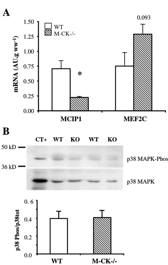
We then attempted to elucidate the signaling pathways involved in mitochondrial biogenesis in gastrocnemius muscle of M-CK-/- mice. In order to assess calcineurin transcriptional activity, we measured the level of transcription of the myocyte-enriched calcineurin interacting protein 1 (MCIP1), which contains 15 repeats of the NFAT binding site and thus has been shown to be the most sensitive indicator of calcineurin transcriptional activity (Yang et al., 2000). MCIP1 expression exhibited a 3-fold decrease suggesting decreased rather than increased transcriptional activity of calcineurin in M-CK-/- mice (Figure 3A). Expression level of the MEF2C transcription factor which acts in synergy with NFAT to regulate muscle fiber type [32] was slightly but not significantly increased in M-CK-/- mice. On the other hand, we examined whether the p38 MAPK pathway was activated in gastrocnemius muscle of M-CK-/- mice by western blotting with antibodies specific for the phosphorylated or the total p38 MAPK (Figure 3B). No increase in p38 MAPK phosphorylation was observed in gastrocnemius muscle of CK-/- mice suggesting that this pathway does not participate in mitochondrial biogenesis.
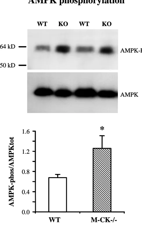
Finally AMPK was also shown to induce mitochondrial biogenesis in muscle, in response to metabolic stress and exercise. AMPK phosphorylation was significantly increased in M-CK-/- gastrocnemius muscle (Figure 4).
Discussion
This study was aimed at investigating the transcriptional cascade involved in mitochondrial biogenesis induced by creatine kinase depletion in fast skeletal muscle of mice
(Figure 5). The results show that the two-fold increase in mitochondrial mass was accounted for by an increase in both mitochondria and nucleus-encoded mitochondrial proteins, accompanied by a 60% increase in mitochondrial DNA. This could be explained by an increase in mtTFA and NRF2 transcription factors. However, while neither expression of PGC-1α or PPARδ, factors implicated in muscle mitochondrial biogenesis was increased, a slight increase in PRC expression was observed. Moreover, AMPK but not calcineurin or p38 MAPK was activated, evidencing a unique pattern of mitochondrial biogenesis in response to M-CK depletion.
Mitochondrial activity and content
Creatine kinase deficiency is a unique model of metabolic deficiency. It leads to a massive metabolic remodeling especially in the fast gastrocnemius muscle. Indeed, a doubling of oxidative capacity and mitochondrial volume density was described in gastrocnemius of M-CK or M-CK and mi-CK deficient mice [6,8,33]. As mi-CK is almost absent from fast fibers [1], this mitochondrial remodeling results from M-CK deficiency. At the same time, mitochondrial DNA is increased by only 60%, showing the complexity of mitochondrial biogenesis. On the other hand, as a more than two-fold increase in CS activity was measured, this suggests that not only mitochondrial mass but also mitochondrial specific activity was increased, in line with increased state 3 respiration rates in mitochondria isolated from CK-/- gastrocnemius muscle [34]. Mitochondrial function depends on the proper assembly of complexes of the electron transport chain (ETC) that are embedded within the inner mitochondrial membrane. The most widely studied ETC complex is COX which converts oxygen to water and provides part of protons required for ATP synthesis. COX assembly depends on COX subunits that are both nuclear- and mitochondria-encoded. Both the mitochondria encoded COX I and the nuclear encoded COX IV subunits were upregulated by a factor of 2 in M-CK deficient mice. This led to a 3.5 increase in COX activity. Moreover
this increase in COX activity exceeded that of CS as a significant 1.5 increase in COX/CS activity was observed. Indeed the COX/CS activity ratio can vary in a muscle type specific manner being much higher in oxidative (2.3 in heart, 2.6 in soleus) [35] than in fast glycolytic muscle (0.3 in gastrocnemius), showing the complex regulation of mitochondrial protein expression in muscles. Interestingly, in M-CK-/- mice the COX/CS ratio was significantly increased to 0.5 toward that of an oxidative muscle, but still much lower than WT soleus muscle.
The structural and functional adaptations of the mitochondrial network in challenged skeletal muscles result not only from changes in the mitochondrial protein expression and proper assembly but also involve mitochondrial dynamics. Training induced mitochondrial biogenesis in humans involve the coordinate increase in both Mfn2, a protein involved in fusion, and Drp1, a protein involved in mitochondrial fission, in vastus lateralis muscle [18]. Here we see a significant 50% increase in expression of Drp1, while the expression of Mfn2 remained unchanged. This suggests that in this model, the increase in mitochondrial number results from growing and fission of preexisting mitochondria.
Mitochondrial biogenesis transcription cascade
It is now well documented that mitochondrial biogenesis involves the integration of multiple transcriptional regulatory pathways controlling both the activation of nuclear and mitochondrial genomes. The mitochondrial genome however should not only be expressed but also replicated. This can be achieved by the nuclear encoded transcription factor mtTFA that binds to the mitochondrial DNA and induces both replication and transcription [13]. Accordingly mtTFA expression was significantly enhanced in M-CK-/- gastrocnemius muscle. Upstream of mtTFA, the NRFs are thought to increase the expression of multiple mitochondrial proteins by binding to specific recognition sites. The expression of NRF2α, the main isoform involved in rodent mitochondrial biogenesis [36] was also increased in M-CK-/-
mice, but with lower significance (p<0.057). Upstream of NRFs, PGC-1α is thought to orchestrate mitochondrial biogenesis in most tissues [37]. Moreover, PGC-1α expression correlates with oxidative capacity of cardiac and skeletal muscles [28]. It was thus surprising that its expression was not increased in gastrocnemius muscle of M-CK-/- mice, despite massive increase in mitochondrial content. Loss-of-function experiments have established that PGC-1α is not an absolute requirement for mitochondrial biogenesis. KO mice have preserved volume density of mitochondria in both heart and skeletal muscle [38], but reduced mRNA for mitochondrial proteins [38]. In a similar study however, Leone et al described slightly different muscle phenotype with a 30% decrease in mitochondrial volume and mRNA expression of mitochondrial proteins in soleus muscle and decrease in respiration rate [39]. Although results differ between the two studies, they show that the absence of PGC-1α can be partly overcome by other mitochondrial biogenesis pathways. PGC-1α is a member of an important family of transcriptional co-activators that includes PGC-1β and PGC-related co-activator (PRC) [40]. Those transcription factors share a high degree of homology and redundancy with PGC-1α with respect to the manner they activate mitochondrial biogenesis although they seem to respond differently to activators of mitochondrial biogenesis. Thus the slight increase in both PGC-1β (P<0.069) and PRC (P<0.05) could participate in increased mitochondrial biogenesis in M-CK-/- gastrocnemius. Finally, PPARδ whose invalidation was also shown to alter mitochondrial content [31,41] was unchanged in M-CK mice. However, the slight increase in transcription of members of PPAR and PGC transcription factor families may not be sufficient to explain the robust increase in gastrocnemius mitochondrial content (Figure 5).
Signaling pathways
It is likely that Ca2+ and energy depletion signals act in concert to promote metabolic
plasticity in CK-/- fast skeletal muscle. Indeed, altered calcium movements have been described in skeletal muscle of M-CK and CK-/- gastrocnemius muscle. CK-/- fast muscle fibers have increased resistance to fatigue and prolonged calcium transient [5]. It was thus proposed that calcium homeostasis impairment due to CK deficiency may have played a role in directing the increase in mitochondrial capacity [34]. The calcium-regulated serine/threonine protein phosphatase calcineurin has been implicated in the transduction of motor neuron signals to alter gene expression programs in skeletal muscle. However, assessment of the transcriptional activity of calcineurin by MCIP1 expression showed a decreased rather than increased calcineurin activity. Moreover, MEF2C was not up-regulated to significant level in these mice. This suggests that calcium is not primarily involved in gastrocnemius increase in mitochondrial biogenesis. The absence of increased calcineurin activation could indeed be linked to the low level of physical activity and thus of contraction-induced calcium increase in M-CK-/- mice [35].
Evidences are accumulating that the transcriptional activity of PGC-1α can be regulated by posttranslational modifications including phosphorylations. Transcriptional activity of PGC-1α can be enhanced by phosphorylation and disruption of a PGC-1α/p160MBP interaction through activation of the p38 MAPK pathway [42,43]. However, no activation of p38 MAPK was observed in this study suggesting that p38 MAPK is not involved in mitochondrial biogenesis in this model.
Finally, AMPK can activate mitochondrial biogenesis during long term exercise or energetic deficiency in muscle. It has been shown that AMPK can activate the transcription of PGC-1α indicating that its mitochondriogenic action can partly originate from enhancement of PGC-1α expression [26,44]. Moreover, skeletal muscle of transgenic mice expressing a
dominant-negative mutant form of AMPK have blunted response to energy deprivation by β -GPA feeding, showing that AMPK is necessary for mitochondrial biogenesis in response to metabolic stress [26]. Low frequency stimulation that mimics exercise training induces AMPK-PGC-1α signaling pathway [45]. AMPK is phosphorylated and activated in response to the increase in the AMP/ATP ratio. Indeed, both a decrease in ATP and an increase in AMP have been shown in hindlimb muscle of M-CK KO mice [3]. The present results show that AMPK phosphorylation is robustly increased in the gastrocnemius muscle of M-CK-/- mice. However, AMPK activation did not lead to increased expression of PGC-1α. It was shown very recently that AMPK can directly interact with and phosphorylate PGC-1α showing a posttranslational regulation of mitochondrial biogenesis by AMPK; moreover, AMPK phosphorylation of PGC-1alpha initiates many of the important gene regulatory functions of AMPK in skeletal muscle [46]. This strongly suggests that M-CK deficiency creates an energy deprivation state in gastrocnemius muscle of M-CK-/- mice that induces basal activation of AMPK, AMPK-dependent phosphorylation of PGC-1α and increased transcriptional activity of PGC-1α. Interestingly, activation of AMPK induces decreased skeletal muscle fiber size and increased mitochondrial biogenesis [47] in S6K deficient mice. Basal activation of AMPK in M-CK-/- mice could be a major factor involved in muscle atrophy and decreased exercise capacity of CK deficient mice [35].
Thus, following M-CK deletion, fast skeletal muscle undergoes a metabolic remodeling and fiber atrophy. These events seem to be triggered by energy depletion-induced AMPK activation, rather than by calcium-dependent processes (Figure 5).
Acknowledgments
We thank Dr. R. Fischmeister for continuous support. RV-C is supported by “Centre National de la Recherche Scientifique”. We thank Dr. B. Wieringa for kind support with the mice. We thank Claudine Deloménie and the “Plateforme transcriptome” for their help in RT-PCR experiments and Valérie Domergue and the animal care center for taking care of the animals.
References
1. Qin, W. N., Khuchua, Z., Boero, J., Payne, R. M., and Strauss, A. W., Oxidative myocytes of heart and skeletal muscle express abundant sarcomeric mitochondrial creatine kinase, Histochemical Journal Histochem J 31 (1999) 357-365.
2. Ventura-Clapier, R., Kuznetsov, A., Veksler, V., Boehm, E., and Anflous, K.,
Functional coupling of creatine kinases in muscles: Species and tissue specificity, Mol Cell Biochem 184 (1998) 231-247.
3. van Deursen, J., Heerschap, A., Oerlemans, F., Ruitenbeek, W., Jap, P., ter Laak, H., and Wieringa, B., Skeletal muscles of mice deficient in muscle creatine kinase lack burst activity, Cell 74 (1993) 621-631.
4. Gorselink, M., Drost, M. R., Coumans, W. A., van Kranenburg, G. P., Hesselink, R. P., and van der Vusse, G. J., Impaired muscular contractile performance and adenine nucleotide handling in creatine kinase-deficient mice, Am J Physiol Endocrinol Metab 281 (2001) E619-E625.
5. Dahlstedt, A. J. and Westerblad, H., Inhibition of creatine kinase reduces the rate of fatigue-induced decrease in tetanic Ca(2+), J Physiol 533 (2001) 639-49.
6. Veksler, V. I., Kuznetsov, A. V., Anflous, K., Mateo, P., van Deursen, J., Wieringa, B., and Ventura-Clapier, R., Muscle creatine kinase-deficient mice.2. Cardiac and skeletal muscles exhibit tissue-specific adaptation of the mitochondrial function., J Biol Chem 270 (1995) 19921-19929.
7. Kaasik, A., Veksler, V., Boehm, E., Novotova, M., and Ventura-Clapier, R., From energy store to energy channeling: a study in creatine kinase deficient fast skeletal
muscle, FASEB J 17 (2003) 708-710.
8. Novotova, M., Pavlovicova, M., Veksler, V., Ventura-Clapier, R., and Zahradnik, I., Ultrastructural remodeling of fast skeletal muscle fibers induced by invalidation of creatine kinase, Am J Physiol Cell Physiol 291 (2006) C1279-C1285.
9. de Groof, A. J., Smeets, B., Groot Koerkamp, M. J., Mul, A. N., Janssen, E. E., Tabak, H. F., and Wieringa, B., Changes in mRNA expression profile underlie phenotypic adaptations in creatine kinase-deficient muscles, FEBS Lett 506 (2001) 73-8.
10. de Groof, A. J., Oerlemans, F. T., Jost, C. R., and Wieringa, B., Changes in glycolytic network and mitochondrial design in creatine kinase-deficient muscles, Muscle Nerve 24 (2001) 1188-96.
11. Wu, Z. D., Puigserver, P., Andersson, U., Zhang, C. Y., Adelmant, G., Mootha, V., Troy, A., Cinti, S., Lowell, B., Scarpulla, R. C., and Spiegelman, B. M., Mechanisms controlling mitochondrial biogenesis and respiration through the thermogenic
coactivator PGC-1, Cell 98 (1999) 115-124.
12. Knutti, D. and Kralli, A., PGC-1, a versatile coactivator, Trends Endocrinol Metab 12 (2001) 360-365.
13. Scarpulla, R. C., Transcriptional activators and coactivators in the nuclear control of mitochondrial function in mammalian cells, Gene 286 (2002) 81-89.
14. Larsson, N. G., Wang, J., Wilhelmsson, H., Oldfors, A., Rustin, P., Lewandoski, M., Barsh, G. S., and Clayton, D. A., Mitochondrial transcription factor A is necessary for mtDNA maintenance and embryogenesis in mice, Nat Genet 18 (1998) 231-236.
form and function., Rev Physiol Biochem Pharmacol 146 (2003) 159-216.
16. Reznick, R. M. and Shulman, G. I., The Role of AMP-Activated Protein Kinase in Mitochondrial Biogenesis, J Physiol 574 (2006) 33-39.
17. Hood, D. A., Irrcher, I., Ljubicic, V., and Joseph, A. M., Coordination of metabolic plasticity in skeletal muscle, J Exp Biol 209 (2006) 2265-2275.
18. Garnier, A., Fortin, D., Zoll, J., N'Guessan, B., Mettauer, B., Lampert, E., Veksler, V., and Ventura-Clapier, R., Coordinated changes in mitochondrial function and biogenesis in healthy and diseased human skeletal muscle., FASEB J 19 (2005) 43-52.
19. Chin, E. R., Olson, E. N., Richardson, J. A., Yang, Q., Humphries, C., Shelton, J. M., Wu, W., Bassel-Duby, R., and Williams, R. S., A calcineurin-dependent transcriptional pathway controls skeletal muscle fiber type, Genes and Development 12 (1998) 2499-2509.
20. Lin, J., Wu, H., Tarr, P. T., Zhang, C. Y., Wu, Z., Boss, O., Michael, L. F., Puigserver, P., Isotani, E., Olson, E. N., Lowell, B. B., Bassel-Duby, R., and Spiegelman, B. M., Transcriptional co-activator PGC-1alpha drives the formation of slow- twitch muscle fibres, Nature 418 (2002) 797-801.
21. Garcia-Roves, P. M., Huss, J., and Holloszy, J. O., Role of calcineurin in exercise-induced mitochondrial biogenesis, Am J Physiol Endocrinol Metab 290 (2006) E1172-E1179 .
22. Bigard, X., Sanchez, H., Zoll, J., Mateo, P., Rousseau, V., Veksler, V., and Ventura-Clapier, R., Calcineurin Co-regulates contractile and metabolic components of slow muscle phenotype, J Biol Chem 275 (2000) 19653-60.
23. Akimoto, T., Pohnert, S. C., Li, P., Zhang, M., Gumbs, C., Rosenberg, P. B., Williams, R. S., and Yan, Z., Exercise stimulates Pgc-1alpha transcription in skeletal muscle through activation of the p38 MAPK pathway, J Biol Chem 280 (2005) 19587-19593.
24. Wu, H., Kanatous, S. B., Thurmond, F. A., Gallardo, T., Isotani, E., Bassel-Duby, R., and Williams, R. S., Regulation of mitochondrial biogenesis in skeletal muscle by CaMK, Science 296 (2002) 349-352.
25. Bergeron, R., Ren, J. M., Cadman, K. S., Moore, I. K., Perret, P., Pypaert, M., Young, L. H., Semenkovich, C. F., and Shulman, G. I., Chronic activation of AMP kinase results in NRF-1 activation and mitochondrial biogenesis, Am J Physiol 281 (2001) E1340-E1346.
26. Zong, H., Ren, J. M., Young, L. H., Pypaert, M., Mu, J., Birnbaum, M. J., and Shulman, G. I., AMP kinase is required for mitochondrial biogenesis in skeletal muscle in
response to chronic energy deprivation, Proc Natl Acad Sci U S A 99 (2002) 15983-15987.
27. Jorgensen, S. B., Wojtaszewski, J. F., Viollet, B., Andreelli, F., Birk, J. B., Hellsten, Y., Schjerling, P., Vaulont, S., Neufer, P. D., Richter, E. A., and Pilegaard, H., Effects of alpha-AMPK knockout on exercise-induced gene activation in mouse skeletal muscle, FASEB J 19 (2005) 1146-1148.
28. Garnier, A., Fortin, D., Delomenie, C., Momken, I., Veksler, V., and Ventura-Clapier, R., Depressed mitochondrial transcription factors and oxidative capacity in rat failing cardiac and skeletal muscles., J Physiol 551 (2003) 491-501.
(2002) 200-204.
30. Puigserver, P., Wu, Z., Park, C. W., Graves, R., Wright, M., and Spiegelman, B. M., A cold-inducible coactivator of nuclear receptors linked to adaptive thermogenesis, Cell 92 (1998) 829-839.
31. Wang, Y. X., Zhang, C. L., Yu, R. T., Cho, H. K., Nelson, M. C., Bayuga-Ocampo, C. R., Ham, J., Kang, H., and Evans, R. M., Regulation of muscle fiber type and running endurance by PPARdelta, PLoS Biol 2 (2004) e294.
32. Wu, H., Naya, F. J., McKinsey, T. A., Mercer, B., Shelton, J. M., Chin, E. R., Simard, A. R., Michel, R. N., BasselDuby, R., Olson, E. N., and Williams, R. S., MEF2 responds to multiple calcium-regulated signals in the control of skeletal muscle fiber type, EMBO Journal 19 (2000) 1963-1973.
33. Kaasik, A., Veksler, V., Boehm, E., Novotova, M., Minajeva, A., and Ventura-Clapier, R., Energetic crosstalk between organelles: architectural integration of energy
production and utilization, Circ Res 89 (2001) 153-159.
34. ter Veld, F., Jeneson, J. A., and Nicolay, K., Mitochondrial affinity for ADP is twofold lower in creatine kinase knock-out muscles. Possible role in rescuing cellular energy homeostasis, FEBS J 272 (2005) 956-965.
35. Momken, I., Lechene, P., Koulmann, N., Fortin, D., Mateo, P., Doan, B. T., Hoerter, J., Bigard, X., Veksler, V., and Ventura-Clapier, R., Impaired voluntary running capacity of creatine kinase-deficient mice, J Physiol 565 (2005) 951-964.
36. Scarpulla, R. C., Nuclear activators and coactivators in mammalian mitochondrial biogenesis, Biochim Biophys Acta 1576 (2002) 1-14.
37. Puigserver, P. and Spiegelman, B. M., Peroxisome proliferator-activated receptor-gamma coactivator 1 alpha (PGC-1 alpha): transcriptional coactivator and metabolic regulator., Endocr Rev 24 (2003) 78-90.
38. Arany, Z., He, H., Lin, J., Hoyer, K., Handschin, C., Toka, O., Ahmad, F., Matsui, T., Chin, S., Wu, P. H., Rybkin, I. I., Shelton, J. M., Manieri, M., Cinti, S., Schoen, F. J., Bassel-Duby, R., Rosenzweig, A., Ingwall, J. S., and Spiegelman, B. M., Transcriptional coactivator PGC-1 alpha controls the energy state and contractile function of cardiac muscle, Cell Metab 1 (2005) 259-271.
39. Leone, T. C. , Lehman, J. J., Finck, B. N., Schaeffer, P. J., Wende, A. R., Boudina, S., Courtois, M. , Wozniak, D. F., Sambandam, N., Bernal-Mizrachi, C., Chen, Z., Holloszy, J. O., Medeiros, D. M., Schmidt, R. E., Saffitz, J. E., Abel, E. D.,
Semenkovich, C. F., and Kelly, D. P., PGC-1alpha deficiency causes multi-system energy metabolic derangements: muscle dysfunction, abnormal weight control and hepatic steatosis., PLoS Biol 3 (2005) e101.
40. Lin, J., Handschin, C., and Spiegelman, B. M., Metabolic control through the PGC-1 family of transcription coactivators, Cell Metab 1 (2005) 361-370.
41. Luquet, S., Lopez-Soriano, J., Holst, D., Fredenrich, A., Melki, J., Rassoulzadegan, M., and Grimaldi, P. A., Peroxisome proliferator-activated receptor delta controls muscle development and oxidative capability, FASEB J 17 (2003) 2299-301.
42. Puigserver, P., Rhee, J., Lin, J., Wu, Z., Yoon, J. C., Zhang, C. Y., Krauss, S., Mootha, V. K., Lowell, B. B., and Spiegelman, B. M., Cytokine stimulation of energy
expenditure through p38 MAP kinase activation of PPARgamma coactivator-1, Mol Cell 8 (2001) 971-982.
43. Fan, M., Rhee, J., St-Pierre, J., Handschin, C., Puigserver, P., Lin, J., Jaeger, S., Erdjument-Bromage, H., Tempst, P., and Spiegelman, B. M., Suppression of mitochondrial respiration through recruitment of p160 myb binding protein to PGC-1alpha: modulation by p38 MAPK., Genes Dev 18 (2004) 278-289.
44. Jorgensen, S. B., Richter, E. A., and Wojtaszewski, J. F., Role of AMPK in Skeletal Muscle Metabolic Regulation and Adaptation In Relation to Exercise, J Physiol 574 (2006) 17-31.
45. Atherton, P. J., Babraj, J., Smith, K., Singh, J., Rennie, M. J., and Wackerhage, H., Selective activation of AMPK-PGC-1alpha or PKB-TSC2-mTOR signaling can explain specific adaptive responses to endurance or resistance training-like electrical muscle stimulation, FASEB J 19 (2005) 786-788.
46. Jager, S., Handschin, C., St-Pierre, J., and Spiegelman, B. M., AMP-activated protein kinase (AMPK) action in skeletal muscle via direct phosphorylation of PGC-1{alpha}, Proc Natl Acad Sci USA 104 (2007) 12017-12022.
47. Aguilar, V., Alliouachene, S., Sotiropoulos, A., Sobering, A., Athea, Y., Djouadi, F., Miraux, S., Thiaudiere, E., Foretz, M., Viollet, B., Diolez, P., Bastin, J., Benit, P., Rustin, P., Carling, D., Sandri, M., Ventura-Clapier, R., and Pende, M., S6 Kinase Deletion Suppresses Muscle Growth Adaptations to Nutrient Availability by Activating AMP Kinase, Cell Metab 5 (2007) 476-487.
Figure Legends
Figure 1: Mitochondrial enzyme activity and expression in gastrocnemius muscle of WT (n=6) and M-CK-/- (n=6) mice. A. Increase in cytochrome oxidase and citrate synthase
activity measured by spectrophotometry. B. Increased mRNA expression of subunit I (COXI, mitochondria-encoded) and subunit IV (COXIV, nuclear-encoded) of the cytochrome oxidase and of dynamin-related protein 1 (Drp1) but not mitofusin 2 (Mfn2) two proteins that are involved in mitochondrial dynamics. Values are means±sem. ** p<0.01 versus WT.
Figure 2: Mitochondrial transcription cascade in gastrocnemius muscle of WT (n=6) and M-CK-/- (n=6) mice. A. Real-time RT-PCR analysis of the nuclear respiratory factor2
(NRF2) and the mitochondrial transcription factor A (mtTFA). B. mRNA expression of the peroxisome proliferator-activated receptor (PPAR) gamma coactivator 1α (PGC-1α) or 1β (PGC-1β) isoforms, the PGC-1-related coactivator (PRC), and the delta isoform of PPAR. Values are means±sem and are shown as arbitrary units normalized to the glucocerebrosidase and to the amount of mRNA. * p<0.05, ** p<0.01 versus WT.
Figure 3: Calcium signaling pathways of mitochondrial biogenesis in gastrocnemius muscle of WT (n=6) and M-CK-/- (n=6) mice. A. Calcineurin activation was assessed by
the level of expression of the modulatory calcineurin-interacting protein 1 (MCIP1) and the myocyte-specific enhancer factor 2 (MEF2C). B. Activation of p38 MAPK has been assessed by the ratio of signals obtained by immunoblots of the phosphorylated and total forms; upper part representative immunoblots; lower part mean values. Values are means±sem. * p<0.05 versus WT.
Figure 4: AMPK activation in gastrocnemius muscle of WT (n=6) and M-CK-/- (n=6) mice. Activation of AMP-activated protein kinase (AMPK) has been assessed by the ratio of
signals obtained by immunoblots of the phosphorylated and total forms; upper part representative immunoblots; lower part mean values. * p<0.05 versus WT.
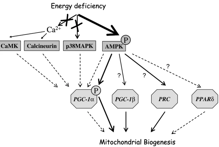
Figure 5: A model for mitochondrial biogenesis in gastrocnemius muscle of CK deficient mice. Energy deficiency induced by M-CK deletion lead to increased mitochondrial
biogenesis. This effect was not accompanied by increased activity of calcineurin or phosphorylation of p38 MAPK suggesting that neither calcium signaling through calcineurin or CaMK, nor p38 signaling was involved. AMPK appeared phosphorylated suggesting a direct link between energy depletion, change in AMP/ATP ratio and mitochondrial biogenesis. A slight increase in PGC-1β and PRC but no change in PGC-1α or PPARδ expression, the two main regulators of mitochondrial biogenesis in muscle, was observed. However, PGC-1α may be involved in mitochondrial biogenesis by direct phosphorylation and activation by AMPK.
Table 1. Primers used for real-time PCR amplification
Gene GenBank AN Forward primer Reverse primer (5’-3’) PCR product size (pb) Annealing temperature (°C)
PGC-1a NM_008904 CAC CAA ACC CAC AGA GAA CAG GCA GTT CCA GAG AGT TCC ACA
210 58
PGC-1ß NM_133249 TGG AAA GCC CCT GTG AGA GT TTG TAT GGA GGT GTG GTG GG
202 60
PRC XM_358330 AGG AAA CTC AGG CAG CAT TG
GGC GGT GGA TTT AGG AGA TT
178 60
NRF2a NM_008065 AGGTGACGAGATGGGCTGC
CGTTGTCCCCATTTTTGCG
604 65
mtTFA NM_009360 GCT GAT GGG TAT GGA GAA G GAG CCG AAT CAT CCT TTG C
161 56
COX I NC_006914 CAC TAA TAA TCG GAG CCC CA TTC ATC CTG TTC CTG CTC CT
129 60
COX IV NM_053091 TGG GAG TGT TGT GAA GAG TGA GCA GTG AAG CCG ATG AAG AAC
273 58
Drp1 NM_152816 CTG ACG CTT GTG GAT TTA CC CCC TTC CCA TCA ATA CAT CC
277 58
Mfn2 NM_133201 ACG AGC AAT GGG AAG AGC AC
TCC ATC AGC ACG AGG TCA TC
284 60
MCIP1 NM_019466 CAG CGA AAG TGA GAC CAG GG ACG GGG GTG GCA TCT TCT AC
309 60
MEF2C NM_025282 CAG GGA ACG GGT ATG GCA ATC
CAA TGA CTG AGC CGA CTG GGA
239 60
PPARd NM_011145 GCC TCC ATC GTC AAC AAA GA TCT ACC TGG GGC ACA TTC AT
230 60
GCB NM_008094 CCC ATT TCA CTC TTT GCC AG AGG TTC ATT CTC CGC TGT CA
198 60
PGC-1α and PGC-1β: peroxisome proliferator-activated receptor gamma co-activator-1α and β; PRC: PGC-1α related co-activator NRF2α: nuclear respiratory factor-2α; mtTFA, mitochondrial transcription factor A; COXI and IV: cytochrome oxidase subunits I and IV; Drp1: dynamin related protein 1; Mfn2: mitofusin 2; MCIP1: modulatory calcineurin interacting protein 1; MEF2C: myocyte-specific enhancer factor 2; PPARδ: peroxisome proliferator activated receptor δ; GCB: glucocerebrosidase.