HAL Id: hal-02177806
https://hal-univ-rennes1.archives-ouvertes.fr/hal-02177806
Submitted on 17 Sep 2019HAL is a multi-disciplinary open access archive for the deposit and dissemination of sci-entific research documents, whether they are pub-lished or not. The documents may come from teaching and research institutions in France or abroad, or from public or private research centers.
L’archive ouverte pluridisciplinaire HAL, est destinée au dépôt et à la diffusion de documents scientifiques de niveau recherche, publiés ou non, émanant des établissements d’enseignement et de recherche français ou étrangers, des laboratoires publics ou privés.
subcloning and biochemical characterization
Mateja Senicar, Laurent Legentil, Vincent Ferrières, Svetlana Eliseeva,
Stéphane Petoud, Kaoru Takegawa, Pierre Lafite, Richard Daniellou
To cite this version:
Mateja Senicar, Laurent Legentil, Vincent Ferrières, Svetlana Eliseeva, Stéphane Petoud, et al.. Galactofuranosidase from JHA 19 Streptomyces sp:. subcloning and biochemical characterization. Carbohydrate Research, Elsevier, 2019, 480, pp.35-41. �10.1016/j.carres.2019.05.011�. �hal-02177806�
M
AN
US
CR
IP
T
AC
CE
PT
ED
Galactofuranosidase from JHA 19 Streptomyces sp.: subcloning and
biochemical characterization.
Mateja Senicar1,2, Laurent Legentil3, Vincent Ferrières3, Svetlana V. Eliseeva2, Stéphane Petoud2, Kaoru Takegawa4, Pierre Lafite1 and Richard Daniellou1,*
1
ICOA UMR CRNS 7311, Université d’Orléans, rue de Chartres, BP 6759, 45067 Orléans cedex 2, France.
2
Centre de Biophysique Moléculaire, CNRS UPR 4301, 45071 Orléans, France.
3
Univ Rennes, Ecole Nationale Supérieure de Chimie de Rennes, CNRS, ISCR - UMR 6226, F-35000 Rennes, France.
4
Department of Bioscience and Biotechnology, Faculty of Agriculture, Kyushu University, Moto-oka 744, Nishi-ku, Fukuoka, Japan.
* Corresponding author; E-mail address: richard.daniellou@univ-orleans.fr; Tel: +33-2-38-49-49-78.
M
AN
US
CR
IP
T
AC
CE
M
AN
US
CR
IP
T
AC
CE
PT
ED
Highlights• Galf-ase from JHA 19 Streptomyces sp. was subcloned.
• Recombinant Galf-ase was obtained with a yield of 0.5 mg/litre of culture. • Galf-ase exhibits the best reported KM for Galf moiety.
• Galf-ase can be competitively inhibited by thiogalactofuranosides analogues. • Galf-ase can be envisioned in biocatalysis.
M
AN
US
CR
IP
T
AC
CE
PT
ED
AbstractDespite the crucial role of the rare galactofuranose (Galf) in many pathogenic micro-organisms and our increased knowledge of its metabolism, there is still a lack of recombinant and efficient galactofuranoside hydrolase available for chemo-enzymatic synthetic purposes of specific galactofuranosyl-conjugates. Subcloning of the Galf-ase from JHA 19
Streptomyces sp. and its further overexpression lead us to the production of this enzyme with
a yield of 0.5 mg/litre of culture. It exhibits substrate specificity exclusively towards pNP β-D -Galf, giving a KM value of 250 µM, and the highest enzymatic efficiency ever observed of 14
mM-1.s-1. It proved to be stable to temperature up to 60°C and to at least 4 freeze-thaw’s
cycles. Thus, Galf-ase demonstrated to be an efficient and stable biocatalyst with greatly improved specificity toward the galactofuranosyl entity, thus paving the way to the further development of transglycosylation and thioligation reactions.
Keywords: β-D-galactofuranosidase, Subcloning, Glycoside hydrolase,
M
AN
US
CR
IP
T
AC
CE
PT
ED
1. IntroductionD-Galactose (D-Gal) is extensively distributed in nature as constituent of oligosaccharides and glycoconjugates.[1] However, under its furanose form, galactofuranose (Galf) is totally absent from mammals, but is present in many pathogenic microorganisms.[2],[3] Indeed, there are
three main and specific enzymes involved in the biosynthesis and the metabolism of the Galf-containing molecules[4]: UDP-galactopyranose mutase (UGM),[5] galactofuranosyltransferase (GalfT)[6] and galactofuranosidase (Galf-ase).[7] The glycobiology of Galf and enzymes involved in its metabolism have thus become attractive targets for the development of antimicrobial agents.[8]
Although the metabolism of β-D-galactofuranosyl conjugates has been extensively studied, mostly in infectious microorganisms such as Mycobacteria, Trypanosoma, Leishmania and
Aspergillus, there are only few reports related to these enzymes, especially Galf-ase. Over the
past forty years, Galf-ase has been identified as responsible for the degradation of the D-Galf
containing glycoconjugates.[8] The first Galf-ase identified was an extracellular exo-β-D
-galactofuranosidase (exo-β-D-Galf-ase) isolated and partially purified from Penicillium
fellutanum (ex-type of Penicillium charlesii) culture filtrates.[7] Although described back in
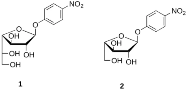
1977 and later, several exo- and endo-Galf-ases were purified from the culture supernatants and cell lysates of filamentous fungi,[7]-[9] bacteria[10] and protozoa,[11] but the genes encoding these enzymes have not been identified and expressed earlier, nor their amino acid sequence determined. Therefore, reported chemo-enzymatic procedures in literature all used cloned α-L-arabinofuranosidases as biocatalytic tools, based on the structure similarity between pNP β-D-Galf 1 and its analogue pNP α-L-Araf 2 (Fig 1.).[12][13][14] However, given the poor catalytic efficiency of theses biocatalysts toward pNP β-D-Galf 1, there is still a
crucial need for the discovery of improved biocatalysts that will allow the community to easily access to furanosyl conjugates (and analogues) owing potential biological properties.[3]
M
AN
US
CR
IP
T
AC
CE
PT
ED
Fig. 1. Chemical structures of pNP β-D-Galf 1 and its analogue pNP α-L-Araf 2.
Only recently, in 2015, based on the draft genome sequence analysis of soil Gram-positive bacteria Streptomyces sp., strain JHA19, an open reading frame that encoded Galf specific enzyme was identified and cloned, and the enzyme characterized.[15], [16] Another gene
coding for a putative Galf-ase was also found later in the genome of Streptomyces sp., strain JHA26.[16][17], However this first cloned Galf-ase protein was co-expressed with a large
fusion NusA[18] (N-utilization substance A) tag as solubility enhancer and two His-tags for affinity purification purpose, thus leading to a large protein of around 150 kDa (Figure
2A).[15] We thus rapidly thought that these multiple tags might hamper i) the overexpression
of the Galf-ase leading to low yield, and ii) the physico-chemical and enzymatic properties of this original biocatalyst. In addition, the obtained low yield of protein was incompatible with the development of this biocatalyst.
In view of the previous research considering Streptomyces Galf-ase investigations,[15] we wish to report herein the optimization of the overexpression and the purification to homogeneity of recombinant ORF1110 β-D-galactofuranosidase including only one His-tag (Figure 2B), with detailed and improved biochemical and kinetic properties, as well as preliminary inhibition studies of this enzyme. This Galf-ase will thus constitute the basis for the further development of biocatalyzed incorporation of Galf moieties in more complex glycoconjugates. O OH OH OH O NO2 OH O OH OH OH O NO2 2 1
M
AN
US
CR
IP
T
AC
CE
PT
ED
Fig. 2. Schematic representation of pET plasmid constructs cloning/expression regions
bearing Galf-ase gene insert. A) pET-50b(+) plasmid construct ORF coding for N-terminal Nus protein and double 6xHis-tagged Galf-ase. B) pET-28a(+) plasmid construct ORF coding for N-terminal single peptide 6xHis-tagged Galf-ase.
2. Materials and Methods
2.1. Biological and chemical reagents
Restriction enzymes and T4 DNA ligase were purchased from Thermo ScientificTM, E. coli
RosettaTM (DE3) and plasmid vector pET-28a(+) from Novagen®. All p-nitrophenyl
monosaccharides (pNP sugars) were purchased from Carbosynth (Compton, UK). Galactofuranosides 3-6 were chemically synthesized as previously described.[19][20] All other laboratory chemicals used as the starting compounds, reagents and solvents were analytical grade purity and commercially available, unless otherwise specified.
M
AN
US
CR
IP
T
AC
CE
PT
ED
2.2. Subcloning, overexpression, and protein purification
Plasmid construct pET-50b(+), encoding wild type Galf-ase gene was digested with EcoRI and HindIII restriction enzymes and the resulting 2.5 kb fragment containing Galf-ase gene was agarose-gel purified and ligated into expression vector pET-28a(+) (Novagen®). Ligation
was verified by restriction enzyme analysis, and the gene sequence integrity was confirmed by DNA sequencing performed by Eurofins Genomics. E. coli RosettaTM (DE3) (Novagen®)
expression strain was transformed with Galf-ase encoding pET-28a(+) plasmid construct by heat shock method and cultured at 37°C overnight on LB agar plates supplemented with chloramphenicol (34 µg/mL) and kanamycin (30 µg/mL). One single positive colony was used to inoculate fresh lysogeny broth (LB) (10 mL) supplemented with the same antibiotics and cultured overnight with agitation at 37°C. The preculture was then inoculated into LB broth (1L) containing corresponding antibiotics and shaker incubated (250 rpm) at 37°C. Cells were grown to mid-exponential phase (OD600: 0.6), shortly cooled on ice and protein
expression was induced by the addition of β-D-thiogalactopyranoside (IPTG) (100 µL; 1 M)
and left on shaker incubator (250 rpm) for further 16 hours at 15°C. Cells were harvested by centrifugation (4255 g, 30 min, 4 °C) and the cell pellets were resuspended in lysis buffer solution (1:10 v/v; 100 mM NaCl, 50 mM Tris/HCl pH 8, 1 mM phenylmethanesulfonyl fluoride (PMSF), 5% glycerol, 0.1% Triton X-100, lysozyme 1 mg/L). Suspension was incubated by stirring for 20 min at 4°C, lysed by three freeze-thaw cycles and subsequently sonicated ([2 min on/2 min pause]x3, 50% cycle) on ice. Lysate was centrifuged (30 000 g, 20 min, 4°C), supernatant filtered (0.45 µm pore filter) and the recombinant protein was then purified from the clarified lysates using pre-equilibrated (10 mL; 50 mM Tris, 200 mM NaCl, 10 mM Imidazole) Thermo Scientific HisPurTM Ni-NTA Chromatography Cartridge (1 mL).
The bound protein was eluted by an imidazole gradient (10-500 mM) and an aliquot of eluted fractions were analysed by sodium dodecyl sulfate-polyacrylamide gel electrophoresis
(SDS-M
AN
US
CR
IP
T
AC
CE
PT
ED
PAGE) on 8% separating gel according to Laemmli’s method[21], and protein bands were visualized by staining with Coomassie Brilliant Blue G250. Fractions containing pure protein were collected, concentrated by ultrafiltration (30 000 MWCO, Sartorius Vivaspin® sample
concentrator) and protein quantity was determined using colorimetric Bio-Rad protein assay based on the Bradford dye-binding method, with a bovine serum albumin (BSA) as a standard.
2.3. Substrate specificity through hydrolysis of pNP sugars
The hydrolysis of 21 pNP-linked monosaccharide substrates (pNP α and β-D-Glc, -Man, -Gal,
-GlcNAc, -Xyl,-L-Fuc, -L-Ara, pNP α-L-Rha, pNP β-D-GalNAc and pNP β-D-GlcA, and 3 furanoses namely pNP β-D-Galf, pNP α-L-Araf and pNP β-D-Ribf) was assayed in reaction mixture (50 µL) containing purified enzyme (5 µL; 0.053 mg/mL), pNP monosaccharide substrates (5 µL; 10 mM), buffer (8 µL; 0.1 M Citric acid/0.2 M Na2HPO4 pH 4.5) and water
(32 µL). Residual spontaneous hydrolysis of the substrate was determined on sample containing H2O instead of enzyme. For paranitrophenol (pNP) containing substrates, after 20
min of incubation at 30°C, 150 µ L of sodium carbonate 1 M were added, and produced pNP (ɛ405 = 19 500 M−1cm−1) was quantified by absorbance measurement at 405 nm (Thermo
Scientific™ Multiskan™ GO). All kinetics parameters were calculated by fitting of saturation curves (as mean of triplicate measurements) with standard Michaelis-Menten equation, using Prism 6 (GraphPad). One unit (U) of enzyme activity was defined as the amount of enzyme required to liberate 1 µmol of pNP per min.
2.4. Effects of pH and temperature
The optimum pH was determined by incubating the enzyme in various pH adjusted buffers (0.1 M Citric acid/HCl pH 2; 0.1 M Citric acid/0.2 M Na2HPO4 pH 2.5 – 6.5; 0.1 M Tris/HCl
M
AN
US
CR
IP
T
AC
CE
PT
ED
performed separately in each buffer system containing purified enzyme (5 µL; 0.05 mg/mL),
pNP β-D-Galf substrate (5 µL; 10 mM), buffer (8 µL; pH 2 – 9.5) and water (32 µL). After 20 min of incubation at 30°C (water bath), reaction was terminated (150 µL; 1 M Na2CO3) and
absorbance (405 nm) of released pNP was measured using spectrophotometer (Thermo Scientific™ Multiskan™ GO). All the reactions were assayed in triplicate and absorbance values were corrected for the spontaneous hydrolysis of the substrate.
The effect of temperature on the enzyme activity was investigated at temperatures ranging from 10 to 80°C. The reaction mixtures (50 µL), containing purified enzyme (5 µL; 0.05 mg/mL), pNP β-D-Galf substrate (5 µL; 10 mM), buffer (8 µL; 0.1 M Citric acid/0.2 M Na2HPO4 pH 4.5) and water (32 µL), were incubated 20 min in thermocycler (Esco Swift
MiniPro Thermal Cycler). Afterwards, reactions were stopped (150 µL; 1 M Na2CO3). Then,
the absorbance (405 nm) of released pNP was measured (Thermo Scientific™ Multiskan™ GO) and values were corrected for the spontaneous hydrolysis of the substrate. All the reactions were assayed in triplicate and absorbance values were corrected for the spontaneous hydrolysis of the substrate.
2.5. Effect of freeze-thaw cycles
Three samples (100 µL), containing previously purified enzyme (50 µL; 0.235 mg/mL) different concentrations of glycerol (0 %, 10%, 20%) and water, were prepared. The samples were precooled on ice (30 min), frozen at -20°C and were removed from -20°C after four days, thawed on ice (30 min) and the enzymatic activity of a sample aliquot (10 µL) was assayed in reaction mixture (200 µL) containing pNP β-D-Galf substrate (20 µL; 10 mM),
buffer (35 µL; 0.1 M citric acid/0.2 M Na2HPO4 pH 4.5) and water (135 µL) at 37°C (water
bath), during 10 min, according to previously described enzymatic activity protocol. This predefined freeze and thaw procedure and activity assessment were repeated in four cycles
M
AN
US
CR
IP
T
AC
CE
PT
ED
to freezing. All the reactions were assayed separately in duplicate and absorbance values were corrected for the spontaneous hydrolysis of the substrate.
2.6. Kinetic studies
Kinetic parameters were determined with pNP β-D-Galf as a substrate in different
concentration ranges (0.01 mM-5 mM). The reaction (200 µL) containing buffer (20 µL; 0.1 M citric acid/0.2 M Na2HPO4 pH 4.5) and water, if required, was started by addition of the
enzyme (2 µL; 0.13 mg/mL) and after 20 min of incubation at 37°C (water bath), reaction was terminated (100 µL; 1 M Na2CO3) and absorbance (405 nm) of released pNP was measured.
All the reactions were assayed in triplicate and absorbance values were corrected for the spontaneous hydrolysis of the substrate. The kinetic parameters (KM, Vm, kcat) were calculated
using GraphPad Prism 5 software (GraphPad Software, San Diego, CA, USA).
2.7. Inhibition studies
IC50 assay was performed in the presence of different concentration ranges of inhibitors 3 – 6
(0.1 mM-7 mM), pNP β-D-Galf substrate (3.75 µL; 10 mM), buffer (20 µL; citric
acid/Na2HPO4, pH 4.5) and water. The reaction (150 µL) was started by addition of the
enzyme (11.2 µL; 0.067 mg/mL), and after 20 min of incubation at 37°C (water bath), reaction was terminated (100 µL; 1 M Na2CO3) and absorbance of released pNP was
measured (405 nm). Control enzyme activity reactions were carried out and absorbance values were corrected for the spontaneous hydrolysis of the substrate. The type of inhibition for 4 was tested in the presence of different concentration ranges of pNP β-D-Galf substrate (0.05 mM-1 mM) and with different concentrations of 4 (0.1 mM-1 mM) in the reaction conditions identical to the one previously described. The IC50 value for each inhibitor, the type of the
M
AN
US
CR
IP
T
AC
CE
PT
ED
Software, San Diego, CA, USA). KI value for compounds 3, 5 and 6 was corroborated by the
following Cheng & Prusoff equation: KI = IC50/(1+[S]/Km).[22]
3. Results and Discussion
3.1. Subcloning, expression and purification of recombinant Galf-ase
Plasmid construct pET-50b(+), containing the gene insert coding for β-D-galactofuranosidase
(Galf-ase), was double digested with EcoRI and HindIII restriction enzymes and the resulting 2.4 kb Galf-ase gene fragment was excised and purified from agarose-gel. The gene fragment was ligated to EcoRI and HindIII linearized pET-28a(+) expression vector. Newly obtained plasmid construct was checked for integrity by restriction enzyme analysis and the gene sequence was confirmed by DNA sequencing performed by Eurofins Genomics. Plasmid construct was transformed into E. coli RosettaTM (DE3) (Novagen®) expression system.
Galf-ase was expressed as soluble recombinant protein bearing only N-terminal peptide hexahistidine fusion tag (6xHis-tag). 6xHis-tag facilitated selective recombinant protein purification from the bacterial cellular environment by immobilized metal-affinity chromatography (IMAC) and, due to its small size, was less likely to interfere with protein activity and structure.[23] The 6xHis-tagged Galf-ase was expressed and purified by IMAC with a Ni2+ immobilized on a nitrilotriacetic acid matrix (Ni-NTA) column with a yield of 0.5
mg/litre of bacterial culture.[24],[25] The purified enzyme displayed an intense band on the
SDS-PAGE gel at an apparent molecular weight (90.3 kDa), consistent with that theoretically predicted for the Galf-ase fusion protein (Fig. 3.). This purified Galf-ase was further used for biochemical characterization.
M
AN
US
CR
IP
T
AC
CE
PT
ED
Fig. 3. SDS-PAGE analysis of Galf-ase after IPTG induction and Ni-NTA chromatography
purification. Lane 1: protein marker. Lanes 2 & 3: cell’s extract before and after IPTG’s induction. Lane 4: purified Galf-ase.
3.2. Substrate specificity
A Simple one-step colorimetric assay which commonly employs synthetic pNP sugars as artificial substrates was used for the assessment of Galf-ase activity. The assay directly correlates the release of formed pNP with the hydrolytic activity of tested enzyme and is quantified spectrophotometrically at 405 nm. The hydrolytic activity and substrate specificity of Galf-ase was screened against 21 commercially available pNP pyranosyl and furanosyl substrates at 1 mM (Fig. S1.). The only hydrolysed substrate was pNP β-D-Galf and the
Galf-ase exhibited no hydrolytic activity towards any other of the 20 tested pNP sugars. It is also noteworthy that, in these experimental conditions, no activity was observed with the pNP α-L -araf analogue nor the pNP β-D-Ribf. This result is in total agreement with our previous observation.[15]
M
AN
US
CR
IP
T
AC
CE
PT
ED
3.3. Temperature and pH properties
The biochemical properties of purified Galf-ase were investigated with pNP β-D-Galf as a substrate in different buffer systems covering the pH intervals between pH 2 and 9.5 and temperature intervals between 10 and 80 °C. The optimum pH was comprised in a narrow range between 3.5 and 5, significantly decreasing above pH 5. The peak of maximum activity is present at pH 4.5 (Fig. 4. Left). Once the optimum pH conditions were established, the influence of different temperatures on activity was also measured. For 20 min reactions, the optimal temperature was at 60°C with a clear peak of activity (Fig. 4. Right). Temperatures above the optimum value resulted in dramatic loss of activity. However, due to practical constraints, all the following activity assays were assessed at the physiological temperature of 37°C. Still, this current Galf-ase biocatalyst exhibits different and improved physico-chemical properties toward the previously reported one, with a maximum pH shift from 5.5 to 4.5 and a maximum temperature at 60 °C, thus demonstrating the drawback of large fusion tags.[15]
Fig. 4. A: Influence of pH on Galf-ase hydrolytic activity towards pNP β-D-Galf 1. B:
Influence of temperature on Galf-ase hydrolytic activity towards pNP β-D-Galf 1. Error bars
M
AN
US
CR
IP
T
AC
CE
PT
ED
3.4. Freeze-thaw stabilityGalf-ase stability towards multiple freeze and thaw cycles was assayed over a different storage conditions, without and in the presence of glycerol (10% and 20%), and throughout 16 days. Percent of activity was normalized to an initial activity measured for each storage condition sample. Galf-ase exhibited stability across all storage conditions tested and maintained minimally 70% activity after three cycles (Fig. S2.). After four cycles, Galf-ase demonstrated low activity (40%) over all storage conditions and exhibited higher sensitivity to inactivation due to multiple freezing. No cryoprotectant was included in this assay to test the stability of crude Galf-ase sample which showed to be resistant to inactivation to multiple freeze-thaw cycles and retaining 80% activity after three cycles. The addition of glycerol did not significantly improve or conserve enzyme stability when compared to one observed in the absence of a cryoprotectant. Glycerol is a common cryoprotectant which protects proteins from inactivation during freezing and thawing by inhibiting the formation of ice crystals upon freezing.[26] Also, it is worth mentioning that the purified enzyme remains fairly active over a minimum of three weeks when stored at 4°C without presence of any additives such are cryoprotectants, protease inhibitors, reducing agents, metal chelators or antimicrobial agents. All together these experiments demonstrate the good stability of the Galf-ase under storage condition, which will be highly convenient in order to use it during chemo-enzymatic synthesis.
3.5. Kinetic analysis
To determine catalytic parameters, Galf-ase activity was further investigated with pNP β-D
-Galf as a substrate at optimum pH and at appropriate temperature. The -Galf-ase exhibited typical Michaelis-Menten kinetics, with KM value of 250 µM and with kcat value of 3.5 s-1
M
AN
US
CR
IP
T
AC
CE
PT
ED
also extended to pNP α-L-Araf, despite displaying no hydrolysis when screened for substrate
specificity. The pNP α-L-Araf did not appear to show saturation kinetics with concentrations as high as 15 mM (Fig. S3.). The assay did not yield interpretable kinetic parameters since the hydrolysis rate was too low to give accurate values. These observations emphasized that Galf-ase has a really poor Araf-Galf-ase activity as it badly recognised the α-L-arabinofuranose moiety, thus demonstrating the crucial role of the C-6 hydroxymethyl group in the specific recognition by the Galf-ase. In addition, when compared to the recombinant α-L-Araf hydrolases AbfD3
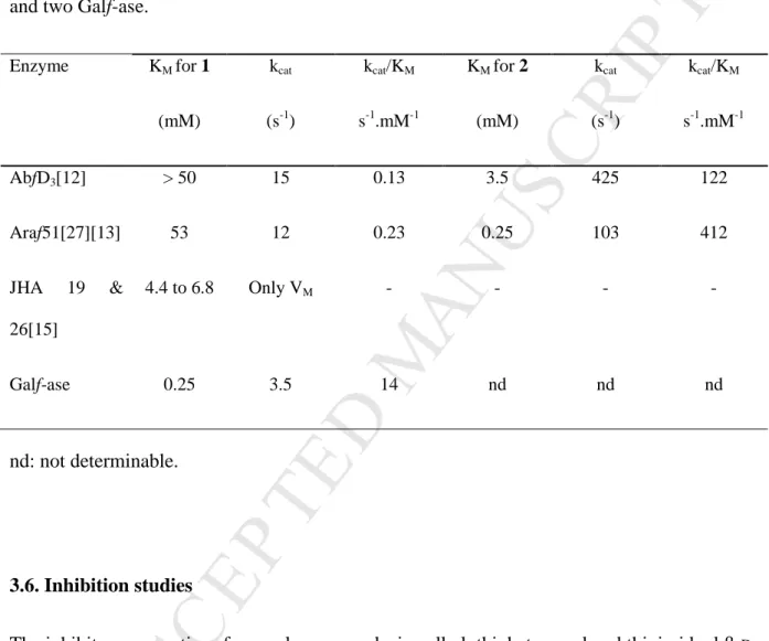
and Araf51 owing dual specificity for α-L-Araf and β-D-Galf (Table 1, lines 1 & 2), the
specificity of Galf-ase is incredibly increased toward the hexofuranosyl moiety by a factor of about 100-fold. When compared to the only two reported cloned Galf-ases (line 3), from
Streptomyces sp. strains JHA19 and JHA26, the values are not in the same order of
magnitude. For Galf-ase JHA19 and JHA26, KM value ranges from 4.4 to 6.8 mM
respectively,[15] yielding at least a 18-fold lower substrate affinity compared to Galf-ase’s KM
which is in the µM level (line 4). Finally, the Galf-ase parameters described herein are the best reported to date and fully compatible with the development of the specific and efficient biocatalyzed incorporation of Galf entities into more complex glycoconjugates.
0 2000 4000 6000 0 5.0××××10-6 1.0××××10-5 1.5××××10-5 [pNP ββββ-D-Galf] (µµµµM) v0 /M m in -1
M
AN
US
CR
IP
T
AC
CE
PT
ED
Fig. 5. Michaelis-Menten plot of pNP β-D-Galf 1 hydrolysis reaction catalysed by Galf-ase.
Mean values and SD error bars were calculated from three independent experiments and from a single protein preparation. Error bars correspond to the SD of 3 independent measurements.
Table 1. KM for synthetic pNP furanoses 1 and 2 toward two different arabinofuranosidases
and two Galf-ase.
Enzyme KM for 1 (mM) kcat (s-1) kcat/KM s-1.mM-1 KM for 2 (mM) kcat (s-1) kcat/KM s-1.mM-1 AbfD3[12] > 50 15 0.13 3.5 425 122 Araf51[27][13] 53 12 0.23 0.25 103 412 JHA 19 & 26[15] 4.4 to 6.8 Only VM - - - - Galf-ase 0.25 3.5 14 nd nd nd nd: not determinable. 3.6. Inhibition studies
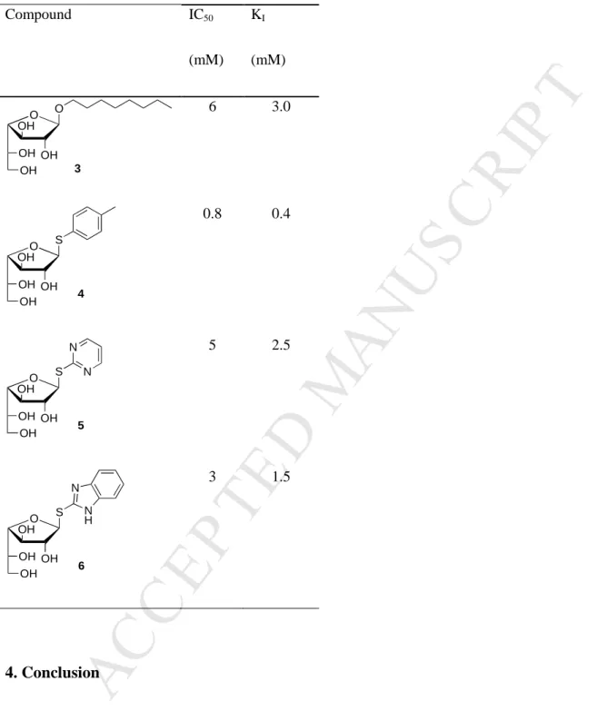
The inhibitory properties of several compounds, i.e. alkyl, thioheteroaryl and thioimidoyl β-D -galactofuranosides 3-6, were investigated by incubating the enzyme, Galf-ase, in the presence of both the substrate pNP β-D-Galf and compounds 3-6 (Fig. S4.). These compounds were tested as inhibitors because (i) the glycone part, that is β-D-Galf, is recognized by the active
site, (ii) the aglycone part is mimicking a carbohydrate moiety and (iii) to probe the stability of the thioglyosidic bond.[20] It has been reported previously that alkyl, aryl and heteroaryl 1-thio-β-D-galactofuranoside derivatives are good inhibitors of exo β-D-galactofuranosidase,
M
AN
US
CR
IP
T
AC
CE
PT
ED
isolated from P. fellutanum culture media, and D-galactono-1,4-lactone has been evaluated as
a reference inhibitor with an IC50 value of 0.02 mM.[28] In our hands, compounds 3, 5 and 6
showed weak inhibitory activity with IC50 values of 6, 5 and 3 mM respectively, while
Galf-ase was more sensitive to the inhibition by thiogalactofuranoside 4 (Table 2.). This latter showed the best inhibition activity, with IC50 value of 0.8 mM. The analysis of the
Lineweaver–Burk plot (Fig. 6.) indicated that the compound 4 is a competitive inhibitor of the Galf-ase with KI value of 0.4 mM. Nevertheless, compared to the only reference L
-arabino-1,4-lactone inhibitor previously probed on Galf-ase,[15] 4 exhibited a 100 times increased in inhibition, that can be mostly attributed to its structural analogy to the artificial substrate.
Fig. 6. Lineweaver–Burk plot of competitive inhibition of Galf-ase hydrolytic activity towards
pNP β-D-Galf by 4. Mean values and SD error bars were calculated from two independent
M
AN
US
CR
IP
T
AC
CE
PT
ED
Table 2. Inhibition studies with galactofuranosides 3-6: determination of their respective IC50
& KI. Compound IC50 (mM) KI (mM) 6 3.0 0.8 0.4 5 2.5 3 1.5 4. Conclusion
In this work, we provided a complete biochemical and kinetic characterization of a subcloned recombinant Galf-ase since the recent publication of the first ever cloned one. This enzyme was obtained with a reasonable yield of 0.5 mg/liter of culture, and optimally active at pH 4.5 and at 60°C. It is inhibited by substrate’s structural thio analogue (IC50 = 0.8 mM), which
O OH OH OH O OH 3 O OH OH OH S OH 4 O OH OH OH S N N OH 5 O OH OH OH S OH 6 N H N
M
AN
US
CR
IP
T
AC
CE
PT
ED
proved to be a moderate competitive inhibitor (KI = 0.4 mM). It exhibits substrate specificity
exclusively towards pNP β-D-Galf, giving a KM value of 250 µM, and the highest enzymatic
efficiency ever observed of 14 mM-1.s-1. Since to date, only a few furanosidases have been
involved in the chemoenzymatic approaches, it would be of great interest to explore and evaluate the potential synthetic ability of this enzyme to act both as wild-type in transglycosylation[29] or after site-directed mutagenesis as an efficient thioligase[30] in the efficient preparation of galactofuranosyl conjugates. Works are currently under progress in this direction and results will be reported in due course.
Funding
This work was supported by the APR IR NeoLect project from the Région Centre-Val de Loire, France. S. P. acknowledges support from Institut National de la Santé et de la Recherche Médicale (INSERM).
References
[1] A. Varki, Biological roles of oligosaccharides: all of the theories are correct, Glycobiology. 3 (1993) 97–130. doi:10.1093/glycob/3.2.97.
[2] M.R. Richards, T.L. Lowary, Chemistry and biology of galactofuranose-containing polysaccharides, ChemBioChem. 10 (2009) 1920–1938. doi:10.1002/cbic.200900208.
[3] P. Peltier, R. Euzen, R. Daniellou, C. Nugier-Chauvin, V. Ferrières, Recent knowledge and innovations related to hexofuranosides: structure, synthesis and applications, Carbohydr. Res. 343 (2008). doi:10.1016/j.carres.2008.02.010.
[4] I. Chlubnova, L. Legentil, R. Dureau, A. Pennec, M. Almendros, R. Daniellou, C. Nugier-Chauvin, V. Ferrières, Specific and non-specific enzymes for furanosyl-containing conjugates: Biosynthesis, metabolism, and chemo-enzymatic synthesis,
M
AN
US
CR
IP
T
AC
CE
PT
ED
Carbohydr. Res. 356 (2012). doi:10.1016/j.carres.2012.04.002.
[5] P.M. Nassau, S.L. Martin, R.E. Brown, A. Weston, D. Monsey, M.R. Mcneil, K. Duncan, Galactofuranose biosynthesis in Escherichia coli K-12: Identification and cloning of UDP-galactopyranose mutase, J. Bacteriol. 178 (1996) 1047–1052. doi:10.1128/jb.178.4.1047-1052.1996.
[6] J. Ati, C. Colas, P. Lafite, R.P. Sweeney, R.B. Zheng, T.L. Lowary, R. Daniellou, The LPG1x family from Leishmania major is constituted of rare eukaryotic
galactofuranosyltransferases with unprecedented catalytic properties, Sci. Rep. 8 (2018) 17566. doi:10.1038/s41598-018-35847-w.
[7] M. Rietschel-Berst, N.H. Jentoft, P.D. Rick, C. Pletcher, F. Fang, J.E. Gander, Extracellular exo-Galactofuranosidase from Penicillium charlesii, J. Biol. Chem. 252 (1977) 3219–3226. http://www.ncbi.nlm.nih.gov/pubmed/863879.
[8] C. Marino, C. Gallo-Rodriguez, R.M. de Lederkremer, Galactofuranosyl-containing glycans: Occurrence, synthesis and biochemistry, 2012. doi:10.1063/1.3243690.
[9] H.J. Kamphuis, G.A. De Ruiter, F.M. Rombouts, P. Mischnick, J.H. Van Boom, A.W. Van Bruggen-Van Der Lugt, New structural features of the antigenic extracellular polysaccharides of Penicillium and Aspergillus species revealed with exo-beta-D-galactofuranosidase., J. Bacteriol. 174 (2016) 6096–6102. doi:10.1128/jb.174.19.6096-6102.1992.
[10] N. Ramli, M. Fujinaga, M. Tabuchi, K. Takegawa, S. Iwahara, Isolation and
Characterization of a Novel Endo- β -galactofuranosidase from Bacillus sp., Biosci. Biotechnol. Biochem. 59 (1995) 1856–1860. doi:10.1271/bbb.59.1856.
M
AN
US
CR
IP
T
AC
CE
PT
ED
Lederkremer, Evidence for exo β-D-galactofuranosidase in Trypanosoma cruzi, Mol. Biochem. Parasitol. 127 (2003) 85–88. doi:10.1016/S0166-6851(02)00307-9.
[12] R. Euzen, G. Lopez, C. Nugier-Chauvin, V. Ferrières, D. Plusquellec, C. Rémond, M. O’Donohue, A Chemoenzymatic Approach for the Synthesis of Unnatural
Disaccharides Containing D-Galacto- or D-Fucofuranosides, European J. Org. Chem. 2005 (2005) 4860–4869. doi:10.1002/ejoc.200500525.
[13] I. Chlubnová, D. Filipp, V. Spiwok, H. Dvoáková, R. Daniellou, C. Nugier-Chauvin, B. Králová, V. Ferrières, Enzymatic synthesis of oligo-D-galactofuranosides and L-arabinofuranosides: From molecular dynamics to immunological assays, Org. Biomol. Chem. 8 (2010). doi:10.1039/b926988f.
[14] I. Chlubnová, B. Králová, H. Dvořáková, P. Hošek, V. Spiwok, D. Filipp, C. Nugier-Chauvin, R. Daniellou, V. Ferrières, The versatile enzyme Araf51 allowed efficient synthesis of rare pathogen-related β-D-galactofuranosyl-pyranoside disaccharides, Org. Biomol. Chem. 12 (2014). doi:10.1039/c3ob42519c.
[15] E. Matsunaga, Y. Higuchi, K. Mori, N. Yairo, T. Oka, S. Shinozuka, K. Tashiro, M. Izumi, S. Kuhara, K. Takegawa, Identification and characterization of a novel galactofuranose-specific β-D-galactofuranosidase from Streptomyces species, PLoS One. 10 (2015) 1–16. doi:10.1371/journal.pone.0137230.
[16] E. Matsunaga, Y. Higuchi, K. Mori, K. Tashiro, S. Kuhara, K. Takegawa, Draft genome sequence of Streptomyces sp. JHA19, a strain that possesses β-D-galactofuranosidase activity, Genome Announc. 3 (2015) 3–4.
doi:10.1128/genomeA.01171-15.
[17] E. Matsunaga, Y. Higuchi, K. Mori, N. Yairo, S. Toyota, T. Oka, K. Tashiro, K. Takegawa, Characterization of a PA14 domain-containing galactofuranose-specific
β-M
AN
US
CR
IP
T
AC
CE
PT
ED
D-galactofuranosidase from Streptomyces sp., Biosci. Biotechnol. Biochem. 81 (2017) 1314–1319. doi:10.1080/09168451.2017.1300518.
[18] G.D. Davis, C. Elisee, D.M. Newham, R.G. Harrison, New fusion protein systems designed to give soluble expression in Escherichia coli., Biotechnol. Bioeng. 65 (1999) 382–8. http://www.ncbi.nlm.nih.gov/pubmed/10506413 (accessed April 12, 2019).
[19] L. Legentil, J.-L. Audic, R. Daniellou, C. Nugier-Chauvin, V. Ferrières, Studies of a furanoside as antimycobacterial agent loaded into a biodegradable PBAT/sodium caseinate support, Carbohydr. Res. 346 (2011). doi:10.1016/j.carres.2011.04.025.
[20] G. Lopez, R. Daniellou, M. O’Donohue, V. Ferrières, C. Nugier-Chauvin, Thioimidoyl furanosides as first inhibitors of the α-L-arabinofuranosidase AbfD3, Bioorganic Med. Chem. Lett. 17 (2007) 434–438. doi:10.1016/j.bmcl.2006.10.032.
[21] U.K. Laemmli, Cleavage of structural proteins during the assembly of the head of bacteriophage T4., Nature. 227 (1970) 680–5.
http://www.ncbi.nlm.nih.gov/pubmed/5432063 (accessed May 16, 2019).
[22] C. Yung-Chi, W.H. Prusoff, Relationship between the inhibition constant (KI) and the concentration of inhibitor which causes 50 per cent inhibition (I50) of an enzymatic reaction, Biochem. Pharmacol. 22 (1973) 3099–3108.
doi:10.1016/0006-2952(73)90196-2.
[23] G.L. Rosano, E.A. Ceccarelli, Recombinant protein expression in Escherichia coli: Advances and challenges, Front. Microbiol. 5 (2014) 1–17.
doi:10.3389/fmicb.2014.00172.
[24] J.J. Winzerling, P. Berna, J. Porath, How to use immobilized metal ion affinity chromatography, Methods. 4 (1992) 4–13. doi:10.1016/1046-2023(92)90052-A.
M
AN
US
CR
IP
T
AC
CE
PT
ED
[25] V. Gaberc-porekar, V. Menart, Perspectives of immobilized-metal affinity chromatography, J. Biochem. Biophys. Methods. 49 (2001) 335–360.
[26] N.J. Alves, K.B. Turner, I.L. Medintz, S.A. Walper, Protecting enzymatic function through directed packaging into bacterial outer membrane vesicles, Sci. Rep. 6 (2016). doi:10.1038/srep24866.
[27] E.J. Taylor, N.L. Smith, J.P. Turkenburg, S. D’souza, H.J. Gilbert, G.J. Davies, Structural insight into the ligand specificity of a thermostable family 51
arabinofuranosidase, Araf 51, from Clostridium thermocellum, Biochem. J. 395 (2006) 31–37. doi:10.1042/BJ20051780.
[28] E. Repetto, C. Marino, O. Varela, Synthesis of the (1→6)-linked thiodisaccharide of galactofuranose: Inhibitory activity against a β-galactofuranosidase, Bioorganic Med. Chem. 21 (2013) 3327–3333. doi:10.1016/j.bmc.2013.02.057.
[29] A. Pennec, R. Daniellou, P. Loyer, C. Nugier-Chauvin, V. Ferrières, Araf51 with improved transglycosylation activities: One engineered biocatalyst for one specific acceptor, Carbohydr. Res. 402 (2015) 50–55. doi:10.1016/j.carres.2014.10.031.
[30] M. Almendros, D. Danalev, M. Franois-Heude, P. Loyer, L. Legentil, C. Nugier-Chauvin, R. Daniellou, V. Ferrières, Exploring the synthetic potency of the first
furanothioglycoligase through original remote activation, Org. Biomol. Chem. 9 (2011) 8371–8378. doi:10.1039/c1ob06227a.