HAL Id: inserm-03037871
https://www.hal.inserm.fr/inserm-03037871
Submitted on 3 Dec 2020HAL is a multi-disciplinary open access archive for the deposit and dissemination of sci-entific research documents, whether they are pub-lished or not. The documents may come from teaching and research institutions in France or abroad, or from public or private research centers.
L’archive ouverte pluridisciplinaire HAL, est destinée au dépôt et à la diffusion de documents scientifiques de niveau recherche, publiés ou non, émanant des établissements d’enseignement et de recherche français ou étrangers, des laboratoires publics ou privés.
Association of Partial Chromosome 3 Deletion in Uveal
Melanomas With Metastasis-Free Survival
Manuel Rodrigues, Khadija Ait Rais, Flore Salviat, Nathalie Algret,
Fatoumata Simaga, Raymond Barnhill, Sophie Gardrat, Vincent Servois,
Pascale Mariani, Sophie Piperno-Neumann, et al.
To cite this version:
Manuel Rodrigues, Khadija Ait Rais, Flore Salviat, Nathalie Algret, Fatoumata Simaga, et al.. As-sociation of Partial Chromosome 3 Deletion in Uveal Melanomas With Metastasis-Free Survival. JAMA Ophthalmology, American Medical Association 2020, 138 (2), pp.182. �10.1001/jamaoph-thalmol.2019.5403�. �inserm-03037871�
1
Association of Partial Chromosome 3 Deletion in Uveal Melanomas with
1
Metastasis-free Survival
2
Manuel Rodrigues1,2*, M.D. Ph.D., Khadija Ait-Rais3, Flore Salviat4, M.D., Nathalie 3
Algret4, Fatoumata Simaga3, Raymond Barnhill5,6, M.D., Sophie Gardrat1,5, M.D., 4
Vincent Servois7, M.D., Pascale Mariani8, M.D., Sophie Piperno-Neumann2, M.D., 5
Sergio Roman-Roman9, Pharm.D. Ph.D., Olivier Delattre1,3, M.D. Ph.D., Nathalie 6
Cassoux6,10, M.D. Ph.D., Alexia Savignoni4, M.D. Ph.D., Marc-Henri Stern1,3, M.D. 7
Ph.D., Gaëlle Pierron3, Ph.D. 8
9
1 - Unit 830 « Cancer, heterogeneity, instability and plasticity » INSERM, Institut 10
Curie, PSL Research University, Paris, 75248, France 11
2 - Department of Medical Oncology, Institut Curie, PSL Research University, Paris, 12
75248, France 13
3- Department of Genetics, Institut Curie, PSL Research University, Paris, 75248, 14
France 15
4 - Department of Biometry, Institut Curie, PSL Research University, Paris, 75248, 16
France 17
5- Department of Biopathology, Institut Curie, PSL Research University, Paris, 18
75248, France 19
6- Faculty of Medicine, University of Paris Descartes, Paris, 75006, France 20
7- Department of Medical Imaging, Institut Curie, PSL Research University, Paris, 21
75248, France 22
8- Department of Surgical Oncology, Institut Curie, PSL Research University, Paris, 23
75248, France 24
2
9- Department of Translational Research, Institut Curie, PSL Research University, 25
Paris, 75248, France 26
10- Department of Ocular Oncology, Institut Curie, PSL Research University, Paris, 27
75248, France 28
29
*Correspondence to: Dr. Manuel Rodrigues, Institut Curie 26 rue d’Ulm, 75248
30
Paris Cedex 05. email: manuel.rodrigues@curie.fr Tel: +33144324672 Fax: 31 +33153104041 32 33 Word count: 2,729 34 35 36 37
3
Key Points
38
Question: What is the association of partial chromosome 3 deletion in uveal
39
melanomas with metastasis-free survival? 40
Findings: In this retrospective study, partial deletions of chromosome 3
41
encompassing the BAP1 locus were associated with a lower metastasis-free survival 42
at 60 months compared to uveal melanomas without such deletion. 43
Meaning: These findings suggest that uveal melanomas carrying a partial deletion of
44
chromosome 3 encompassing the BAP1 locus have a poor prognosis. 45
4
Abstract
47
Importance
48
Studies on uveal melanomas (UMs) demonstrated the prognostic value of 8q gain 49
and monosomy 3, but the prognosis of UMs with partial deletion of chromosome 3 50
remains to be defined. 51
Objective
52
To determine the association of partial chromome 3 deletion in uveal melanomas with 53
metastasis-free survival. 54
Design
55
Retrospective cohort of consecutive comparative genomic hybridization arrays from 56
May 2006 to July 2015. 57
Setting
58
Monocentric study in a referral center. 59
Participants
60
Patients presenting with UMs with and without partial loss of chromosome 3. 61
Main Outcomes and Measures
62
Metastasis-free survival and overall survival at 60 months. 63
Results
64
Of the 1,088 consecutive comparative genomic hybridization arrays that were 65
performed, 43 UMs (4%) carried partial deletions of chromosome 3. Median follow-up 66
was 66 months. Metastasis-free survival at 60 months was 34% (95% confidence 67
interval [CI], 15.8 to 71.4) for UMs carrying a deletion of the BAP1 (BRCA1 68
associated protein-1) locus (BAP1del; 24 tumors) and 81% (95% CI, 64.8 to 100) for
69
UMs without the loss of the BAP1 locus (BAP1 normal; BAP1nl; 19 tumors; log-rank 70
p-value = .001). Overall survival at 60 months was 65% (95% CI, 43.5 to 95.8) versus 71
5
84% (95% CI, 69.0 to 100) in the BAP1del and the BAP1nl groups, respectively (log-72
rank p-value < .001). In these 43 cases, metastasis-free survival at 60 months was 73
100% for UMs without loss of the BAP1 locus or 8q gain, 70% (95% CI, 50.5 to 96.9) 74
for UMs carrying one of these alterations and 13% for those carrying both (95% CI, 75
2.1 to 73.7; log-rank p-value < .001). Similarly, overall survival at 60 months was 76
100%, 81% (95% CI, 63.3 to 100) and 47% (95% CI, 23.3 to 93.6) in these three 77
groups, respectively (log-rank p-value < .001). 78
Conclusions and Relevance
79
These findings suggest that partial deletion of chromosome 3 encompassing the 80
BAP1 locus is associated with poor prognosis. A cytogenetic classification of UMs
81
could be proposed based on the status of the BAP1 locus instead of chromosome 3, 82
locus, while also taking chromosome 8q into account. 83
6
Introduction
85
Uveal melanoma (UM) is the most common primary malignant ocular tumor in adults 86
of European ancestry1. Despite efficient treatment, up to 50% of the patients will 87
eventually develop metastases2-4. Reliable prognostic assessment allows a closer 88
monitoring of high-risk patients. Pathological prognostic factors include large tumor 89
basal diameter, thickness, ciliary body involvement, extraocular extension, epithelioid 90
cell histology, high mitotic rate and lymphocytic infiltration5. The gene expression 91
profile DecisionDx-UM (GEP; Castle Biosciences, Friendswood, TX), based on the 92
expression level of 12 genes, is frequently used in North America to complete the 93
prognostic assessment6,7. 94
In the early 1990s, recurrent cytogenetic aberrations including monosomy 3 (M3), 95
gain of 6p and 8q were identified in UM samples8. In 1996, M3 was empirically shown 96
to be a robust prognostic factor9. Since then, genomic arrays have become routine 97
tools to refine pathological prognosis along with the GEP. We previously refined the 98
prognostic value of M3 and gain of 8q by defining three groups: (i) high-risk patients 99
whose tumors present a M3 and an 8q gain with a 2-year metastasis-free interval 100
(2y-MFI) of 37%; (ii) intermediate-risk with either a M3 or an 8q gain (2y-MFI: ~85%) 101
and (iii) low-risk with neither M3 nor 8q gain (2y-MFI: ~100%)10. 102
The most common hypothesis to explain the poor prognosis of M3 tumors is the 103
presence of one or more tumor suppressor genes (TSG) on chromosome 3. BAP1 104
(BRCA1 associated protein-1), a TSG located on the 3p21.1 cytoband, is now 105
established as a main actor of UM malignant transformation as it is frequently 106
mutated in M3 tumors and germline mutations are associated with UM 107
predisposition11-16. However, all or most BAP1-mutated UMs intriguingly present a 108
M3 (or a loss of heterozygosity of the whole chromosome 3 due an isodisomy) 109
7
suggesting that the role of chromosome 3 loss in UM tumorigenesis may not be 110
restricted to BAP1 inactivation. Therefore, prognostication of UM samples with partial 111
deletions of chromosome 3, as sometimes observed in our daily practice and by 112
other authors, is problematic17. The goals of the present study were to explore these 113
UMs with partial deletions of chromosome 3, as assessed by comparative genomic 114
hybridization (array-CGH), in order to assess their prognosis and to determine the 115
minimal region of deletion associated with poor prognosis. 116
8
Materials and methods
117
Patients
118
This study was approved by our institutional ethics committee. Written informed 119
consent for the use of tissues and data for research was signed by each patient. The 120
study complied with the principles of the Declaration of Helsinki. All patients were 121
referred to our institution and followed up by our physicians. Clinical diagnosis of 122
uveal melanoma was based on the presence of typical clinical findings as previously 123
described10. Local treatment consisted of proton beam radiotherapy, iodine 125 124
brachytherapy or enucleation, depending on the size and location of tumors. Tumor 125
samples were obtained by enucleation, endoresection or fine-needle aspiration at the 126
time of clip or plaque positioning. Liver ultrasound, liver magnetic resonance imaging 127
or body computed tomography were performed at diagnosis and every 6 months 128
afterwards. Diagnosis of metastasis was systematically confirmed by a biopsy. 129
Genomic analysis
130
Tumor DNA was extracted and processed as previously described10. Array-CGH was 131
performed on three different platforms according to the period when the test was 132
performed: bacterial artificial chromosome arrays as previously described18, 133
NimbleGen 4×72 K arrays (Roche NimbleGen, Madison, Wisconsin, USA) and 134
Agilent 180K CGH/LOH custom chip (Santa Clara, California, USA). Array-CGH were 135
interpreted by three of the authors (MR, KAR, GP). Partial deletion of chromosome 3 136
was defined as the loss of at least one region of chromosome 3, but not the totality, 137
whatever its size and location. Genomic positions in this article are defined in hg18 138
human genome assembly. 139
Statistical analysis
9
Clinical, pathological and genomic data at diagnosis and follow-up events (local and 141
distant recurrences, second cancers, death from UM or from any other cause) were 142
collected. The French Death Registry was consulted for patients lost to follow-up. 143
The metastasis-free survival (MFS) at 60 months was defined as the proportion of 144
patients alive and free of metastasis at 60 months of follow-up after local treatment of 145
primary UM. The overall survival (OS) at 60 months was defined as the proportion of 146
patients alive at 60 months of follow-up after local treatment of primary UM, whatever 147
the cause of death. Survival distributions were estimated by the Kaplan–Meier 148
method and compared using the log-rank test. All tests were bilateral and performed 149
with a significant level of 5%. In order to identify variables associated with MFS, a 150
Cox regression analysis of candidate prognostic factors was performed using a 151
forward stepwise selection procedure. The added value of each variable to the Cox 152
model was determined using a likelihood ratio test with a significant level of 5%. 153
Statistical analysis was performed using R software V3.3 (http://www.r-project.org/). 154
10
Results
156
We prospectively re-analyzed the array-CGH profiles in 1,088 UMs which had been 157
processed between May 2006 and July 2015, and detected 43 cases (4.0%) 158
harboring a partial deletion of chromosome 3 (eTable 1 in the supplement). Median 159
follow-up in these 43 cases was 66 months (range: 1.2-126.2 months). Median age 160
was 58 years-old (range 12-79), median tumor diameter was 16 millimeters (range: 161
10-22) and median thickness was 10 millimeters (range: 5.3-18.2). Ciliary body and 162
optic nerve were involved in 33% (14/43) and in 9% (4/43) of cases, respectively. Cell 163
morphology was epithelioid or mixed in 30% of cases (13/43). Primary tumors were 164
treated by enucleation in 42% (18/43) of cases. MFS and OS at 60 months were 61% 165
(95% confidence interval [CI], 46 to 79.7) and 76% (95% CI, 62.8 to 92.3), 166
respectively. A global overview of copy number profiles is provided in eFigure 1 in the 167
supplement. Size of deletions ranged from 1.36 to 110.88 megabases. 168
169
We first explored survival data in an unsupervised manner and observed three 170
recurrently lost regions in at least eight metastatic samples: (i) from 3pter to p22.2, (ii) 171
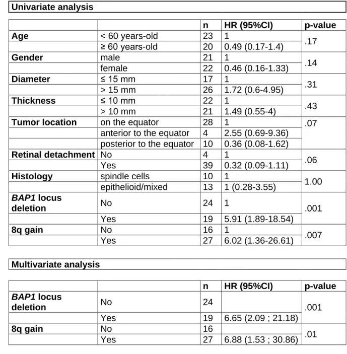
from 3p22.1 to p14.2 and (iii) from 3q13.2 to q24 (Figure 1). Of these, two regions 172
were more frequently lost in metastatic cases than in non-metastatic ones: the 3pter-173
p22.2 region (8/13 versus 6/30 cases, respectively; p=.013; odds ratio [OR]=6.1; 95% 174
CI, 1.2 to 34.1) and the 3p22.1-p14.2 region, which encompasses BAP1 (10/13 175
versus 9/30 cases, respectively; p=.007; OR=7.4; 95% CI, 1.5 to 51.8). These two
176
regions were close and highly correlated between each other, as 8 out of 10 177
metastatic cases presenting a 3p22.1-p14.2 loss also presented a 3pter-p22.2 loss. 178
The 3p22.1-p14.2 region carries 290 other genes beside BAP1, but no recurrent 179
mutations of these 290 genes were found in public and in-house databases12,19,20. 180
11 181
We then hypothesized that BAP1 loss was the main driver of poor prognosis in M3. 182
To explore this hypothesis, we compared tumors with a chromosome 3 partial 183
deletion encompassing the BAP1 locus (24 tumors; BAP1del) and tumors with a 184
chromosome 3 partial deletion not encompassing the BAP1 locus (19 tumors; 185
BAP1nl). Tumors carrying a loss of the BAP1 locus frequently showed large losses of 186
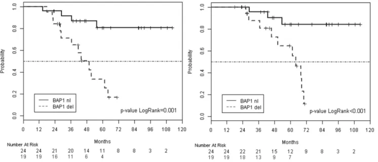
the short arm of chromosome 3 (Figure 2). MFS at 60 months was 81% (95% CI, 187
64.8 to 100) for the BAP1nl genomic group and 34% (95% CI, 15.8 to 71.4) for the 188
BAP1del group (Figure 3; p=.001). OS at 60 months was 84% (95% CI, 69.0 to 100) 189
for the BAP1nl genomic group and 65% (95% CI, 43.5 to 95.8) for the BAP1del group 190
(p<.001). The only variables associated with MFS in univariate analysis were loss of 191
the BAP1 locus and gain of 8q. These two variables independently contributed to 192
MFS in multivariate analysis (Table 1). 193
194
We defined four groups depending on the BAP1 locus (lost/not lost) and 8q 195
(gained/not gained) statuses. Prognoses of the BAP1 locus lost/8q normal and BAP1 196
locus not lost/8q gained were similar so we merged these two groups, as in our 197
previous classification (eFigure 2 in the supplement)10. By analogy with our previous 198
work, we defined three prognosis groups as follows: (i) a group at low risk of 199
metastasis without loss of the BAP1 locus or 8q gain (9 cases), (ii) an intermediate 200
risk group with tumors carrying either loss of the BAP1 locus (7 cases) or 8q gain (15 201
cases) and (iii) a high risk group with loss of the BAP1 locus and 8q gain (12 cases). 202
MFS at 60 months were 100%, 70% (95% CI, 50.5 to 96.9) and 13% (95% CI, 2.1 to 203
73.7) for the low-, intermediate- and high-risk groups, respectively (Figure 4; p<.001). 204
12
OS at 60 months were 100%, 81% (95% CI, 63.3 to 100) and 47% (95% CI, 23.3 to 205
93.6) for the low-, intermediate- and high-risk groups, respectively (p<.001). 206
13
Discussion
208
In this work, we explored a relatively large series of UMs with partial deletion of 209
chromosome 3 and showed that loss of the BAP1 locus is likely to explain the poor 210
prognosis of M3 UM. This result was obtained by two different approaches 211
investigating indirectly the prognostic value of the most frequently deleted regions of 212
chromosome 3 and then directly assessing the prognostic value of the loss of the 213
BAP1 locus in this series. The first consequence is to provide a potentially more
214
accurate estimation of the prognosis of UMs presenting a partial deletion of 215
chromosome 3. Our classification suggested efficiency in predicting metastatic 216
outcome, identifying a group with a very good MFS with no recurrence and a group 217
with a high risk of 92% of recurrences with a median follow-up of more than 5 years. 218
Survival rates were close to what we observed in a previous series of UMs 219
presenting either a M3 or a disomy 3, associated or not with 8q gain10. This 220
hypothesis has yet to be verified in subsequent studies because direct comparison 221
could not be done here. 222
223
Other teams are using different genomic technologies to assess UM prognosis. 224
Fluorescence in situ hybridization (FISH) is widely used but it may miss the loss of 225
the BAP1 locus if the probe is not centered on this gene, as observed in several 226
publications2,21-24. Furthermore, FISH is often performed without chromosome 8q 227
assessment leading to suboptimal prognosis estimation. Multiplex ligation-dependent 228
probe amplification (MLPA) assay covering the BAP1 locus is a good alternative to 229
characterize recurrent genomic imbalances in UM but MLPA, as well as FISH and 230
array-CGH, only evaluate copy number and, consequently does not identify 231
isodisomic cases25,26. GEP is a transcriptomic prognosis assay that is widely used in 232
14
United States7. This assay distinguishes two subsets of UMs either at low or high risk 233
of metastasis by assessing the expression of 12 genes, including four that are 234
located on the short arm of chromosome 3 (EIF1B, LMCD1, ROBO1, SATB1) and 235
one on the 3q (FXR1). Underexpression of these genes, possibly due to M3, is 236
associated with poor prognosis. A more accurate prediction by GEP is possible by 237
adding the expression of PRAME, a gene located on an instable region of 238
chromosome 22 exposed to duplication, which was correlated to the 8q status in the 239
pivotal paper6. To our knowledge, GEP has never been specifically tested in a large 240
series of UMs with partial chromosome 3 deletions. Furthermore, GEP has never 241
been compared to the combined M3/8q signature in a large cohort, impeding any 242
conclusion on the superiority of one modality on the other. BAP1 243
immunohistochemistry is an alternative way to assess the prognosis of UMs27,28. 244
However, immunohistochemistry for BAP1 does not correlate in all cases to the 245
BAP1 mutational status in UM, and is therefore not a perfect surrogate27. 246
247
In the present series, partial deletions of chromosome 3 were found in 4% of cases, 248
which is comparable with some previous series29-31 but lower than others17,32,33. 249
Recruitment bias may explain part of this discrepancy but it is most probably 250
explained by the variety of technologies, as well as the different classifications that 251
were used. Comparison of all these studies is therefore limited. Similarly, the 252
prognosis of these tumors was not clear as a discrepancy was observed with some 253
series associating partial loss with good prognosis17,29,32 while others associated it 254
with intermediate or poor prognosis25,33,34. These differences may be explained by 255
the absence of distinction depending on the loss of the BAP1 locus compared to 256
other losses. 257
15 258
One explanation for the low MFS associated with the loss of this locus may be that 259
the loss of one BAP1 allele contributes to the inactivation of this gene and 260
subsequent aggressiveness of the tumor. However, the minimal region of deletion we 261
found associated with the lowest MFS in our series (3p22.1-p14.2) includes 291 262
genes. Even though this region encompasses BAP1, it cannot be excluded that other 263
important genes are present there and that haploinsufficiency of these genes affects 264
tumorigenesis. The two alleles of a TSG are commonly inactivated in the two-hit 265
model by a combination of different mechanisms, including total or partial loss of a 266
chromosome, deleterious point mutations, short insertions/deletions, large-scale 267
insertions/deletions and promoter methylations35. It is highly intriguing that, BAP1 268
inactivation is so frequently associated with monosomy 3 in UM, contrary to renal 269
clear-cell carcinomas and mesotheliomas, which rather carry losses of the short arm 270
of chromosome 3 only or deleterious mutations of both alleles16. Furthermore, 271
haploinsufficiency of other genes on chromosome 3, possibly on its long arm, may 272
play a role on UM tumorigenesis. This hypothesis may be of particular interest and 273
should be put in perspective with the recent discovery of MBD4 (3q21.3) recurrent, 274
inactivating mutations in UM36-38. 275
276
There is, for now, no standard treatment in the metastatic setting, but new drugs are 277
being developed in UM39. When an efficient treatment will be available, the following 278
step will consist in testing this treatment in the adjuvant setting in high-risk patients40. 279
Accurate prognosis evaluation is essential for such trials and assays able to assess 280
the status of the BAP1 locus and 8q status may then be required. Next-generation 281
sequencing appears to be the best option in the near future as it not only assesses 282
16
copy number, heterozygosity and mutational statuses of UMs at low cost and with a 283
lower amount of DNA, but also allow to follow circulating tumor DNA41-43. Moving 284
towards the implementation of such technologies in our daily practice will allow ocular 285
oncology to enter the modern age of precision medicine while reducing costs and 286 refining UM prognosis. 287 288 Limitations 289
The conclusions of this work are limited by its retrospective nature, but prospective 290
series are unrealistic given the rarity of such tumors. Instead, the present work 291
provides evidence to refine the current UM genomic classification, which may help 292
ophthalmologists to better predict the metastatic evolution of their patients. Before 293
generalization, other series from different centers are required. Furthermore, one 294
could argue that our series, composed of large tumors (median diameter of 16mm 295
and median thickness of 10mm) is not reflecting the overall population of UM 296
patients, particularly as larger UMs are known to host a greater frequency of genomic 297
alterations, including 8q gains37,44. Other centers have reported genomic studies on 298
biopsies of smaller UMs45. However, this procedure is not consensual and must not 299
be undertaken in inexperienced ocular oncology centers because of potential surgical 300
complications. Multicenter collaborative studies of small UM genomics are required to 301
address the question of partial chromosome 3 loss frequency at this stage of primary 302
UM development. Another limitation of this study is that the array-CGH technology is 303
not adapted to detect chromosome 3 isodisomy, an infrequent alteration in UM, 304
probably associated with poor prognosis. SNP-array can resolve this issue but, in the 305
future, next-generation sequencing will probably be the privileged technology to 306
circumvent this issue. More importantly, although the BAP1 locus hypothesis is a 307
17
logical hypothesis, we cannot definitely affirm that BAP1 is indeed the target of such 308
deletions. Chromosome 3 is dense in cancer genes and the BAP1 region, for 309
instance, encompasses the tumor-suppressor gene PBRM1, which was recently 310
found mutated in rare UMs. To confirm the BAP1 locus hypothesis and the 311
classification, validation series are required, ideally together with further work 312
sequencing BAP1 to confirm the presence of a second hit. 313
314
Conclusions
315
These findings suggest that partial deletion of chromosome 3 encompassing the 316
BAP1 locus is associated with poor prognosis. Consequently, a new cytogenetic
317
classification of UMs is proposed, based on the status of the BAP1 locus instead of 318
chromosome 3. The very frequent loss of the whole chromosome 3 in UMs raises the 319
possibility of other genes associated with UM tumorigenesis on this chromosome. 320
321 322
18
Funding/Support: This research was funded by the INCa/ITMO/AVIESAN PhD
323
fellowship program “Formation à la recherche translationelle” (M. Rodrigues), the 324
Institut National de la Santé et de la Recherche Médicale (INSERM), the Ligue 325
Nationale Contre le Cancer (Labellisation) and the Institut Curie. 326
327
Role of the Funder/Sponsor: None of the funders had any role in the design and
328
conduct of the study; collection, management, analysis, and interpretation of the 329
data; preparation, review, or approval of the manuscript; and the decision to submit 330
the manuscript for publication. 331
332
Acknowledgments: We thank the patients and their family members. We thank
333
Sylvain Dureau for his help with review. 334
335
Author Contributions: M. Rodrigues had full access to all the data in the study and
336
take responsibility for the integrity of the data and the accuracy of the data analysis. 337
Concept and design: Rodrigues, Savignoni, Stern, Pierron 338
Acquisition, analysis, or interpretation of data: Rodrigues, Ait-Rais, Salviat, Algret, 339
Simaga, Barnhill, Gardrat, Servois, Mariani, Piperno-Neumann, Roman-Roman, 340
Delattre, Cassoux, Savignoni, Stern, Pierron. 341
Drafting of the manuscript: Rodrigues, Savignoni, Stern, Pierron. 342
Critical revision of the manuscript for important intellectual content: Rodrigues, Ait-343
Rais, Salviat, Algret, Simaga, Barnhill, Gardrat, Servois, Mariani, Piperno-Neumann, 344
Roman-Roman, Delattre, Cassoux, Savignoni, Stern, Pierron 345
Supervision: Rodrigues, Savignoni, Stern, Pierron. 346
19
Conflicts of Interest: The authors declare no conflict of interest.
20
References
349
1. Mahendraraj K, Lau CS, Lee I, Chamberlain RS. Trends in incidence, survival, 350
and management of uveal melanoma: a population-based study of 7,516 patients 351
from the Surveillance, Epidemiology, and End Results database (1973-2012). Clin 352
Ophthalmol. 2016;10:2113-2119.
353
2. Desjardins L, Levy-Gabriel C, Lumbroso-Lerouic L, et al. [Prognostic factors 354
for malignant uveal melanoma. Retrospective study on 2,241 patients and recent 355
contribution of monosomy-3 research]. Journal francais d'ophtalmologie. Sep 356
2006;29(7):741-749. 357
3. Singh AD, Turell ME, Topham AK. Uveal melanoma: trends in incidence, 358
treatment, and survival. Ophthalmology. Sep 2011;118(9):1881-1885. 359
4. Kujala E, Makitie T, Kivela T. Very long-term prognosis of patients with 360
malignant uveal melanoma. Investigative ophthalmology & visual science. Nov 361
2003;44(11):4651-4659. 362
5. Brierley JD, Gospodarowicz MK, Wittekind C. The TNM Classification of 363
Malignant Tumours. 8th edition.2016.
364
6. Field MG, Decatur CL, Kurtenbach S, et al. PRAME as an Independent 365
Biomarker for Metastasis in Uveal Melanoma. Clinical cancer research : an official 366
journal of the American Association for Cancer Research. Mar 1
2016;22(5):1234-367
1242. 368
7. Onken MD, Worley LA, Ehlers JP, Harbour JW. Gene expression profiling in 369
uveal melanoma reveals two molecular classes and predicts metastatic death. 370
Cancer research. Oct 15 2004;64(20):7205-7209.
21
8. Prescher G, Bornfeld N, Becher R. Nonrandom chromosomal abnormalities in 372
primary uveal melanoma. Journal of the National Cancer Institute. Nov 21 373
1990;82(22):1765-1769. 374
9. Prescher G, Bornfeld N, Hirche H, Horsthemke B, Jockel KH, Becher R. 375
Prognostic implications of monosomy 3 in uveal melanoma. Lancet. May 4 376
1996;347(9010):1222-1225. 377
10. Cassoux N, Rodrigues MJ, Plancher C, et al. Genome-wide profiling is a 378
clinically relevant and affordable prognostic test in posterior uveal melanoma. The 379
British journal of ophthalmology. Jun 2014;98(6):769-774.
380
11. Abdel-Rahman MH, Pilarski R, Cebulla CM, et al. Germline BAP1 mutation 381
predisposes to uveal melanoma, lung adenocarcinoma, meningioma, and other 382
cancers. Journal of medical genetics. Dec 2011;48(12):856-859. 383
12. Robertson AG, Shih J, Yau C, et al. Integrative Analysis Identifies Four 384
Molecular and Clinical Subsets in Uveal Melanoma. Cancer Cell. Jan 8 385
2018;33(1):151. 386
13. Field MG, Durante MA, Anbunathan H, et al. Punctuated evolution of 387
canonical genomic aberrations in uveal melanoma. Nature communications. Jan 9 388
2018;9(1):116. 389
14. Rai K, Pilarski R, Boru G, et al. Germline BAP1 alterations in familial uveal 390
melanoma. Genes, Chromosomes & Cancer. Feb 2017;56(2):168-174. 391
15. Harbour JW, Onken MD, Roberson ED, et al. Frequent mutation of BAP1 in 392
metastasizing uveal melanomas. Science. Dec 3 2010;330(6009):1410-1413. 393
16. Wiesner T, Obenauf AC, Murali R, et al. Germline mutations in BAP1 394
predispose to melanocytic tumors. Nature genetics. Aug 28 2011;43(10):1018-1021. 395
22
17. Abdel-Rahman MH, Christopher BN, Faramawi MF, et al. Frequency, 396
molecular pathology and potential clinical significance of partial chromosome 3 397
aberrations in uveal melanoma. Modern pathology : an official journal of the United 398
States and Canadian Academy of Pathology, Inc. Jul 2011;24(7):954-962.
399
18. Trolet J, Hupe P, Huon I, et al. Genomic profiling and identification of high-risk 400
uveal melanoma by array CGH analysis of primary tumors and liver metastases. 401
Investigative ophthalmology & visual science. Jun 2009;50(6):2572-2580.
402
19. Furney SJ, Pedersen M, Gentien D, et al. SF3B1 mutations are associated 403
with alternative splicing in uveal melanoma. Cancer discovery. Oct 2013;3(10):1122-404
1129. 405
20. Johansson P, Aoude LG, Wadt K, et al. Deep sequencing of uveal melanoma 406
identifies a recurrent mutation in PLCB4. Oncotarget. Jan 26 2016;7(4):4624-4631. 407
21. Mensink HW, Vaarwater J, de Keizer RJ, et al. Chromosomal aberrations in 408
iris melanomas. The British journal of ophthalmology. Mar 2011;95(3):424-428. 409
22. Worley LA, Onken MD, Person E, et al. Transcriptomic versus chromosomal 410
prognostic markers and clinical outcome in uveal melanoma. Clinical cancer research 411
: an official journal of the American Association for Cancer Research. Mar 1
412
2007;13(5):1466-1471. 413
23. van Gils W, Lodder EM, Mensink HW, et al. Gene expression profiling in uveal 414
melanoma: two regions on 3p related to prognosis. Investigative ophthalmology & 415
visual science. Oct 2008;49(10):4254-4262.
416
24. Singh AD, Aronow ME, Sun Y, et al. Chromosome 3 status in uveal 417
melanoma: a comparison of fluorescence in situ hybridization and single-nucleotide 418
polymorphism array. Investigative ophthalmology & visual science. Jun 5 419
2012;53(7):3331-3339. 420
23
25. Damato B, Dopierala J, Klaasen A, van Dijk M, Sibbring J, Coupland SE. 421
Multiplex ligation-dependent probe amplification of uveal melanoma: correlation with 422
metastatic death. Investigative ophthalmology & visual science. Jul 2009;50(7):3048-423
3055. 424
26. Larsen AC, Holst L, Kaczkowski B, et al. MicroRNA expression analysis and 425
Multiplex ligation-dependent probe amplification in metastatic and non-metastatic 426
uveal melanoma. Acta ophthalmologica. Sep 2014;92(6):541-549. 427
27. Kalirai H, Dodson A, Faqir S, Damato BE, Coupland SE. Lack of BAP1 protein 428
expression in uveal melanoma is associated with increased metastatic risk and has 429
utility in routine prognostic testing. British journal of cancer. Sep 23 430
2014;111(7):1373-1380. 431
28. van Essen TH, van Pelt SI, Versluis M, et al. Prognostic parameters in uveal 432
melanoma and their association with BAP1 expression. Br J Ophthalmol. Aug 21 433
2014. 434
29. Thomas S, Putter C, Weber S, Bornfeld N, Lohmann DR, Zeschnigk M. 435
Prognostic significance of chromosome 3 alterations determined by microsatellite 436
analysis in uveal melanoma: a long-term follow-up study. British Journal of Cancer. 437
Mar 13 2012;106(6):1171-1176. 438
30. Tschentscher F, Prescher G, Horsman DE, et al. Partial deletions of the long 439
and short arm of chromosome 3 point to two tumor suppressor genes in uveal 440
melanoma. Cancer research. Apr 15 2001;61(8):3439-3442. 441
31. Cross NA, Rennie IG, Murray AK, Sisley K. The identification of chromosome 442
abnormalities associated with the invasive phenotype of uveal melanoma in vitro. Clin 443
Exp Metastasis. 2005;22(2):107-113.
24
32. Shields CL, Ganguly A, Bianciotto CG, Turaka K, Tavallali A, Shields JA. 445
Prognosis of uveal melanoma in 500 cases using genetic testing of fine-needle 446
aspiration biopsy specimens. Ophthalmology. Feb 2011;118(2):396-401. 447
33. Damato B, Dopierala JA, Coupland SE. Genotypic profiling of 452 choroidal 448
melanomas with multiplex ligation-dependent probe amplification. Clinical cancer 449
research : an official journal of the American Association for Cancer Research. Dec
450
15 2010;16(24):6083-6092. 451
34. Ewens KG, Kanetsky PA, Richards-Yutz J, et al. Genomic profile of 320 uveal 452
melanoma cases: chromosome 8p-loss and metastatic outcome. Invest Ophthalmol 453
Vis Sci. Aug 23 2013;54(8):5721-5729.
454
35. Knudson AG. Two genetic hits (more or less) to cancer. Nat Rev Cancer. Nov 455
2001;1(2):157-162. 456
36. Rodrigues M, Mobuchon L, Houy A, et al. Outlier response to anti-PD1 in 457
uveal melanoma reveals germline MBD4 mutations in hypermutated tumors. Nature 458
communications. May 14 2018;9(1):1866.
459
37. Rodrigues M, Mobuchon L, Houy A, et al. Evolutionary routes in metastatic 460
uveal melanomas depend on MBD4 alterations. Clin Cancer Res. Jun 21 2019. 461
38. Johansson PA, Stark A, Palmer JM, et al. Prolonged stable disease in a uveal 462
melanoma patient with germline MBD4 nonsense mutation treated with 463
pembrolizumab and ipilimumab. Immunogenetics. May 2019;71(5-6):433-436. 464
39. Carvajal RD, Schwartz GK, Tezel T, Marr B, Francis JH, Nathan PD. 465
Metastatic disease from uveal melanoma: treatment options and future prospects. 466
The British journal of ophthalmology. Jan 2017;101(1):38-44.
467
40. Piperno-Neumann S, Rodrigues MJ, Servois V, et al. A randomized 468
multicenter phase 3 trial of adjuvant fotemustine versus surveillance in high risk uveal 469
25
melanoma (UM) patients (FOTEADJ). Journal of Clinical Oncology. 470
2017;35(15_suppl):9502-9502. 471
41. Smit KN, van Poppelen NM, Vaarwater J, et al. Combined mutation and copy-472
number variation detection by targeted next-generation sequencing in uveal 473
melanoma. Mod Pathol. May 2018;31(5):763-771. 474
42. Afshar AR, Damato BE, Stewart JM, et al. Next-Generation Sequencing of 475
Uveal Melanoma for Detection of Genetic Alterations Predicting Metastasis. Transl 476
Vis Sci Technol. Mar 2019;8(2):18.
477
43. Matet A, Ait Rais K, Malaise D, et al. Comparative Cytogenetic Abnormalities 478
in Paired Choroidal Melanoma Samples Obtained Before and After Proton Beam 479
Irradiation by Transscleral Fine-Needle Aspiration Biopsy and Endoresection. 480
Cancers (Basel). Aug 14 2019;11(8).
481
44. Shain AH, Bagger MM, Yu R, et al. The genetic evolution of metastatic uveal 482
melanoma. Nature Genetics. Jul 2019;51(7):1123-1130. 483
45. Angi M, Kalirai H, Taktak A, et al. Prognostic biopsy of choroidal melanoma: 484
an optimised surgical and laboratory approach. The British journal of ophthalmology. 485
Aug 2017;101(8):1143-1146. 486
487 488
26
Tables
489
Table 1. Univariate and multivariate analyses of risk factors for metastasis.
490
HR (95%CI): hazard ratio (95% confidence interval); mm: millimeters; n: number of 491 cases. 492 Univariate analysis n HR (95%CI) p-value Age < 60 years-old 23 1 .17 ≥ 60 years-old 20 0.49 (0.17-1.4) Gender male 21 1 .14 female 22 0.46 (0.16-1.33) Diameter ≤ 15 mm 17 1 .31 > 15 mm 26 1.72 (0.6-4.95) Thickness ≤ 10 mm 22 1 .43 > 10 mm 21 1.49 (0.55-4)
Tumor location on the equator 28 1 .07
anterior to the equator 4 2.55 (0.69-9.36)
posterior to the equator 10 0.36 (0.08-1.62)
Retinal detachment No 4 1
.06
Yes 39 0.32 (0.09-1.11)
Histology spindle cells 10 1
1.00 epithelioid/mixed 13 1 (0.28-3.55) BAP1 locus deletion No 24 1 .001 Yes 19 5.91 (1.89-18.54) 8q gain No 16 1 .007 Yes 27 6.02 (1.36-26.61) Multivariate analysis n HR (95%CI) p-value BAP1 locus deletion No 24 .001 Yes 19 6.65 (2.09 ; 21.18) 8q gain No 16 .01 Yes 27 6.88 (1.53 ; 30.86) 493
27
Figures
494
Figure 1. Copy number profiles in metastatic versus non-metastatic cases.
495
Frequencies of losses at a given position are shown at the bottom. Light gray: non-496
metastatic cases (Met-; n=30); dark gray: metastatic cases (Met+; n=13). 497
498
Figure 2. Copy number profiles in BAP1del cases versus BAP1nl. Frequencies
499
of deletion at a given position are shown at the bottom. Light gray: BAP1nl cases 500
(n=19); dark gray: BAP1del cases (n=24). 501
502
Figure 3. Metastasis-free and overall survivals according to the loss of the
503
BAP1 locus. Metastasis-free survival (left) and overall survival (right) curves in UMs
504
with a partial loss of chromosome 3 encompassing the BAP1 locus or not. BAP1del: 505
deletion of the BAP1 locus; BAP1nl: absence of loss of the BAP1 locus. 506
507
Figure 4. Metastasis-free and overall survivals according to the three different
508
prognosis groups. Metastasis-free survival (left) and overall survival (right) curves
509
in UMs with a partial loss of chromosome 3 according to the three different prognosis 510
groups. 511
Met- Met+ pter qter BAP1 3pter-p22.2 3p22.1-p14.2 3q13.2-q24 25% 50% 75% Fre qu e nc y of lo s s 0% 100%
BAP1nl BAP1del pter qter BAP1 3p22.1-p14.2 25% 50% 75% Frequency of loss 0% 100%