Applications of CRISPR–Cas systems in neuroscience
The MIT Faculty has made this article openly available.
Please share
how this access benefits you. Your story matters.
Citation
Heidenreich, Matthias and Zhang, Feng. "Applications of CRISPR–
Cas systems in neuroscience." Nature Reviews Neuroscience 17
(January 2016): 36-44 © 2016 Macmillan Publishers Limited
As Published
http://dx.doi.org/10.1038/nrn.2015.2
Publisher
Nature Publishing Group
Version
Author's final manuscript
Citable link
http://hdl.handle.net/1721.1/112734
Terms of Use
Creative Commons Attribution-Noncommercial-Share Alike
Applications of CRISPR-Cas systems in neuroscience
Matthias Heidenreich1,2 and Feng Zhang1,2,3
1Broad Institute of MIT and Harvard, Cambridge, MA 02142, USA
2McGovern Institute for Brain Research, Department of Brain and Cognitive Sciences,
Department of Biological Engineering, Massachusetts Institute of Technology, Cambridge, MA 02139, USA
3Stanely Center for Psychiatric Research, Broad Institute of Harvard and MIT, Cambridge, MA 02142, USA
Abstract
Genome editing tools, and in particular those based on CRISPR-Cas systems, are accelerating the pace of biological research and enabling targeted genetic interrogation in virtually any organism and cell type. These tools have opened the door to the development of new model systems for studying the complexity of the nervous system, including animal and stem cell-derived in vitro models. Precise and efficient gene editing using CRISPR-Cas systems has the potential to advance both basic and translational neuroscience research.
Our understanding of brain function at the cellular and circuit level has been greatly
advanced by functional genomics and the availability of a variety of genetic tools to decipher neuronal diversity and function and to model human brain disorders in non-mammalian and mammalian organisms. Just as chemical DNA mutagens1 and RNA interference (RNAi)2 led to huge leaps in the fields of genetics and developmental biology — mainly as a result of research in non-mammalian organisms such as flies, worms, and fish3–5 — precise genetic modifications introduced by homologous recombination in embryonic stem cells (ESCs)6 paved the way for studying the mammalian brain and modeling human diseases in mice and rats. For example, many neurological disorders, such as Alzheimer’s disease, are associated with genetic risk factors that can be introduced and studied in animal models7. In addition, novel approaches based on human ESCs and induced pluripotent stem cells (iPSCs) are changing the way that we model cellular processes under normal and pathological
conditions in vitro. For example, human stem cells can be differentiated into neurons or glia to genetically dissect the molecular mechanisms of complex brain disorders in vitro8–12. Genome editing technologies are allowing researchers to take full advantage of both animal and cellular models and to work more easily with non-traditional model organisms for neuroscience research.
Genome editing tools based on site-specific DNA nucleases including zinc finger (ZF) nucleases (ZFNs)13–15, transcription activator-like effector (TALE) nucleases
HHS Public Access
Author manuscript
Nat Rev Neurosci
. Author manuscript; available in PMC 2016 June 09. Published in final edited form as:Nat Rev Neurosci. 2016 January ; 17(1): 36–44. doi:10.1038/nrn.2015.2.
A
uthor Man
uscr
ipt
A
uthor Man
uscr
ipt
A
uthor Man
uscr
ipt
A
uthor Man
uscr
ipt
(TALENs)16–19 and the Cas effector proteins of clustered regularly interspaced short palindromic repeat (CRISPR) systems such as Cas920–25 and Cpf126, 27 have been
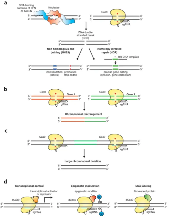
developed to facilitate site-specific genomic modifications. In addition, ZFs28, TALEs29, and enzymatically inactive versions of Cas9 (also known as dead Cas9 (dCas9))30 can be coupled to functionally different enzymatic domains30–35 or fluorophor proteins36 to achieve targeted transcriptional control, epigenetic modification, and DNA labeling (FIG. 1).
ZFNs and TALENs recognize specific DNA sequences through protein-DNA interactions, whereas the DNA specificity of Cas proteins is RNA guided. To target Cas proteins to specific genomic loci, dual- or single-guide RNAs (sgRNAs)24, 25, 27, 37, 38 can be designed and generated quickly. Another key advantage of Cas proteins is that multiple sgRNAs can be simultaneously used to edit multiple genes, which can be useful for studying genetic interactions and modelling multigenic disorders, something that previously required multiple cloning and complex protein engineering steps to achieve with ZFNs and TALENs.
The benefits of using CRISPR-Cas systems to study the nervous system are highlighted by several successful applications in a variety of animal species and cell types to study synaptic and circuit function39–41, neuronal development42–45 and diseases41, 46. Here, we describe how genome editing tools, and in particular those based on CRISPR-Cas enzymes, are opening new avenues for neuroscience and biomedical research via the generation of new model systems, both in vivo and in vitro, and discuss the challenges and possible future applications of this technology for understanding the brain.
Overview of genome editing strategies
Site-specific nucleases including ZFNs, TALENs, and Cas proteins enable precise genetic modifications by inducing double-strand DNA breaks (DSBs) at target locations in the genome. Two highly conserved DNA repair machinery pathways typically restore DSBs that would otherwise result in cell death: non-homologous end joining (NHEJ) and homology-directed recombination (HDR)14, 47–55 (FIG. 1a). The highly error-prone NHEJ pathway induces insertions and deletions (indels) of various lengths that can result in frameshift mutations and, consequently, gene knockout. By contrast, the HDR pathway directs a precise recombination event between a homologous DNA donor template and the damaged DNA site, resulting in accurate correction of the DSB. Therefore, HDR-repair can be used to introduce specific mutations or transgenes into the genome. Because ZFNs and TALENs achieve specific DNA binding via protein domains, individual nucleases have to be synthesized for each target site. By contrast, Cas proteins are guided by a specificity-determining guide RNA sequence (CRISPR RNA (crRNA)) that is associated with a trans-activating crRNA (tracrRNA) and forms Watson-Crick base pairs with the complementary DNA target sequence, resulting in a site-specific DSB 22, 23, 37, 56. A simple two-component system (consisting of Cas9 from the bacterium strains Streptococcus pyogenes (SpCas9) or Staphylococcus aureus (SaCas9), and a fusion of the tracrRNA:crRNA duplex to a single guide RNA (sgRNA))37 has been engineered for expression in eukaryotic cells and can achieve DNA cleavage at any genomic locus of interest24, 25, 57. More recently, Cpf1, a single-RNA guided nuclease, has also been adapted for genome editing27. Hence, different Cas proteins can be targeted to specific DNA sequences simply by changing the short
A
uthor Man
uscr
ipt
A
uthor Man
uscr
ipt
A
uthor Man
uscr
ipt
A
uthor Man
uscr
ipt
specificity-determining part of the guide RNA, which can be easily achieved in one cloning step.
Gene editing across species
Non-human animal models provide an experimental platform to dissect the complexity of the brain and study the cellular and molecular underpinnings of brain disorders.
Neuroscience in particular benefits from exploiting a wide diversity of species including worms, flies, fish and mammals as well as non-traditional model systems such as birds and amphibians58. Disrupting gene expression is a common approach to study gene function and understand loss-of-function disease mutations. For many years, RNAi was the gold standard for gene silencing and studying gene function in vitro and in vivo59, 60; however, genome editing based on engineered designer nucleases offers several advantages over RNAi (TABLE 1). For example, genome editing tools can be modified to allow for more refined control gene expression beyond simple gene knockdown, adding to their versatility (FIG. 1d).
Multiplying the power of simple model organisms
At the molecular level, non-mammalian model systems can provide important information about fundamental features of the nervous system as a result of their well-characterized genetic and cellular organization and amenability to a variety of genetic tools. For example, many evolutionarily conserved genes involved in human neurological disorders such as Alzheimer’s or Parkinson’s disease have been extensively studied using flies, worms, and
fish61–63. For years, studies using these simple model organisms relied mainly on genetic
screens using chemical mutagenesis and RNAi3–5 or imprecise methods for transposon excision and retroviral insertion64–66. More precise genetic modifications have been achieved using ZFNs67–69, TALENs70–73, and Cas9 (reviewed in 74). In the case of Cas proteins, large numbers of RNA guides can be easily synthesized to study gene function on a large scale. By contrast, generating large libraries based on ZFNs and TALENs is
challenging due to difficulties in designing and synthesizing these proteins with varying DNA binding specificities. In a proof-of-concept study a hundred genes were screened with Cas9 and novel loci involved in electrical synapse formation in zebrafish were identified43. Such in vivo screening approaches in small model organisms offer an accessible platform to identify the genes involved in various aspects of nervous system function and dysfunction.
Rapid generation of mammalian models
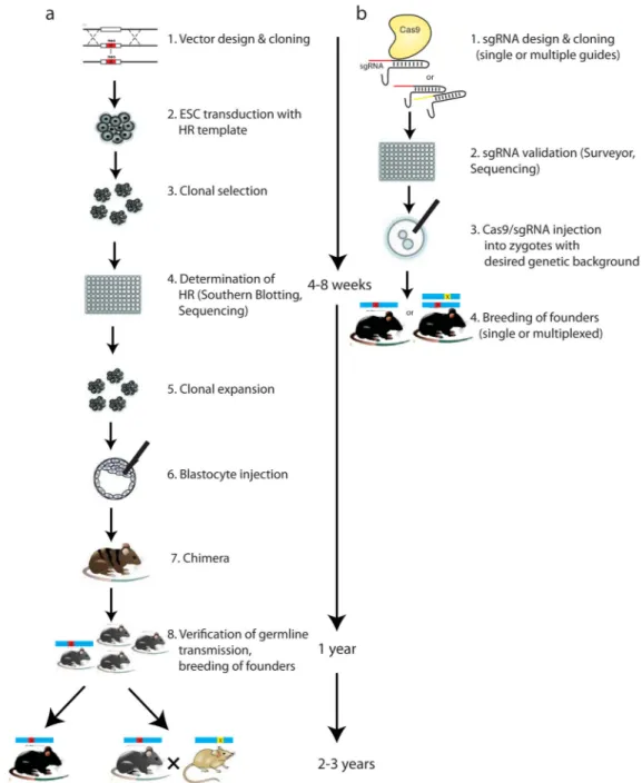
The development of methods enabling homologous recombination in embryonic stem cells (ESCs)6 enabled neuroscientists to study the effects of gene knockouts mainly in mice. This approach has been significantly enhanced by genome editing technologies (FIG. 2). Genome editing in single-cell embryos has been used to generate mouse75, rat76, and primate
models77, 78 that can be used to study the role of specific proteins in nervous system function. Mouse and rat models thus provide a bridge between our understanding of the molecular underpinnings of the nervous system gleaned from studies in non-mammalian systems and the complex phenotypes observed in brain disorders. In some cases, however, a comprehensive understanding of the human brain will require primate models, which are
A
uthor Man
uscr
ipt
A
uthor Man
uscr
ipt
A
uthor Man
uscr
ipt
A
uthor Man
uscr
ipt
more similar to the human brain in terms of neuroanatomical, physiological, perceptual, and behavioral characteristics.
Transgenic approaches in primates are generally very inefficient. However, successful insertion of transgenic alleles in primates, including macaques79, 80 and the common marmoset81, has been achieved using retroviral and lentiviral approaches in early embryos. For example, the viral insertion of a disease-related version of the human huntingtin gene (HTT) into the macaque genome recapitulated clinical features of Huntington’s disease80, representing an important step forward for genetic disease modelling in non-human primates. TALENs have also been successfully used in monkeys to model mutations in MECP2, an X-linked Rett-syndrome gene77, and genome engineered primates have been generated by precise disruption of single and multiple genes with Cas978. The simplicity of the use of Cas proteins relative to ZFNs and TALENs and the ability to modify multiple genes simultaneously is a breakthrough that is already catalyzing molecular interrogation of neurological and psychiatric dysfunctions in disease-relevant brain circuits using primate models78, 82. The ability to examine brain function in genetically modified non-human primates has the potential to contribute significantly to our understanding of higher cognitive functions and to the development of new therapeutic strategies for diseases that cannot be adequately modeled in rodents. Such research, however, raises important bioethical questions, and requires careful consideration of the costs and benefits before moving forward.
In vivo gene editing in the brain
In vivo gene editing allows the systematic genetic dissection of neuronal circuits and the ability to model pathological conditions while bypassing the need to engineer germline modified mutant strains. This experimental approach is fast, independent of genetic
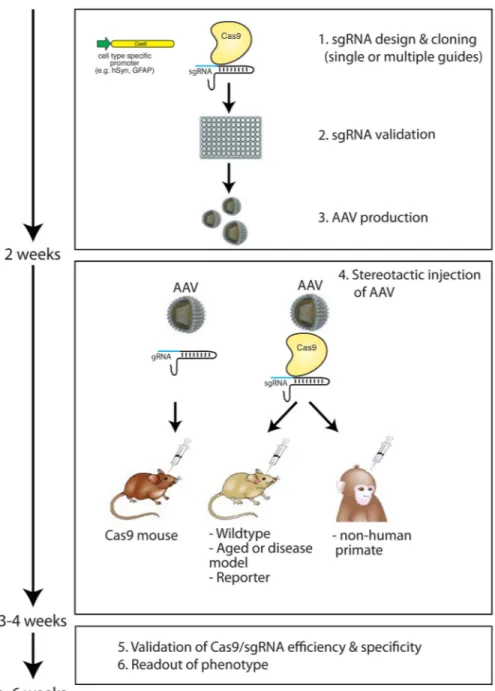
background, animal species, and availability of ESCs, and can be applied to existing disease models and transgenic strains as well as aged animals to study age-related neurological changes (FIG. 3). In vivo methods based on RNAi have been commonly used to reduce the expression of genes in the brain83. In addition, alternative methods based on DNA antisense oligonucleotides (ASOs) can be used for gene silencing and have been shown to be
promising therapeutic molecules for suppressing pathogenic protein aggregates in the brain84, 85. However, both strategies do not allow the generation of stable gene knockouts and site-specific epigenetic modifications (TABLE 1). In the mouse brain, histone
modifications and transcriptional control have been achieved using ZFs86 and TALEs28, and Cas9 has been used to induce indel mutations in neurons in order to achieve stable gene knockouts in living animals39, 41. This demonstrates the capacity for spatial and temporal control of gene expression in fully developed circuits and also opens the door to probing epigenetic dynamics30–33, 35 in the brain. Epigenetic control is of particular interest as there is increasing evidence that epigenetic mechanisms such as histone modifications and DNA methylation play a role in learning and memory formation and the pathology of
neuropsychiatric disorders87. Using Cas proteins, functional domains of DNA methylation or demethylation enzymes or histone modifiers can be easily targeted to specific DNA sequences to edit the epigenome with high spatial and temporal specificity in vivo (FIG. 1d).
A
uthor Man
uscr
ipt
A
uthor Man
uscr
ipt
A
uthor Man
uscr
ipt
A
uthor Man
uscr
ipt
Delivery to the brain
Viral vectors are a promising mode for delivery of Cas proteins to the brain. Viral vectors have defined, tissue-specific or cell type-specific tropism and can be admitted either locally to the brain or through the bloodstream to achieve more systematic tissue penetration88. The most attractive gene delivery vectors are adeno-associated viruses (AAV), which afford long-term expression without genomic integration, are relatively safe, and are
non-pathogenic89, 90. AAV vectors, however, have limited transgene capacity, and the large size of the commonly used Streptococcus pyogenes Cas9 variant poses a significant challenge for AAV-mediated delivery41, 91. AAV-mediated delivery may become even more challenging when Cas9 is enlarged by the fusion of additional functional domains. Smaller Cas9 orthologs, such as those derived from Staphylococcus aureus, are easier to pack57, making them an attractive option for in vivo genome editing in the brain.
Other techniques have been also used to deliver Cas9 and RNA guides to the brain, such as in utero electroporation39 and polyethylenimine (PEI)-mediated transfection46. In rodents, electroporation and PEI-transfection are easy to use, fast, and efficient at delivering large plasmid DNA into a high number of neurons. However, two drawbacks of these techniques are their low spatial accuracy of transgene expression and the necessity of prenatal
intervention, which often results in low viability and targeting of mitotic neuronal precursors instead of post-mitotic differentiated neurons.
Alternatively, Cas9 protein itself, rather than the DNA or RNA that encodes it, could be delivered, an approach that is particularly interesting for protein-based therapeutics. The anionic nature of sgRNA allows the integration of Cas9 protein–sgRNA complexes into cationic liposomes, a commonly used DNA, RNA, and protein delivery tool. Liposome Cas9 protein–sgRNA complexes have already been successfully used to achieve genome editing in the mouse inner ear92. Therefore, lipid-mediated delivery of Cas9 may also serve as powerful tool for genome editing in the brain in the future.
Cell type specific genome editing
In the mammalian brain there are probably several hundred neuronal subtypes, each with distinct morphological, biophysical, biochemical and computational functions. Thus, cell type specific tools are required to dissect this heterogeneous tissue. Research has shown that malfunction of specific cell types in different brain regions contributes to diverse symptoms usually connected with neuropsychiatric symptoms, such as hallucinations, depression, or repetitive motor behavior93. This highlights the need to pinpoint causal relationships between cell types within the context of relevant neuronal networks, genetics, and behavioral dysfunction, which will require precise genome editing in specific cellular subtypes. Site-specific Cre-LoxP recombination elements that enable the control of the spatio-temporal expression of Cas9 have been introduced in fish94 and mouse embryos91, 95 and similar approaches could achieve precise gene editing in defined cell types in vivo. The vast number of established Cre-driver mouse lines96 and inducible Cas9 systems97–99 can, when
combined with conditional gene targeting strategies, provide enormous combinatorial power to decipher the logic of complex neuronal networks and their role in neurological disorders in vivo.
A
uthor Man
uscr
ipt
A
uthor Man
uscr
ipt
A
uthor Man
uscr
ipt
A
uthor Man
uscr
ipt
In vivo efficiency and specificity
In postmitotic neurons, Cas9 has been successfully used to introduce single39–41, 46, 91 and multiple DSBs41, 46 resulting in NHEJ and efficient formation of indel mutations. For example, AAV delivery of Cas9 and sgRNA targeting Mecp2 in the adult mouse brain resulted in MeCP2 protein loss of more than 70%, which was sufficient to recapitulate phenotypes observed in classic Mecp2 mouse models and patients41. In another study, Cas9-mediated deletion of common tumour suppressor genes such as Ptch1, Trp53, Pten and Nf1 in the cerebellum or forebrain efficiently induced the formation of medulloblastoma or glioblastoma tumors, respectively46. Despite this success, the validation of Cas-mediated gene editing in the brain is still challenging, and sensitive methods are required for analyzing Cas efficiency and specificity in targeted brain regions (BOX 1).
Box 1
Practical considerations for validating Cas nuclease efficiency and specificity in the mammalian brain
Validating Cas nuclease efficiency and specificity is particularly challenging in the mammalian brain because of its complex architecture and cellular diversity. To precisely validate nuclease efficiency and specificity, targeted cells first have to be identified and sorted out from the heterogeneous cell population in the brain. Recently, an easy and efficient method in which fluorescent activated cell sorting (FACS) is used to isolate fluorphor-tagged nuclei of targeted cells to purify and analyze genomic DNA and nuclear RNA with high resolution and sensitivity, was developed41.
Cas efficiency
Cas nuclease efficiency and specificity can be validated using enzymatic DNA cleavage assays (SURVEYOR®)111 or DNA sequencing41, 46, 91. DNA sequencing analysis provides a complete picture of indel frequency, types of frame-shift and in-frame mutations, length and exact sequence of insertions and deletions, as well as information about mono- and bi-allelic modifications when applied to single cells41. In addition, RNA levels of the targeted gene can be determined using quantitative PCR (qPCR) or RNA sequencing methods. Depending on the targeted exon (that is, whether it is an early or late exon), truncated transcripts might be expressed from the target gene and should also be considered when qPCR probes are designed. Ideally, effective protein knockdown should also be measured using histological, biochemical, or functional (e.g.,
electrophysiology, enzymatic activity assays) readouts.
Cas specificity
Similar to ZFNs and TALENs, Cas proteins can cleave off-target sites in the genome. Many software tools predict potential off-target effects and help to choose optimal target sequences to reduce off-target activity (a noncomprehensive list of online tools can be found in ‘Further information’). On-target specificity can be further improved by using double-nicking112, 113 or truncated sgRNA approaches114. In addition, sensitive readout methods for identifying genome-wide Cas9 off-target activity have been developed that
A
uthor Man
uscr
ipt
A
uthor Man
uscr
ipt
A
uthor Man
uscr
ipt
A
uthor Man
uscr
ipt
provide useful tools for evaluating specificity and safety of Cas9 in basic and clinical research57, 115, 116
Selected online off-target prediction tools
CRISPR Design (http://crispr.mit.edu/), sgRNA designer (http://www.broadinstitute.org/ rnai/public/analysis-tools/sgrna-design), CHOPCHOP (https://
chopchop.rc.fas.harvard.edu/), Benchling (https://benchling.com/crispr), CasOT (http:// eendb.zfgenetics.org/casot/), E-CRISP (http://www.e-crisp.org/E-CRISP/), ZiFiT (http:// zifit.partners.org/ZiFiT/, DESKGEN (https://www.deskgen.com/landing/#/), COSMID (http://omictools.com/cosmid-s9890.html).
Although NHEJ in postmitotic neurons has been demonstrated to be active, it remains unclear how efficient HDR is in postmitotic cells. It is commonly believed that HDR predominantly occurs in the S and G2 phases of the cell cycle100, 101, and is therefore thought to be rare in non-dividing cells such as neurons. Introduction or correction of precise genetic mutations via HDR in the brain would validate disease mutations in vivo and open the door to therapeutic applications of genome editing in brain disorders. Thus, future work should focus on identifying and activating signaling pathways required for triggering HDR in differentiated cells. It should also be noted, however, that gene insertion has been achieved through NHEJ pathways, which may allow us to insert DNA in neurons and glia102.
In contrast to precise gene knockout and insertion, genome editing aimed at transcriptional regulation and epigenetic modulation may be less challenging in the brain, as these approaches are independent of DNA repair pathways. Achieving epigenetic control in neurons can aid in the study of the molecular mechanisms of natural gene silencing in the nervous system and to better understand neurological disorders associated with gene imprinting, such as Angelman syndrome103.
Gene editing in human iPSCs
Combinatorial approaches based on iPSC technology and genome editing offer another approach to model human neurological disorders in vitro. A key advantage of this approach is that genetic modifications can be studied in different human genetic backgrounds because iPSCs retain all of the individual donor’s genetic information. For complex neurological disorders this is particularly important because genetic variants associated with such diseases act in concert with many other alleles. Another advantage is that the genetically modified cells can be differentiated into virtually any cell type, including those that are not easily accessible in patients such as neurons and glia.
iPSC-based disease models have been generated for several neurological disorders including Parkinson’s10, 11, Alzheimer’s9, and Huntington’s8 disease and have been proven to closely mimic cellular and molecular features of human diseases. Genome editing tools applied to these models can be used to examine the genetic link between risk variants and cellular pathways involved in multigenic neurological disorders in a high-throughput manner (FIG. 4). Furthermore, specific signaling pathways involved in the pathogenesis of the disease can
A
uthor Man
uscr
ipt
A
uthor Man
uscr
ipt
A
uthor Man
uscr
ipt
A
uthor Man
uscr
ipt
be precisely dissected to gain insight into the molecular mechanisms of the disease and to identify new drug targets10. Gene editing may be performed either in iPSCs or later in differentiated cells97, 99, allowing for the investigation of phenotypes that arise during cell differentiation, which may be relevant when studying neurodevelopmental aspects of a disease such as in Rett-syndrome104–106. On the other side, inducing or rescuing a phenotype in differentiated cells will be useful for validating potential therapeutic applications.
Future perspectives
Genome editing technologies allow for the introduction of genetic modifications into virtually any cell type and organism. For example, Cas9 has been already used to alter genes in species such as killifish107 and salamander108, which are commonly used to study aging and tissue regeneration, respectively. It may also open up the possibility of developing models in other species of interest to neuroscience research, such as social insects or songbirds58, which have been intractable to genetic modification. In addition to generation of new model systems, including iPSC-derived in vitro models, genome editing in
combination with single-cell transcriptomics109 provides a route to understanding cell type specific gene function within a heterogeneous tissue, allowing for precise dissection of genetic networks in the brain. Furthermore, together with genome-wide association studies, in vivo genome editing holds potential for personalized therapeutic applications for brain disorders110. To realize these advances, however, several open challenges have to be addressed. First, existing methods for delivering Cas proteins and RNA guides to the brain must be optimized and new methods developed to achieve sufficient levels of specificity and efficiency. Second, new methods for stimulating efficient gene insertion and correction in postmitotic cells have to be established. Third, safety and ethical concerns have to be carefully addressed. Nevertheless, we believe that novel genome editing technologies based on CRISPR-Cas systems, together with powerful readout methods, will help us better understand the logic of neuronal circuits and unravel some of the mysteries of complex neurological disorders in the near future.
Suggested glossary terms
Functional genomics Studying gene functions and interactions in relationship to RNA transcripts and protein products using genome-wide data, and often involving high-throughput methods.
RNA interference (RNAi) A technique used to knock down the expression of a specific gene by introducing a double stranded RNA molecule that complements the gene of interest and triggers the degradation of the target mRNA.
Homologous recombination (HR)Exchange of homologous DNA strands between similar DNA molecules, which naturally occurs during meiosis to generate genetic variation. HR is used to direct error-free repair of DNA double-strand breaks induced by
A
uthor Man
uscr
ipt
A
uthor Man
uscr
ipt
A
uthor Man
uscr
ipt
A
uthor Man
uscr
ipt
DNA nucleases such as ZFNs, TALENs, and Cas proteins.
Embryonic stem cells (ESCs) Totipotent cells derived from embryos that can be genetically manipulated in vitro to generate transgenic, knockin and knockout mice. ESCs can also be directed to differentiate into a variety of cell types in vitro including neurons and glial cells.
Induced pluripotent stem cells (iPSCs)Pluripotent cells derived from reprogrammed
differentiated adult cells with similar properties as ESCs, and therefore can be differentiated in principle into all cell types of the body.
Epigenetic mechanisms Multilayered cellular processes that modulate gene expression and function in response to interoceptive and environmental stimuli during development, adult life and ageing, including DNA methylation, post-translational histone modifications, ATP-dependent nucleosome and higher-order chromatin remodelling, non-coding RNA deployment and nuclear reorganization.
Liposomes A lipid vesicle artificially formed by sonicating lipids in an aqueous solution. Liposomes can be packed with negatively charged molecules to deliver them into cells, and are therefore promising vehicles for therapeutic applications.
Cre-LoxP recombination A site-specific recombination system derived from Escherichia coli bacteriophage P1. Two short DNA sequences (loxP sites) are engineered to flank the target DNA. Activation of the Cre-recombinase enzyme catalyses recombination between the loxP sites, leading to excision of the intervening sequence.
References
1. Lewis EBBF. Methods for feeding ethyl methane sulphonate (EMS) to Drosophila males. Drosoph Inf Serv. 1968; 43
2. Fire A, et al. Potent and specific genetic interference by double-stranded RNA in Caenorhabditis elegans. Nature. 1998; 391:806–11. [PubMed: 9486653]
3. St Johnston D. The art and design of genetic screens: Drosophila melanogaster. Nat Rev Genet. 2002; 3:176–88. [PubMed: 11972155]
4. Jorgensen EM, Mango SE. The art and design of genetic screens: caenorhabditis elegans. Nat Rev Genet. 2002; 3:356–69. [PubMed: 11988761]
5. Patton EE, Zon LI. The art and design of genetic screens: zebrafish. Nat Rev Genet. 2001; 2:956–66. [PubMed: 11733748]
6. Thomas KR, Capecchi MR. Site-directed mutagenesis by gene targeting in mouse embryo-derived stem cells. Cell. 1987; 51:503–12. [PubMed: 2822260]
A
uthor Man
uscr
ipt
A
uthor Man
uscr
ipt
A
uthor Man
uscr
ipt
A
uthor Man
uscr
ipt
7. Oddo S, et al. Triple-transgenic model of Alzheimer’s disease with plaques and tangles: intracellular Abeta and synaptic dysfunction. Neuron. 2003; 39:409–21. [PubMed: 12895417]
8. Jeon I, et al. Neuronal properties, in vivo effects, and pathology of a Huntington’s disease patient-derived induced pluripotent stem cells. Stem Cells. 2012; 30:2054–62. [PubMed: 22628015] 9. Yagi T, et al. Modeling familial Alzheimer’s disease with induced pluripotent stem cells. Hum Mol
Genet. 2011; 20:4530–9. [PubMed: 21900357]
10. Ryan SD, et al. Isogenic human iPSC Parkinson’s model shows nitrosative stress-induced dysfunction in MEF2-PGC1alpha transcription. Cell. 2013; 155:1351–64. [PubMed: 24290359] 11. Soldner F, et al. Generation of isogenic pluripotent stem cells differing exclusively at two early
onset Parkinson point mutations. Cell. 2011; 146:318–31. References 10 and 11 combine ZFN-mediated genome editing and human stem cell technologies for studying neuological disorders in vitro. [PubMed: 21757228]
12. Pak C, et al. Human Neuropsychiatric Disease Modeling using Conditional Deletion Reveals Synaptic Transmission Defects Caused by Heterozygous Mutations in NRXN1. Cell Stem Cell. 2015; 17:316–28. [PubMed: 26279266]
13. Kim YG, Cha J, Chandrasegaran S. Hybrid restriction enzymes: zinc finger fusions to Fok I cleavage domain. Proc Natl Acad Sci U S A. 1996; 93:1156–60. [PubMed: 8577732] 14. Bibikova M, et al. Stimulation of homologous recombination through targeted cleavage by
chimeric nucleases. Mol Cell Biol. 2001; 21:289–97. This early study shows the use of ZFNs in
Xenopus laevis for stimulating homologous recombination. [PubMed: 11113203]
15. Urnov FD, et al. Highly efficient endogenous human gene correction using designed zinc-finger nucleases. Nature. 2005; 435:646–51. [PubMed: 15806097]
16. Boch J, et al. Breaking the code of DNA binding specificity of TAL-type III effectors. Science. 2009; 326:1509–12. [PubMed: 19933107]
17. Moscou MJ, Bogdanove AJ. A simple cipher governs DNA recognition by TAL effectors. Science. 2009; 326:1501. [PubMed: 19933106]
18. Christian M, et al. Targeting DNA double-strand breaks with TAL effector nucleases. Genetics. 2010; 186:757–61. [PubMed: 20660643]
19. Miller JC, et al. A TALE nuclease architecture for efficient genome editing. Nat Biotechnol. 2011; 29:143–8. This paper uses an improved TALEN architecture to introduce gene knockouts in human cells. [PubMed: 21179091]
20. Bolotin A, Quinquis B, Sorokin A, Ehrlich SD. Clustered regularly interspaced short palindrome repeats (CRISPRs) have spacers of extrachromosomal origin. Microbiology. 2005; 151:2551–61. [PubMed: 16079334]
21. Barrangou R, et al. CRISPR provides acquired resistance against viruses in prokaryotes. Science. 2007; 315:1709–12. This work is a first experimental demonstration of the adaptive immune function of the CRISPR-Cas9 system in bacteria. [PubMed: 17379808]
22. Deltcheva E, et al. CRISPR RNA maturation by trans-encoded small RNA and host factor RNase III. Nature. 2011; 471:602–7. [PubMed: 21455174]
23. Garneau JE, et al. The CRISPR/Cas bacterial immune system cleaves bacteriophage and plasmid DNA. Nature. 2010; 468:67–71. This paper demonstrates that Cas9 facilitates DNA cleavage in bacteria. [PubMed: 21048762]
24. Cong L, et al. Multiplex genome engineering using CRISPR/Cas systems. Science. 2013; 339:819– 23. [PubMed: 23287718]
25. Mali P, et al. RNA-guided human genome engineering via Cas9. Science. 2013; 339:823–6. References 24 and 25 describe the succesful harnessing of the CRISPR-Cas9 system for editing the mammalian genome in cell lines. [PubMed: 23287722]
26. Makarova KS, Koonin EV. Annotation and Classification of CRISPR-Cas Systems. Methods Mol Biol. 2015; 1311:47–75. [PubMed: 25981466]
27. Zetsche B, et al. Cpf1 Is a Single RNA-Guided Endonuclease of a Class 2 CRISPR-Cas System. Cell. 2015
28. Beerli RR, Segal DJ, Dreier B, Barbas CF 3rd. Toward controlling gene expression at will: specific regulation of the erbB-2/HER-2 promoter by using polydactyl zinc finger proteins constructed
A
uthor Man
uscr
ipt
A
uthor Man
uscr
ipt
A
uthor Man
uscr
ipt
A
uthor Man
uscr
ipt
from modular building blocks. Proc Natl Acad Sci U S A. 1998; 95:14628–33. [PubMed: 9843940]
29. Zhang F, et al. Efficient construction of sequence-specific TAL effectors for modulating mammalian transcription. Nat Biotechnol. 2011; 29:149–53. [PubMed: 21248753] 30. Gilbert LA, et al. CRISPR-mediated modular RNA-guided regulation of transcription in
eukaryotes. Cell. 2013; 154:442–51. [PubMed: 23849981]
31. Qi LS, et al. Repurposing CRISPR as an RNA-guided platform for sequence-specific control of gene expression. Cell. 2013; 152:1173–83. [PubMed: 23452860]
32. Konermann S, et al. Optical control of mammalian endogenous transcription and epigenetic states. Nature. 2013; 500:472–6. [PubMed: 23877069]
33. Kearns NA, et al. Functional annotation of native enhancers with a Cas9-histone demethylase fusion. Nat Methods. 2015; 12:401–3. [PubMed: 25775043]
34. Konermann S, et al. Genome-scale transcriptional activation by an engineered CRISPR-Cas9 complex. Nature. 2015; 517:583–8. [PubMed: 25494202]
35. Hilton IB, et al. Epigenome editing by a CRISPR-Cas9-based acetyltransferase activates genes from promoters and enhancers. Nat Biotechnol. 2015; 33:510–7. [PubMed: 25849900] 36. Chen B, et al. Dynamic imaging of genomic loci in living human cells by an optimized
CRISPR/Cas system. Cell. 2013; 155:1479–91. [PubMed: 24360272]
37. Jinek M, et al. A programmable dual-RNA-guided DNA endonuclease in adaptive bacterial immunity. Science. 2012; 337:816–21. This study together with reference 56 demonstrates that Cas9 cleaves genomic DNA in vitro using a single guide RNA chimera containing a truncated tracrRNA. [PubMed: 22745249]
38. Hsu PD, et al. DNA targeting specificity of RNA-guided Cas9 nucleases. Nat Biotechnol. 2013; 31:827–32. [PubMed: 23873081]
39. Straub C, Granger AJ, Saulnier JL, Sabatini BL. CRISPR/Cas9-mediated gene knock-down in post-mitotic neurons. PLoS One. 2014; 9:e105584. [PubMed: 25140704]
40. Incontro S, Asensio CS, Edwards RH, Nicoll RA. Efficient, complete deletion of synaptic proteins using CRISPR. Neuron. 2014; 83:1051–7. This paper reports the delivery of Cas9 and guide RNAs in organotypic brain slice cultures and the disruption NMDA- and AMPA receptor subunits. [PubMed: 25155957]
41. Swiech L, et al. In vivo interrogation of gene function in the mammalian brain using CRISPR-Cas9. Nat Biotechnol. 2015; 33:102–6. This paper demonstrates the delivery of Cas9 and guide RNAs into the mouse brain using adesno-associated virus, and single and multiplex gene editing in vivo. It also shows a purification method of genetically tagged Cas9-targeted cell nuclei for DNA- and RNA-sequencing. [PubMed: 25326897]
42. Shen Z, et al. Conditional knockouts generated by engineered CRISPR-Cas9 endonuclease reveal the roles of coronin in C. elegans neural development. Dev Cell. 2014; 30:625–36. This paper describes a conditional knockout strategy using Cas9 in Caenorhabditis elegans for studying gene function in neural development. [PubMed: 25155554]
43. Shah AN, Davey CF, Whitebirch AC, Miller AC, Moens CB. Rapid reverse genetic screening using CRISPR in zebrafish. Nat Methods. 2015; 12:535–40. This paper represents a useful application of Cas9 for studying neurodevelopmental processes on a large scale in zebrafish. [PubMed:
25867848]
44. Auer TO, Duroure K, De Cian A, Concordet JP, Del Bene F. Highly efficient CRISPR/Cas9-mediated knock-in in zebrafish by homology-independent DNA repair. Genome Res. 2014; 24:142–53. [PubMed: 24179142]
45. Jao LE, Wente SR, Chen W. Efficient multiplex biallelic zebrafish genome editing using a CRISPR nuclease system. Proc Natl Acad Sci U S A. 2013; 110:13904–9. [PubMed: 23918387]
46. Zuckermann M, et al. Somatic CRISPR/Cas9-mediated tumour suppressor disruption enables versatile brain tumour modelling. Nat Commun. 2015; 6:7391. This paper describes methods for delivering Cas9 and guide RNA into the brain of newborn mice and embryos. By targeting multiple tumour suppressor genes the development of medulla- and glioblastoma was induced. [PubMed: 26067104]
A
uthor Man
uscr
ipt
A
uthor Man
uscr
ipt
A
uthor Man
uscr
ipt
A
uthor Man
uscr
ipt
47. Plessis A, Perrin A, Haber JE, Dujon B. Site-specific recombination determined by I-SceI, a mitochondrial group I intron-encoded endonuclease expressed in the yeast nucleus. Genetics. 1992; 130:451–60. [PubMed: 1551570]
48. Rudin N, Sugarman E, Haber JE. Genetic and physical analysis of double-strand break repair and recombination in Saccharomyces cerevisiae. Genetics. 1989; 122:519–34. [PubMed: 2668114] 49. Fishman-Lobell J, Haber JE. Removal of nonhomologous DNA ends in double-strand break
recombination: the role of the yeast ultraviolet repair gene RAD1. Science. 1992; 258:480–4. [PubMed: 1411547]
50. Fishman-Lobell J, Rudin N, Haber JE. Two alternative pathways of double-strand break repair that are kinetically separable and independently modulated. Mol Cell Biol. 1992; 12:1292–303. [PubMed: 1545810]
51. Liang F, Han M, Romanienko PJ, Jasin M. Homology-directed repair is a major double-strand break repair pathway in mammalian cells. Proc Natl Acad Sci U S A. 1998; 95:5172–7. [PubMed: 9560248]
52. Johnson RD, Liu N, Jasin M. Mammalian XRCC2 promotes the repair of DNA double-strand breaks by homologous recombination. Nature. 1999; 401:397–9. [PubMed: 10517641] 53. Bibikova M, Beumer K, Trautman JK, Carroll D. Enhancing gene targeting with designed zinc
finger nucleases. Science. 2003; 300:764. [PubMed: 12730594]
54. Rouet P, Smih F, Jasin M. Introduction of double-strand breaks into the genome of mouse cells by expression of a rare-cutting endonuclease. Mol Cell Biol. 1994; 14:8096–106. [PubMed: 7969147] 55. Rouet P, Smih F, Jasin M. Expression of a site-specific endonuclease stimulates homologous
recombination in mammalian cells. Proc Natl Acad Sci U S A. 1994; 91:6064–8. [PubMed: 8016116]
56. Gasiunas G, Barrangou R, Horvath P, Siksnys V. Cas9-crRNA ribonucleoprotein complex mediates specific DNA cleavage for adaptive immunity in bacteria. Proc Natl Acad Sci U S A. 2012; 109:E2579–86. [PubMed: 22949671]
57. Ran FA, et al. In vivo genome editing using Staphylococcus aureus Cas9. Nature. 2015; 520:186– 91. [PubMed: 25830891]
58. Brenowitz EA, Zakon HH. Emerging from the bottleneck: benefits of the comparative approach to modern neuroscience. Trends Neurosci. 2015
59. Elbashir SM, et al. Duplexes of 21-nucleotide RNAs mediate RNA interference in cultured mammalian cells. Nature. 2001; 411:494–8. [PubMed: 11373684]
60. Hommel JD, Sears RM, Georgescu D, Simmons DL, DiLeone RJ. Local gene knockdown in the brain using viral-mediated RNA interference. Nat Med. 2003; 9:1539–44. [PubMed: 14634645] 61. Wittenburg N, et al. Presenilin is required for proper morphology and function of neurons in C.
elegans. Nature. 2000; 406:306–9. [PubMed: 10917532]
62. Geling A, Steiner H, Willem M, Bally-Cuif L, Haass C. A gamma-secretase inhibitor blocks Notch signaling in vivo and causes a severe neurogenic phenotype in zebrafish. EMBO Rep. 2002; 3:688–94. [PubMed: 12101103]
63. Clark IE, et al. Drosophila pink1 is required for mitochondrial function and interacts genetically with parkin. Nature. 2006; 441:1162–6. [PubMed: 16672981]
64. Cooley L, Kelley R, Spradling A. Insertional mutagenesis of the Drosophila genome with single P elements. Science. 1988; 239:1121–8. [PubMed: 2830671]
65. Gaiano N, et al. Insertional mutagenesis and rapid cloning of essential genes in zebrafish. Nature. 1996; 383:829–32. [PubMed: 8893009]
66. Bessereau JL, et al. Mobilization of a Drosophila transposon in the Caenorhabditis elegans germ line. Nature. 2001; 413:70–4. [PubMed: 11544527]
67. Beumer KJ, et al. Efficient gene targeting in Drosophila by direct embryo injection with zinc-finger nucleases. Proc Natl Acad Sci U S A. 2008; 105:19821–6. [PubMed: 19064913]
68. Morton J, Davis MW, Jorgensen EM, Carroll D. Induction and repair of zinc-finger nuclease-targeted double-strand breaks in Caenorhabditis elegans somatic cells. Proc Natl Acad Sci U S A. 2006; 103:16370–5. [PubMed: 17060623]
A
uthor Man
uscr
ipt
A
uthor Man
uscr
ipt
A
uthor Man
uscr
ipt
A
uthor Man
uscr
ipt
69. Doyon Y, et al. Heritable targeted gene disruption in zebrafish using designed zinc-finger nucleases. Nat Biotechnol. 2008; 26:702–8. [PubMed: 18500334]
70. Bedell VM, et al. In vivo genome editing using a high-efficiency TALEN system. Nature. 2012; 491:114–8. [PubMed: 23000899]
71. Wood AJ, et al. Targeted genome editing across species using ZFNs and TALENs. Science. 2011; 333:307. [PubMed: 21700836]
72. Sander JD, et al. Targeted gene disruption in somatic zebrafish cells using engineered TALENs. Nat Biotechnol. 2011; 29:697–8. [PubMed: 21822241]
73. Katsuyama T, et al. An efficient strategy for TALEN-mediated genome engineering in Drosophila. Nucleic Acids Res. 2013; 41:e163. [PubMed: 23877243]
74. Sander JD, Joung JK. CRISPR-Cas systems for editing, regulating and targeting genomes. Nat Biotechnol. 2014; 32:347–55. [PubMed: 24584096]
75. Carbery ID, et al. Targeted genome modification in mice using zinc-finger nucleases. Genetics. 2010; 186:451–9. [PubMed: 20628038]
76. Geurts AM, et al. Knockout rats via embryo microinjection of zinc-finger nucleases. Science. 2009; 325:433. [PubMed: 19628861]
77. Liu H, et al. TALEN-mediated gene mutagenesis in rhesus and cynomolgus monkeys. Cell Stem Cell. 2014; 14:323–8. [PubMed: 24529597]
78. Niu Y, et al. Generation of gene-modified cynomolgus monkey via Cas9/RNA-mediated gene targeting in one-cell embryos. Cell. 2014; 156:836–43. References 77 and 78 describe the succesfull generation of genetically modified non-human primates using genome editing technologies in early embryos. [PubMed: 24486104]
79. Chan AW, Chong KY, Martinovich C, Simerly C, Schatten G. Transgenic monkeys produced by retroviral gene transfer into mature oocytes. Science. 2001; 291:309–12. [PubMed: 11209082] 80. Yang SH, et al. Towards a transgenic model of Huntington’s disease in a non-human primate.
Nature. 2008; 453:921–4. [PubMed: 18488016]
81. Sasaki E, et al. Generation of transgenic non-human primates with germline transmission. Nature. 2009; 459:523–7. [PubMed: 19478777]
82. Belmonte JC, et al. Brains, Genes, and Primates. Neuron. 2015; 86:617–631. [PubMed: 25950631] 83. Xia H, Mao Q, Paulson HL, Davidson BL. siRNA-mediated gene silencing in vitro and in vivo.
Nat Biotechnol. 2002; 20:1006–10. [PubMed: 12244328]
84. Kordasiewicz HB, et al. Sustained therapeutic reversal of Huntington’s disease by transient repression of huntingtin synthesis. Neuron. 2012; 74:1031–44. [PubMed: 22726834]
85. Smith RA, et al. Antisense oligonucleotide therapy for neurodegenerative disease. J Clin Invest. 2006; 116:2290–6. [PubMed: 16878173]
86. Garriga-Canut M, et al. Synthetic zinc finger repressors reduce mutant huntingtin expression in the brain of R6/2 mice. Proc Natl Acad Sci U S A. 2012; 109:E3136–45. [PubMed: 23054839] 87. Sweatt JD. The emerging field of neuroepigenetics. Neuron. 2013; 80:624–32. [PubMed:
24183015]
88. Murlidharan G, Samulski RJ, Asokan A. Biology of adeno-associated viral vectors in the central nervous system. Front Mol Neurosci. 2014; 7:76. [PubMed: 25285067]
89. Burger C, Nash K, Mandel RJ. Recombinant adeno-associated viral vectors in the nervous system. Hum Gene Ther. 2005; 16:781–91. [PubMed: 16000060]
90. Taymans JM, et al. Comparative analysis of adeno-associated viral vector serotypes 1, 2, 5, 7, and 8 in mouse brain. Hum Gene Ther. 2007; 18:195–206. [PubMed: 17343566]
91. Platt RJ, et al. CRISPR-Cas9 knockin mice for genome editing and cancer modeling. Cell. 2014; 159:440–55. This paper describes how the CRISPR-Cas9 knockin mouse can be used for cell type specific gene editing in the brain. [PubMed: 25263330]
92. Zuris JA, et al. Cationic lipid-mediated delivery of proteins enables efficient protein-based genome editing in vitro and in vivo. Nat Biotechnol. 2015; 33:73–80. [PubMed: 25357182]
93. Akil H, et al. Medicine. The future of psychiatric research: genomes and neural circuits. Science. 2010; 327:1580–1. [PubMed: 20339051]
A
uthor Man
uscr
ipt
A
uthor Man
uscr
ipt
A
uthor Man
uscr
ipt
A
uthor Man
uscr
ipt
94. Yin L, et al. Multiplex Conditional Mutagenesis Using Transgenic Expression of Cas9 and sgRNAs. Genetics. 2015
95. Yang H, et al. One-step generation of mice carrying reporter and conditional alleles by CRISPR/ Cas-mediated genome engineering. Cell. 2013; 154:1370–9. This paper together with reference 121 describes the generation of genetically modified mice using Cas9. [PubMed: 23992847] 96. Harris JA, et al. Anatomical characterization of Cre driver mice for neural circuit mapping and
manipulation. Front Neural Circuits. 2014; 8:76. [PubMed: 25071457]
97. Polstein LR, Gersbach CA. A light-inducible CRISPR-Cas9 system for control of endogenous gene activation. Nat Chem Biol. 2015; 11:198–200. [PubMed: 25664691]
98. Dow LE, et al. Inducible in vivo genome editing with CRISPR-Cas9. Nat Biotechnol. 2015; 33:390–4. [PubMed: 25690852]
99. Zetsche B, Volz SE, Zhang F. A split-Cas9 architecture for inducible genome editing and transcription modulation. Nat Biotechnol. 2015; 33:139–42. [PubMed: 25643054]
100. Pardo B, Gomez-Gonzalez B, Aguilera A. DNA repair in mammalian cells: DNA double-strand break repair: how to fix a broken relationship. Cell Mol Life Sci. 2009; 66:1039–56. [PubMed: 19153654]
101. van Gent DC, van der Burg M. Non-homologous end-joining, a sticky affair. Oncogene. 2007; 26:7731–40. [PubMed: 18066085]
102. Maresca M, Lin VG, Guo N, Yang Y. Obligate ligation-gated recombination (ObLiGaRe): custom-designed nuclease-mediated targeted integration through nonhomologous end joining. Genome Res. 2013; 23:539–46. [PubMed: 23152450]
103. Vu TH, Hoffman AR. Imprinting of the Angelman syndrome gene, UBE3A, is restricted to brain. Nat Genet. 1997; 17:12–3. [PubMed: 9288087]
104. Kim KY, Hysolli E, Park IH. Neuronal maturation defect in induced pluripotent stem cells from patients with Rett syndrome. Proc Natl Acad Sci U S A. 2011; 108:14169–74. [PubMed: 21807996]
105. Cheung AY, et al. Isolation of MECP2-null Rett Syndrome patient hiPS cells and isogenic controls through X-chromosome inactivation. Hum Mol Genet. 2011; 20:2103–15. [PubMed: 21372149]
106. Marchetto MC, et al. A model for neural development and treatment of Rett syndrome using human induced pluripotent stem cells. Cell. 2010; 143:527–39. [PubMed: 21074045] 107. Harel I, et al. A platform for rapid exploration of aging and diseases in a naturally short-lived
vertebrate. Cell. 2015; 160:1013–26. [PubMed: 25684364]
108. Flowers GP, Timberlake AT, McLean KC, Monaghan JR, Crews CM. Highly efficient targeted mutagenesis in axolotl using Cas9 RNA-guided nuclease. Development. 2014; 141:2165–71. [PubMed: 24764077]
109. Zeisel A, et al. Brain structure. Cell types in the mouse cortex and hippocampus revealed by single-cell RNA-seq. Science. 2015; 347:1138–42. [PubMed: 25700174]
110. Cox DB, Platt RJ, Zhang F. Therapeutic genome editing: prospects and challenges. Nat Med. 2015; 21:121–131. [PubMed: 25654603]
111. Qiu P, et al. Mutation detection using Surveyor nuclease. Biotechniques. 2004; 36:702–7. [PubMed: 15088388]
112. Ran FA, et al. Double nicking by RNA-guided CRISPR Cas9 for enhanced genome editing specificity. Cell. 2013; 154:1380–9. [PubMed: 23992846]
113. Mali P, et al. CAS9 transcriptional activators for target specificity screening and paired nickases for cooperative genome engineering. Nat Biotechnol. 2013; 31:833–8. [PubMed: 23907171] 114. Fu Y, Sander JD, Reyon D, Cascio VM, Joung JK. Improving CRISPR-Cas nuclease specificity
using truncated guide RNAs. Nat Biotechnol. 2014; 32:279–84. [PubMed: 24463574]
115. Tsai SQ, et al. GUIDE-seq enables genome-wide profiling of off-target cleavage by CRISPR-Cas nucleases. Nat Biotechnol. 2015; 33:187–97. [PubMed: 25513782]
116. Kim D, et al. Digenome-seq: genome-wide profiling of CRISPR-Cas9 off-target effects in human cells. Nat Methods. 2015; 12:237–43. 1–243. [PubMed: 25664545]
A
uthor Man
uscr
ipt
A
uthor Man
uscr
ipt
A
uthor Man
uscr
ipt
A
uthor Man
uscr
ipt
117. Blasco RB, et al. Simple and rapid in vivo generation of chromosomal rearrangements using CRISPR/Cas9 technology. Cell Rep. 2014; 9:1219–27. [PubMed: 25456124]
118. Maddalo D, et al. In vivo engineering of oncogenic chromosomal rearrangements with the CRISPR/Cas9 system. Nature. 2014; 516:423–7. [PubMed: 25337876]
119. Xiao A, et al. Chromosomal deletions and inversions mediated by TALENs and CRISPR/Cas in zebrafish. Nucleic Acids Res. 2013; 41:e141. [PubMed: 23748566]
120. Essletzbichler P, et al. Megabase-scale deletion using CRISPR/Cas9 to generate a fully haploid human cell line. Genome Res. 2014; 24:2059–65. [PubMed: 25373145]
121. Wang H, et al. One-step generation of mice carrying mutations in multiple genes by CRISPR/Cas-mediated genome engineering. Cell. 2013; 153:910–8. [PubMed: 23643243]
A
uthor Man
uscr
ipt
A
uthor Man
uscr
ipt
A
uthor Man
uscr
ipt
A
uthor Man
uscr
ipt
Figure 1. Genome editing applications of CRISPR-Cas9
(a) Non-homologous end-joining (NHEJ) and homology-directed repair (HDR) after DNA double-strand break (red arrowheads) induced by zinc finger nucleases (ZFNs) or
transcription activator-like effector nucleases (TALENs) (left) and Cas9 (right). ZFNs and TALENs recognize their DNA binding site via protein domains (indicated in blue) that can be modularly assembled for each DNA target sequence. Cas9 recognizes its DNA binding site via RNA-DNA interactions mediated by the short guide RNA (sgRNA), which can be easily designed and cloned. The error-prone NHEJ repair pathway53 can result in
introduction of indel mutations that can lead to a frame shift, introduction of a premature
A
uthor Man
uscr
ipt
A
uthor Man
uscr
ipt
A
uthor Man
uscr
ipt
A
uthor Man
uscr
ipt
stop codon and consequently gene knockout. The alternative HDR repair pathway14, 47–53 can be used to introduce precise genetic modifications if a homologous DNA template is present. (b) Two different sgRNAs guide Cas9 to induce DNA cleavage at two different genes, resulting in chromosomal rearrangements117, 118. (c) Two proximate sgRNAs guide Cas9 to induce DNA cleavage at two different loci of the same gene, introducing large deletions119, 120. (d) The nuclease inactivated version of Cas9 (dead Cas9 (dCas9)) can be fused to different functional enzymatic domains in order to mediate transcriptional control, epigenetic modulation editing, or fluorescent DNA labeling of specific genetic loci30–36.
A
uthor Man
uscr
ipt
A
uthor Man
uscr
ipt
A
uthor Man
uscr
ipt
A
uthor Man
uscr
ipt
Figure 2. Methods for generating genetically modified rodents
Comparison of the timelines of traditional gene targeting using classic homologous
recombination (HR) in embryonic stem cells (ESCs) or Cas9 in one-cell embryos. (a) There are two main time- and cost-intensive phases of the HR approach. The first, is the design and cloning the targeting vector, ESC transduction and selection, and generation of chimeras. The second is the backcrossing of mice to a desired background and/or crossbreeding in order to generate multiple genetically modified animals. (b) By contrast, cloning of sgRNA into targeting vector, verification of sgRNA on-target efficiency, Cas9/sgRNA
microinjection, and founder identification are relatively easy and fast95, 121. Because
A
uthor Man
uscr
ipt
A
uthor Man
uscr
ipt
A
uthor Man
uscr
ipt
A
uthor Man
uscr
ipt
embryos can be obtained from any mouse strain and multiple genes can be targeted simultaneously, no genetic backcrossing and crossbreeding is required.
A
uthor Man
uscr
ipt
A
uthor Man
uscr
ipt
A
uthor Man
uscr
ipt
A
uthor Man
uscr
ipt
Figure 3. In vivo genome editing strategies using viral delivery of Cas9 in the mammalian brain
Cas9 nucleases enable precise in vivo genome editing of specific cell types in the mammalian brain on a relatively short time-scale. Cas9 is cloned under the control of cell type specific promoters and sgRNA efficiency is validated in vitro before being packaged into viral vectors such as adeno-associated viruses (AAV). sgRNA can then be
stereotactically delivered into the brain of mice that express endogenous Cas9 expression (Cas9 mice, (left))91, or together with Cas9 into wildtype mice41 or rats, aged and disease models, or reporter lines. In vivo genome editing in the brain is not limited to rodents and can be theoretically applied to other mammalian systems including non-human primates (right). hSyn: human Synapsin promoter; GFAP: glial fibrillary acidic protein.
A
uthor Man
uscr
ipt
A
uthor Man
uscr
ipt
A
uthor Man
uscr
ipt
A
uthor Man
uscr
ipt
Figure 4. In vitro application of Cas-based genome editing in human induced pluripotent stem cells (iPSCs)
(a) Evaluation of disease candidate genes from large-population genome-wide association studies (GWAS). Human primary cells such as neurons are not easily available and are difficult to expand in culture. By contrast, iPSCs derived from somatic cells (such as fibroblasts) of healthy individuals or patients with neurological disorders can be differentiated into neurons and cultured in vitro8–12. Disease candidate genes can be examined in two ways. Site-specific homologous recombination (HDR) of the candidate gene using Cas nucleases can be applied in disease-affected cells (top). If this rescues disease phenotypes (as for candidate gene B in the example shown) the validity of the
A
uthor Man
uscr
ipt
A
uthor Man
uscr
ipt
A
uthor Man
uscr
ipt
A
uthor Man
uscr
ipt
candidate gene is confirmed. Alternatively, candidate genes can be mutated in healthy cells (bottom). Where this recapitulates disease pathogenesis in vitro (as in the case of candidate gene B) the validity of the candidate gene is confirmed. (b) The contribution of specific genetic loci to multigenic disorders such as Alzheimer’s or Parkinson’s disease can be systematically evaluated using Cas-mediated single and multiplex genome editing. This may enable the dissection of possible synergistic effects (as shown for candidate genes A and B) and screening for functional correlations between disease phenotypes and distinct gene mutations.
A
uthor Man
uscr
ipt
A
uthor Man
uscr
ipt
A
uthor Man
uscr
ipt
A
uthor Man
uscr
ipt
A
uthor Man
uscr
ipt
A
uthor Man
uscr
ipt
A
uthor Man
uscr
ipt
A
uthor Man
uscr
ipt
T ab le 1 Comparison of RN Ai, ASO, ZF(N), TALEN and CRISPR-Cas.
Molecular tar
get
Result of tar
geting
Ease of generating tar
get specif icity Off-tar geting Ease of multi-plexing T ranscriptional
and epigenetic contr
ol
Ease of deli
v
ery
into the mammalian CNS Ease to generate lar
ge-scale libraries Costs RN Ai RN A re v ersible knockdo wn
easy; simple oligo synthesis and cloning steps; chemical modif
ications to enhance RN A de gradation limited high high
direct control not possible
easy; deli
v
ered by
nanoparticles, bioconjug
ates, cell
penetrating peptides, viral vectors high; simple oligo synthesis and cloning required
lo w ASO RN A re v ersible knockdo wn
easy; simple oligo synthesis and cloning steps; often chemically modif
ied to
enhance RN
A
binding and ASO stability
high
high
direct control not possible; Translation- suppressing oligonucleotides (TSOs) interfere with protein translation
easy; deli
v
ered by
nanoparticles, bioconjug
ates, cell
penetrating peptides, viral vectors high; simple oligo synthesis and cloning required
lo w ZFN DN A irre v ersible knock out dif ficult;
substantial cloning and protein engineering required
moderate
lo
w
DN
A binding ZF-
domains can be fused to ne
w functional domains moderate; deli v ered by AA V v ectors lo w; comple x
protein engineering required for each gene
high T ALEN DN A irre v ersible knock out
moderate; substantial cloning steps required
lo
w
moderate
DN
A binding
domains can be fused to ne
w functional domains moderate; deli v ered by AA V v ectors, lar ge size mak es packaging into viral v ectors challenging
moderate; technically challenging cloning steps
moderate CRISPR-Cas DN A irre v ersible knock out
easy; simple oligo synthesis and cloning steps
lo
w
high
ne
w functional
domains can be fused to enzymatically inacti
v
e dCas9
moderate; deli
v
ered by
electroporation, PEI-transfection, nanoparticles and AA
V v
ectors
high; simple oligo synthesis and cloning required
lo
w
RN
Ai: RN
A interference; ASO: DN
A antisense oligonucleotides; ZF(N): Zinc f
inger (nuclease); T ALEN: T ranscription acti v ator -lik e ef
fector nuclease; CNS: central nerv
ous system; dCas9: dead Cas9;
PEI: polyeth
ylenimine; AA
V


