HAL Id: hal-02660033
https://hal.inrae.fr/hal-02660033
Submitted on 26 May 2021HAL is a multi-disciplinary open access
archive for the deposit and dissemination of sci-entific research documents, whether they are pub-lished or not. The documents may come from teaching and research institutions in France or abroad, or from public or private research centers.
L’archive ouverte pluridisciplinaire HAL, est destinée au dépôt et à la diffusion de documents scientifiques de niveau recherche, publiés ou non, émanant des établissements d’enseignement et de recherche français ou étrangers, des laboratoires publics ou privés.
Sequence analysis of the ribosomal internal transcribed
spacers region in spider mites (Prostigmata:
Tetranychidae) occurring in citrus orchards in Eastern
Spain: use for species discrimination
M.A. Hurtado, T. Ansaloni, Sandrine Cros-Arteil, J.A. Jacas, Maria Navajas
Navarro
To cite this version:
M.A. Hurtado, T. Ansaloni, Sandrine Cros-Arteil, J.A. Jacas, Maria Navajas Navarro. Sequence anal-ysis of the ribosomal internal transcribed spacers region in spider mites (Prostigmata: Tetranychidae) occurring in citrus orchards in Eastern Spain: use for species discrimination. Annals of Applied Probability, Institute of Mathematical Statistics (IMS), 2008, 153 (2), pp.167-174. �10.1111/j.1744-7348.2008.00250.x�. �hal-02660033�
Sequence analysis of the ribosomalInternal Transcribed Spacers region in spider mites (Prostigmata: Tetranychidae) occurring in citrus orchards in Eastern Spain: use for species discrimination.
MA Hurtado1, T Ansaloni1, S Cros-Arteil2, JA Jacas1 and M Navajas2.
1
Departament de Ciències Agràries i del Medi Natural; Universitat Jaume I; Campus del Riu Sec; E-12071- Castelló de la Plana (Spain).
2
INRA-CBGP; Campus International de Baillarguet; CS 30016; F-34988-Montferrier- sur-Lez Cedex (France).
Author for correspondence:
Mónica Hurtado Ruiz ; Universitat Jaume I ; Departament de Ciències Agràries i del Medi Natural; Campus del Riu Sec; E-12071-Castelló de la Plana (Spain) Phone: +34 964 729404 ; Fax: +34 964 728066 ; Email: mhurtado@camn.uji.es
Summary
Tetranychus urticae is a polyphagous mite which is an important pest of citrus worldwide. This mite can be found feeding on many plant species occurring in the citrus agrosystem moving from weeds to trees. Because field samples consist of a mixture of different Tetranychidae species, as a first step necessary to further implement population characterization of T. urticae, species-discriminating criteria based on molecular techniques are needed. In this study, the nucleotide variation of the Internal Transcribed Spacers (ITS) 1 and 2 and the intergenic 5.8S fragment of nuclear ribosomal DNA of T. urticae, T. turkestani, T. evansi, T. ludeni and P. citri, have been determined. Results demonstrate that for these species, the rDNA ITS2 regions are much more conserved than the corresponding rDNA ITS1. The high homogeneity of the ITS2 sequence observed among the specimens of T. urticae obtained from the same eco-region makes this DNA-sequence an excellent tool for species
discrimination. ITS sequences differentiate not only species but also specimens from different geographical origin. Furthermore, PCR-RFLP analysis of the ITS2 proved adequate for a quick screening of high numbers of field samples.
Key Words: Pest management, Citrus reticulata, molecular species discrimination, Panonychus citri., Tetranychus spp., Internal Transcribed Spacer, phylogenetic relationships.
Introduction
Tetranychus urticae Koch (Acari: Tetranychidae) is an important pest of citrus in Spain (Aucejo-Romero et al. 2004; Ansaloni et al. 2007) as well as in some citrus growing areas, especially on mandarins under Mediterranean climate (Bodenheimer, 1951; Talhouk, 1975; Swirski, 1977; McMurtry, 1985; Vacante, 1986; Hmimina et al, 1995; Souliotis et al, 1997).This mite constitutes the key pest of clementine mandarins, Citrus reticulata Blanco, in the region of La Plana, the area around the city of Castelló de la Plana (39º 59’N; 00º 02’W), where Spanish clementine production concentrates (around 1.5 million tons from 60,000 ha).Mite infestations in clementines result in chlorotic spots on leaves, but more importantly, in fruit scarring, which decreases its commercial value.Moreover, severe infestations can result in massive, sudden leaf drop (Martínez-Ferrer et al., 2006). Tetranychus urticae can be found feeding on many plant species occurring in the citrus agrosystem (Aucejo et al., 2003), where several other tetranychid species coexist (Tetranychus evansi Baker & Pritchard, T. turkestani (Ugarov & Nikolshi), T. ludeni Zacher and Panonychus citri (McGregor) (same authors, unpublished results).
Different molecular techniques, such as microsatellites (Bailly et al., 2004) or the mitochondrial DNA gene coding for cytochrome oxidase I (COI) (Xie et al., 2006) have already been used for population genetic studies in tetranychid mites. However, before these techniques can be applied to mites occurring in citrus, reliable techniques to separate the different species, while usable material is preserved for further genetic analysis, are needed.
Although morphologically-based taxonomic characters to distinguish between the five Tetranychidae species found in our citrus groves exist, these characters require the mounting of whole adult mites (around 400 µm in size), obviously dead, which makes these specimens useless for further genetic studies. As an alternative, different biochemical and molecular techniques have been recently applied to mites not only for species diagnostics, but also to solve questions about both inter- and
intraspecific variation among populations (Navajas, 1998; Navajas et al., 1998, 1999, 2000; Navajas & Fenton 2000; Hinomoto & Takafuji 2001; Tixier et al., 2002a,b; Xie et al., 2006; Ben-David et al., 2007).
In this study, the nucleotide variation of the Internal Transcribed Spacers (ITS1 and ITS2) and the intergenic 5.8S fragment of nuclear ribosomal DNA (rDNA) of T. urticae, T. turkestani, T. evansi, T. ludeni and P. citri, were determined. We also investigated intraspecific variation from specimens collected from different locations. We then used PCR-RFLP of the ITS rDNA as a fast and easy method to identify T. urticae among citrus-associated tetranychid mites. Several field mite samples were analyzed to determine the potential of this genetic marker as a species-specific discrimination molecular tool.
Materials and Methods
Mite Sampling for ITS sequencing
The orchards sampled were located along the Mediterranean eastern coast of Spain where citrus production is mainly located, between 39º 30’ and 41º 00’ N latitude. The study included six different commercial orchards located within a distance of 350 km (Figure 1), which were sampled at different times. At each sampling date, 30 T. urticae infested leaves from different trees (either clementine or lemon) were taken. Citrus leaves were kept in separate plastic bags, refrigerated and transported to the laboratory where they were further processed. Additionally, we processed a few mite samples from other places: T. urticae from France and Florida, T. evansi from Madeira and T. turkestani from France (Table 1).
DNA preparation and sequencing
The ribosomal DNA ITS region of T. urticae, T. ludeni, T. turkestani, T. evansi and P. citri from each location were sequenced. Genomic DNA was prepared by crushing individual fresh mites with a plastic pestle in a 1.5 ml microcentrifuge tube
using a CTAB based extraction method (Navajas et al. 1999). The DNA pellet was resuspended in 20 µl of Mili-Q water.
The region including the ITS 1, 5.8S and ITS 2 was amplified using the polymerase chain reaction (PCR) with the primers 5’
AGAGGAAGTAAAAGTCGTAACAAG 3’ (annealing 18S) and 5’
ATATGCTTAAATTCAGGGGG 3’ (annealing 28S). In a final volume of 25 µl, PCR conditions were as follows: 2.5 µl of 10x reaction buffer Magnesium free (Promega; Madison, Wisconsin, USA), 250 µM of each dNTP, 2.5 mM of MgCl2, 0.5 µM of each primer, 1 unit of Taq DNA polymerase in storage buffer A (Promega; Madison,
Wisconsin, USA), and 2 µl of DNA template. PCR amplifications were performed with a MJ Research PTC-200 thermocycler and consisted of an initial denaturing step of 4 min at 94°C; followed by 35 cycles of 1 min at 92°C, annealing of 1 min at 50 °C, and extension of 1 min 30 sec at 72°C; with a final extension at 72°C for 10 min.The length of PCR products was estimated by electrophoresison 1 % agarose gel stained with ethidium bromide using a molecular weight marker of 50 bp DNA Ladder (Invitrogen; Carlsbad, California, USA).
PCR fragments of the appropriated size were excised from agarose gels and fragments were recovered using the QIAquick gel extraction kit (Qiagen; Venlo, The Netherlands) and sequenced. To obtain the complete sequence spanning both ITS 1 and ITS 2 regions, in addition to the PCR primers defined in the 18S and the 28S we used two internal ones: 5’ GAT CAC TCG AAT TAC CAA TCG 3’ and 5’ CGA TTG GTA ATT CGA GTG ATC 3’. All primers were published in Navia et al. (2005). Fragments were sequenced in both directions using the ABI PRSIM® 3100 Genetic Analyzer.
In order to obtain only the ITS 1 fragment for sequencing we used the
combination of primers 5’ AGA GGA AGT AAA AGT CGT AAC AAG 3’ (annealing 18S) and 5’ CGT TCT TCA TCG ATT GGT A 3’ (annealing in 5,8S) following the same PCR
conditions described above with a time reduction in elongation at 72º C of 30 sec each cycle. Fragments were sequenced in both directions.
To test the homogeneity of the ribosomal DNA ITS region in T. urticae,
specimens from Florida (Gainesville, 29º66’N 82º45’W, on citrus) and France (Alenyà, 42º63’N 2º98’E, on beans) were included. Three specimens from each location (Table 1) were sequenced. Additionally, we have sequenced T. evansi specimens from Portugal (Madeira, 32º44’ N 17º2’W, on Solanum nigrum). Already published sequences of T. urticae and T. ludeni from Japan (Osakabe et al., 2002; 2006), and sequences of T. evansi from Africa and South America (Knapp et al., 2003) were included for comparison.
Mite Sampling for ITS-based PCR-RFLP species determination
Seven different orchards located within the same geographical range as before (Figure 1) were sampled at different times (Table 2). At each sampling date, 30 T. urticae infested leaves from different trees (either clementine or lemon) were taken. Simultaneously, mites on any of the weeds selected for this study (Parietaria officinalis D.C., Lavatera trimestris L., Lamium amplexicaule L. and Solanum nigrum L.), if present, were also sampled. Each weed sample consisted of 30 different plants taken randomly from the orchard. Samples were further processed as described by Navajas et al. (1999).
Restriction profiles
The ITS2 region chosen for the PCR-RFLP analysis of field samples was amplified with primers: 5’ TAC CAA TCG ATG AAG AAC GTA GC 3’ (annealing in the 5,8S) and 5’ ATA TGC TTA AAT TCA GGG GG 3’ (annealing in the 28S). The PCR conditions were as described for ITS1. Based on the restriction map of the ITS2 sequences of the different species, the restriction enzyme RsaI was chosen to discriminate between taxa. Digestions were performed at 37º C during 1h 30 min in a final volume of 15 µl containing 10 µl of the PCR product, 1.5 µl of 10x buffer-C (Promega), 1.5 µl of acetyled bovine serum albumin10x (10 µg/µl), 10 units of
restriction enzyme RsaI (Promega). The restriction fragments were separated by electrophoresis on a 2 % agarose gel.
Data analysis
Sequences were edited and compared using the BioEdit software (Hall 1999). Restriction maps were designed using the same software package. Final sequence alignment was performed using GeneDoc Version 2.6.002 (Nicholas et al., 1997). A distance matrix was calculated using the Kimura 2 parameters pairwise distance analysis (Kumar et al., 2004).
Results
The complete sequences of the rDNA region including ITS1, 5.8S rRNA, ITS2 and partial fragments of 18S and 28S rRNA of the five Tetranychidae species
commonly found in citrus orchards in Eastern Spain were obtained. The nucleotide sequences are available from the EMBL database (Table 1).
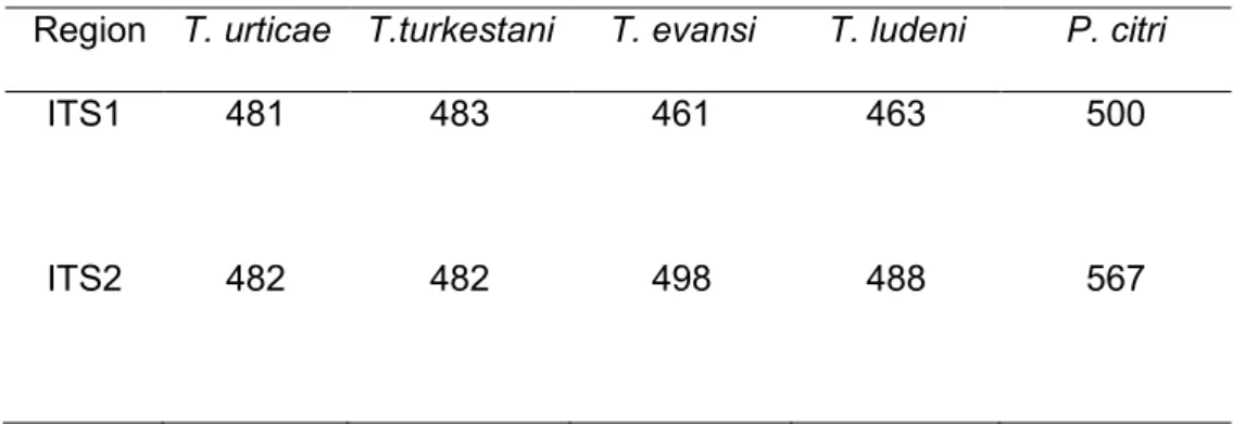
Size range of ITS1 was 461 to 500 bp, for ITS2 was 482 to 567 bp (Table 3). The 5.8 S gene length was fixed at 160 bp in all species.
Alignments of sequences of the ITS1 and ITS2 of Tetranychus species remained unambiguous, despite some insertions and deletions. The nucleotide divergence betweenT. urticae and the other three Tetranychus species was 11 % for T. evansi and T. ludeni and 3 % for T. turkestani. As expected, the nucleotide
divergence between P. citri and the other four Tetranychus species was much higher: 42% for T. urticae and T. ludeni; 41% for T. turkestani and T. evansi. All samples collected in both Spain and France showed the same ITS2 sequence. Despite a low sequence divergence between these samples and both Florida and Japan ones, this divergence did not affect the RFLP pattern.
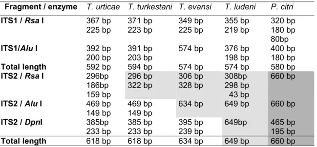
Three restriction enzymes (RsaI, AluI and DpnI) in combination produced restriction fragments that distinguished all species (Table 4). Restriction patterns using RsaI and AluI are shown in Figure 2.
Comparison of sequences using optimal global pairwise alignment (BioEdit software) (Hall 1999) showed that the most interspecific variation appeared on the ITS1, with a sequence divergence ranging from 14 to 16% between the four Tetranychusspecies considered. ITS2 showed less variation among species: 11% between T. urticae and T. evansi, 11% between T. urticae and T. ludeni and 1-3 % between T. urticae and T. turkestani. A pairwise nucleotide distance matrix for the five mite species considered in this study was calculated. The range of sequence
divergence observed within and among taxa are presented in Table 5.
Intraspecific variation was estimated based in the comparison of the sequences of T. urticae collected in 4 different countries (Spain, France, Portugal and the United States) plus already published sequences from Japan. The ITS1 region showed 2 to 3% nucleotide divergence in comparisons between specimens collected in Europe and from other continents (America and Asia). By contrast, intraspecific comparisons of ITS2 sequences of T. urticae, revealed only a nucleotide substitution in position 361 in the sample from Castelló which differentiates this sequence from specimens coming from different continents: America (Florida) and Asia (Japan) (Osakabe et al., 2002; 2006).
There was no difference between the ITS2 region from T. evansi specimens coming from Castelló and Madeira. Nevertheless, comparison between the ITS2 region of T. evansi from Castelló and the strains obtained by Knapp et al. (2003) in three different locations (Brasil, Zimbabwe and Kenya), detected several point mutations between sequences of individuals originated from different locations, which represents a sequence divergence 1-2%, higher than the one observed for the T. urticae ITS2 region.
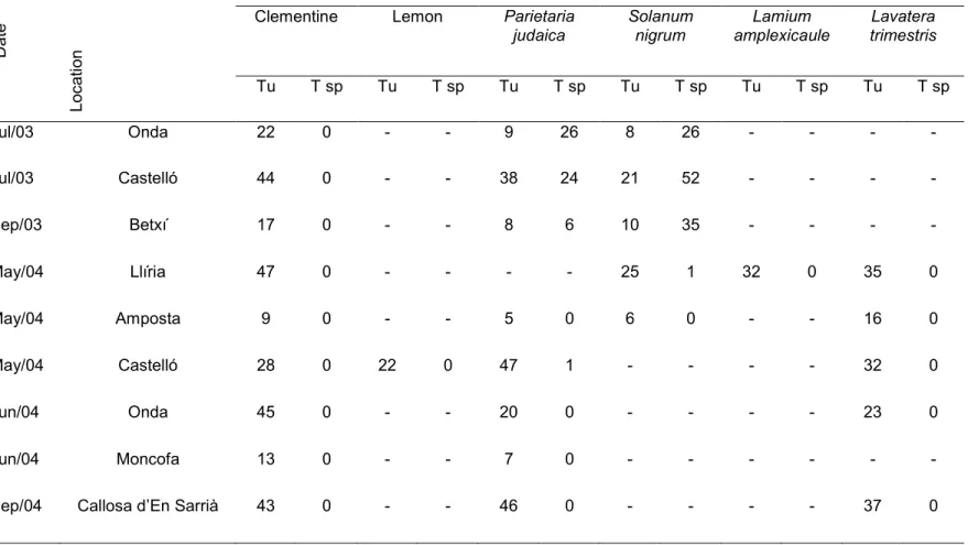
PCR-RFLP was used to screen a series of field samples collected on 7 different locations from May 2003 to December 2004 (Table 2). We processed a total of 886 individuals: 268 from clementine trees, 237 from Parietaria judaica, 184 from Solanum nigrum L., 143 from Lavatera trimestris L., 32 individuals from Lamium amplexicaule L.
and 22 from lemon trees. All the Tetranychidae specimens feeding on citrus analyzed in this study were identified as T. urticae. The proportion of T. urticae on P. judaica was highly variable, depending on both the sampling location and the date. By contrast, less than 30 % of the specimens collected on S. nigrum during the summer were T. urticae. The results obtained by RFLP analysis during summer showed that T. urticae
represented 25.8 % and 49.5 % on S. nigrum and P. judaica, respectively.
Discussion
Interspecific ribosomal ITS1 and ITS2 sequence variation
The sizes of the ITS1 and ITS2 regions of the mites considered in this study (Table 3) are in the range of other tetranychid mites reported in the literature (Navajas et al., 1999; Knapp et al., 2003; Osakabe et al., 2002; Ben-David et al., 2007). The
divergence obtained between P. citri and the other four Tetranychus species (around 40 %) was much higher than the 14% found between P. citri and P. ulmi (Hsu et al., 2004; Ben-David et al., 2007), which was close to what was observed among the species belonging to the genus Tetranychus.
ITS sequence variation was further investigated based on comparisons between present and already published Tetranychidae sequences (Navajas et al., 1998; Osakabe et al., 2002; Knapp et al., 2003, Ben-David et al., 2007). Pairwise distance matrices (Table 5) show that the ITS1 had more interspecific variation than ITS2. As expected, ITS2 variation between T. urticae and T. turkestani, which are very closely related species (Navajas & Boursot, 2003; Ben-David et al., 2007) was very low. Therefore, the low sequence divergence in ITS2 between species allows us to use these small differences as species specific markers.
Intraspecific ribosomal ITS1 and ITS2 sequence variation
The 2-3% intraspecific variation found for ITS1 of T. urticae from different locations exceeds the 2% variation threshold in use for spider mite species diagnosis (Ben-David et al., 2007) and is of the same magnitude as that found between two
distinct species of Phytoseiid mites, Neoseiulus fallacis (Garman) and N. californicus (McGregor) (Navajas et al., 1999). Therefore, the variability of this region is too high to be consistently used for species discrimination.
Navajas & Boursot (2003) showed that ITS2 sequences from European specimens of T. urticae were perfectly homogeneous, as did Ben-David et al. (2007) for Israeli mites. These results are in agreement with the ITS2-T. urticae sequences from specimens collected along the 350 km in the Mediterranean Eastern coast of Spain from Amposta (39º97’º N 0º05’W) to Callosa d’en Sarrià (38º6’N 0º05’W), which present no sequence variation except for some insertions and deletions which resulted in different fragment sizes (Table 1). However, these variations never affected the enzyme restriction sites. Interestingly, previous studies had revealed that this homogeneity extended worldwide (Navajas et al., 1998). Our results provide
confirmation of lack of variation in ITS2 among Northwestern Mediterranean specimens (France and Spain). However the intercontinental variation found (Japan and Florida versus Europe) deserves further investigation.
Intraspecific polymorphism has been reported in the ITS2 region from T.
turkestani originated from several locations in Europe and the United States (Navajas & Boursot 2003). We have only found one point mutation (one A>G transition) on 116 bp position comparing T. turkestani from Castelló and France, but intraspecific ITS1 variation was more important. It consisted of one deletion, two transitions (C>T) and two transversions (G>T and A>T).
The observed ITS2 intraspecific variation differentiates between geographically long distant localities, whereas specimens from the same area showed homogenization of sequences. This makes ITS2 PCR-RFLP tests useful for species discrimination of Tetranychidae specimens, as also suggested by Ben-David et al. (2007).
Recognition sites and patterns
Restriction enzymes were selected that isolated species-specific nucleotide differences (Table 4). Among all potential restriction enzymes, RsaI separates T.
urticae from the rest of species found in citrus orchards independently of the
geographical origin of the specimen (Figure 2). Because T. urticae is the main pest in citrus and it will be the target for further population genetic studies, ITS2 PCR -RFLP using RsaI will be useful for broad screening of mites collected in field surveys to determine species identity.
The RsaI restriction profile for the five species (Figure 2) shows the existence of two restriction sites for both T. urticae and T. ludeni, whereas only one restriction site exists for the remaining species. The size of the restriction fragment unambiguously identifies these two species (Table 4). However T. evansi, T. ludeni and T. turkestani RsaI restriction patterns appear similar on agarose gels. Therefore, additional enzymes were selected to identify these species. AluI identifies T. turkestani (Figure 2) and DpnI separates T. evansi from T. ludeni.
Although the existence of intraspecific variation could impair the use of this technique for species-discrimination, Rsal restriction profiles have proved to
unambiguously separate T. urticae from the remaining four tetranychid species found in citrus (Figure 2). Furthermore, this study did not detect any ITS2 sequence variation for T. urticae specimens collected along the Spanish Mediterranean coast, whereas the differences with Florida and Japan samples does not interfere with RsaI profile. PCR –RFLP: as a quick species diagnostic of field samples
The results obtained from the PCR-RFLP analyses (Table 2) are consistent with those obtained by Aucejo et al. (2002) for mites on the same plant species and season on previous years. In their study T. urticae represented 11.8 % and 50.0 % of total summer mite catches, on P. judaica and S. nigrum, respectively, whereas in spring T. urticae clearly predominated, and represented 94.1 % and 98.6 % on RFLP samples of S. nigrum and P. judaica, respectively.
The PCR-RFLP approach is a promising method to screen high numbers of samples to separate T.urticae from other mite species and quantify its relative
abundance. These results will pave the way to further studies aimed at evaluating the genetic structure of populations of T. urticae in citrus orchards.
Acknowledgments
The authors thank B. Sabater (IVIA-Montcada) for her helping during the sequence analyses, M. Foó, S. Aucejo and P. Troncho (UJI) for their help with the sampling and B. Hurtado (UJI) for graphical design support. This work was partially funded by the Agència Valenciana de Ciència i Tecnología (code CTESPP/2003/075), the Fundació Bancaixa – Caixa de Castelló (code P1 1B2003-06) and the Ministerio de Ciencia y Tecnología (AGL2004-07464-C03-01/AGR).
References Cited
Ansaloni, T., Pascual-Ruiz, S. Hurtado, M.A., Jacas, J.A. (2007). Can summer and fall vegetative growth regulate the incidence of Tetranychus urticae Koch on
clementine fruit? Crop Protection doi:10.1016/j.cropro.2007.07.016
Aucejo, S., Foó M., Gimeno E., Gómez-Cadenas A., Monfort R., Obiol F., Prades E., Ramis M., Ripollés J.L., Tirado V., Zaragozà L., Jacas J.A., Martínez-Ferrer M.T. (2003) Management of Tetranychus urticae in citrus in Spain: acarofauna
associated to weeds. Integrated Control in Citrus Fruit Crops. IOBC wprs Bulletin, 26(6), 213-220.
Aucejo-Romero S., Gómez-Cadenas A., Jacas-Miret J.A. (2004) Effects of NaCl- stressed citrus plants on life-history parameters of Tetranychus urticae (Acari : Tetranychidae). Experimental and Applied Acarology, 33,1-2, 55-67.
Bailly X., Migeon A., Navajas M. (2004). Analysis of microsatellite variation in the spider mite Tetranychus turkestani (Acari: Tetranychidae) reveals population
genetic structure and raises questions about related ecological factors. Biological Journal of the Linnean Society, 82, 69-78.
Ben-David T., Melamed S., Gerson U., Morin S. (2007). ITS2 sequences as barcodes for identifying and analyzing spider mites (Acari: Tetranychidae). Experimental and Applied Acarology, 41, 169-181.
Bodenheimer F.S. (1951). Citrus entomology. Dr. W Junk Publishers.
Hall T.A. (1999). Bioedit, a user-friendly biological sequences alignment editor and analysis program for Windows 95/98/NT. Nucleic Acids Symposium Series, 41, 95-98
Hinomoto N., Takafuji A. (2001). Genetic diversity and phylogeny of the Kanzawa spider mite, Tetranychus kanzawai, in Japan. Experimental and Applied Acarology, 25, 355-370.
Hmimina M., Allam L., Ougass Y., Marmouche A. (1995). Circonstances des pullulations de Tetranychus urticae Koch (Tetranychidae: Acarina) en verger d'agrumes. IOBC wprs Bulletin 18 (5), 28-35.
Hsu K., Hua T., Chang N.T., Yeh W.B. (2004). Nuclear ribosomal DNA sequence in tetranychid mites: a conflict phylogeny to mitochondrial cytochrome oxidase I. Gene Bank accession # AY750708.
Kumar S., Tamura K., Nei M. (2004) MEGA3: Integrated software for Molecular Evolutionary Genetics Analysis and sequence alignment. Briefings in Bioinformatics, 5,150-163.
Martínez-Ferrer M.T., Jacas J.A., Ripollés J.L., Aucejo S. (2006). Approaches for sampling the two spotted spider mite Tetranychus urticae on clementines in Spain. Journal of Economic Entomology, 99, 1490-1499.
Navajas M., Boursot P. (2003). Nuclear ribosomal DNA monophyly versus
mitochondrial DNA polyphyly in two closely related mite species: the influence of life history and molecular drive. Proceedings of the Royal Society of London Series B-Biological Sciences, 270, S124-S127.
Navajas M., Gutierrez J., Lagnel J., Boursot P. (1996). Mitochondrial cytochrome oxidase I in tetranychid mites: A comparison between molecular phylogeny and changes of morphological and life history traits. Bulletin of Entomological Research, 86, 407-417.
Navajas M. (1998). Host plant associations in the spider mite Tetranychus urticae (Acari: Tetranychidae): insights from molecular phylogeography. Experimental and Applied. Acarology, 22, 201-214.
Navajas M., Lagnel J., Gutierrez J., Boursot P. (1998). Species-wide homogeneity of nuclear ribosomal ITS2 sequences in the spider mite Tetranychus urticae
contrasts with extensive mitochondrial COI polymorphism. Heredity, 80, 742-752. Navajas M., Lagnel J., Fauvel G., De Moraes G. (1999). Sequence variation of
ribosomal internal transcribed spacers (ITS) in commercially important phytoseiidae mites. Experimental and Applied Acarology, 23, 851-859.
Navajas M., Fenton B. (2000). The application of molecular markers in the study of diversity in acarology: A review. Experimental and Applied Acarology, 24,751- 774.
Navajas M., Tsagkarakov A., Lagnel J., Perrot-Minnot M.J. (2000). Genetic
differentiation in Tetranychus urticae (Acari : Tetranychidae): polymorphism, host races or sibling species? Experimental and Applied Acarology, 24, 365-376. Navajas M., Perrot-Minnot M.J., Lagnel J., Migeon A., Bourse T., Cornuet J.M. (2002).
Genetic structure of a greenhouse population of the spider mite Tetranychus urticae: spatio-temporal analysis with microsatellite markers. Insect Molecular Biology, 11, 157-165.
Navia D., de Moraes G., Roderick G.K., Navajas M. (2005). The invasive coconut mite, Aceria guerreronis (Acari: Eriophyidae): origin and invasion sources inferred from mitochondrial (16S) and ribosomal (ITS) sequences. Bulletin of
Nicholas, K.B., Nicholas H.B. Jr., Deerfield, D.W. II. (1997). GeneDoc: Analysis and Visualization of Genetic Variation, EMBNEW.NEWS 4:14.
Osakabe M., Hirose T., Sato M. (2002). Discrimination of four Japanese Tetranychus species (Acari : Tetranychidae) using PCR-RFLP of the inter-transcribed spacer region of nuclear ribosomal DNA. Applied Entomology and Zoology, 37, 399-407. Osakabe,M., Kotsubo,Y., Tajima,R. and Hinomoto,N. 2006. Phylogenetic Relationship
and RFLP Catalogue for Molecular Identification of Tetranychus Spider Mites (Acari: Tetranychidae) in Japan. GenBank.
Souliotis P., Tsagkarakou A., Nomikou, M. (1997). Field observations and eco- ethological aspects of Phytoseiid mites in greek citrus groves. Acarologia XXXVIII, 29-37.
Swirski, E. (1977). Integrated control of mites in Israel, in ‘O. Carpens (Ed.), I Congreso Mundial de Citricultura, CEBAS Murcia-Valencia 1973’. Vol. 2 pp. 477-480. Tixier M.S., Kreiter S., Auger P. (2002a). How can molecular data contribute to analyse
the colonisation of vineyards by Kampimodromus aberrans ? Acari: Phylogeny and evolution. Adaptations in mites and ticks: 331-340.
Tixier M.S., Kreiter S., Croft B.A., Auger P. (2002b). Colonization of vineyards by Kampimodromus aberrans: dispersal from surrounding plants as indicated by random amplified polymorphism DNA typing. Agricultural and Forest Entomology, 4, 255-264.
Vacante V. (1986). Influence of white mineral oil treatments on Eastern Sicily. CEC Experts Meeting. Acireale 1985. In ‘Integrated Pest Control in Citrus Groves’. Balkema, Rotterdam. pp. 423-431.
Xie L., Hong X-Y, Xue Y-F. (2006). Population genetic structure of the twospotted spider mite (Acari: Tetranychidae) from China. Annals Entomological Society of America, 99, 959-965.
Table 1. Collection sites of Tetranychidae mites considered in this study. The host plant and the ITS region sequenced with the respective EMBL accession number and the total length of the amplified DNA region are indicated.
Species Sample abbreviation
Location Host plant ITS1 ITS2 Accession number and
total length Tetranychus
urticae
Tu Cs Spain (Castelló) Citrus reticulata Blanco x x AM408030 1223 bp Tu Am Spain (Amposta) Citrus reticulata Blanco x AM408042 484 bp Tu Be Spain (Betxi) Citrus reticulata
Blanco
x AM408043 482 bp Tu Ca Spain (Callosa) Citrus reticulata
Blanco x AM408044 481 bp Tu Mo Spain (Moncofar) Citrus reticulata Blanco x AM408045 484 bp Tu On Spain (Onda) Citrus reticulata
Blanco x AM408046 481 bp Tu Fr France (Alenyà) Solanum lycopersicum L * x x AM408031 1220 bp Tu Jp1 Japan Citrullus lanatus (Thunb) x x AB076369 AB257738 AB257737 AB257736 Consensus 1205 bp Tu Fl Florida (Gainesville)
Citrus sinensis L. x x AM408035 1163 bp Tetranychus
evansi
Te Cs Spain (Castelló) Solanum nigrum L. x x AM408033 1218 bp Te Ma Portugal
(Madeira)
Solanum nigrum L. x AM408036 462 bp x AM408047 498 bp Te Br2 Brazil (Piracaiba) S. lycopersicum L. x AJ419833 497 bp Te Ky2 Kenya S. lycopersicum L. x AJ419833 493 bp Te Zb2 Zimbabwe S. lycopersicum L. x AJ419833 496 bp Tetranychus ludeni
TlCs Spain (Castelló) Paretaria judaica x AM408037 464 bp x AM408040 488bp TlJp1 Japan - x x AB076371 1197 bp Tetranychus turkestani Tt Fr France (Codognan) Solanum melongena L. * x x AM408032 1223 bp Tt Cs Spain (Castelló) Convolvulus
arvensis L.
x AM408038 479 bp
x AM408041 483 bp
Panonychus citri
Pc Cs Spain (Castelló) Citrus reticulata Blanco x x AM408034 1282 bp Pc Mo Spain (Moncofa) Citrus reticulata Blanco x AM408039 434 bp 1
Data from Osakabe et al., 2002 and 2006 2
Data from Knapp et al., 2003 * Reared on Phaseolus vulgaris L.
Table 3. The Internal Transcribed Spacers (ITS1 and ITS2) sizes (bp) of Tetranychus urticae, T. turkestani, T. evansi, T. ludeni and Panonychus citri present in citrus orchards from Castelló (Spain).
Region T. urticae T.turkestani T. evansi T. ludeni P. citri
ITS1 481 483 461 463 500
ITS2 482 482 498 488 567
Table 2. Species discrimination results of PCR-RFLP analysis of the ribosomal ITS2 (650 bp) using the RsaI enzyme for mites collected on several host plants and localities in Spain. For each sampling date, the number of specimens identified is presented. Tu: Tetranychus urticae; T sp: Tetranychus spp. other than T. urticae.
Da te L o c a ti o n Plant species Clementine Lemon Parietaria
judaica Solanum nigrum Lamium amplexicaule Lavatera trimestris Tu T sp Tu T sp Tu T sp Tu T sp Tu T sp Tu T sp Jul/03 Onda 22 0 - - 9 26 8 26 - - - - Jul/03 Castelló 44 0 - - 38 24 21 52 - - - - Sep/03 Betxí 17 0 - - 8 6 10 35 - - - - May/04 Llíria 47 0 - - - - 25 1 32 0 35 0 May/04 Amposta 9 0 - - 5 0 6 0 - - 16 0 May/04 Castelló 28 0 22 0 47 1 - - - - 32 0 Jun/04 Onda 45 0 - - 20 0 - - - - 23 0 Jun/04 Moncofa 13 0 - - 7 0 - - - -
Sep/04 Callosa d’En Sarrià 43 0 - - 46 0 - - - - 37 0
Table 4. ITS1 and ITS2 restriction fragment length differences between Tetranychus urticae, T. turkestani, T. evansi, T. ludeni and Panonychus citri from Castelló (Spain) using different enzymes. For ITS2 (the only fragment used for species discrimination), within a line, same color cells indicate species not differentiated using the
corresponding enzyme. Total length corresponds to the sizes of the PCR product including primers.
Fragment / enzyme T. urticae T. turkestani T. evansi T. ludeni P. citri ITS1 / Rsa I 367 bp 225 bp 371 bp 223 bp 349 bp 225 bp 355 bp 219 bp 320 bp 180 bp 80bp ITS1/Alu I 392 bp 200 bp 391 bp 203 bp 574 bp 376 bp 198 bp 400 bp 180 bp Total length 592 bp 594 bp 574 bp 574 bp 580 bp ITS2 / Rsa I 296bp 186bp 159 bp 296 bp 322 bp 306 bp 328 bp 308bp 298 bp 43 bp 660 bp ITS2 / Alu I 469 bp 149 bp 469 bp 149 bp 634 bp 649 bp 660 bp ITS2 / DpnI 385bp 233 bp 385 bp 233 bp 395 bp 239 bp 649bp 465 bp 195 bp Total length 618 bp 618 bp 634 bp 649 bp 660 bp
Table 5. Pairwise distances using the Kimura 2 parameter between Tetranychidae mites based on differences in nucleotide sequences of the ribosomal ITS regions (first line corresponds to the 28S-ITS1-5,8S-ITS2-18S nucleotide distance comparison). Tetranychidae mites are from different origins: Castelló (Cs), France (Fr), Florida (Fl), Japan (Jp, Osakabe et al., 2002 and 2006), Madeira (Ma) and Brasil, Kenya and Zimbabwe (Br, Ky and Zb, respectively, Knapp et al., 2003). Grayed areas indicate intraspecific comparisons.
Tu (Cs, Fr, Fl, Jp) Tt (Cs, Fr) Te (Cs, Ma, Br, Ky, Zb) Tl (Cs, Jp) Pc (Cs) Tu (Cs, Fr, Fl, Jp) ITS 0.0000 - 0.0091 0.0103 – 0.0148 0.0620 – 0.0694 0.0603 – 0.0712 0.3514 – 0.3542 ITS1 0.0000 - 0.0116 0.0186 – 0.0232 0.0905 – 0.0974 0.0766 – 0.0882 0.4362 – 0.4385 ITS2 0.0000 - 0.0021 0.0083 – 0. 0124 0.0669 – 0.0729 0.0818 – 0.0861 0.4116 – 0.4142 Tt (Cs, Fr) ITS 0.0000 0.0712 – 0.0731 0.0686 – 0.0721 0.3516 – 0.3529 ITS1 0.0000 - 0.0093 0.1021 – 0.1090 0.0905 – 0.0951 0.4362 – 0.4455 ITS2 0.0000 - 0.0021 0.0689 – 0.0728 0.0798 – 0.0837 0.4163 – 0.4176 Te (Cs, Ma, Br, Ky, Zb) ITS 0 .0000 – 0.0009 0.0442 – 0.0483 0.3614 – 0.3624 ITS1 0.0000 - 0.0023 0.0537 – 0.0580 0.4617 - 0.4640 ITS2 0.0000 – 0.0061 0.0515 – 0.0646 0.4121 – 0.4182 Tl (Cs, Jp) ITS 0.0000 – 0.0036 0.3673 – 0.3677 ITS1 0.0000 - 0.0023 0.4710 – 0.4733 ITS2 0.0000 - 0.0062 0.4195 – 0.4225
Figure Captions
Figure 1. Location map of the study area. For each locality the host plant sampled and sampling date are indicated.
Figure 2. Ribosomal ITS2 restriction fragment length differences in the species T. urticae (Tu), T. ludeni (Tl ), T. turkestani (Tt), T. evansi (Te) and Panonychus citri (Pc) digested with RsaI (R) and AluI (A). Molecular weight marker: 50 bp DNA Ladder (Invitrogen).
Figure 1. Figure 1. Amposta Castelló de la Plana Onda Betxí Llíria Montcada
Callosa d’en Sarrià
EURO PE Clementine Lavatera Parietaria Clementine Solanum Clementine Lavatera Solanum Clementine Lavatera Parietaria Lemon Clementine Lavatera Solanum Parietaria 39.97ºN 0.05ºW 40.72ºN 0.58ºE 38.60ºN 0.05ºW 39.55ºN 0.40ºW 39.62ºN 0.60ºW Clementine Lavatera Solanum Parietaria 39.95ºN 0.27ºW Clementine 39.56ºN 0.01ºW Amposta Castelló de la Plana Onda Betxí Llíria Montcada
Callosa d’en Sarrià
EURO PE Clementine Lavatera Parietaria Clementine Solanum Clementine Lavatera Solanum Clementine Lavatera Parietaria Lemon Clementine Lavatera Solanum Parietaria 39.97ºN 0.05ºW 40.72ºN 0.58ºE 38.60ºN 0.05ºW 39.55ºN 0.40ºW 39.62ºN 0.60ºW Clementine Lavatera Solanum Parietaria 39.95ºN 0.27ºW Clementine 39.56ºN 0.01ºW
Figure 2.


![Whole-body [18F]-fludeoxyglucose positron emission tomography in endocarditis: the story of a rare diagnosis.](data:image/gif;base64,R0lGODlhAQABAIAAAP///wAAACH5BAEAAAAALAAAAAABAAEAAAICRAEAOw==)