DOI 10.1007/s00417-005-1165-2 Alexander Meyenberg David Goldblum Jean-Marc Zingg Angelo Azzi Kalanithi Nesaretnam Monika Kilchenmann Beatrice E. Frueh Received: 19 November 2004 Revised: 26 January 2005 Accepted: 14 February 2005 Published online: 21 May 2005
# Springer-Verlag 2005
Tocotrienol inhibits proliferation of human
Tenon’s fibroblasts in vitro: a comparative
study with vitamin E forms and mitomycin C
Abstract Purpose: To evaluate the potential of the vitamin E compound α-tocotrienol as antifibrotic agent in vitro. Methods: Using human Te-non’s capsule fibroblast cultures, the antiproliferative and cytotoxic effects of the different vitamin E forms α-tocopherol, α-tocopheryl acetate, α-tocopheryl succinate and α-toco-trienol were compared with those of mitomycin C. To mimic subconjunc-tival and regular oral application in vivo, exposure time of serum-stimu-lated and serum-restimuserum-stimu-lated fibro-blasts (SF and RF, respectively) to vitamin E forms was set at 6 days. Cultures were only exposed for 5 min to mitomycin C due to its known acute toxicity and to mimic the short-time intraoperative administration. Proliferation (expressed as % of con-trol) was determined by DNA content quantification on days 2, 4 and 6, whereas cytotoxicity was assessed by cell morphology and glucose 6-phos-phate dehydrogenase (G6PD) release
after 24 h. Results: α-Tocopherol andα-tocopheryl acetate stimulated growth of SF, but not RF. Reduction of fibroblast content byα-tocopheryl succinate was accompanied by in-creased G6PD release and necrosis. Contrary toα-tocopheryl succinate, 50μM or repeatedly 20 μM of α-tocotrienol significantly inhibited proliferation without causing cellular toxicity (maximal effect: 46.8%). RF were more sensitive to this effect than SF. Mitomycin C 100–400 μg/ml showed a stronger antiproliferative effect thanα-tocotrienol (maximal effect: 13.8%). Morphologic charac-teristics of apoptosis were more commonly found under treatment with mitomycin C. Conclusions: Of the vitamin E forms tested, only α-tocotrienol significantly inhibited growth at non-toxic concentrations. In this in vitro study, antiproliferative effects of mitomycin C were stronger than those ofα-tocotrienol.
Introduction
Failure of glaucoma filtration surgery is mostly due to fibrosis of the surgical fistula and the filtering bleb [1,14]. Fibroblasts from the conjunctival-Tenon’s capsule-epi-scleral interface migrate to the surgical site, proliferate, and secrete extracellular matrix components, ultimately leading to excessive scar formation [14]. Unfortunately, the antiinflammatory and antiproliferative effects of topical steroids are often not sufficient to avoid the induction of this process.
Mitomycin C is currently the most frequently used agent to inhibit this cicatrisation [18, 35]. However, adjunctive topical chemotherapy with mitomycin C is associated with severe and potentially sight-threatening complications such as ocular hypotony and endophthalmitis [15,23,26]. Cy-totoxic effects to various cell types seem to be responsible for these serious side effects [27,33,36,42].
In the search for therapeutic alternatives we focused on vitamin E, the most important lipid-soluble antioxidant in the human body. Vitamin E is a generic name. Tocopherols and tocotrienols represent the two subclasses within the A. Meyenberg . D. Goldblum .
M. Kilchenmann . B. E. Frueh (*) Departement of Ophthalmology, University of Berne, Inselspital Berne, 3010 Berne, Switzerland
e-mail: [email protected] Tel.: +41-31-6322111
Fax: +41-31-3824779 J.-M. Zingg . A. Azzi
Institute for Biochemistry and Molecular Biology, University of Berne,
Buehlstrasse 28, 3012 Berne, Switzerland K. Nesaretnam
Malaysian Palm Oil Board, PO Box 10620, 50720 Kuala Lumpur, Malaysia
vitamin E family of compounds, both of them consisting of , β-, γ-, and δ-isoforms. Esterized forms such as α-tocopheryl acetate or succinate also exhibit vitamin E ac-tivity. Lipid-rich plant products and vegetable oils are the main natural sources of vitamin E.
Vitamin E protects biological membranes from oxida-tion by acting as a radical chain breaking molecule. Im-portant novel neuroprotective [28], antithrombotic [48], antiinflammatory [16], hypocholesterolemic [53], antiath-erogenic [46], and antineoplastic effects [24, 32, 50] of vitamin E cannot all be attributed to the antioxidant activity alone. Increasing evidence suggests involvement of mul-tiple intracellular signaling pathways [4,19,45]. It has to be emphasized that the biological functions strongly de-pend on the vitamin E form applied, and that not all cell types are as susceptible to these effects.
The vitamin E formsα-tocopherol, α-tocopheryl acetate andα-tocopheryl succinate have been previously tested in in vitro and in animal model studies for potential appli-cations in glaucoma filtration surgery and in the prevention of proliferative vitreoretinopathy [10, 11, 20–22, 29, 37,
38, 40, 41]. However, results were conflicting [30], and there is a need to compare directly the antiproliferative and cytotoxic effects of these different vitamin E forms with mitomycin C, the gold standard of antiscarring drugs in glaucoma filtration surgery.
α-Tocotrienol is one of the major vitamin E compounds in palm oil. Similar to tocopherols, the antiproliferative effects of tocotrienols are mediated by inhibition of protein kinase C [44]. In human and murine tumor cells, toco-trienols induce a cell-cycle arrest in the G-1 phase and apoptosis [54]. To the best of our knowledge, the potential of tocotrienols as antifibrotic agents has not been evaluated. By using human primary Tenon’s capsule fibroblast cultures, we therefore compared the antiproliferative and cytotoxic effects oftocopherol, tocopheryl acetate, α-tocopheryl succinate, andα-tocotrienol with those of mito-mycin C.
Materials and methodes
Reagents
α-Tocopherol (RRR-α-tocopherol) was obtained from Cognis (Duesseldorf, Germany),α-tocopheryl acetate (dl-α-toco-pherol acetate) andα-tocopheryl succinate (d-α-tocopherol succinate) were from Supelco (distributed by Fluka Chemie, Buchs, Switzerland). RRR-α-Tocotrienol was a kind gift from the Malaysian Palm Oil Board (Kuala Lumpur, Ma-laysia). Vitamin E forms were dissolved in ethanol absolute and then stored light-protected at 4°C. Mitomycin C (Mi-tomycin-C Kyowa) was purchased from Kyowa (distrib-uted by Roche Pharma, Reinach, Switzerland) and dissolved freshly in phosphate-buffered saline (PBS) before use.
Cell cultures
Explants of human Tenon’s capsule were obtained from seven patients (aged 68–93 years) at the time of cataract surgery. None of the donors had had antiglaucomatous or antimetabolite treatment, or ocular surgery before. All pa-tients gave their informed consent before inclusion in the study, which was approved by local ethics committee and conformed with the provisions of the Declaration of Helsiniki. The specimens were dissected and placed in 25 cm2tissue culture flasks containing Dulbecco’s minimal essential medium (DMEM) supplemented with L
-gluta-mine (584 mg/l), sodium pyruvate (100 mg/l), glucose (1000 mg/l), penicillin (60 IU/ml), streptomycin (60μg/ml) and 10% fetal calf serum (FCS). Cultures were main-tained in a humidified 5% CO2incubator at 37°C.
Fibro-blasts were characterised morphologically and confluent cultures were trypsinized, centrifuged and repassaged. The above medium was changed twice per week. Quantified by trypan blue dye exclusion method, viability was always more than 95%. For the assays, third- to sixth-passage cells were used.
Cell proliferation assay
Two experiments were performed with all seven cell lines in triplicate. Substances were either applied to serum-stim-ulated fibroblasts (SF) in the exponential growth phase, or to previously quiescent serum-restimulated fibroblasts (RF) with a beginning proliferative response. For the ex-periments with SF and RF, 2000 and 1500 cells/well were seeded in 96-well tissue culture plates (black/clear-bottom, Corning Life Sciences), respectively, each well containing 200μl of culture medium. Preliminary studies showed this difference to be suitable for achieving similar cell densities at the time of compound application. Fibroblasts were then allowed to settle for 24 h in the incubator.
Growth of serum-stimulated fibroblasts (SF) Cells were rinsed with PBS and fresh DMEM/10% FCS was added. Vitamin E forms were diluted to the indicated concentra-tions in above media. To mimic subconjunctival injection and a regular oral vitamin E supplement intake in vivo, exposure time to all vitamin E forms was set at 6 days, and application of α-tocopherol and α-tocotrienol to the cell cultures was repeated at days 2 and 4, respectively. In vivo, the estersα-tocopheryl acetate and succinate are naturally cleaved intestinally, resulting in adsorption of the active formα-tocopherol. Repeated application was performed by pipetting an identical amount of compounds as initially used into the corresponding wells without changing media. Cells treated with an equivalent amount of ethanol absolute were also included as controls. The cumulative concentra-tion of ethanol was less than 0.6%, which had no effect on
proliferation and viability (data not shown). Mitomycin C was dissolved in PBS to concentrations of 10, 100, 200 and 400μg/ml. Due to its known acute toxicity and to mimic the short-time intraoperative administration, SF were only ex-posed for exactly 5 min to 100 μl/well mitomycin C so-lution. Cells were then gently washed with PBS alone and fed with 200 μl/well DMEM/10% FCS again. Multiple rinsing with PBS was not performed, since a decrease in cell density was observed in preliminary studies. PBS-treated cultures served as control. The plates were then incubated for 6 days at 37° C in 5% CO2in a humidified air
atmosphere. No media was changed during this time. Growth of serum-restimulated fibroblasts (RF) Fibroblasts were washed with PBS 24 h after plating and incubated for another 48–72 h with DMEM containing 0.2% FCS. As previously described [10], this procedure induced growth arrest on human Tenon’s capsule fibroblasts in vitro. Cell growth was then restimulated by changing this medium to one containing 10% FCS. Incubation with vitamin E forms followed as described above. RF were exclusively exposed to 100 μg/ml mitomycin C due to the almost complete growth inhibition found in the SF experiment at this concentration.
Morphology of SF and RF was studied daily using phase contrast light microscopy (Leica DMIRB research micro-scope, Leica Microsystems, Wetzlar, Germany). Photomi-crographs were obtained with a color camera (Kappa CF15 MC, Kappa Messtechnik, Gleichen, Germany) connected to a video printer (Sony UP-5200MDP, Sony, Schlieren, Switzerland).
Measurement of DNA content A fluorometric assay (CyQUANT Cell Proliferation Assay Kit, Molecular Probes, distributed by JURO Supply, Lucerne, Switzerland) was used to determine cell density on days 2, 4 and 6 by DNA content quantification. Briefly, CyQuant GR dye exhibited green fluorescence enhancement when bound to cellular nucleic acids. Fluorescence due to dye binding to RNA was eliminated by pretreating samples with 4 μl RNase (DNase-free, Roche Diagnostics, Rotkreuz, Switzerland) per ml cell lysis buffer containing 1 mM ethylenediamine-tetraacetate (EDTA). The fluorescent dye and its solutions were always handled light-protected. A reference standard curve was constructed by plotting the fluorescence signal against the DNA content of 50–10,000 human Tenon’s capsule fibroblasts/well. The relation was strongly linear (Pearson correlation coefficient r=0.99). On the indicated days, plates were gently inverted and blotted onto paper towels to remove medium from the wells. Wells were rinsed with PBS, and dry plates were frozen at−70°C for up to 2 weeks. As stated by the manufacturer, the freezing step is important for efficient cell lysis. In order to measure sample fluorescence, plates were thawed, 150μl of cell lysis buffer/ EDTA/RNase was added to each well, and plates were
placed for one hour at 37° C in 5% CO2in a humidified air
atmosphere. Next, 50μl of 4-fold concentrated fluorescent dye/cell lysis buffer/EDTA was added to each well. After light-protected incubation for exactly 3 1/2 minutes, sample fluorescence was measured with a fluorescence microplate reader (Cytofluor 2300; Millipore, Volketswil, Switzerland) with 485 nm excitation and 530 nm emission wave lenghts.
Cytotoxicity assay
Damaged cells release the cytosolic enzyme glucose 6-phosphate dehydrogenase (G6PD) into the surrounding medium. G6PD-release 24 h after incubation with vitamin E forms or 24 h after 5-min exposure to mitomycin C was determined fluorometrically by the Vybrant Cytotoxicity Assay Kit (Molecular Probes, distributed by JURO Supply, Lucerne, Switzerland). By plotting the fluorescence signal against the G6PD medium content of 500 to 10,000 lysed human Tenon’s capsule fibroblasts/well (reference standard curve), a strongly linear relation was observed (Pearson correlation coefficient r=0.98). Substances containing the fluorescent dye were always processed light-protected. The assay was performed with six different cell lines in trip-licate (one cell line was lost due to contamination). In all, 7500 cells/well were seeded to optimise sensitivity of the assay. The further procedure was identical to the prolifer-ation assay with SF until 24 h after applicprolifer-ation of the in-dicated compounds. Tissue culture plates (96-well) were then gently shaken, 50μl medium of each well was trans-ferred to separate plates, and 50μl/well 2-fold concentrated resazurin/reaction mixture was added to the collected media. After light-protected incubation for 20 minutes at 37°C, measurements were made by using the fluorescence microplate reader with excitation at 530 nm and emission detection at 590 nm. To determine the maximal G6PD content, all fibroblasts in separate wells were lysed by addition of 1μl 100-fold concentrated cell lysis buffer/well 50 min before fluorescence measurement. Total cell lysis was checked microscopically.
Statistical analysis
Results of the proliferation and cytotoxicity assay are presented as mean percentage of control values and mean percentage of total cell lysis values (mean±SD %), respec-tively. Differences between control and compound values were analyzed by Friedman’s test followed by the non-parametric Dunnett’s test based on rank sum as post hoc analysis. If only a single compound concentration was used in the proliferation assay, results were compared with con-trols using the Wilcoxon signed-rank test. The criterion for statistical significance was P<0.05.
Results
Cell proliferation and morphology
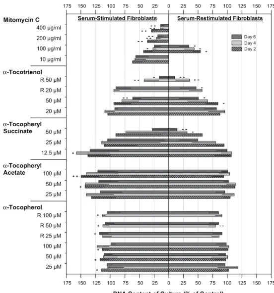
Vitamin E forms were dissolved in ethanol absolute, and they all created small lipid droplets on the medium surface after application. α-Tocopheryl succinatecs tiny droplets were disappearing first within 24 h, implicating a higher solubility in comparison with the other vitamin E forms (their bigger droplets were visible for up to 48 h). The results of the experiments with continiously growing and restimulated cultures are presented in Fig.1.
Effect of α-tocopherol
Compared with control cultures, single and multiple ap-plication ofα-tocopherol slightly stimulated growth of SF but not RF. A mild inhibitory effect was only detected on
day 4 after twice adding 50μM α-tocopherol to RF (80.0± 11.8%, P<0.01). Cell morphology remained identical to control cultures during the study.
Effect ofα-tocopheryl acetate
Growth of continuously growing cultures was markedly promoted by 25–100 μM α-tocopheryl acetate. Prolifera-tion of restimulated cultures was not affected. No degen-erative cell changes were seen in light microscopy.
Effect ofα-tocopheryl succinate
α-Tocopheryl succinate 12.5 μM demonstrated a strong growth stimulating effect in SF cultures (maximal effect: day 4, 157.6±56.6%). Again, proliferation of restimulated cultures was not promoted. Higher concentrations of
α-DNA Content of Culture (% of Control)
-175 -150 -125 -100 -75 -50 -25 0 25 50 75 100 125 150 175 -175 -150 -125 -100 -75 -50 -25 0 25 50 75 100 125 150 175 Day 4 Day 2 α-Tocopheryl Succinate α-Tocopheryl Acetate α-Tocopherol α-Tocotrienol
Mitomycin C Serum-Stimulated Fibroblasts Serum-Restimulated Fibroblasts
10 µg/ml 100 µg/ml 200 µg/ml 400 µg/ml 20 µM 50 µM R 20 µM R 50 µM 25 µM 50 µM 12.5 µM 100 µM 50 µM 25 µM 100 µM 50 µM 25 µM R 25 µM R 50 µM R 100 µM -- -- -- -- -- - - - -+ -+ + + + + + + + -Day 6 +
Fig. 1 Effects of incubation with vitamin E forms for 6 days or exposure to mitomycin C for 5 min on human Tenon’s fibroblast proliferation. Two experiments were performed: compounds (increasing concen-trations upwards) were added to serum-stimulated cells, shown at left, and serum-restimulated previously quiescent cells, shown at right. To mimic regular oral supplement intake in vivo, application ofα-tocopherol and α-tocotrienol was further re-peated on days 2 and 4 (con-centrations marked with R). A fluorometric assay determined proliferation by DNA content quantification on days 2, 4 and 6. Note the positive % of control values for both experiments. Bars with lines represent means; -SD of seven different cell cultures in triplicate. Significant growth inhibiting or stimulating effects, compared with control, are marked with minus and plus, respectively: ±P<0.05,−−/++ P<0.01
tocopheryl succinate affected cell morphology and viabil-ity. Compared to control cultures, fibroblasts exposed to 25 μM of α-tocopheryl succinate were transiently more granulated between day 2 and 5 (Fig.2b), accompanied by an increased cell lysate content in the culture media. Severe accumulation of intracytoplasmatic granules was visible after exposure to 50μM α-tocopheryl succinate for 24 h. Adherent fibroblasts then retracted their long processes, and became swollen and round shaped (Fig.2c). The dying cells finally released their content through ruptured plasma membranes into the surrounding culture media. Depending on the ability to adapt to this toxic stress and to delay cell death, the DNA content varied strongly between the seven different fibroblast cultures after incubation with 25 and 50 μM of α-tocopheryl succinate. On day 6, the marked decrease of DNA content in RF cultures by 25μM (49.5± 31.2%) and SF cultures by 50μM α-tocopheryl succinate (27.9±34.0%) was therefore not significant. Nevertheless, the cytotoxic effect of 50μM α-tocopheryl succinate was dramatic; necrotic RF cultures showed on day 4 and 6 significantly reduced DNA contents (maximal effect: 13.9± 11.6%, P<0.01).
Effect of α-tocotrienol
Single application of 50 μM α-tocotrienol significantly inhibited growth of both stimulated and restimulated cell cultures. This effect was most pronounced on day 6 (61.4± 14.0% for SF and 50.7±32.8% for RF, respectively). Fur-ther, the DNA content of RF but not of SF cultures was strongly reduced on day 6 after repeated incubation with 20μM α-tocotrienol (46.8±27.6%, P<0.05). By phase con-trast microscopic observation, no degenerative cell changes were accompanying these antiproliferative effects (Fig.2d). Cytotoxic effects were only observed after repeated ex-posure to 50μM of α-tocotrienol. After twice adding the compound (day 4), fibroblasts showed increased
cytoplas-mic granulation, and some of them did get round shaped (Fig.2e). The DNA content of continuously proliferating (41.8±24.5%) and restimulated cultures (35.5±23.5%) was markedly decreased (P<0.01), probably partly due to an overlap of antiproliferative and cytotoxic effects. After the third application of 50μM α-tocotrienol, overt necrosis as seen during incubation with 50μM α-tocopherol succinate developed. Therefore, the DNA content decreased drasti-cally on day 6 in both experiments (SF and RF).
Effect of mitomycin C
Growth of continously proliferating cultures was signifi-cantly and strongly inhibited on all DNA measurement days after 5-minute exposure to 200 and 400μg/ml mitomycin C, and on day 4, if treated with 100 μg/ml mitomycin C. The antiproliferative effect of mitomycin C occurred in a dose-dependent manner (maximal effect: 400μg/ml, day 6, 13.8±5.9%). After 6 days, only a 17.4% growth was found in 100 μg/ml mitomycin C treated SF cultures. RF were therefore exclusively exposed to this antimetabolite con-centration. Restimulated cultures were then significantly inhibited by 100μg/ml mitomycin C on all measurement days (maximal effect: day 6, 34.2±16.2%).
No degenerative cell changes were seen in fibroblast cultures exposed to mitomycin C concentrations as high as 200μg/ml. However, 5-minute treatment with 400 μg/ml mitomycin C resulted in a steady decrease of the absolute DNA content over the 6 day observation period (mean difference between day 0 and 6: −33.3%). Induction of apoptosis by 400μg/ml mitomycin C was assumed, as its morphologic characteristics like nuclear and cytoplasmic condensation, and nuclear fragmentation were observed more often than in control cultures. In contrast to the cyto-toxic effects of tocopherol succinate (50 μM) and α-tocotrienol (repeatedly 50μM), which affected to a varying degree virtually all cells, most fibroblasts remaining after
Fig. 2 Phase contrast photomi-crographs of serum-stimulated human Tenon fibroblasts (SF) on day 4 of the proliferation assay. (a) control; (b) 25μM α-tocopheryl succinate; (c) 50μM α-tocopheryl succinate; (d) 50μM α-tocotrienol; (e) R 50μM α-tocotrienol (applica-tion every 2nd day); (f) 400μg/ ml mitomycin C. Arrows: shrunken degenerated cells. Photographs were obtained from fibroblasts in the center of the well. Magnification: (a), (d), (f) ×93; (b), (c), (e), ×186
treatment with 400 μg/ml mitomycin C showed normal morphologic features (Fig.2f).
In general, the anti-/proliferative response to vitamin E forms differed between continuously proliferating and re-stimulated fibroblast cultures. RF cultures seemed to be more susceptible to antiproliferative effects, whereas growth stimulating effects were only observed in SF cultures. To summarize the results of the proliferation assay, 50μM and repeatedly 20μM of α-tocotrienol were the only vitamin E forms/concentrations proving a relevant inhibitory effect without affecting cell morphology and viability. The anti-proliferative effect of 100–400 μg/ml mitomycin C was stronger and earlier apparent, compared withα-tocotrienol.
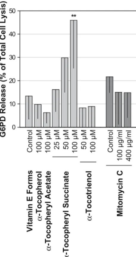
Glucose 6-phosphate dehydrogenase release
Accumulation of intracytoplasmatic granules was observed already 24 h after incubation with 50 μM α-tocopherol succinate. To evaluate for early cytoplasmic membran rup-ture, the G6PD release into the culture media was measured
24 h after incubation with vitamin E forms or 5-min ex-posure to mitomycin C (Fig.3).
Since dysmorphic cell changes were found from day 4 on after twice adding 50μM of α-tocotrienol, 100 μM of the compound was additionally tested to correspond with maximum drug accumulation. This was confronted with the effect of 100 μM α-tocopherol succinate on G6PD release.
Of the vitamin E forms, only 50 μM (29.8±14.8% of total cell lysis) and 100 μM of α-tocopheryl succinate (46.0±26% of total cell lysis, P<0.01) showed increased G6PD culture medium levels, compared to controls (13.4± 10.0% of total cell lysis). Due to the extended handling procedure with a possible negative effect on cell viability, G6PD culture media concentration in PBS treated wells (control of mitomycin C) was higher than in vitamin E control. G6PD levels of 100 and 400μg/ml mitomycin C were not increased in comparison with controls.
Discussion
A comparison between tocopherol and mitomycin C has been reported previously [11]. In the present study, α-tocotrienol and additional vitamin E forms were compared with mitomycin C, based on their effects on proliferation, morphology and viability of cultured human Tenon’s cap-sule fibroblasts. The study did not account for the effects on cell migration, attachment and contraction, collagen syn-thesis and angiogenesis, which are further critical steps involved in surgical bleb failure. No cultures of eyes with increased risk of trabeculectomy failure, such as young age or long-term use of topical anti-glaucomatous drugs, were included in this study.
α-Tocopherol inhibits proliferation by reducing protein kinase C activation, independent of its antioxidant proper-ties [4,45]. By adsorbing this lipophilic compound to FCS [5], our group previously found a strong antiproliferative effect on restimulated human Tenon’s capsule fibroblasts and retinal pigment epithelium cells [10,29]. Other authors dissolved vitamin E in ethanol to inhibit growth of similar cell types [38,40]. In the present study, we used ethanol for reasons of comparability. Instead of inhibiting prolifer-ation, both α-tocopherol and α-tocopheryl acetate then stimulated growth (SF cultures), or had no effect on pro-liferation (RF cultures). The former could be explained by the removal of lipoperoxide-induced inhibition of growth. Higher concentrations than 100μM were not tested, since α-tocopherol becomes water-insoluble and precipitates in the form of micellar aggregates in vivo. As compound and cell type were identical in our present and previous study, the different carrier substances (ethanol versus FCS) might have contributed to the conflicting results to a great extent. Lipid droplets on the culture medium surface indicated in-homogenous distribution of the vitamin E forms previously dissolved in ethanol. Prolonged exposure to oxygen and
0 10 20 30 40 50 ** V it a min E Forms Co nt ro l 100 µM α -Toc ophe rol α -Toc ophe ry l Ac e ta te 100 µM 25 µM 50 µM 100 µM α -Toc ophe ry l S u c c in a te 50 µ M 100 µM α -Toc o tri e nol Cont ro l 100 µg/ m l 400 µg/ m l M it o m yci n C G 6 P D R e le as e ( % o f T o ta l C e ll L y si s)
Fig. 3 Relative glucose 6-phosphate dehydrogenase (G6PD) culture media levels 24 h after incubation with vitamin E forms and 5-min exposure to mitomycin C. Dying cells release G6PD through rup-tured cytoplasmic membranes. Note that 100μM α-tocopheryl suc-cinate and 100 μM α-tocotrienol were double the concentrations used in the proliferation assay. Data are presented as % of total cell lysis. Bars with lines indicate means; -SD of six different cell cul-tures in triplicate. Significant differences, compared with control, are marked with ** P<0.01
heat might have inactivated a considerable amount of the compounds prior to cellular uptake.
Together with dietary fat, all unesterized vitamin E forms are directly absorbed in the human intestine.α-Tocopheryl acetate and succinate are naturally cleaved intestinally, resulting in adsorption of the active form α-tocopherol. Only α-tocotrienol and α-tocopherol were therefore re-peatedly applied to the cultures to mimic a regular oral vitamin E supplement intake in vivo. However, an unpre-dicted accumulation of vitamin E could not be fully ex-cluded, since the relation of oxidative loss and uptake into the cells remained unknown.
α-Tocopheryl succinate induces differentiation, DNA synthesis arrest, or apoptosis in malignant cells, but not in most normal cell lines [39]. Involvement of transforming growth factor β-, Fas-, and c-Jun-NH2-terminal kinase
signaling pathways is suggested [3]. In the years 1999 and 2000, Larrosa, Pinilla and colleagues published in vitro, animal model and histologic studies evaluating the poten-tial application of α-tocopheryl succinate and acetate in glaucoma surgery [21, 22, 37, 38]. Both compounds in-jected subconjunctivally improved the outcome of glauco-ma filtration surgery in rabbits. In our in vitro study, any possible antiproliferative effect of α-tocopheryl succinate was strongly overlaid by its cytotoxic effects. Increased cytoplasmic granulation, cell swelling, and early plasma membrane leakage strongly pointed to simple primary ne-crosis [17]. In vivo, simple primary necrosis leads to in-flammation of surrounding tissue. Other authors exposing ocular cell types and tissues to comparable concentrations of α-tocopheryl succinate (up to 100 μM) did not report relevant cytotoxic effects [9, 38, 40, 41]. No major com-plications were reported in a rabbit model of glaucoma filtration surgery with adjunctive topicalα-tocopheryl suc-cinate and acetate therapy [22,37].
In our study,α-tocotrienol was the only vitamin E form showing a relevant inhibitory effect on both serum-stim-ulated and -restimserum-stim-ulated fibroblasts in the absence of cytotoxicity. By using 50μM or repeatedly 20 μM of α-tocotrienol, a roughly 50% growth inhibition was achieved after 4–6 days of incubation. Restimulated cultures were more sensitive to this effect. We therefore conclude that α-tocotrienol better inhibits activation of fibroblasts than ongoing proliferation. Antiproliferative effects of higher α-tocotrienol concentrations orγ-/δ-isoforms were not tested, but they might be even stronger [25].
Toxic effects ofα-tocotrienol were only observed from day 4 on after twice exposing cultures to 50 μM of the compound. In contrast to α-tocopheryl succinate, G6PD levels after incubation with 100 μM α-tocotrienol were not increased. Due to the different onset of degenerative changes, the tolerability and the mechanisms of the cyto-toxic effects seem not to be identical for both substances. Considering the possible administration routes of α-toco-trienol in patients, subconjunctival injection or intraopera-tive topical application seems favorable since sufficiently
high tissue concentrations can be assumed. Penetration of tocotrienols through skin and of instilled α-tocopheryl acetate into the anterior chamber and lens has been shown in animal models [31,47]. No toxic intraocular effects of α-tocotrienol are known.
Vitamin E supplements to human diet are considered to be safe. Caution is required in cases of regular aspirin intake or in patients with vitamin K deficiency, as the risk of bleeding is increased by vitamin E. No significant ad-verse effects were reported in studies of long-time daily supplementation of human individuals with palm oil toco-trienol [46,49,51,52]. No teratogenic potential is known. However, it is remains unclear if appropriate concentra-tions of α-tocotrienol (25–50 μM in this study) can be achieved at the surgical site after systemic medication. α-Tocopherol was shown to predominantly accumulate in the retina after oral supplementation, compared with other oc-ular tissues [2,43]. Plasma concentrations ofα-tocotrienol only increased from less than 1μM to approximately 8 μM in humans supplemented with a palm oil concentrate [52]. However, accumulation ofα-tocotrienol in many solid or-gans/tissues is well known. α-Tocotrienol concentrations in the skin and adipose tissue can be tenfold to those in the plasma [12,13,34].
Induction of apoptotic cell death in human Tenon’s cap-sule fibroblasts after 5-min exposure to 400 μg/ml mito-mycin C has been described by Crowston and colleagues [6,7]. After 6 days, 27% of the fibroblasts were reported to exhibit morphologic characteristics of apoptosis [6]. Under very similar conditions, we detected a comparable cell content decrease by 33% and morphologic alterations such as nuclear and cytoplasmic condensation, indicating an apoptotic effect of 400 μg/ml mitomycin C. It should be noted that the methods applied prove cell death, but not its mechanism.
Apoptosis has been shown to be physiologically involved in scar formation [8]. As apoptosis does in general not provoke an inflammatory reponse in vivo [17], death of fibrolasts and inflammatory cells by apoptosis should be an acceptable event in the postoperative period, leading to reduction of scar tissue. However, complications such as bleb leak and infection may occur if the bleb tissue is too extensively devitalized.
Comparing α-tocotrienol and mitomycin C, fibroblast proliferation was earlier and stronger inhibited after 5-min exposure to the antimetabolite. Almost complete growth inhibition of SF cultures was achieved by using 100μg/ml mitomycin C. Furthermore, mitomycin C induced growth arrest is irreversible based on its DNA alkylating proper-ties. As α-tocotrienol regulates the activity of cytosolic enzymes, its antiproliferative effect is not likely to last for a long time after medication is discontinued. However, mitomycin C induced toxicity to ocular structures is well known. Alternative drugs with a reversible antiproliferative effect may be advantageous in wound healing situations where the healing stimulus is maximal only during a short
period that can be covered by treatment. Due to the different antiproliferative mechanisms, a synergistic effect of α-tocotrienol and mitomycin C is further conceivable, po-tentially allowing lower mitomycin C concentrations and shorter exposure-time to increase drug safety. Our findings suggest that α-tocotrienol deserves further evaluation in animal model studies to clarify its impact on the outcome of filtering procedures.
Acknowledgements The authors thank Milko E. Iliev, MD, for helpful discussions and critical comments on the manuscript.
Financial interests: A. Meyenberg, none; D. Goldblum, none; J.-M. Zingg, none; A. Azzi, none; K. Nesaretnam, employee of the Malay-sian Palm Oil Board, no proprietary interests; M. Kilchenmann, none; B. E. Frueh, none.
The authors have full control of all primary data and they agree to allow Graefe’s Archive for Clinical and Experimental Ophthalmol-ogy to review their data if requested.
References
1. Addicks EM, Quigley HA, Green WR, Robin AL (1983) Histologic character-istics of filtering blebs in glaucomatous eyes. Arch Ophthalmol 101:795–798 2. Alvarez RA, Liou GI, Fong SL,
Bridges CD (1987) Levels of alpha-and gamma-tocopherol in human eyes: evaluation of possible role of IRBP in intraocular alpha-tocopherol transport. Am J Clin Nutr 46:481–487
3. Anderson K, Simmons-Menchaca M, Lawson KA, Atkinson J, Sanders BG, Kline K (2004) Differential response of human ovarian cancer cells to induction of apoptosis by vitamin E succinate and vitamin E analogue, alpha-TEA. Can-cer Res 64:4263–4269
4. Boscoboinik D, Szewczyk A, Hensey C, Azzi A (1991) Inhibition of cell proliferation by alpha-tocopherol. Role of protein kinase C. J Biol Chem 266:6188–6194
5. Chan AC, Tran K (1990) The uptake of α-tocopherol by human endothelial cells in culture. Lipids 25:17–21 6. Crowston JG, Akbar AN, Constable
PH, Occleston NL, Daniels JT, Khaw PT (1998) Antimetabolite-induced ap-optosis in Tenon’s capsule fibroblasts. Invest Ophthalmol Vis Sci 39:449–454 7. Crowston JG, Chang LH, Constable
PH, Daniels JT, Akbar AN, Khaw PT (2002) Apoptosis gene expression and death receptor signaling in mitomycin-C treated human tenon capsule fibro-blasts. Invest Ophthalmol Vis Sci 43:692–699
8. Desmouliere A, Redard M, Darby I, Gabbiani G (1995) Apoptosis mediates the decrease in cellularity during the transition between granulation tissue and scar. Am J Pathol 146:56–66 9. Fallor MK, Silverman CA, Yoshizumi
MO (1994) Ocular toxicity of experi-mental intravitreal vitamin E. J Toxicol 3:337–345
10. Haas AL, Boscoboinik D, Mojon DS, Bohnke M, Azzi A (1996) Vitamin E inhibits proliferation of human Tenon’s capsule fibroblasts in vitro. Ophthalmic Res 28:171–175
11. Haas AL, Boscoboinik DO, Bohnke M (1996) Inhibition of Tenon fibroblast proliferation. Comparison between to-copherol and mitomycin-C. Invest Ophthalmol Vis Sci 37(suppl):877S 12. Hayes KC, Pronczuk A, Liang JS
(1993) Differences in the plasma transport and tissue concentrations of tocopherols and tocotrienols: observa-tions in humans and hamsters. Proc Soc Exp Biol Med 202:353–359
13. Ikeda S, Tohyama T, Yoshimura H, Hamamura K, Abe K, Yamashita K (2003) Dietary alpha-tocopherol de-creases alpha-tocotrienol but not gamma-tocotrienol concentration in rats. J Nutr 133:428–434
14. Jampel HD, McGuigan LJ, Dunkelberger GR, Hernault NL, Quigley HA (1988) Cellular proliferation after experimental glaucoma filtration surgery. Arch Ophthalmol 106:89–94
15. Jampel HD, Pasquale LR, Dibernardo C (1992) Hypotony maculopathy fol-lowing trabeculectomy with mitomycin C. Arch Ophthalmol 110:1049–1050 16. Jiang Q, Ames BN (2003)
Gamma-tocopherol, but not alpha-Gamma-tocopherol, decreases proinflammatory eicosanoids and inflammation damage in rats. FASEB J 17:816–822
17. Kanduc D, Mittelman A, Serpico R et al (2002) Cell death: apoptosis versus necrosis. Int J Oncol 21:165–170 18. Kitazawa Y, Kawase K, Matsushita H,
Minobe M (1991) Trabeculectomy with mitomycin: a comparison study with fluorouracil. Arch Ophthalmol 109:1693–1698
19. Kline K, Yu W, Sanders BG (2001) Vitamin E: mechanisms of action as tumor cell growth inhibitors. J Nutr 131:161–163
20. Larrosa JM, Veloso AA Jr, Leong FL, Refojo MF (1997) Antiproliferative effect of intravitreal alpha-tocopherol and alpha-tocopheryl-acid-succinate in a rabbit model of PVR. Curr Eye Res 16:1030–1035
21. Larrosa JM, Polo V, Ramirez T, Pinilla I, Pablo LE, Honrubia FM (2000) Alpha-tocopherol derivatives and wound healing in an experimental model of filtering surgery. Ophthalmic Surg Lasers 31:131–135
22. Larrosa JM, Polo V, Ramirez T et al. (2000) Histopathological evaluation of vitamin E in surgical fistula closure. Arch Soc Esp Oftalmol 75:523–528 23. Lehmann OJ, Bunce C, Matheson MM
et al. (2000) Risk factors for develop-ment of post-trabeculectomy endoph-thalmitis. Br J Ophthalmol 84:1349–1353 24. Malafa MP, Fokum FD, Mowlavi A,
Abusief M, King M (2002) Vitamin E inhibits melanoma growth in mice. Surgery 131:85–91
25. McIntyre BS, Briski KP, Gapor A, Sylvester PW (2000) Antiproliferative and apoptotic effects of tocopherols and tocotrienols on preneoplastic and neo-plastic mouse mammary epithelial cells. Proc Soc Exp Biol Med 224:292– 301
26. Membrey WL, Poinoosawmy DP, Bunce C, Hitchings RA (2000) Glau-coma surgery with or without adjunc-tive antiproliferaadjunc-tives in normal tension glaucoma: 1 intraocular pressure con-trol and complications. Br J Ophthal-mol 84:586–590
27. Mietz H, Addicks K, Diestelhorst M, Kriegelstein GK (1994) Extraocular application of mitomycin C in a rabbit model: cytotoxic effects on the ciliary body and epithelium. Ophthalmic Surg 25:204–244
28. Mishima K, Tanaka T, Pu F et al (2003) Vitamin E isoforms alpha-tocotrienol and gamma-tocopherol prevent cerebral infarction in mice. Neurosci Lett 337:56–60
29. Mojon D, Boscoboinik D, Haas A, Bohnke M, Azzi A (1994) Vitamin E inhibits retinal pigment epithelium cell proliferation in vitro. Ophthalmic Res 26:304–309
30. Mojon D, Boscoboinik D, Haas A, Bohnke M, Azzi A (1997) Inhibition of bovine RPE cells by vitamin E succi-nate (comment). Graefes Arch Clin Exp Ophthalmol 235:190–191
31. Nagata M, Kawazu K, Midori Y, Kojima M, Shirasawa E, Sasaki K (2001) Intracameral and lenticular penetration of locally applied stable isotope-labeled vitamin E. Jpn J Ophthalmol 45:125–127
32. Nesaretnam K, Guthrie N, Chambers AF, Carroll KK (1995) Effect of tocotrienols on the growth of a human breast cancer cell line in culture. Lipids 30:1139–1143
33. Nuyts RM, Pels E, Greve EL (1992) The effects of 5-fluorouracil and mito-mycin C on the corneal endothelium. Curr Eye Res 11:565–570
34. Ohrvall M, Tengblad S, Vessby B (1994) Tocopherol concentrations in adipose tissue. Relationships of tocopherol concentrations and fatty acid composition in serum in a refer-ence population of Swedish men and women. Eur J Clin Nutr 48:212–218 35. Palmer SS (1991) Mitomycin as
adjunct chemotherapy with trabeculectomy. Ophthalmology 98:317–321
36. Park HK, Lee KW, Choi JS, Joo CK (2002) Mitomycin C-induced cell death in mouse lens epithelial cells. Oph-thalmic Res 34:213–219
37. Pinilla I, Larrosa JM, Polo V, Honrubia FM (1999) Alpha-tocopherol deriva-tives in an experimental model of filtering surgery. Ophthalmic Res 31:440–445
38. Pinilla I, Piazuelo E, Jimenez P et al (2000) Inhibitory effect of alpha tocopherol succinate on fibroblast wound healing. Arch Soc Esp Oftalmol 75:383–388
39. Prasad KN, Kumar B, Yan XD, Hanson AJ, Cole WC (2003) Alpha-tocopheryl succinate, the most effective form of vitamin E for adjuvant cancer treat-ment: a review. J Am Coll Nutr 22:108–117
40. Sakamoto T, Hinton DR, Kimura H, Spee C, Gopalakrishna R, Ryan SJ (1996) Vitamin E succinate inhibits proliferation and migration of retinal pigment epithelial cells in vitro: thera-peutic implication for proliferative vi-treoretinopathy. Graefes Arch Clin Exp Ophthalmol 234:186–192
41. Sakamoto T, Oshima Y, Ishibashi T, Inomata H (1996) Inhibitory effect of Vitamin E succinate on the proliferation of cultured bovine choroidal endothe-lial cells. J Jpn Ophthalmol Soci 100:777–782
42. Smith S, D’Amore PA, Dreyer EB (1994) Comparative toxicity of mito-mycin C and 5-fluorouracil in vitro. Am J Ophthalmol 118:332–337 43. Stephens RJ, Negi DS, Short SM, van
Kuijk FJ, Dratz EA, Thomas DW (1988) Vitamin E distribution in ocular tissues following long-term dietary depletion and supplementation as determined by microdissection and chromatography-mass spectrometry. Exp Eye Res 47:237–245
44. Sylvester PW, McIntyre BS, Gapor A, Briski KP (2001) Vitamin E inhibition of normal mammary epithelial cell growth is associated with a reduction in protein kinase C(alpha) activation. Cell Prolif 34:347–357
45. Tasinato A, Boscoboinik D, Bartoli GM, Maroni P, Azzi A (1995) d- α-Tocopherol inhibiton of vascular smooth muscle cell proliferation occurs at physiological concentrations, corre-lates with protein kinase C inhibition, and is independent of its antioxidant properties. Proc Natl Acad Sci USA 92:12190–12194
46. Tomeo AC, Geller M, Watkins TR, Gapor A, Bierenbaum ML (1995) An-tioxidant effects of tocotrienols in pa-tients with hyperlipidemia and carotid stenosis. Lipids 30:1179–1183
47. Traber MG, Rallis M, Podda M, Weber C, Maibach HI, Packer L (1998) Pen-etration and distribution of alpha-toco-pherol, alpha- or gamma-tocotrienols applied individually onto murine skin. Lipids 33:87–91
48. Unchern S, Laoharuangpanya N, Phumala N et al. (2003) The effects of vitamin E on platelet activity in beta-thalassaemia patients. Br J Haematol 123:738–744
49. Wahlqvist ML, Krivokuca-Bogetic Z, Lo CS, Hage H, Smith R, Lukito W (1992) Differential serum responses of tocopherols and tocotrienols during vitamin supplementation in hypercho-lesterolaemic individuals without changes in coronary risk factors. Nutr Res 12(suppl):181S–201S
50. Wu K, Shan YJ, Zhao Y, Yu JW, Liu BH (2001) Inhibitory effects of RRR-alpha-tocopheryl succinate on benzo(a) pyrene (B(a)P)-induced forestomach carcinogenesis in female mice. World J Gastroenterol 7:60–65
51. Qureshi AA, Qureshi N, Wright JJ et al. (1991) Lowering of serum cholesterol in hypercholesterolemic humans by tocotrienols (palmvitee). Am J Clin Nutr 53(suppl):1021S–1026S 52. Qureshi AA, Bradlow BA, Brace L
et al. (1995) Response of hypercholes-terolemic subjects to administration of tocotrienols. Lipids 30:1171–1177 53. Qureshi AA, Sami SA, Salser WA,
Khan FA (2002) Dose-dependent suppression of serum cholesterol by tocotrienol-rich fraction (TRF25) of rice bran in hypercholesterolemic humans. Atherosclerosis 161:199–207 54. Yu W, Simmonss-Menchaca M, Gapor
A, Sanders BG, Kline K (1999) In-duction of apoptosis in human breast cancer cells by tocopherols and toco-trienols. Nutr Cancer 33:26–32