REVIEW
The diverse functional LINCs of the nuclear envelope
to the cytoskeleton and chromatin
Andrea Rothballer
&Ulrike Kutay
Received: 28 February 2013 / Revised: 14 May 2013 / Accepted: 15 May 2013 / Published online: 5 June 2013 # The Author(s) 2013. This article is published with open access at Springerlink.com
Abstract The nuclear envelope (NE) is connected to the
different types of cytoskeletal elements by linker of
nucleoskeleton and cytoskeleton (LINC) complexes. LINC
complexes exist from yeast to humans, and have preserved
their general architecture throughout evolution. They are
composed of SUN and KASH domain proteins of the inner
and the outer nuclear membrane, respectively. These SUN
–
KASH bridges are used for the transmission of forces across
the NE and support diverse biological processes. Here, we
review the function of SUN and KASH domain proteins in
various unicellular and multicellular species. Specifically,
we discuss their influence on nuclear morphology and
cy-toskeletal organization. Further, emphasis is given on the
role of LINC complexes in nuclear anchorage and migration
as well as in genome organization.
Keywords LINC complex . SUN domain . KASH . Nesprins .
Nuclear migration . Nuclear envelope
Introduction
Linker of nucleoskeleton and cytoskeleton (LINC)
com-plexes are built from members of two conserved protein
families, SUN and KASH domain proteins. SUN domain
proteins are integral to the inner nuclear membrane (INM),
whereas KASH domain proteins reside in the outer nuclear
membrane (ONM). SUN and KASH proteins directly bind
each other in the perinuclear space (PNS), thereby forming a
bridge across the nuclear envelope (NE). On the
nucleoplas-mic face of the NE, SUN domain proteins engage into
interactions with other INM proteins, chromatin, and, in
metazoans, the nuclear lamina. In the cytoplasm, KASH
domain proteins associate with all major cytoskeletal
ele-ments, i.e., actin, intermediate filaele-ments, and microtubules.
Based on their bridging molecular architecture, LINC
com-plexes serve as handles for the cytoskeleton on the nucleus,
as well as for force transduction between nucleo- and
cyto-plasm. Consistently, functions of LINC complexes involve
mechanical action and crosstalk between both sides of the
NE (Burke and Roux
2009
; Razafsky and Hodzic
2009
;
Starr and Fridolfsson
2010
).
SUN domain proteins are type II membrane proteins.
They comprise an N-terminal, nucleo/cytoplasmic portion
and one transmembrane segment, followed by a coiled-coil
region and the SUN domain in the C-terminal, luminal part
of the protein. On the sequence level, the SUN domain is
highly conserved, while other regions of SUN proteins are
rather diverse (Hiraoka and Dernburg
2009
; Razafsky and
Hodzic
2009
).
KASH domain proteins are tail-anchored membrane
pro-teins. They are composed of an N-terminal, cytoplasmic
part, and the C-terminal KASH domain, which includes
both the membrane anchor and a luminal peptide of
typical-ly 20-30 amino acids. The cytoplasmic portions of KASH
domain proteins differ in size, structure, and function. The
KASH domain, in contrast, is conserved both in the
trans-membrane segment and in the luminal peptide (Razafsky
and Hodzic
2009
; Starr and Fischer
2005
). Importantly, a
motif of two or three prolines followed by one variable
amino acid (PPPX) features the very C terminus of typical
metazoan KASH peptides, and hydrophobic residues are
conserved further upstream in the peptide sequence. Both
elements are essential for interaction between KASH and
SUN domains, as well as for SUN-mediated localization of
KASH to the ONM (Sosa et al.
2012
; Stewart-Hutchinson et
al.
2008
). In contrast to metazoan KASH domains, ONM
constituents of LINC complexes in yeast contain atypical
luminal peptides with low sequence conservation (Razafsky
A. Rothballer
:
U. Kutay (*)Department of Biology, Institute of Biochemistry, ETH Zurich, Schafmattstrasse 18,
8093 Zurich, Switzerland
and Hodzic
2009
; Starr and Fischer
2005
). Based on their
preserved biological role, the term KASH domain protein
will here be used for both metazoan and yeast ONM
components.
The hallmark of LINC complexes is the interaction of
SUN domains and KASH peptides in the PNS. In crystal
structures of mammalian LINC, this centerpiece of the
com-plex is hexameric, composed of three protomers of each
SUN and KASH. The three SUN domains form a globular
assembly, which is organized by a preceding trimeric
coiled-coil. This arrangement of SUN domains is essential for
KASH binding, and each SUN trimer harbors three binding
sites for KASH peptides at the interfaces of neighboring
SUN domains (Sosa et al.
2012
).
The repertoire of LINC components is evolutionary
di-verse. One SUN domain protein has been identified in both
budding and fission yeast, two are found in Caenorhabditis
elegans and Drosophila melanogaster, and mammals posses
at least five family members. Mammalian SUN1 and SUN2
are expressed in most tissues and organs, whereas SUN3 to
5 are specific to the male germline (Hiraoka and Dernburg
2009
; Razafsky and Hodzic
2009
).
KASH domain proteins have diverged even further than
their INM counterparts. Two KASH proteins have been
iden-tified in both budding and fission yeast. Three family
mem-bers are found in C. elegans, and two in D. melanogaster. In
vertebrates, six KASH domain proteins have been described:
Nesprin-1 to Nesprin-4, KASH5, and LRMP (Mellad et al.
2010
; Morimoto et al.
2012
; Lindeman and Pelegri
2012
;
Noegel and Neumann
2011
). While Nesprin-1, Nesprin-2,
and Nesprin-3 are expressed ubiquitously, Nesprin-4 is
spe-cific to epithelial cells and KASH5 is only expressed in
meiosis (Morimoto et al.
2012
; Mellad et al.
2010
). LRMP
has originally been described as lymphoid-restricted protein in
mammals and its zebrafish homolog has recently been
char-acterized in the zygote and the early embryo (Behrens et al.
1994
; Lindeman and Pelegri
2012
). Mammalian Nesprin-1
and Nesprin-2 exist in various splice isoforms, including giant
variants of up to 800 MDa as well as many smaller ones. Most
Nesprins reside in the ONM and form part of classical LINC
complexes that connect the nucleus to the cytoskeleton. Some
isoforms have also been described at the INM, and soluble
Nesprin variants lacking the KASH domain were found in the
nucleo- and cytoplasm (Mellad et al.
2010
; Warren et al.
2005
;
Noegel and Neumann
2011
).
Little is known about specificities in SUN–KASH
associ-ation. With some exceptions, SUN and KASH domain
pro-teins seem to bind each other promiscuously. Some LINC
constituents are, however, tissue- or cell-type-specific and
their expression patterns hence determine the composition of
complexes. Plasticity and regulation of LINC complexes have
so far remained unexplored (Burke and Roux
2009
; Razafsky
and Hodzic
2009
; Starr and Fridolfsson
2010
).
In their position as NE bridges and linkers of nucleo- and
cytoskeleton, SUN–KASH assemblies form integral
ele-ments of cellular architecture. Here, we summarize the
influence of LINC complexes on nuclear and cytoskeletal
structure, and review their functions in nuclear positioning
and chromatin organization.
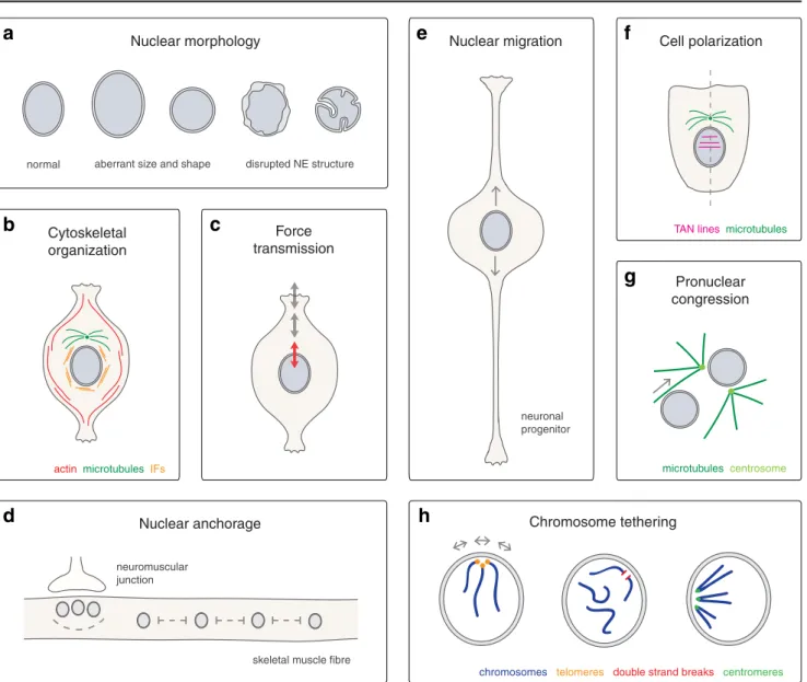
LINC complexes determine nuclear morphology
and membrane structure
Size and shape of the cell nucleus are largely determined by its
structural constituents—the lamina, NE proteins, and
chroma-tin in the first place (Walters et al.
2012
). Aberrant nuclear
morphologies are a hallmark of laminopathic diseases,
indi-cating that the maintenance of nuclear structure may be critical
for function (Worman et al.
2010
). As integral NE
compo-nents, SUN and KASH domain proteins are thought to
orga-nize the nuclear membrane system and the nucleus as a whole
(Fig.
1
). Size and shape of both animal and plant nuclei are
influenced by LINC complexes (Table
1
) (Luke et al.
2008
;
Lu et al.
2012
; Zhou et al.
2012
; Zhang et al.
2007a
). In
cultured mammalian cells, an interplay between different
KASH domain proteins has been implicated in nuclear size
control. Giant isoforms of Nesprin-1 and Nesprin-2 were
shown to bind to Nesprin-3, and disruption of Nesprin
interchain associations led to larger nuclei (Taranum et al.
2012
; Lu et al.
2012
). Furthermore, LINC complexes are
required to maintain the even spacing of the nuclear
mem-branes. LINC disruption in Hela cells caused irregular
expan-sions of the PNS and bulges of the ONM (Crisp et al.
2006
). In
fact, structural analyses suggest that SUN–KASH assemblies
would be well suited to determine the width of the NE (Sosa et
al.
2012
; Rothballer et al.
2013
).
LINC complexes organize the cytoskeleton
LINC complexes also act as cytoskeletal organizers (Fig.
1
). In
vertebrate cells, Nesprins determine the structure and
distribu-tion of perinuclear actin and intermediate filaments with
im-pact on overall cellular shape and mechanics (Table
1
) (Khatau
et al.
2009
; Lombardi et al.
2011
; Schneider et al.
2011
;
Morgan et al.
2011
; Postel et al.
2011
; Chambliss et al.
2013
). Giant isoforms of Nesprin-1 and Nesprin-2 directly
bind to actin via N-terminal calponin-homology (CH) domains
(Fig.
2
) (Zhang et al.
2002
; Zhen et al.
2002
; Padmakumar et
al.
2004
). Intermediate filaments are connected to the nucleus
by Nesprin-3 and the cytoskeletal crosslinker plectin (Fig.
2
)
(Ketema et al.
2007
; Wilhelmsen et al.
2005
). Consistent with
their role in cytoskeletal organization, LINC components were
found to influence organelle positioning, cell polarity, and
T able 1 Functions of LINC complexes Cytoplasm ic factors KASH domain proteins SUN domain pr oteins Nucleoplasmi c factors Function s References V ertebrates Actin Nesprin -1 giant SUN1 /2 nd Nuclear anchorage in muscle fibers (Grady et al. 2005 ; Zhang et al. 2010 ; Zhang et al. 2007b ; Lei et al. 2009 ) Dynein, kinesin-1 Nesprin -1 /2 SUN1 /2 nd Nucleus -centrosome attachment and nuclear migration in neural progenitors (Zhang et al. 2009 ) Dynein, kinesin-1 Nesprin -2 SUN1 /2 nd Nuclear migration in photorecept or progenitors (Y u et al. 201 1 ; Razafsky et al. 2012 ) Actin Nesprin -2 giant SUN2 Lamin A/C Formation of T A N lines, polarizati on of cultured cells (Folker et al. 201 1; Luxton et al. 2010 ; Luke et al. 2008 ) Kinesin-1 Nesprin -2 SUN1/2 Lamin A/C Cytoskelet al or ganization , nucleus-centro some attachment, polarizati on of cultured cells (Schneider et al. 201 1 ; Lombardi et al. 201 1 ; Rashmi et al. 201 1 ) nd Nesprin -2 SUN1/2 nd Cell proliferation and differentiation during wound healing in vivo (Rashmi et al. 201 1 ) nd Nesprin -1 , other Nesprins SUN1/2 nd Mechan ics and force transduc tion in cultured cells (Stewart-Hutch inson et al. 2008 ; Lombardi et al. 201 1 ; Anno et al. 2012 ) Plectin Nesprin -3 α SUN1/2 Lamin A/C Cytoskelet al or ganization , nucleus-centrosome attachme nt, polarization of cultured cells (Ketema et al. 2007 ; Morgan et al. 201 1 ; W ilhelmsen et al. 2005 ; Postel et al. 201 1 ) nd Nesprin-1/2/3 SUN1 /2 nd Or ganization of NE morphology in cultured cells (Crisp et al. 2006 ) nd Nesprin -1 /2 giant and Nesprin -3 SUN1/2 nd Nuclear size control in cultured cells (Lu et al. 2012 ) nd nd SUN1 /2 DNAPK DNA damage response in primary cells (Lei et al. 2012 ) Kinesin-1 Nesprin -4 SUN1 nd Nuclear positioning in outer hair cells of the cochlea (Roux et al. 2009 ; Horn et al. 2013 ) Dynein KASH5 SUN1 nd T ethering of telomeres to the NE in meiosis (Morimoto et al. 2012 ; Ding et al. 2007 ; Boateng et al. 2013 ) nd LRMP nd nd Pronucleus-centrosome attachment and pronuclear congression in the zygote (Lindeman and Pelegri 2012 ) nd Nesprin -3 SUN1 η nd Sperm developmen t (Gob et al. 2010 ) nd Nesprin -1 SUN3 nd Sperm developmen t (Gob et al. 2010 ) C . elegans Actin ANC -1 UNC -84 nd Nuclear anchorage in syncytia (Starr and Han 2002 ; Malone et al. 1999 ) Dynein, kinesin-1 UNC -83 UNC -84 nd Nuclear migration in various cell types (Starr et al. 2001 ; Fridolfss on et al. 2010 ; McGee et al. 2006 ; Meyerzon et al. 2009a ; Malone et al. 1999 ) Dynein, ZYG-12A ZYG -12 SUN -1 nd Pronucleus-centrosome attachment and pronuclear congressi on in the zygote (Malone et al. 1999 ; Minn et al. 2009) Dynein ZYG -12 SUN-1 nd Cytoskelet al or ganization and nuclear positioning in the gonad (Zhou et al. 2009) Dynein ZYG -12 SUN -1 HIM-8/ZIM1 -3 T ethering of chromosome s to the NE in meiosis (Labella et al. 201 1 ; Penkner et al. 2007 ; Penkner et al. 2009 ; Sato et al. 2009 ) nd KDP -1 SUN -1 / UNC-84 nd Cell cycle regulation in the germline and the embryo (McGee et al. 2009 ) nd nd SUN -1 nd Apoptosis in the embryo (Tzur et al. 2006 )
T able 1 (continued) Cytoplasm ic factors KASH domain proteins SUN domain pr oteins Nucleoplasmi c factors Function s References D . melanogaste r Actin, microtubules MSP -300 , Klarsicht Klaroid nd Nuclear anchorage in muscle fibers (T echnau and Roth 2008 ; Rosenberg-Hasson et al. 1996; V olk 1992 ; Elhanany-T amir et al. 2012 ) Microtubu les Klarsicht Klaroid Lamin Dm(0) Nucleus -centrosome attachment and nuclear migration during eye develop ment (Kracklauer et al. 2007 ; Mosley-Bish op et al. 1999 ; Patterson et al. 2004 ) Y uri Gagarin, dynein nd Spag -4 nd Nucleus -centrosome attachment during spermatogenesi s (Kracklauer et al. 2010 ) S. pombe nd Kms1, Kms2 Sad1 nd SPB integrity , mitotic spindle formation (Hagan and Y anagida 1995 ; Miki et al. 2004 ; Shimanuk i et al. 1997 ) nd nd Sad1 Csi1 T ethering of centromeres to the NE in interphase (Hou et al. 2012 ) Dynein Kms1 Sad1 Bqt1, Bqt2 T ethering of telomeres to the NE in meiosis (Chikashige et al. 2006 ; Shimanuki et al. 1997 ) S. cerevisia e nd Mps2 Mps3 nd SPB integrity and duplication, mitotic spindle formation (Jaspersen et al. 2002 ; Jaspersen et al. 2006 ; Munoz-Cent eno et al. 1999 ; W iney et al. 1991 ; Friederichs et al. 201 1 ) nd nd Mps3 Sir4 T ethering of telomeres to the NE during vegetative growth (Bupp et al. 2007 ) nd nd Mps3 Sir4, Ebp2, Rrs1 Clustering of tethered telomeres at the NE (Horigome et al. 201 1 ) nd nd Mps3 T elomerase, Ku70/80 T ethering of telomeres to the NE during vegetative growth (Schober et al. 2009 ; Antoniacci et al. 2007 ) nd nd Mps3 T elomerase, Ku70/Ku8 0 T ethering of DNA double strand breaks to the NE (Oza et al. 2009 ) nd nd Mps3 TFIIIC T ethering of extra TFIIIC sites to the NE (Hiraga et al. 2012 ) nd nd Mps3 Ctf7 Sister chromatid cohesion (Antoniacci et al. 2004 ) nd nd Mps3 Replication factor C, Htz1 Potentia lly sister chr omatid cohesion or DNA repair (Haas et al. 2012 ) Actin Csm4 Mps3 Ndj1 T ethering of telomeres to the NE in meiosis (Conrad et al. 2008 ; Conrad et al. 2007 ; Kosaka et al. 2008 ; W anat et al. 2008 ) A . thaliana AtRanGA P1 AtWIP1 -3 AtSUN1 /2 nd Anchorage of AtRanGA P1 at the NE, maintenance of nuclear shape (Zhou et al. 2012 ) The table lists characterized functions of LINC complexes in model org anisms, the involved SUN and KASH domain proteins, as well as their cytoplasmic and nucleoplasmic interaction partners. For SUN and KASH domain proteins written in bold, the respective functions have directly been demonstrated; others have been implicated indirectly nd not defined
actin microtubules IFs
TAN lines microtubules Nuclear morphology Cytoskeletal organization Force transmission Cell polarization Nuclear migration Nuclear anchorage Pronuclear congression Chromosome tethering
chromosomes telomeres double strand breaks centromeres microtubules centrosome
d
c
b
a
e
f
g
h
skeletal muscle fibre neuromuscular
junction
aberrant size and shape disrupted NE structure
neuronal progenitor normal
Fig. 1 Cellular functions of LINC complexes. a Nuclear morphology. LINC complexes are required to maintain nuclear size and shape in mam-mals (Lu et al.2012; Luke et al.2008) and in A. thaliana (Zhou et al.2012), as well as structure and integrity of the mammalian NE (Crisp et al.2006; Zhang et al.2007a). b Cytoskeletal organization. LINC complexes influ-ence structure and distribution of perinuclear actin and intermediate fila-ments (IFs) in vertebrate cells (Khatau et al.2009; Lombardi et al.2011; Schneider et al.2011; Morgan et al.2011; Postel et al.2011; Chambliss et al. 2013), and tether centrosomes to the NE in various metazoans (Schneider et al.2011; Zhang et al.2009; Malone et al.2003; Morgan et al.2011; Roux et al.2009; Patterson et al.2004). c Force transmission. LINC complexes transmit forces across the NE and affect mechanical properties of cultured mammalian cells (Lombardi et al.2011; Anno et al. 2012; Stewart-Hutchinson et al.2008). d Nuclear anchorage. LINC com-plexes mediate anchorage and positioning of nuclei in syncytial systems of various metazoans (Zhang et al.2010; Zhang et al.2007b; Grady et al. 2005; Lei et al.2009; Starr and Han2002; Malone et al.1999; Elhanany-Tamir et al.2012). In mammalian skeletal muscle (shown here), LINC complexes are required for the even spacing of extrasynaptic nuclei throughout myotubes, as well as for the clustering of synaptic nuclei beneath the neuromuscular junction (Zhang et al.2010; Zhang et al. 2007b; Grady et al.2005; Lei et al.2009). e Nuclear migration. LINC complexes function in nuclear migration during various metazoan devel-opmental events (Yu et al.2011; Zhang et al.2009; Malone et al.1999; McGee et al.2006; Starr et al.2001; Meyerzon et al.2009a; Fridolfsson et
al. 2010; Mosley-Bishop et al. 1999; Fischer-Vize and Mosley 1994; Patterson et al.2004; Kracklauer et al.2007). In neural progenitors of the mammalian neocortex and retina (shown here), LINC complex-mediated nuclear migration processes are essential for proliferation and differentia-tion (Yu et al.2011; Zhang et al.2009). f Cell polarization. LINC com-plexes are required for nuclear positioning and orientation of the nuclear– centrosomal axis during fibroblast polarization (Lombardi et al. 2011; Luxton et al.2010). LINC complexes form transmembrane actin-associated nuclear (TAN) lines to couple the nucleus to retrograde actin flow (Luxton et al. 2010). g Pronuclear congression. LINC complexes function in congression of male and female pronuclei in the fertilized zygote of C. elegans and vertebrates. Dedicated KASH domain proteins connect pronuclei to microtubule asters and the centrosome to allow their migration towards each other (Malone et al.2003; Lindeman and Pelegri2012). h Chromosome tethering. LINC complex-mediated tethering of chromo-somes to the NE plays a role in various biological processes. In S. cerevisiae, tethering of telomeres and DNA double strand breaks has been implicated in silencing, stabilization, and repair (Bupp et al.2007; Schober et al.2009; Oza et al.2009). In S. pombe, tethering of centromeres is important for mitotic chromosome segregation (Hou et al.2012). Meiotic chromosomes are anchored to the NE via LINC complexes in both yeast and metazoans with impact on homolog pairing and recombination (Conrad et al.2008; Conrad et al.2007; Kosaka et al.2008; Wanat et al.2008; Chikashige et al.2006; Shimanuki et al.1997; Penkner et al.2007; Penkner et al.2009; Sato et al.2009; Ding et al.2007; Morimoto et al.2012)
1982
; Starr and Han
2002
; Dawe et al.
2009
; Elhanany-Tamir
et al.
2012
).
Importantly, metazoan SUN–KASH pairs mediate the
at-tachment of the centrosome to the nucleus, which is thought to
be essential during nuclear and cell migration (Table
1
)
(Schneider et al.
2011
; Zhang et al.
2009
; Malone et al.
2003
;
Morgan et al.
2011
; Roux et al.
2009
; Patterson et al.
2004
).
Similarly, yeast SUN and KASH domain proteins function at
the spindle pole body (SPB; Table
1
) (Jaspersen et al.
2002
;
Jaspersen et al.
2006
; Winey et al.
1991
; Hagan and Yanagida
1995
). SPBs are multi-subunit protein complexes integrated
into or closely associated with the NE. They serve as the main
microtubule organizing centers (MTOC) in yeast cells.
Duplication of the SPB occurs via formation and maturation
of a cytoplasmic complex, which is subsequently inserted into
the NE. In Saccharomyces cerevisiae, SPB insertion is coupled
to its duplication, and mature complexes remain
membrane-integrated throughout the cell cycle. Schizosaccharomyces
pombe SPBs, in contrast, lie closely attached to the ONM
during interphase and integrate into the NE only during mitosis
to organize the intranuclear microtubule spindle (Ding et al.
1997
; Jaspersen and Winey
2004
).
SUN and KASH domain proteins are structural
compo-nents of the SPB in S. cerevisiae (Munoz-Centeno et al.
1999
;
Jaspersen et al.
2002
), and form part of the MTOC attachment
site in the NE in S. pombe (Hagan and Yanagida
1995
; King et
al.
2008
; Shimanuki et al.
1997
; Miki et al.
2004
). In both
yeasts, they are essential for SPB integrity and duplication,
and consequently for mitotic spindle formation and
chromo-some segregation (Jaspersen et al.
2002
; Jaspersen et al.
2006
;
Winey et al.
1991
; Hagan and Yanagida
1995
). The S.
cerevisiae SUN domain protein Mps3 has been implicated
specifically in the insertion of SPBs into the NE (Friederichs
et al.
2011
). SPB insertion requires a local fusion between
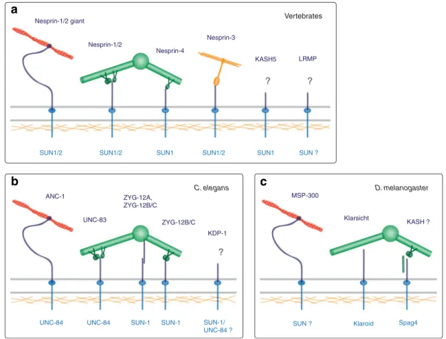
Nesprin-4 Nesprin-1/2 giant Nesprin-1/2 Nesprin-3 LRMP KASH5
SUN1/2 SUN1/2 SUN1 SUN1/2 SUN1 SUN ?
ZYG-12A, ZYG-12B/C ANC-1 UNC-83 KDP-1 ZYG-12B/C SUN-1 SUN-1/ UNC-84 ? UNC-84 UNC-84 SUN-1
? ? Vertebrates C. elegans D. melanogaster ? ? MSP-300 Klarsicht KASH ?
SUN ? Klaroid Spag4
a
b
c
Fig. 2 Nucleo-cytoskeletal interactions through LINC complexes. SUN-KASH pairs (dark and light blue) and their connections to the cytoskeleton in mammals (a), C. elegans (b), and D. melanogaster (c). Trimeric organization of SUN domain proteins (Sosa et al.2012) has been omitted for clarity. Giant KASH domain proteins directly bind to actin (red) (Zhang et al.2002; Zhang et al.2001; Zhen et al.2002; Padmakumar et al.2004; Volk1992; Starr and Han2002). Intermediate filaments (orange) and microtubules (green) are linked via plectin (Ketema et al. 2007; Wilhelmsen et al. 2005) and motor proteins
(Schneider et al.2011; Yu et al.2011; Zhang et al.2009; Meyerzon et al.2009a; Fridolfsson et al.2010), respectively. C. elegans ZYG-12A directly associates with the centrosome (Malone et al.2003). D. melanogaster Klarsicht colocalizes with microtubules, but their molec-ular connection has not been characterized (Fischer et al.2004). Spag4 cooperates with the coiled-coil protein Yuri Gagarin and dynein, po-tentially via a KASH domain protein (Kracklauer et al.2010). Question marks indicate that the specific protein or structure involved in the depicted complexes has not yet been defined
INM and ONM to generate an aqueous pore in the double
membrane into which complexes are embedded (Jaspersen
and Ghosh
2012
). Mps3 was shown to influence the lipid
composition of the NE, which was proposed to modulate
biophysical membrane properties required for pore formation.
Alternatively, Mps3 may function by recruiting membrane
fusion factors to the site of SPB integration (Friederichs et
al.
2011
; Jaspersen and Ghosh
2012
).
LINC complexes transmit forces at the nuclear envelope
The cytoskeleton is the major determinant of the physical and
mechanical properties of a cell, and mediates its responses to
respective cues from the surrounding. LINC complexes
con-nect the nucleus to the cytoskeleton, and modulate cellular
mechanics and force transmission throughout the cell (Fig.
1
;
Table
1
) (Lombardi et al.
2011
; Anno et al.
2012
;
Stewart-Hutchinson et al.
2008
). In cultured mammalian cells, LINC
disruption was shown to affect the stiffness of the cytoplasm
(Stewart-Hutchinson et al.
2008
). Biophysical assays that
directly measure force propagation in cells further revealed a
role of SUN–KASH complexes in the mechanical coupling of
nucleus and cytoskeleton. Forces were applied either at the
cell surface or in the cytoplasm, e.g., by stretching of the
growth substrate, and resulting changes in cellular and nuclear
shape were analyzed. Deformation of the nucleus was strongly
impaired upon LINC disruption indicating that the complexes
transmit forces at the NE (Lombardi et al.
2011
; Anno et al.
2012
). Activation of mechanosensitive genes, however, was
unaffected. LINC complexes thus seem to be critical for
physical responses to mechanical cues but dispensable for
mechanotransduction signaling (Lombardi et al.
2011
).
LINC complexes anchor and move the nucleus
Many founding members of the SUN and KASH domain
protein families have initially been identified in model
organ-isms based on mutant phenotypes affecting nuclear anchorage
or migration. The anchorage of nuclei in cells or syncytia
mostly relies on the actin cytoskeleton. Nuclear migration
events, in contrast, often involve microtubules, motor
pro-teins, and centrosomes (Table
1
). From yeast to humans,
nuclear anchorage and migration are essential for
reproduc-tion, development, and differentiation (Burke and Roux
2009
;
Starr and Fridolfsson
2010
; Razafsky et al.
2011
).
Nuclear anchorage in syncytia
Nuclear anchorage is particularly important in syncytia,
multinucleate cells developed through cell fusions (Daubenmire
1936
) (Fig.
1
). Anchorage is generally achieved by dedicated
KASH domain proteins, which tether nuclei to the actin
cyto-skeleton (Fig.
2
; Table
1
). C. elegans ANC-1 (Starr and Han
2002
), D. melanogaster MSP-300 (Volk
1992
), and the giant
isoforms of vertebrate Nesprin-1 and Nesprin-2 (Zhang et al.
2002
; Zhang et al.
2001
; Zhen et al.
2002
; Padmakumar et al.
2004
), share an enormous size of
∼800 kDa as well as their
functional domain structure. They contain a tandem repeat of
N-terminal actin-binding domains, an extended middle region built
from repetitive elements, as well as the C-terminal KASH
do-main. While the middle regions of MSP-300 and Nesprins are
composed of spectrin repeats, coiled-coils constitute the
back-bone of ANC-1 (Zhang et al.
2002
; Zhang et al.
2001
; Zhen et al.
2002
; Padmakumar et al.
2004
; Volk
1992
; Starr and Han
2002
).
Adult C. elegans are covered with several large syncytia
containing more than a hundred nuclei in total (Sulston and
Horvitz
1977
). LINC complexes composed of ANC-1 and
the SUN domain protein UNC-84 anchor syncytial nuclei to
the actin cytoskeleton and aid to maintain their equal
distri-bution. In ANC-1 or UNC-84 mutants, nuclei float freely in
the cytoplasm or accumulate in clusters (Hedgecock and
Thomson
1982
; Starr and Han
2002
; Malone et al.
1999
).
Similarly, mammalian skeletal muscle is composed of
syn-cytial myotubes. Nuclei are evenly spaced throughout
myotubes with some specialized synaptic nuclei clustered
beneath the neuromuscular junction (NMJ) (Bruusgaard et
al.
2003
). Nuclear anchorage and positioning in muscle cells
relies primarily on Nesprin-1, in conjunction with SUN1 and
SUN2, as deduced from knockout mice phenotypes (Zhang et
al.
2010
; Zhang et al.
2007b
; Grady et al.
2005
; Lei et al.
2009
). The two SUN domain proteins seem to fulfill dedicated
functions in muscle cells. SUN2 is expressed in both synaptic
and extrasynaptic nuclei, whereas high levels of SUN1 could
only be detected outside the synapse. Localization of both
SUN2 and Nesprin-1 throughout the NE of synaptic, but not
of extrasynaptic nuclei depended on integrity of the Lamin A
network, suggesting that LINC complex defects may
contrib-ute to NMJ phenotypes observed in laminopathic diseases
(Mejat et al.
2009
).
Organization of nuclei in muscle fibers has recently also
been characterized in D. melanogaster. A complex interplay
between both membrane-bound and KASH-less isoforms of
MSP-300, as well as the microtubule-associated KASH
domain protein Klarsicht has been revealed
(Elhanany-Tamir et al.
2012
; Volk
2012
). A function of MSP-300 in
nuclear positioning in fly oocytes has also been suggested
(Yu et al.
2006
), but its role remains controversial (Xie and
Fischer
2008
; Technau and Roth
2008
).
Nuclear migration in development
Nuclear migration events are critical at diverse steps of
metazo-an development (Fig.
1
; Table
1
) (Burke and Roux
2009
; Starr
SUN–KASH pair UNC-83/UNC-84 has been implicated in
nuclear migration in a variety of tissues including epidermal
precursors and P cells, which give rise to vulval cells and
neurons. Mutants of either UNC protein have uncoordinated
movement and egg-laying-defective phenotypes due to failure
of P cell nuclear migration (Horvitz and Sulston
1980
; Sulston
and Horvitz
1981
; Malone et al.
1999
; McGee et al.
2006
; Starr
et al.
2001
). UNC-83 interacts with kinesin-1 as well as with two
distinct cytoplasmic dynein adaptor complexes (Fig.
2
)
(Meyerzon et al.
2009a
; Fridolfsson et al.
2010
). Based on
mutant phenotypes, differential roles in nuclear migration have
been ascribed to the motor proteins. Kinesin-mediated
move-ment towards microtubule plus ends is thought to constitute the
main driving force, whereas minus end-directed movement via
dynein may overcome hindrances or modulate migration
(Fridolfsson et al.
2010
; Meyerzon et al.
2009a
).
In D. melanogaster, nuclear migration is important for
eye development (Tomlinson
1985
). The KASH domain
protein Klarsicht, in conjunction with the SUN domain
protein Klaroid, have been implicated in this process
(Mosley-Bishop et al.
1999
; Fischer-Vize and Mosley
1994
; Patterson et al.
2004
; Kracklauer et al.
2007
).
Klarsicht was shown to colocalize with microtubules and
is thought to mediate nuclear movements via motor proteins
(Fig.
2
) (Welte
2004
; Welte et al.
1998
; Mosley-Bishop et al.
1999
; Patterson et al.
2004
; Fischer et al.
2004
).
Direct homologs of UNC-83 or Klarsicht are absent in
mammals (Mellad et al.
2010
; Razafsky and Hodzic
2009
;
Starr and Fischer
2005
; Starr and Fridolfsson
2010
).
However, small Nesprin-1 and Nesprin-2 isoforms as well
as Nesprin-4 seem to fulfill analogous functions (Fig.
2
)
(Roux et al.
2009
; Yu et al.
2011
; Zhang et al.
2009
).
Nesprin-1/-2, together with SUN1/2, function in nuclear
migration during neuronal development (Yu et al.
2011
;
Zhang et al.
2009
). The characteristic multi-layered structure
of the neocortex and the retina are established by coordinated
events of nuclear and cell migration (Baye and Link
2008
;
Schaar and McConnell
2005
). Two fundamental processes
are interkinetic nuclear migration (IKNM) and radial
neuro-nal migration (RNM). IKNM in neural progenitor cells
de-scribes movements of the nucleus between the basal and the
apical surface of the neuroepithelium in coordination with
the cell cycle. The centrosome remains apical during IKNM.
During G2, the nucleus moves towards centrosomes and
mitosis occurs at the apical surface. In G1, the nucleus moves
back to the basal side, where DNA replication takes place.
Apical and basal nuclear movements were suggested to
involve dynein and kinesin, respectively, but alternative
models also exist (Kosodo
2012
; Meyer et al.
2011
; Spear
and Erickson
2012
). IKNM is essential for the asymmetric
division of progenitor cells and for neuronal differentiation.
RNM specifies the migration of neurons from the
neuroepithelium into outer layers of the cortex (Lambert de
Rouvroit and Goffinet
2001
). The centrosome is constantly
positioned behind the leading edge of the migrating cell,
whereas the nucleus follows in a saltatory mode. Nuclear
movement during RNM depends on dynein (Bellion et al.
2005
; Tsai et al.
2007
; Schaar and McConnell
2005
). RNM
critically contributes to formation of the striated structure of
neocortex and retina (Lambert de Rouvroit and Goffinet
2001
).
LINC complexes composed of Nesprin-1/-2 and SUN1/2
function in both IKNM and RNM. Double knockout of either
both KASH or both SUN domain proteins in mice causes
reduced brain size, severe cortical malformation, and lethality.
Mice show depletion of neural progenitor pools and
detach-ment of the centrosome from the nucleus in migrating neurons
(Zhang et al.
2009
). Similar phenotypes are observed in the
retina upon disruption of Nesprin-2 or SUN1 and SUN2 (Yu et
al.
2011
). Importantly, Nesprin-2 was found to colocalize and
interact with both dynein and kinesin-1 during cortical and
retinal development (Fig.
2
), strongly supporting a direct role
of LINC complexes in neuronal nuclear migration (Yu et al.
2011
; Zhang et al.
2009
).
Nesprin-4 also binds to kinesin-1 (Fig.
2
). Nesprin-4 is
specific to epithelial cells, and its heterologous expression in
Hela cells induces microtubule plus end-directed movement
of nuclei into the cell periphery (Roux et al.
2009
). Mouse
knockout studies revealed that Nesprin-4, in collaboration
with SUN1, is essential for hearing. In absence of either
LINC component, the outer hair cells of the cochlea
exhibited nuclear positioning defects and degeneration
eventually leading to deafness (Horn et al.
2013
).
Nuclear movement during cell polarization
Major reorganization of cells occurs during polarization as for
instance in fibroblasts that prepare for cell migration (Fig.
1
).
Rearrangements include positioning of the nucleus and
orien-tation of the nuclear–centrosomal axis. Specifically, the
nu-cleus moves away from the leading edge towards the rear of
the cell, while the centrosome stays in place. Nuclear
move-ment is driven by retrograde actin flow, and depends on
actin-associated LINC complexes built of Nesprin-2 giant and
SUN2 (Fig.
2
) (Gomes et al.
2005
; Lombardi et al.
2011
;
Luxton et al.
2010
; Luxton and Gundersen
2011
).
Interestingly, during fibroblast polarization in wounded cell
monolayers, LINC components organize into linear arrays in
the NE, termed transmembrane actin-associated nuclear
(TAN) lines. TAN lines align with actin cables perpendicular
to the leading edge, and are thought to couple the nucleus to
retrograde actin flow (Luxton et al.
2010
; Luxton et al.
2011
).
Pronuclear congression in the zygote
A particular type of nuclear migration takes place in
fertil-ized oocytes, where the male and female pronuclei congress
to unite their haploid genomes and initiate the first mitotic
division (Fig.
1
). In many species, the centrosome is closely
associated with the male pronucleus, and the female
pronu-cleus migrates towards it along astral microtubules (Reinsch
and Gonczy
1998
; Schatten
1994
). In the C. elegans zygote,
nucleus-centrosome attachment and pronuclear migration
are mediated by the KASH domain protein ZYG-12 and
the SUN domain protein SUN-1 (Wood et al.
1980
; Malone
et al.
2003
). ZYG-12 contains an N-terminal Hook domain,
which anchors organelles to microtubules, and interacts with
dynein (Walenta et al.
2001
; Malone et al.
2003
). KASH
domain-containing isoforms of ZYG-12 localize to the
ONM, whereas KASH-less forms are found at the
centro-some (Fig.
2
) (Malone et al.
2003
). Pronucleus–centrosome
association is thought to involve two steps.
Membrane-bound ZYG-12 first captures microtubule asters via dynein
and pulls them towards the nucleus (Malone et al.
2003
;
Meyerzon et al.
2009b
). Attachment is then completed by
direct interaction between ZYG-12 isoforms at the
centro-some and the ONM (Malone et al.
2003
).
Vertebrates do not posses a direct ZYG-12 homolog.
Another potential KASH domain protein, LRMP, however,
seems to fulfill the same principal task as recently
charac-terized in zebrafish (Lindeman and Pelegri
2012
). In
ab-sence of LRMP, the centrosome detaches from the male
pronucleus, and male and female pronuclei fail to congress.
LRMP localizes at the NE in the zygote and early embryo,
and concentrates in microtubule-associated membrane
re-gions, suggesting a direct function in NE–microtubule
at-tachment. The existence of LRMP-containing LINC
com-plexes and participating SUN domain proteins still await
their identification.
LINC complexes organize the genome
Although the center stage for nuclear anchorage and
migra-tion seems to be placed on the cytoplasmic face of the NE,
functions of LINC complexes in the nuclear interior are not
less intricate. From yeast to humans, SUN and KASH
domain proteins connect chromatin to the NE (Fig.
1
).
Diverse physical links between SUN domain proteins and
chromosomes have been revealed in recent years, and many
of them could be implicated in chromatin organization and
function (Table
1
).
Chromosome tethering to the nuclear envelope in vegetative
and somatic cells
The spatial organization of the genome within the nucleus is
not random. In general, the nuclear periphery is considered
as repressive environment associated with heterochromatin
and inactive genes. Lamins and INM proteins have been
implicated
in
chromatin
condensation
and
silencing
(Mekhail and Moazed
2010
; Towbin et al.
2009
; Van de
Vosse et al.
2011
). The physical tethering of chromosomes
to the NE is best understood in yeast. In S. cerevisiae,
telomeres are sequestered at the nuclear periphery during
interphase, which aids to suppress telomere transcription
and
subtelomeric
recombination
(Gartenberg
2009
;
Mekhail and Moazed
2010
). Different pathways of NE–
telomere tethering act in parallel, two of which converge at
Mps3 as membrane anchor (Bupp et al.
2007
; Schober et al.
2009
). The first mechanism centers around the Sir complex
of chromatin silencing factors. Sir4 interacts with Mps3, and
is assisted by several other proteins to mediate the
anchor-age and clustering of telomeres at the NE (Bupp et al.
2007
;
Horigome et al.
2011
). The second mechanism requires the
telomere replication machinery, including the telomerase
catalytic core, as well as the Ku70/Ku80 DNA-binding
sub-units (Schober et al.
2009
). Linkage to the NE is thought to
involve the interaction between Mps3 and the telomerase
subunit Est1 (Antoniacci et al.
2007
).
Interestingly, DNA double strand breaks (DSBs) seem to
be handled by yeast cells similarly to telomeres. Persistent
DSBs are shuttled to the nuclear periphery, where they are
retained by Mps3 in cooperation with Ku70/Ku80 (Oza et
al.
2009
). Sequestration at the NE is thought to separate
DSBs from bulk chromatin to allow for their processing
along several possible routes. DSBs may be stabilized by
the de novo addition of telomeres, they may be repaired by
homologous recombination, or they may be passed on to
NPCs to meet nuclear basket-associated DNA repair
path-ways (Gartenberg
2009
; Oza and Peterson
2010
). The
spec-trum of Mps3 interactions with chromatin regulators is
constantly increasing with connections to insulator
ele-ments, cohesion factors, and histone variants uncovered
lately (Antoniacci et al.
2004
; Haas et al.
2012
; Hiraga et
al.
2012
; Gardner et al.
2011
).
Another link between a SUN domain protein and
chro-mosomes has been described in S. pombe (Hou et al.
2012
).
During interphase, centromeres localize at the NE near the
SPB, which is thought to assist their capturing by spindle
microtubules upon mitotic entry. Centromere clustering is
mediated by Sad1 and the nucleoplasmic adaptor Csi1, and
disruption of this anchor was shown to cause defects in
chromosome segregation and mitotic progression (Hou et
al.
2012
).
Connections between vertebrate LINC complexes and
chromatin remain vague. Recently, SUN1 and SUN2 have
been implicated in the DNA damage response (DDR) (Lei et
al.
2012
). SUN1/2 double knockout fibroblasts exhibited
excessive DNA damage, genomic instability, and impaired
activation of the DDR pathway. SUN1 and SUN2 were
found to interact with DNA-dependent protein kinase,
in-cluding the Ku70/Ku80 subunits, involved in DDR and
DNA repair. Although no physical NE–chromatin tether
could be revealed by this study, the principal role of SUN
domain proteins in the handling of DNA damage may well
be conserved from yeast to mammals.
Chromosome tethering to the nuclear envelope in meiosis
From yeast to humans, LINC complexes anchor
chromo-somes at the NE in the prophase of meiosis (Table
1
). In most
organisms, meiotic chromosomes are tethered via telomeres,
which cluster at the NE giving rise to a typical bouquet
configuration of chromosomes. NE tethering is often
accom-panied by dramatic movements of chromosomes within the
nucleus or oscillation of the entire nucleus within the cell.
Different cytoskeletal elements and meiosis-specific nuclear
adaptor proteins cooperate with LINC complexes during these
processes. Importantly, NE attachment and chromosome
movements have been shown to assist homologous pairing
and recombination and are required for faithful meiotic
pro-gression in both yeast and metazoans (Fridkin et al.
2009
;
Hiraoka and Dernburg
2009
; Kracklauer et al.
2013
).
In S. cerevisiae, meiotic bouquet formation is mediated by
the SUN domain protein Mps3 and the meiosis-specific
nucle-ar membrane protein Csm4, a potential atypical KASH domain
protein (Conrad et al.
2008
; Conrad et al.
2007
; Kosaka et al.
2008
; Wanat et al.
2008
). They collaborate with the actin
cytoskeleton (Trelles-Sticken et al.
2005
; Scherthan et al.
2007
; Koszul et al.
2008
), and the meiosis-specific nuclear
adaptor protein Ndj1, which connects Mps3 to telomeres
(Conrad et al.
1997
; Trelles-Sticken et al.
2000
; Conrad et al.
2007
). In meiotic prophase, Mps3 relocalizes from the SPB to
telomere attachment sites at the NE consistent with
establish-ment the molecular tether (Conrad et al.
2007
). Subsequently,
telomeres move along the NE concomitantly with deformation
of the nuclear surface. Both processes are dependent on the
actin cytoskeleton (Conrad et al.
2008
; Scherthan et al.
2007
;
Trelles-Sticken et al.
2005
; Koszul et al.
2008
).
During meiotic prophase of S. pombe, telomeres are
an-chored to the NE by the SUN domain protein Sad1 and the
KASH domain protein Kms1 (Chikashige et al.
2006
;
Chikashige et al.
2007
; Shimanuki et al.
1997
). The
meiosis-specific adaptor proteins Bqt1 and Bqt2 connect Sad1 to
telomeres (Chikashige et al.
2006
), whereas microtubules
and dynein act on the cytoplasmic side (Ding et al.
1998
;
Yamamoto et al.
1999
; Miki et al.
2004
; Goto et al.
2001
; Miki
et al.
2002
). Similar to Mps3, Sad1 disperses from the SPB at
the onset of meiosis and colocalizes with telomeres at the NE
(Chikashige et al.
2006
; Chikashige et al.
2007
). Telomeres
associate with the INM via a distinct mechanism during
interphase (Chikashige et al.
2009
), and are thought to be
captured by Sad1 upon expression of meiotic adaptor proteins
(Chikashige et al.
2006
; Chikashige et al.
2007
). Sad1 and
telomeres subsequently refocus at a site close to the SPB. Led
by this telomere attachment site, the nucleus then oscillates
along the entire length of the cell, a process termed horsetail
movement (Chikashige et al.
1994
; Chikashige et al.
2006
). In
contrast to actin-dependent movements in S. cerevisiae,
nu-clear oscillation in S. pombe is mediated by dynein and
microtubules (Ding et al.
1998
; Yamamoto et al.
1999
).
An exception from telomere-mediated NE attachment is
found in C. elegans, where specific chromosomal regions,
termed pairing centers, are involved (MacQueen et al.
2005
;
Phillips et al.
2005
). Pairing centers are recognized by the
meiosis-specific zinc-finger proteins HIM-8 and ZIM-1 to
ZIM-3, which are required for the association of pairing centers
with the NE (Phillips and Dernburg
2006
; Phillips et al.
2005
).
LINC complexes composed of the SUN domain protein SUN-1
and the KASH domain protein ZYG-12 accumulate at
chromo-some attachment patches at the NE in meiotic prophase, where
they function in chromosome tethering and homolog pairing
(Penkner et al.
2007
; Penkner et al.
2009
; Sato et al.
2009
).
CHK2 and PLK-1/2 kinase activities are necessary for
phos-phorylation of the SUN-1 N terminus and for SUN-1 patch
formation in meiotic prophase (Labella et al.
2011
; Penkner et
al.
2009
). In the cytoplasm, ZYG-12 interacts with dynein
(Fig.
2
) (Malone et al.
2003
), and dynein and microtubules have
also been implicated in meiotic processes (Sato et al.
2009
).
In mammals, the tethering of meiotic chromosomes to the
NE requires SUN1 and KASH5 (Ding et al.
2007
; Morimoto
et al.
2012
). Both SUN1 and SUN2 enrich at telomere
attach-ment sites in meiotic prophase, but only SUN1 is essential for
telomere tethering and homologous recombination in mice
(Ding et al.
2007
; Schmitt et al.
2007
). KASH5 and dynein
colocalize with SUN1 at telomere attachment sites, and are
thought to form a NE bridge that links telomeres to
microtu-bules and mediates meiotic chromosome movements
(Morimoto et al.
2012
). It will be interesting to see which
nuclear adaptor proteins connect SUN1 to telomeres.
LINC components regulate signaling, cell division,
and apoptosis
Besides their structural role in the NE, regulatory functions
of LINC components have been described (Table
1
). The C.
elegans KASH domain protein KDP-1, in conjunction with
SUN-1, regulates cell-cycle progression in the germline and
in early embryos, and is essential for viability and
develop-ment (McGee et al.
2009
). SUN-1 also functions in
apopto-sis. Initiation of apoptosis involves the redistribution of the
proapoptotic factor CED-4 from mitochondria to the NE
(Chen et al.
2000
). CED-4 redistribution depends on
SUN-1, which binds to CED-4 in vitro and affects apoptosis in
vivo (Tzur et al.
2006
). Whether CED-4 enters the nucleus
to contact SUN-1 directly, or is connected via an ONM
KASH domain protein, is not resolved.
Mammalian Nesprin-2 has also been implicated in signal
transduction and gene regulation. Nesprin-2 was shown to
interact with
α- and β-catenin through a conserved spectrin
repeat region present in many isoforms (Neumann et al.
2010
;
Luke et al.
2008
). Catenins are components of cell adhesions,
and
β-catenin additionally functions as a transcription factor
of the Wnt pathway that localizes to the nucleus upon
activa-tion (Pandur et al.
2002
). Nesprin-2 depletion affected nuclear
levels of
β-catenin, target gene expression, and cell
prolifer-ation in cultured cells (Neumann et al.
2010
). In another study,
a nucleoplasmic Nesprin-2 variant lacking the KASH domain
was detected in complex with ERK1/2 (extracellular
signal-regulated kinase 1 and 2) and PML (promyeolocytic leukemia
protein). This isoform was required for association of ERK2
with PML nuclear bodies, and repressed ERK signaling and
cell proliferation (Warren et al.
2010
).
Concluding remarks
Their versatile molecular tasks make SUN and KASH domain
proteins indispensable for cellular function. Not surprisingly
so, disruption of LINC complexes severely affects viability,
development, and reproduction (Kracklauer et al.
2013
; Starr
and Fridolfsson
2010
). Mutations in LINC components have
been associated with laminopathic diseases including
Emery-Dreifuss muscular dystrophy caused by mutation of the LINC
interaction partner Emerin or of Nesprin-1 or Nesprin-2
(Fridkin et al.
2009
; Meinke et al.
2011
; Mejat and Misteli
2011
). An astonishing link between SUN1 and
Hutchinson-Gilford progeria syndrome (HGPS) has recently been
uncov-ered. SUN1 was found to accumulate in HGPS mouse models
as well as in patient cells, and depletion of SUN1 alleviated
progeric phenotypes, indicating that the SUN domain protein
may play a causative role in HGPS (Chen et al.
2012
). A
future challenge will clearly be to dissect the relations between
the molecular functions of LINC complexes in the NE and
their impact on physiology and disease.
Acknowledgments We thank the members of the Kutay lab for helpful discussions, and the Swiss National Science Foundation, and the European Research Council for funding.
Open Access This article is distributed under the terms of the Creative Commons Attribution License which permits any use, distribution, and reproduction in any medium, provided the original author(s) and the source are credited.
References
Anno T, Sakamoto N, Sato M (2012) Role of nesprin-1 in nuclear deformation in endothelial cells under static and uniaxial stretching conditions. Biochem Biophys Res Commun 424(1):94–99
Antoniacci LM, Kenna MA, Skibbens RV (2007) The nuclear enve-lope and spindle pole body-associated Mps3 protein bind telo-mere regulators and function in telotelo-mere clustering. Cell Cycle 6(1):75–79
Antoniacci LM, Kenna MA, Uetz P, Fields S, Skibbens RV (2004) The spindle pole body assembly component mps3p/nep98p functions in sister chromatid cohesion. J Biol Chem 279(47):49542–49550. doi:10.1074/jbc.M404324200
Baye LM, Link BA (2008) Nuclear migration during retinal develop-ment. Brain Res 1192:29–36
Behrens TW, Jagadeesh J, Scherle P, Kearns G, Yewdell J, Staudt LM (1994) Jaw1, A lymphoid-restricted membrane protein localized to the endoplasmic reticulum. J Immunol 153(2):682–690 Bellion A, Baudoin JP, Alvarez C, Bornens M, Metin C (2005)
Nucleokinesis in tangentially migrating neurons comprises two alternating phases: forward migration of the Golgi/centrosome associated with centrosome splitting and myosin contraction at the rear. J Neurosci 25(24):5691–5699
Boateng KA, Bellani MA, Gregoretti IV, Pratto F, Camerini-Otero RD (2013) Homologous Pairing Preceding SPO11-Mediated Double-Strand Breaks in Mice. Dev Cell 24(2):196–205. doi: S1534-5807(12)00575-8
Bruusgaard JC, Liestol K, Ekmark M, Kollstad K, Gundersen K (2003) Number and spatial distribution of nuclei in the muscle fibres of normal mice studied in vivo. J Physiol 551(Pt 2):467–478. doi:10.1113/jphysiol.2003.045328
Bupp JM, Martin AE, Stensrud ES, Jaspersen SL (2007) Telomere anchoring at the nuclear periphery requires the budding yeast Sad1-UNC-84 domain protein Mps3. J Cell Biol 179(5):845–854 Burke B, Roux KJ (2009) Nuclei take a position: managing nuclear
location. Dev Cell 17(5):587–597
Chambliss AB, Khatau SB, Erdenberger N, Robinson DK, Hodzic D, Longmore GD, Wirtz D (2013) The LINC-anchored actin cap connects the extracellular milieu to the nucleus for ultrafast mechanotransduction. Sci Rep 3:1087. doi:10.1038/srep01087 Chen CY, Chi YH, Mutalif RA, Starost MF, Myers TG, Anderson SA,
Stewart CL, Jeang KT (2012) Accumulation of the inner nuclear envelope protein Sun1 is pathogenic in progeric and dystrophic laminopathies. Cell 149(3):565–577
Chen F, Hersh BM, Conradt B, Zhou Z, Riemer D, Gruenbaum Y, Horvitz HR (2000) Translocation of C. elegans CED-4 to nuclear membranes during programmed cell death. Science 287(5457):1485–1489 Chikashige Y, Ding DQ, Funabiki H, Haraguchi T, Mashiko S,
Yanagida M, Hiraoka Y (1994) Telomere-led premeiotic chromo-some movement in fission yeast. Science 264(5156):270–273 Chikashige Y, Haraguchi T, Hiraoka Y (2007) Another way to move
chromosomes. Chromosoma 116(6):497–505. doi: 10.1007/s00412-007-0114-8
Chikashige Y, Tsutsumi C, Yamane M, Okamasa K, Haraguchi T, Hiraoka Y (2006) Meiotic proteins bqt1 and bqt2 tether telomeres to form the bouquet arrangement of chromosomes. Cell 125(1):59– 69
Chikashige Y, Yamane M, Okamasa K, Tsutsumi C, Kojidani T, Sato M, Haraguchi T, Hiraoka Y (2009) Membrane proteins Bqt3 and −4 anchor telomeres to the nuclear envelope to ensure chromo-somal bouquet formation. J Cell Biol 187(3):413–427
Conrad MN, Dominguez AM, Dresser ME (1997) Ndj1p, a meiotic telomere protein required for normal chromosome synapsis and segregation in yeast. Science 276(5316):1252–1255
Conrad MN, Lee CY, Chao G, Shinohara M, Kosaka H, Shinohara A, Conchello JA, Dresser ME (2008) Rapid telomere movement in meiotic prophase is promoted by NDJ1, MPS3, and CSM4 and is modulated by recombination. Cell 133(7):1175–1187
Conrad MN, Lee CY, Wilkerson JL, Dresser ME (2007) MPS3 medi-ates meiotic bouquet formation in Saccharomyces cerevisiae. Proc Natl Acad Sci U S A 104(21):8863–8868
Crisp M, Liu Q, Roux K, Rattner JB, Shanahan C, Burke B, Stahl PD, Hodzic D (2006) Coupling of the nucleus and cytoplasm: role of the LINC complex. J Cell Biol 172(1):41–53
Daubenmire RF (1936) The use of the terms coenocyte and syncytium in biology. Science 84(2189):533
Dawe HR, Adams M, Wheway G, Szymanska K, Logan CV, Noegel AA, Gull K, Johnson CA (2009) Nesprin-2 interacts with meckelin and mediates ciliogenesis via remodelling of the actin cytoskeleton. J Cell Sci 122(Pt 15):2716–2726
Ding DQ, Chikashige Y, Haraguchi T, Hiraoka Y (1998) Oscillatory nuclear movement in fission yeast meiotic prophase is driven by astral microtubules, as revealed by continuous observation of chromosomes and microtubules in living cells. J Cell Sci 111(Pt 6):701–712
Ding R, West RR, Morphew DM, Oakley BR, McIntosh JR (1997) The spindle pole body of Schizosaccharomyces pombe enters and leaves the nuclear envelope as the cell cycle proceeds. Mol Biol Cell 8(8):1461–1479
Ding X, Xu R, Yu J, Xu T, Zhuang Y, Han M (2007) SUN1 is required for telomere attachment to nuclear envelope and gametogenesis in mice. Dev Cell 12(6):863–872
Elhanany-Tamir H, Yu YV, Shnayder M, Jain A, Welte M, Volk T (2012) Organelle positioning in muscles requires cooperation between two KASH proteins and microtubules. J Cell Biol 198(5):833–846
Fischer JA, Acosta S, Kenny A, Cater C, Robinson C, Hook J (2004) Drosophila klarsicht has distinct subcellular localization domains for nuclear envelope and microtubule localization in the eye. Genetics 168(3):1385–1393
Fischer-Vize JA, Mosley KL (1994) Marbles mutants: uncoupling cell determination and nuclear migration in the developing Drosophila eye. Development 120(9):2609–2618
Fridkin A, Penkner A, Jantsch V, Gruenbaum Y (2009) SUN-domain and KASH-domain proteins during development, meiosis and disease. Cell Mol Life Sci 66(9):1518–1533. doi:10.1007/s00018-008-8713-y Fridolfsson HN, Ly N, Meyerzon M, Starr DA (2010) UNC-83 co-ordinates kinesin-1 and dynein activities at the nuclear envelope during nuclear migration. Dev Biol 338(2):237–250
Friederichs JM, Ghosh S, Smoyer CJ, McCroskey S, Miller BD, Weaver KJ, Delventhal KM, Unruh J, Slaughter BD, Jaspersen SL (2011) The SUN protein Mps3 is required for spindle pole body insertion into the nuclear membrane and nuclear envelope homeostasis. PLoS Genet 7(11):e1002365. doi:10.1371/journal.pgen.1002365 Gardner JM, Smoyer CJ, Stensrud ES, Alexander R, Gogol M,
Wiegraebe W, Jaspersen SL (2011) Targeting of the SUN protein Mps3 to the inner nuclear membrane by the histone variant H2A.Z. J Cell Biol 193(3):489–507
Gartenberg MR (2009) Life on the edge: telomeres and persistent DNA breaks converge at the nuclear periphery. Genes Dev 23(9):1027– 1031
Gob E, Schmitt J, Benavente R, Alsheimer M (2010) Mammalian sperm head formation involves different polarization of two novel LINC complexes. PLoS One 5(8):e12072. doi:10.1371/ journal.pone.0012072
Gomes ER, Jani S, Gundersen GG (2005) Nuclear movement regulated by Cdc42, MRCK, myosin, and actin flow establishes MTOC polarization in migrating cells. Cell 121(3):451–463
Goto B, Okazaki K, Niwa O (2001) Cytoplasmic microtubular system implicated in de novo formation of a Rabl-like orientation of chromosomes in fission yeast. J Cell Sci 114(Pt 13):2427– 2435
Grady RM, Starr DA, Ackerman GL, Sanes JR, Han M (2005) Syne proteins anchor muscle nuclei at the neuromuscular junction. Proc Natl Acad Sci U S A 102(12):4359–4364
Haas J, Lemoncelli A, Morozov C, Franke K, Dominder J, Antoniacci LM (2012) Physical links between the nuclear envelope protein
Mps3, three alternate replication factor C complexes, and a vari-ant histone in Saccharomyces cerevisiae. DNA Cell Biol 31(6):917–924. doi:10.1089/dna.2011.1493
Hagan I, Yanagida M (1995) The product of the spindle formation gene sad1+ associates with the fission yeast spindle pole body and is essential for viability. J Cell Biol 129(4):1033–1047
Hedgecock EM, Thomson JN (1982) A gene required for nuclear and mitochondrial attachment in the nematode Caenorhabditis elegans. Cell 30(1):321–330
Hiraga S, Botsios S, Donze D, Donaldson AD (2012) TFIIIC localizes budding yeast ETC. sites to the nuclear periphery. Mol Biol Cell 23(14):2741–2754
Hiraoka Y, Dernburg AF (2009) The SUN rises on meiotic chromo-some dynamics. Dev Cell 17(5):598–605
Horigome C, Okada T, Shimazu K, Gasser SM, Mizuta K (2011) Ribosome biogenesis factors bind a nuclear envelope SUN do-main protein to cluster yeast telomeres. EMBO J 30(18):3799– 3811
Horn HF, Brownstein Z, Lenz DR, Shivatzki S, Dror AA, Dagan-Rosenfeld O, Friedman LM, Roux KJ, Kozlov S, Jeang KT, Frydman M, Burke B, Stewart CL, Avraham KB (2013) The LINC complex is essential for hearing. J Clin Invest. 123 (2):740– 750. doi:10.1172/JCI66911
Horvitz HR, Sulston JE (1980) Isolation and genetic characterization of cell-lineage mutants of the nematode Caenorhabditis elegans. Genetics 96(2):435–454
Hou H, Zhou Z, Wang Y, Wang J, Kallgren SP, Kurchuk T, Miller EA, Chang F, Jia S (2012) Csi1 links centromeres to the nuclear envelope for centromere clustering. J Cell Biol 199(5):735– 744
Jaspersen SL, Ghosh S (2012) Nuclear envelope insertion of spindle pole bodies and nuclear pore complexes. Nucleus 3(3)
Jaspersen SL, Giddings TH Jr, Winey M (2002) Mps3p is a novel component of the yeast spindle pole body that interacts with the yeast centrin homologue Cdc31p. J Cell Biol 159(6):945–956. doi:10.1083/jcb.200208169
Jaspersen SL, Martin AE, Glazko G, Giddings TH Jr, Morgan G, Mushegian A, Winey M (2006) The Sad1-UNC-84 homology domain in Mps3 interacts with Mps2 to connect the spindle pole body with the nuclear envelope. J Cell Biol 174(5):665–675 Jaspersen SL, Winey M (2004) The budding yeast spindle pole body:
structure, duplication, and function. Annu Rev Cell Dev Biol 20:1–28. doi:10.1146/annurev.cellbio.20.022003.114106 Ketema M, Wilhelmsen K, Kuikman I, Janssen H, Hodzic D,
Sonnenberg A (2007) Requirements for the localization of nesprin-3 at the nuclear envelope and its interaction with plectin. J Cell Sci 120(Pt 19):3384–3394
Khatau SB, Hale CM, Stewart-Hutchinson PJ, Patel MS, Stewart CL, Searson PC, Hodzic D, Wirtz D (2009) A perinuclear actin cap regulates nuclear shape. Proc Natl Acad Sci U S A 106(45):19017– 19022
King MC, Drivas TG, Blobel G (2008) A network of nuclear envelope membrane proteins linking centromeres to microtubules. Cell 134(3):427–438
Kosaka H, Shinohara M, Shinohara A (2008) Csm4-dependent telomere movement on nuclear envelope promotes meiotic recombination. PLoS Genet 4(9):e1000196. doi:10.1371/journal.pgen.1000196 Kosodo Y (2012) Interkinetic nuclear migration: beyond a hallmark of
neurogenesis. Cell Mol Life Sci 69(16):2727–2738. doi:10.1007/ s00018-012-0952-2
Koszul R, Kim KP, Prentiss M, Kleckner N, Kameoka S (2008) Meiotic chromosomes move by linkage to dynamic actin cables with transduction of force through the nuclear envelope. Cell 133(7):1188–1201
Kracklauer MP, Banks SM, Xie X, Wu Y, Fischer JA (2007) Drosophila klaroid encodes a SUN domain protein required for