ORIGINAL PAPER
Investigations on the stratification of forestomach
contents in ruminants: an ultrasonographic approach
Andreas Tschuor&Marcus ClaussReceived: 8 January 2008 / Revised: 20 February 2008 / Accepted: 21 February 2008 / Published online: 19 March 2008 # Springer-Verlag 2008
Abstract The degree of stratification in the reticulorumen contents is considered a major explanatory factor for other morphological and physiological differences between ru-minant feeding types. However, information on reticuloru-men (RR) contents is usually gathered from dead animals or indirect such as passage kinetics. We hypothesized that, although the contents of the gastrointestinal tract can usually not be evaluated by ultrasonography, the three typical layers of stratified RR contents (gas dome, fibre mat, fluid) can be demonstrated by this technique. In three domestic cows, the gas dome in the cows’ rumens could be demonstrated sonographically by reverberation lines run-ning in parallel to the line demarcating the rumen mucosa, as is typical for the sonographic image of large, gas-filled spaces. More ventrally, the area behind the rumen mucosa showed typical pattern indicating gaseous inclusions, corresponding to the fibre mat inside the rumen. Further ventrally, in two of the animals, the area behind the demarcation line appeared dark without reverberation lines, as is typical for large fluid-filled spaces. When the technique was applied to a captive, habituated, browse-fed moose, no gas dome could be demonstrated, supporting the
hypothesis that the reticulorumen contents of browsers are less stratified. The results of this study indicate that sonography represents a useful tool for the demonstration of RR contents stratification in live animals.
Keywords Browser . Grazer . Stratification . Digestive physiology . Gas
Introduction
Sonography has proven a useful tool for the investigation of the gastrointestinal tract of cattle (Braun2003). It has been used for the demonstration of physiological states of the reticulum (Braun and Götz 1994; Kaske et al. 1994), the omasum (Braun and Blessing2006), the abomasum (Braun et al.1997) and the small (Braun and Marmier1995) and the large intestines (Braun and Amrein 2001). In particular, the findings on the frequency of contractions of the reticulum (Braun and Götz1994) have been incorporated into models for the mechanistic function of the ruminant forestomach (Seo et al. 2007). In addition, ultrasonography can also be used as a diagnostic tool to detect pathological changes in the gastrointestinal tract—the reticulum (Braun et al.1993; Braun et al.1994; Braun et al.1998; Braun et al.2002a), the omasum, the abomasum (Braun et al. 1995a) and the small (Braun et al. 1995b) and the large intestines (Braun et al. 2002b). While tissue structures and movement can easily be visualized, the ingesta of the forestomach, in particular of the reticulorumen, cannot be imagined normally due to its partly gaseous composition (Braun2003).
However, certain aspects of the ingesta of the ruminant forestomach might be accessible to ultrasonographic inves-tigation nevertheless. In domestic ruminants fed forages, the contents of the rumen are stratified, with a fibre mat on DOI 10.1007/s10344-008-0188-5
Communicated by W. Lutz A. Tschuor
Department of Farm Animals, Vetsuisse Faculty, University of Zurich,
Winterthurerstr. 260, 8057 Zurich, Switzerland e-mail: atschuor@vetclinics.uzh.ch M. Clauss (*)
Clinic for Zoo Animals, Exotic Pets and Wildlife, Vetsuisse Faculty, University of Zurich,
Winterthurerstr. 260, 8057 Zurich, Switzerland e-mail: mclauss@vetclinics.uzh.ch
top of a fluid layer (Grau 1955; Hofmann 1973; Welch 1982). This stratification of the rumen contents is consid-ered a prerogative for the selective retention of particles in the rumen (Beaumont and Deswysen1991; Lechner-Doll et al.1991), as it is characteristic for grazing ruminants such as mouflon (Ovis ammon musimon), sheep, addax antelope (Addax nasomaculatus), cattle, buffalo (Bubalus bubalis) and banteng (Bos javanicus; Clauss and Lechner-Doll 2001; Behrend et al. 2004; Clauss et al. 2006a; Hummel et al.2008a; Schwarm et al. 2008). In contrast, browsing ruminants such as roe deer (Capreolus capreolus), moose (Alces alces), okapi (Okapia johnstoni) and giraffe (Giraffa camelopardalis) appear to have a less distinct selective particle retention in the rumen (Clauss and Lechner-Doll 2001; Behrend et al. 2004; Hummel et al.2005; Clauss et al. 2006a), and it has been repeatedly reported that the rumen contents of these species and other browsers appear not to show the typical stratification (Hofmann 1969; Hofmann 1973; Nygren and Hofmann 1990; Renecker and Hudson1990; Clauss et al.2001). This basic difference between free-ranging animals of different feeding types— stratification in grazing but no stratification in browsing ruminants (Fig. 1)—has been used to explain a series of anatomical and physiological differences observed between various wild ruminant species (Jiang and Hudson 1996; Clauss et al. 2003; Clauss et al. 2006b; Hofmann et al. 2008). However, the presence or absence of stratification in wild ruminants remains to be described in quantitative terms and in live animals.
One particularly interesting difference between grazing and browsing ruminants is the absence of papillae on the dorsal rumen mucosa in grazers (Hofmann1973). Papillae growth is stimulated by volatile fatty acids, in particular butyrate (Warner et al.1956). The continuous presence of a gas dome of CO2 and methane in the dorsal rumen of
grazers (Fig. 1) will prevent significant concentrations of volatile fatty acids in this region, resulting in an unpapil-lated mucosa. In contrast, the rumen of browsing ruminants is usually completely papillated, even in the dorsal area (Hofmann 1973). The resulting hypothesis is that in browsers, no gas dome exists, probably due to more viscous rumen fluid, in which the fermentation gases are trapped (Clauss et al.2006a; Clauss et al.2007), leading to the overall “frothy” appearance of browsers’ rumen contents (Clauss et al.2001).
Differences in rumen contents stratification should be studied in live animals; in particular, the presence or absence of a gas dome can hardly be demonstrated in dead animals, in which forestomach motility ceases at once but bacterial fermentation (and hence gas production) contin-ues. The evaluation of non-invasive techniques for a direct demonstration of rumen contents stratification would represent a major progress in the ongoing investigations of wild ruminant digestive physiology. We predicted that ultrasonography is a useful tool to demonstrate the presence or absence of a gas-filled part of the rumen and the difference between the fibre mat and the underlying, more fluid rumen contents in domestic cattle. Our assumption was that if we could demonstrate either the three major stratification layers in domestic cattle or only the difference between the gas dome and the fibre mat, then sonography might be aptly used to demonstrate a similar stratification— or its absence—in other ruminants, too.
Materials and methods
Three healthy, non-pregnant cows (Swiss braunvieh; 4, 6 and 7.5 years old) owned by the Department of Farm Animals were used for this study. The animals were kept on a grass pasture for about 4 weeks prior to the ultrasonographic examination. They additionally were offered, twice daily, about 5 kg of grass hay and 1 kg of whole-plant maize pellets (Feedstuff 501, Landi AG, Winterthur, Switzerland). One of the three animals had a rumen cannula of approximately 12 cm in diameter and was routinely used for teaching purposes in the veterinary curriculum. The hair on the left flank of each cow was removed, first by clipping and then by the application of a depilatory cream (Depilatorium; Veter-inaria, Zurich, Switzerland) from the hind limb to the twelfth rib and from the lateral vertebral processes to the subcutane-ous abdominal vein. A 5 MHz linear transducer and wide-view panoramic software (Hitachi Ultrasound Scanner Type EUB 6000; Hitachi Medical Systems, Zurich, Switzerland) was used to examine the standing non-sedated cows.
In order to corroborate the findings, the dorsal rumen of the cannulated animal was partially evacuated of its contents. Then, the transition from the fibre mat to the empty dorsal rumen was palpated blindly by hand through the rumen cannula. Afterwards, the intraruminal hand was pressed Fig. 1 Hypothetical differences
between the stratified rumen contents of grazing ruminants (left) and the un-stratified rumen contents of browsing ruminants (right). Modified from Clauss et al. (2003)
against the ruminal wall at the transition point between the gaseous and the fibrous rumen contents, so that the linear transducer could be positioned at the exact spot on the abdominal skin that corresponded to the transition from the fibre mat to the empty dorsal rumen. Reverberation artefacts in the dorsal rumen were interpreted as indicative for a gas dome. The received ultrasonographic images were compared between the cannulated and non-cannulated cows.
After the initial validation of the technique in the domestic cattle, the technique was applied to a captive, adult, female moose. The animal was usually kept on a ration of browse, lucerne hay and approximately 0.5 kg of whole-plant maize pellets and was habituated to the presence of several humans in its enclosure. For 48 h prior to the examination, the animal received only willow browse (Salix spp.) in an unrestricted (ad libitum) amount. For the ultrasonographic examination, the dorsal area of its left flank was shaved. The moose tolerated the presence of the investigators, the application of the pre-warmed contact gel and the ultrasonographic procedure (Fig. 2) without restraint. A portable Tringa Linear Vet (Esaote Biomedical, Neufarn, Germany; linear transducer, 5 MHz) was used for this examination.
Results Domestic cattle
The ruminal wall and the structures between the ruminal wall and the outer skin were clearly visible, but it was not possible to differentiate the layers of the ruminal wall itself. A visual
display of the results, together with the interpretation of the location of the transducer in relation to the rumen contents, is given in Fig.3. For the cannulated animal, the position of the transducer in relation to the transition from the “empty” (“gas dome”) phase to the fibre mat was confirmed by manual palpation of the rumen contents.
The gas dome in the dorsal area of the three cows’ rumens could be demonstrated ultrasonographically by reverberation artefacts running in parallel to the line demarcating the ruminal wall (Fig. 3a), as is typical for ultrasonographic images of gas-filled spaces. The transition from the gas dome to the fibre mat could actually be visualized as an abrupt cessation of the reverberation artefacts in all three cows (Fig. 3b). In the area corresponding to the fibre mat, a typical pattern indicating gaseous inclusions in the material close to the mucosa could be demonstrated (Fig. 3c). Further ventrally, this pattern became smaller and disappeared, with the area behind the ruminal wall appearing dark without reverber-ation artefacts, as is typical for fluid-filled spaces (Fig.3d). This was indicating a disappearance of gaseous inclusions in solid contents. The ultrasonographic differences between the fibre mat and the fluid phase could only be visualised in two of the three cows.
Moose
In the area of the left flank corresponding to the dorsal area of the moose’s rumen, no gas-containing area (i.e. with reverberation artefacts) could be detected (Fig.4). Due to a thick subcutaneous fat layer, the ventral rumen could not be evaluated.
Discussion
The results of this study indicate that ultrasonography represents a useful tool for the demonstration of the rumen contents’ stratification, most particularly the existence of a gas dome, in live animals. The formation of reverberation artefacts when ultrasonography is applied to gas-filled objects is a well-documented feature of this investigation technique. The cause for the appearance of these reverber-ation artefacts is that strong echoes are reflected on the surface of the probe and then sent again into the organ under investigation. This reflection is repeated until the echoes are fully absorbed (Bogner 1992; Kirberger 1995; Pennick1995). As already mentioned before, ultrasonogra-phy does not allow a direct visualisation of the rumen contents (Braun 2003). But the differences in the ultraso-nographic appearance of the space behind the ruminal wall (Fig.3a–d) must be interpreted to reflect a difference in the consistency of the ingesta, respectively the amount of gas in Fig. 2 Ultrasonographic investigation in a captive, adult, female
the ingesta. As physiologically expected, the amount of gas in the ingesta decreased from dorsal to ventral—with a rougher, more gas-containing structure (the fibre mat) in the middle area of the rumen and a less to no gas-containing phase without “rough” ingredients (a fluid phase) in the ventral area.
Our results indicate that, while the presence of a gas dome will be a consistent finding in ultrasonographic investigations of domestic cattle (that are not in the pathological state of “frothy bloat”), a difference between a fibre mat and an underlying fluid phase may be more
difficult to detect. It has been reported that in domestic cattle, the fibre mat is mostly not limited to the lower region of the dorsal rumen, as suggested by schematic drawings like Fig.1, but reaches deep into the ventral rumen (Kovács et al.1997; Ahvenjärvi et al.2001; Hummel et al.2008b). For ultrasonography, this repeated finding has the conse-quence that in many domestic cattle, no difference between ingesta layers may be evident in the ventral rumen. This is most probably also the explanation why we could not differentiate between the fibre mat and the fluid phase in one of the three cows.
Fig. 3 Ultrasonographic find-ings and interpretation with re-spect to the stratification of the rumen contents of pasture-fed domestic cattle; 1 abdominal wall, 2 rumen wall: a reverber-ation lines (3) indicative of a gas-filled space (gas dome); b abrupt transition from gas dome (3) to fibre mat (4); c ingesta with gaseous inclusions (fibre mat; 4) at the rumen wall; d transition from fibre mat (4) to a comparatively sharp demarcat-ing ruminal wall with no signs of gaseous ingesta (fluid layer, 5)
The hypothesis that the rumen contents of a browsing ruminant should not contain a gas dome (Clauss et al.2007) was supported by the ultrasonographic findings in one individual captive moose. For a convincing demonstration of a systematic difference, evidently a larger number of individuals fed their natural diet would have to be investigated. The presence/absence or—quantitatively— the distension of the gas dome is supposedly the most important finding separating wild ruminants of different feeding types—as judged from the difference in the papillation of the dorsal ruminal mucosa (Hofmann 1973; Clauss et al. 2007). Because the according region of the abdominal wall is, as in this case, less likely to include a thick subcutaneous fat layer than the ventral abdominal region, the ultrasonographic approach to the investigation of a gas dome formation should be feasible in a large number of ruminant species.
We can speculate that the finding in moose would correspond to an ultrasonographic finding in domestic cattle with the pathological state of frothy bloat—a state characterized by an absence of a distinct gas dome; instead, the fermentation gases are disseminated throughout the rumen ingesta, which therefore gains a“frothy” appearance. Most likely, it is an increase in the viscosity of the rumen fluid that prevents the formation of a distinct gas dome in domestic cattle with frothy bloat (Meyer and Bartley1971; Meyer and Bartley1972; Clarke and Reid1974; Sakauchi and Hoshino1981). Substances used for a treatment against frothy bloat in domestic cattle reduce rumen fluid viscosity (Meyer and Bartley 1972; Stanford et al. 2001). It is tempting to speculate that the rumen content of browsing
ruminants—which has been described as “frothy” repeat-edly (Hofmann 1973, Nygren and Hofmann 1990, Renecker and Hudson 1990, Clauss et al. 2001)—is of a particularly high viscosity that prevents the formation of a gas dome (Clauss et al.2006a). The underlying mechanism might be both a particularity of browse forage and a particularly viscous saliva in browsing species (Robbins et al. 1995) but remains to be investigated systematically.
In conclusion, this study showed that ultrasonography can be used as a supportive technique for the investigation of potential differences in the morphophysiological adapta-tions of different ruminant species to differences in their natural forage. From the data on the papillation of the rumen mucosa in Hofmann (1973), it appears that the reduction in dorsal rumen mucosa papillation occurs at varying degrees in different species. This could indicate that the extension of the gas dome and its consistent presence might vary between species. In theory, even a further step than the qualitative investigation of this study can be envisioned, where the transition between the different ingesta phases (especially that between the gas dome and the fibre mat) is quantified in terms of its position on the abdominal circumference. As our understanding of differences in wild ruminant digestive physiology increases, measurements performed on live animals become more important. Acknowledgement We thank Prof. Dr. Dr. h.c. Ueli Braun for his support, Jessica Gull for the initial spark for this investigation, Michael Bless for the handling of the domestic cows, Andreas Peemöller for the habituation, preparation and care of the moose used in this study, and Marianne Mathys for the graphics design. This investigation was in accord with current Swiss animal welfare laws.
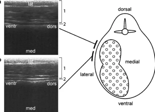
Fig. 4 Ultrasonographic find-ings and interpretation with re-spect to the stratification of the dorsal rumen contents (in two levels, a and b) of a browse-fed, captive moose (A. alces); 1 abdominal wall; 2 rumen wall; no reverberation lines indicative of a gas-filled space (gas dome) could be found
References
Ahvenjärvi S, Skiba B, Huhtanen P (2001) Effect of heterogenous digesta chemical composition on the accuracy of measurements of fibre flow in dairy cows. J Anim Sci 79:1611–1620 Beaumont R, Deswysen AG (1991) Mélange et propulsion du contenu
du réticulo-rumen. Reprod Nutr Dev 31:335–359
Behrend A, Lechner-Doll M, Streich WJ, Clauss M (2004) Seasonal faecal excretion, gut fill, liquid and particle marker retention in mouflon (Ovis ammon musimon), and a comparison with roe deer (Capreolus capreolus). Acta Theriol 49:503–515
Bogner JR (1992) Artefakte. In: Zoller WG, Gresser U, Zöllner N (eds) Einführung in die Ultraschalldiagnostik. Karger, Basel, pp 14–21
Braun U, Götz M, Marmier O (1993) Ultrasonographic findings in cows with traumatic reticuloperitonitis. Vet Rec 133:416–422 Braun U, Flückiger M, Götz M (1994) Comparison of
ultrasono-graphic and radioultrasono-graphic findings in cows with traumatic reticuloperitonitis. Vet Rec 135:470–478
Braun U, Götz M (1994) Ultrasonography of the reticulum in cows. Am J Vet Res 55:325–332
Braun U, Marmier O (1995) Ultrasonographic examinationn of the small intestine of cows. Vet Rec 136:239–244
Braun U, Pusterla N, Schönmann N (1995a) Ultrasonographic findings in cows with left displacement of the abomasum. Vet Rec 141:331–335
Braun U, Marmier O, Pusterla N (1995b) Ultrasonographic examina-tion of the small intestine of cows with ileus of the duodenum, jejunum, or ileum. Vet Rec 137:209–215
Braun U, Wild K, Guscetti F (1997) Ultrasonographic examination of the abomasum of 50 cows. Vet Rec 140:93–98
Braun U, Iselin U, Lischer C, Fluri E (1998) Ultrasonographic findings in five cows before and after treatment of reticular abscesses. Vet Rec 142:184–189
Braun U, Amrein E (2001) Ultrasonographic examination of the caecum and proximal and spiral loop of the colon of cattle. Vet Rec 149:45–48
Braun U, Schweizer G, Flückiger M (2002a) Radiographic and ultrasonographic findings in three cows with reticulo-omasal obstruction due to a foreign body. Vet Rec 150:580–581 Braun U, Amrein E, Koller U, Lischer C (2002b) Ultrasonographic
findings in cows with dilatation, torsion, and retroflexion of the caecum. Vet Rec 150:75–79
Braun U (2003) Ultrasonography in gastrointestinal disease in cattle. Vet J 166:112–124
Braun U, Blessing S (2006) Ultrasonographic examination of the omasum in 30 healthy cows. Vet Rec 159:812–815
Clarke RT, Reid CS (1974) Foamy bloat of cattle. A review. J Dairy Sci 57:753–785
Clauss M, Lechner-Doll M (2001) Differences in selective reticulo-ruminal particle retention as a key factor in ruminant diversifi-cation. Oecologia 129:321–327
Clauss M, Lechner-Doll M, Behrend A, Lason K, Lang D, Streich WJ (2001) Particle retention in the forestomach of a browsing ruminant, the roe deer (Capreolus capreolus). Acta Theriol 46:103–107
Clauss M, Lechner-Doll M, Streich WJ (2003) Ruminant diversifica-tion as an adaptadiversifica-tion to the physicomechanical characteristics of forage. A reevaluation of an old debate and a new hypothesis. Oikos 102:253–262
Clauss M, Hummel J, Streich WJ (2006a) The dissociation of the fluid and particle phase in the forestomach as a physiological characteristic of large grazing ruminants: an evaluation of available, comparable ruminant passage data. Eur J Wildlife Res 52:88–98
Clauss M, Hofmann RR, Hummel J, Adamczewski J, Nygren K, Pitra C, Reese S (2006b) The macroscopic anatomy of the omasum of free-ranging moose (Alces alces) and muskoxen (Ovibos moschatus) and a comparison of the omasal laminal surface area in 34 ruminant species. J Zool Lond 270:346–358
Clauss M, Kaiser T, Hummel J (2007) The morphophysiological adaptations of browsing and grazing mammals. In: Gordon IJ, Prins HHT (eds) The ecology of browsing and grazing. Springer, Heidelberg, pp 47–88
Grau H (1955) Zur Funktion der Vormägen, besonders des Netzmagens, der Wiederkäuer. Berl Münch Tierärztl Wochenschr 15:271–275 Hofmann RR (1969) Zur Topographie und Morphologie des
Wieder-käuermagens im Hinblick auf seine Funktion (nach vergleichen-den Untersuchungen an Material ostafrikanischer Wildarten). Zentralbl Veterinärmed Suppl 10:1–180
Hofmann RR (1973) The ruminant stomach. East African Literature Bureau, Nairobi
Hofmann RR, Streich WJ, Fickel J, Hummel J, Clauss M (2008) Convergent evolution in feeding types: salivary gland mass differences in wild ruminant species. J Morphol 269:240–257 Hummel J, Clauss M, Zimmermann W, Johanson K, Norgaard C,
Pfeffer E (2005) Fluid and particle retention in captive okapi (Okapia johnstoni). Comp Biochem Physiol A 140:436–444 Hummel J, Steuer P, Südekum KH, Hammer S, Hammer C, Streich
WJ, Clauss M (2008a) Fluid and particle retention in the digestive tract of the addax antelope (Addax nasomaculatus)— adaptations of a grazing desert ruminant. Comp Biochem Physiol A 149:142–149
Hummel J, Südekum KH, Bayer D, Ortmann S, Hatt JM, Clauss M (2008b) Physical characteristics of reticuloruminal contents of cattle in relation to forage type and time after feeding. J Anim Physiol Anim Nutr (in press). DOI10.1111/j.1439-0396.2008.00806.x
Jiang Z, Hudson RJ (1996) Digestive responses of wapiti to seasonal forages. Acta Theriol 41:415–425
Kaske M, Midasch A, Rehage J (1994) Sonographic investigation of reticular contractions in healthy sheep, cows, and goats and in cows with traumatic reticulo-peritonitis. J Vet Med Ser A 41:748–756 Kirberger RM (1995) Imaging artifacts in diagnostic ultrasound—a
review. Vet Radiol Ultrasound 36:297–306
Kovács PL, Südekum KH, Stangassinger M (1997) Effects of intake level of a mixed diet on chewing activity and on particle size of ruminated boli, ruminal digesta fractions and faeces of steers. Reprod Nutr Dev 37:517–528
Lechner-Doll M, Kaske M, Engelhardt Wv (1991) Factors affecting the mean retention time of particles in the forestomach of ruminants and camelids. In: Tsuda T, Sasaki Y, Kawashima R (eds) Physiological aspects of digestion and metabolism in ruminants. Academic, San Diego, pp 455–482
Meyer RM, Bartley EE (1971) Bloat in cattle. XV. The relation of viscosity and cell-free polysaccharide content of rumen fluid to feedlot bloat. J Anim Sci 33:1018–1020
Meyer RM, Bartley EE (1972) Bloat in cattle. XVI. Development and application of techniques for selecting drugs to prevent feedlot bloat. J Anim Sci 34:234–240
Nygren K, Hofmann RR (1990) Seasonal variations of food particle size in moose. Alces 26:44–50
Pennick DG (1995) Imaging artifacts in ultrasound. In: Nyland TG, Mattoon JD (eds) Veterinary diagnostic ultrasound. Saunders, Philadelphia, pp 19–29
Renecker LA, Hudson RJ (1990) Digestive kinetics of moose, wapiti and cattle. Anim Prod 50:51–61
Robbins CT, Spalinger DE, Van Hoven W (1995) Adaptations of ruminants to browse and grass diets: are anatomical-based browser-grazer interpretations valid? Oecologia 103:208–213 Sakauchi R, Hoshino S (1981) Effects of monensin on ruminal fluid
microbial activity and population in healthy and bloated feedlot steers. J Anim Physiol Anim Nutr 46:21–33
Schwarm A, Ortmann S, Wolf C, Clauss M (2008) Excretion patterns of fluid and different sized particle passage markers in banteng (Bos javanicus) and pygmy hippopotamus (Hexaprotodon liberiensis): Two functionally different foregut fermenters. Comparative Bio-chemistry and Physiology A (in press). DOI 10.1016/j. cbpa.2008.02.022
Seo S, Lanzas C, Tedeschi LO, Fox DG (2007) Development of a mechanistic model to represent the dynamics of liquid flow out
of the rumen and to predict the rate of passage of liquid in dairy cows. J Dairy Sci 90:840–855
Stanford K, Wang Y, Berg BP, Majak W, McCartney DH, Baron V, McAllister TA (2001) Effects of alcohol ethoxylate and pluronic detergents on the development of pasture bloat in cattle and sheep. J Dairy Sci 84:167–176
Warner RG, Flatt WP, Loosli JK (1956) Dietary factors influencing the development of the ruminant stomach. Agic Food Chem 4:788–792 Welch JG (1982) Rumination, particle size and passage from the