CASE REPORT
Steroid-responsive encephalopathy associated
with Hashimoto thyroiditis
Petra Zimmermann&Enno Stranzinger
Received: 17 May 2011 / Revised: 19 September 2011 / Accepted: 24 September 2011 / Published online: 30 December 2011 # Springer-Verlag 2011
Abstract An 11-year-old girl presented with sudden sensory disturbance and left-sided muscle weakness. MRI revealed ischaemic change in the right lateral thalamus and the right internal capsule. During sonographic work-up of the cervical arteries, inflammation of the thyroid gland was noted. The results of the thyroid function tests and antibody titers con-firmed Hashimoto thyroidits. Under high-dose corticoste-roids, the girl had a full neurological recovery.
Keywords Hashimoto thyroiditis . Cerebrovascular event . Enchephalopathy . Child
Introduction
Hashimoto encephalopathy is a steroid-responsive enceph-alopathy associated with elevated concentrations of antithy-roid antibodies. Patients with the condition are usually euthyroid or mildly hypothyroid. The pathogensis of Hashi-moto encephalopathy is not fully understood, but evidence for autoimmune and vasculitic mechanisms exist [1]. The disease usually responds well to corticosteroids. There are two subtypes. The vasculitic type is characterised by acute stroke-like episodes with focal neurological deficits, myoclonus and seizures. The other subtype is diffuse,
progressive and associated with insidious onset and progres-sive impairment of mental status, such as confusion, hallu-cinations, mood disturbances and psychosis. We report a girl who presented with an acute stroke implying that she suf-fered from the vasculitic type of Hashimoto encephalopathy.
Case report
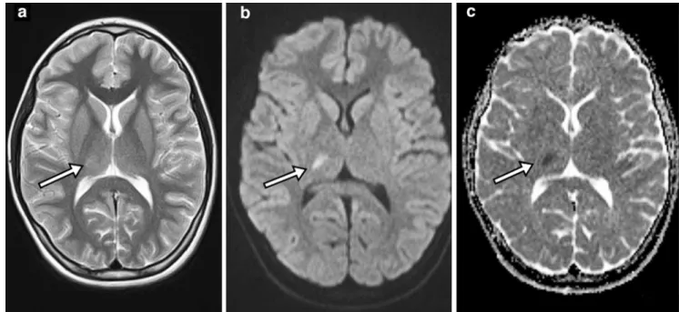
A previously healthy 11-year-old girl presented to the emer-gency room with sudden onset of frontal headache, sensory disturbance and left-sided weakness. There was no history of nausea or vomiting. She had left facial nerve palsy. There was mild hypoaesthesia, hypoalgesia, acrognosis and re-duced muscle strength in her left arm and leg. Diffusion-weighted MRI showed a 10-mm hyperintense area with matching low apparent diffusion coefficients (suggesting restricted diffusion) in the right thalamus and internal cap-sule (Fig.1). This was consistent with an acute stroke. The girl was admitted to hospital and treatment with antiplatelet drugs was started.
Haematological, rheumatological, renal and liver bio-chemical tests were normal. Cerebrospinal fluid, serological tests for borreliosis and ECG were all unremarkable. Ultra-sound (US) was preformed to rule out abnormalities of the arteries in the neck. These were normal, but there was symmetrical diffuse enlargement of the thyroid gland with a hypoechoic, heterogeneous mirconodular echo pattern. On colour Doppler, a diffuse increase in thyroid parenchymal-vascularity was noted (Fig. 2). These findings suggested Hashimoto thyroiditis.
Thyroid function tests showed slightly elevated thyroid-stimulating hormone of 5.71 mU/l (normal range, 0.35–5) with normal levels of free-T4 and free-T3. A markedly elevated antithyroperoxidase antibody titer was observed at
P. Zimmermann (*) Department of Paediatrics,
University Children’s Hospital, Inselspital, Freiburgstrasse, Bern 3010, Switzerland e-mail: [email protected] E. Stranzinger
Institute of Radiology– Paediatric Radiology, University Hospital Bern,
Bern, Switzerland
Pediatr Radiol (2012) 42:891–893 DOI 10.1007/s00247-011-2309-7
2,313 IU/mL (normal range, < 33 IU/ml). The antithyroglo-bulin antibody level was normal at 46 IU/mL (normal range, < 100 IU/mL). These test results together with the US findings led to the diagnosis of a Hashimoto thyreoiditis. There was no clinical goitre.
Intravenous therapy with high-dose corticosteroid was initiated. Subsequently all neurological symptoms and find-ings resolved. After 5 days, she was discharged with anti-platelet drugs and oral corticosteroids to be taken for 3 months. Follow-up MRI after 10 weeks showed a small hyperintense lesion in the right thalamus on T2-weighted imaging, and this was interpreted as gliosis. There was no further abnormality.
Discussion
The differential diagnosis of paediatric strokes is much wider than in adults and includes arteriopathy, cardiac dis-orders, infections and other acute and chronic systemic conditions. Therefore, cerebral MRI, chest radiograph and neck vessel US are part of the investigation.
Corticosteroid-responsive encephalopathy associated with Hashimoto thyroiditis has chiefly been reported in adults, but is a well-recognised differential diagnosis of encephalopathy in children [2] in whom it is often over-looked. As our case illustrates, this condition is treatable, and therefore clinical examination of the thyroid gland
Fig. 1 Cranial MRI in an 11-year-old girl. a Axial T2-weighted images show a 10-mm hyperintense blurred lesion of the right thalamus and internal capsule (arrow). b Diffusion-weighted image demonstrates
hyperintensity related to restricted diffusion in the same area (arrow). c Apparent diffusion coefficient map shows the corresponding low values in the area (arrow)
Fig. 2 Transverse sonogram of the thyroid gland. a B-mode ultrasound (US) reveals hetero-geneous enlargement of the gland with small focal hypoe-choic nodules (arrow) compati-ble with lymphocytic infiltration. b Increased vascularity of the gland is seen on colour Doppler
needs to be performed in children with unclear encephalop-athy. At neck vessel US, it is advisable to include the thyroid. Patients with chronic Hashimoto thyroiditis are usually hypothyroid and show elevated levels of thyroid-stimulating hormone. This has a hypertrophic effect on the thyroid gland and enlargement can be seen on US. Colour Doppler US often reveals hypervascularity within the gland [3], as in our report.
Brain MRI in children with Hashimoto encephalopathy may show focal hyperintensity or volume depletion of the nucleus accumbens, frontal white matter changes, cerebellar atrophy and hippocampal and periventricular lesions. SPECT and MR perfusion imaging may show focal or bilateral hypoperfusion [4]. As in our report, about half of the patients with Hashimoto-associated encephalopathy have nonenhancing MRI abnormalities with increased sig-nal intensity on T2-weighted images and on fast fluid-attenuated inversion recovery (FLAIR) [5]. While FLAIR is more sensitive than T2-weighted images for ischemia-induced cytotoxic oedema, diffusion-weighted imaging (DWI) is the most sensitive imaging sequence for ischaemic injury. MR perfusion imaging is helpful for detecting arterial occlusion and hypoperfusion [3].
Intravenous high-dose corticosteroid treatment is effec-tive, but sequelae are more common in children than in adults and most commonly include neuropsychological dif-ficulties, seizures and behaviour problems [4–6]. Non-response does not exclude Hashimoto enchephalopathy [7].
In conclusion, Hashimoto thyroiditis needs to be con-sidered in children and adolescents with unexplained neuro-logical or psychiatric symptoms [8].
References
1. Watemberg N, Greenstein D, Levine A (2006) Encephalopathy associated with Hashimoto thyroiditis: pediatric perspective. J Child Neurol 21:1–5
2. Hoffmann F, Reiter K, Kluger G et al (2007) Seizures, psychosis and coma: Severe course of Hashimoto encephalopathy in a six-year-old girl. Neuropediatrics 38:197–199
3. Barkovich AJ, Moore KR, Grant E et al. (2007) Pediatric neurora-diology. Elsevier Books, Salt Lake City, Utah
4. Alink J, de Vries TW (2008) Unexplained seizures, confusion or hallucinations: think Hashimoto encephalopathy. Acta Paediatr 97:451–453
5. Grommes C, Griffin C, Downes K et al (2008) Steroid-responsive encephalopathy associated with autoimmune thyroiditis presenting with diffusion MR imaging changes. AJNR 8:1550–1551 6. Vasconcellos E, Piña-Garza JE, Fakhoury T et al (1999) Pediatric
manifestations of Hashimoto’s encephalopathy. Pediatr Neurol 20:394–398
7. Berger I, Castiel Y, Dor T (2010) Paediatric Hashimoto encepha-lopathy, refractory epilepsy and immunoglobulin treatment— unusual case report and review of the literature. Acta Paediatr 99:1903–1905
8. Castro-Gago M, Gómez-Lado C, Maneiro-Freire M et al (2010) Hashimoto encephalopathy in a preschool girl. Pediatr Neurol 42:143–146