Donut-Shaped High-Dose Configuration
for Proton Beam Radiation Therapy
Hans Peter Rutz, Antony J. Lomax
1Background: The authors report on the conception and first clinical application of a donut-shaped high-dose configuration for
proton therapy (PT). This approach allows one to intensify target volume dose coverage for targets encompassing a critical, dose-limiting structure – like here, the cauda equina –, whilst delivering minimal dose to other healthy structures surrounding the target, thereby reducing the integral dose.
Methods and Results: Intensity-modulated PT methods (IMPT) for spot scanning were applied to create and deliver a
donut-shaped high-dose configuration with protons, allowing treating > 75% of the target with at least 95% of the prescribed dose of 72.8 CGE, whilst restricting dose to the cauda equina to 60–65 CGE. Integral dose was lower by a factor of 3.3 as compared to intensity-modulated radiotherapy with photons (IMXT).
Conclusion: IMPT and spot scanning technology allow a potentially clinically useful approach which is also applicable to spare
other critical structures passing through a target volume, including spinal cord, optic nerves, chiasm, brain stem, or urethra.
Key Words: Proton beam · Intensity-modulated · IMPT · IMXT · IMRT · Integral dose Strahlenther Onkol 2005;181:49–53
DOI 10.1007/s00066-005-1280-3
Ringförmige Hochdosiskonfiguration für die Protonentherapie
Hintergrund: Die Autoren berichten über Konzeption und erste klinische Anwendung einer ringförmig konfigurierten Dosis für
die Protonentherapie (PT). Dies erlaubt es, einen möglichst hohen Anteil des Zielvolumens mit der verordneten Dosis zu behan-deln bei gleichzeitiger Schonung kritischer Strukturen, welche das Zielvolumen durchqueren.
Methodik und Ergebnisse: Unter Anwendung intensitätsmodulierter Protonentherapie (IMPT) und „Spot-Scanning“-Technologie
konnte eine Hochdosiskonfiguration in Form eines Donuts generiert und problemlos appliziert werden. Damit konnten > 75% des Zielvolumens mit mindestens 95% der verordneten Dosis von 72,8 CGE behandelt werden bei gleichzeitiger Schonung der Cauda equina, welche mit lediglich 60–65 CGE belastet wurde. Die Integraldosis im gesunden Gewebe war um den Faktor 3,3 kleiner als bei Bestrahlung mit intensitätsmodulierten Photonen (IMXT).
Schlussfolgerung: IMPT und „Spot-Scanning“-Technologie erlauben einen neuen Ansatz für die optimierte PT, der auch zur
Schonung anderer kritischer Strukturen eingesetzt werden kann, welche das Zielvolumen durchqueren, wie z.B. Rückenmark, Nervus opticus, Chiasma, Hirnstamm oder Urethra.
Schlüsselwörter: Protonentherapie · Intensitätsmoduliert · IMPT · IMXT · IMRT · Integraldosis
Received: February 4, 2004; accepted: September 30, 2004
1Division of Radiation Medicine, Paul Scherrer Institute, Villigen PSI, Switzerland.
Introduction
Spot scanning proton therapy (PT) is the result of more than 5 decades of applied particle physics [6] and radiobiology re-search [15]. Here, we report on the conception and first clini-cal application of a novel high-dose configuration in PT, al-lowing an improved treatment – intensification of dose to the
target (PTV), for PTVs encompassing a critical (i.e., dose-lim-iting) structure, like in this case, the cauda equina, whilst at the same time delivering minimal dose to the healthy normal structures surrounding the PTV, reducing integral dose. This goal has been achieved applying intensity modulation meth-ods for PT (IMPT) using spot scanning [9, 16].
Our patient needed adjuvant PT after macroscopically complete resection of a large chordoma of the fifth lumbar vertebra (L5). The vertebra had been removed and titanium implanted for mechanical stabilization. Chordomas are char-acterized by high rates of local failure, even if treated with 60 Gy (photons), or the corresponding equivalence dose (60Co Gy equivalents [CGE]) using charged particles [2, 3, 7, 8, 13, 14, 17, 20, 21]. In PT, the dose is prescribed in CGE, based on a conversion by 1.1 of physical proton dose to account for enhanced biological effectiveness of protons versus photons [15]. However, the cauda region only tolerates a dose of 60–65 CGE; otherwise, the risk of developing clinically appar-ent nerve damage is estimated > 10% [1, 4].
Our goal hence was to deliver 72 CGE to as much as pos-sible of the PTV, which included the cauda, whilst restricting dose delivered to this critical structure. Conventionally, this problem is dealt with in PT by compromising dose to more of the PTV, delivering a modified high-dose configuration in the second treatment series, treating only the proximal aspect of the PTV, which can be reached by protons without pas-sing through the critical structure within, in this case, the cau-da. We decided to explore the possibilities of IMPT to gener-ate a “donut-shaped” high-dose configuration for treatment in the second series, allowing an intensification of PTV dose coverage.
Methods
Intensity modulation methods for PT as well as a first clinical example have been published [9, 10]. In brief, in conventional spot scanning PT, the dose delivered to each individual point (“spot”) in the three-dimensional (3-D) coordinate grid of the region of interest is weighted so as to generate a dose distribu-tion within the PTV, which is as homogeneous as feasible and at least 95% of the reference dose. In IMPT, dose within the PTV is intentionally inhomogeneous within a given field, as defined by the planner. Only the sum of two or three fields adds up to the prescribed treatment dose.
In order to apply PT and IMPT, the positioning of the patient is important. To this end, we perform CT scout view-based controls and correction of patient position before every single treatment [11], allowing to place nonmoving re-gions of interest within 0.5–3 mm as compared to the planning setup. In IMPT, if positioning is not precise, the complemen-tary intensity-modulated (intentionally nonhomogeneous) treatment plans would not match as predicted, causing hidden hot spots in normal tissue, endangering safety, and cold spots within other aspects of the PTV, endangering tumor control.
In this case, the PTV was defined as the preoperative tu-mor bed and the tissue estimated to have been touched during surgery, plus a 5-mm margin in 3-D. CT artifacts from titanium implants were compensated manually according to clinician’s best judgment and assigning water- and bone-equivalent Hounsfield units for dose calculation. The treatment required IMPT already in the first series due to the depth range of the
PTV, which was > 200 mm in water-equivalent density, and a limited number of range shifter plates available at the gantry (depth range of the Bragg peak with a specific energy of the proton beam: 173.5 mm in water-equivalent density). Hence, two energies (166 and 177 MeV) were combined to cover the depth range homogeneously with dose. To spare the cauda (second series), IMPT methods were applied to calculate spot weights generating a dose distribution delivering only 65–75% of the reference dose to the cauda region.
Moreover, PT and the choice of beam directions are re-stricted by respiratory movement, as it would seriously distort dose deposition, unless beam directions are quasi parallel to the movement. In that case, dose distribution is affected only marginally. Safety concerns oblige us to treat the patients with the beam coming from above ± 100° due to the geometric con-struction of our gantry; with beam from below, the patient ta-ble would rise > 3 m above ground, which could be dangerous in the case of panic attacks in patients or an acute medical emergency. We hence treat tumors along the spine (between C3 and the sacrum) with patients positioned in the prone posi-tion and use two beam direcposi-tions, which are ± 10° from poste-rior (above) for treatment, so-called “narrow-angle” PT and IMPT.
This patient, like all our patients, was treated after giving informed written consent. Side effects were scored according to common terminology criteria for adverse events (CTCAE,
http://ctep.cancer.gov/forms/CTCAEv3.pdf). For a
compari-son, an intensity-modulated photon (IMXT) plan (five fields) similarly sparing the cauda was prepared.
Results
The first series to 35 CGE hence did not spare the cauda and consisted of four fractions of 2.0 and 15 fractions of 1.8 CGE. The dose per fraction was reduced because of several unavoid-able implant-associated hot spots of up to 124%. In the second series, we used identical beam directions and fractionation, delivering the donut-shaped high-dose configuration selec-tively sparing the cauda (Figure 1). In the combined treat-ment, a total dose of 72.8 CGE was delivered in 40 fractions (maximum dose: 88.8 CGE; hot spot within implant material). With this approach, it was possible to treat 75.8% of the target to at least 95% (69 GCE) of the reference dose. Of the cauda, 11.4 ml received > 60 CGE; < 1 ml received > 65 CGE (maxi-mum: 70.5 CGE in a hot spot). Without donut configuration generated using IMPT, much less of the target would have re-ceived 69 CGE, both for technical reasons (target depth range > 200 mm in water equivalence) and cauda dose restriction.
The treatment was tolerated without a problem; only a grade 2 skin reaction was noted, yet no other symptom. At the proofreading of the manuscript, 28 months after treatment, the patient is without late symptoms or radiologic evidence for disease.
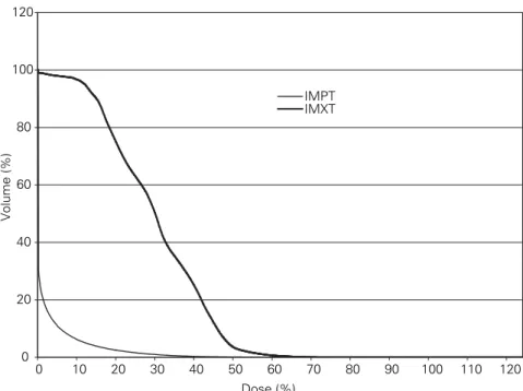
IMXT could achieve a similar level of dose sparing to the cauda (Figure 2), however, only IMPT can do so whilst
simul-taneously sparing surrounding structures very effectively. Fig-ure 3 shows the difference regarding small intestines. The in-tegral dose to non-target tissue was lower by a factor of 3.3 in the case of IMPT versus IMXT.
Discussion
IMPT is suitable to selectively reduce dose delivered to a de-fined volume within a PTV, hence protecting a critical (dose-limiting) structure within the target. This allows an in-tensification of target dose coverage, and therefore, the prob-abilistic chances for a cure over a level that would be possible using conventional spot-scanning PT. Based on current knowl-edge, such dose intensification is presumed to be without gen-erating a higher risk for the development of late normal tissue toxicity and organ dysfunction.
Comparative planning revealed only moderate doses to the intestines at risk, if IMXT had been used for treatment. One can thus say that in this particular case, the expected
tox-icity from delivering the whole treatment with IMXT would have been acceptable. This, nevertheless, is an exception, as the PTV was situated caudal to the kidneys. In the case of a more cranial location of the target (L4 and above), however,
%
PT dose
PTV
Intestines
Cauda
Implants
CT artifact
correction
Figure 1. Proton doses in the middle plane of the target volume (PTV;
green outline) during the second treatment series. Red lines represent anatomic structures and technical volumes. In the first series, the dose inside the target was planned to be > 95% using IMPT to allow com-bining dose delivery by two energies (166 and 177 MeV), to cover the entire depth of the PTV (> 200 mm in water-equivalent density); dose levels outside the target were identical, however. Artifact correction: manual.
Abbildung 1. Protonendosis in der Mittelschicht des Zielvolumens
(PTV; grüne Linie) während der zweiten Bestrahlungsserie (siehe Text). Rote Linien umranden anatomische Strukturen und technische Volu-men. In der ersten Serie war die Dosisverteilung außerhalb des PTV identisch, innerhalb des PTV jedoch mindestens 95% der verordneten Dosis. Dies war nur möglich unter Verwendung der intensitätsmodu-lierten Protonentherapie (IMPT), sodass die beiden Energien (166 und 177 MeV) kombiniert zur Anwendung kommen konnten; so konnte die gesamte Tiefe des PTV (> 200 mm Wasser-äquivalente Distanz) mit Dosis abgedeckt werden. Die Korrektur der Implantat-bedingten Arte-fakte im CT-Datensatz erfolgte manuell.
Figure 2. Radiation doses 9 mm cranially to the middle plane of the
target (PTV; exterior yellow outline) during the second series of the treatment. The inner yellow line shows the preoperative tumor vol-ume and the tissue touched by surgery. Green outlines show cauda as well as a technical volume around the cauda (cauda + 5 mm in 3 D), and intestines. Note that the color code regarding the dose is different from the one used in Figure 1. The upper plan depicts doses generated by IMXT (comparative planning), whereas the lower plan depicts the doses generated using IMPT (as delivered in the second series).
Abbildung 2. Dosisverteilung 9 mm kranial der Mittelschicht des
Ziel-volumens (PTV, äußere gelbe Linie) während der zweiten Bestrah-lungsserie. Die innere gelbe Linie umrandet das Tumorbett sowie das Operationsgebiet. Die grünen Linien umringen die Cauda sowie ein technisches Volumen darum herum (Cauda + 5 mm in 3-D) und Gedär-me. Die Farbkodierung der Dosisverteilung ist ebenfalls anders als in der Abbildung 1. Oben in der Abbildung findet sich die Dosisverteilung bei IMXT, unten jene bei IMPT.
the limited tolerance of the kidneys and lung [1, 4, 23] would have restricted total dose applicable by IMXT alone to 58–60 CGE, as has already been reported regarding IMXT for chordoma of the cervical spine [5], although technically, dose to the spinal cord and cauda equina can be restricted to their respective tolerance levels.
On the other hand, IMXT could be tolerated at these more proximal sites (L4 and above), if the second series were delivered with PT; even conventional photon radiotherapy in the first series could be tolerated, if it were followed by IMPT in the second (or vice versa). The combination of these tech-niques could be considered to increase the number of patients benefiting from PT in terms of achieving a potentially curative target dose. At the Paul Scherrer Institute, patients are treat-ed with protons alone, unless stipulattreat-ed otherwise by the an-nual accelerator shut down for maintenance. In one case, we have actually recently treated a patient with chordoma of L2 with a similar, donut-shaped plan in a first series (IMPT), to be followed by conventional radiotherapy (13 × 2 Gy) at the referring center.
Moreover, irradiation of non-target tissues by IMXT is clearly an avoidable risk for the induction of treatment-in-duced secondary cancer [18], and in this particular case, for example, the integral dose delivered to the tissues outside the PTV would have been much higher (namely, by a factor of 3.3; see above) by using IMXT instead of IMPT. Previous studies have shown similar findings regarding integral dose and lifetime risk for secondary cancer; all were much in favor
of PT and IMPT over XT or IMXT [12, 19, 22]. These considerations, obviously, are particularly significant: when they concern the treatment of children and young adults, especially as some of them may, in fact, be genetically predisposed; or regarding the combination of irradia-tion with chemotherapy.
The concept we present here is also suitable to intensify dose delivered to PTVs at other anatomic sites which are crossed by other critical anatomic struc-tures, like, e.g., spinal cord, optic nerves, chiasm, brain stem, and esophagus; and it is applicable to treatments with other positively charged particles, such as car-bon ions.
Acknowledgments
We thank our colleagues for referring pa-tients as well as stimulating discussions. Spe-cial thanks to Shinji Sugahara for critical reading of the revised manuscript and his very helpful comments. This study was funded by the Paul Scherrer Institute.
References
1. Burman C, Kutcher GJ, Emami B, et al. Fitting of normal tissue toler-ance data to an analytic function. Int J Radiat Oncol Biol Phys 1991;21: 123–35.
2. Catton C, O’Sullivan B, Bell R, et al. Chordoma: long-term follow-up after radical photon irradiation. Radiother Oncol 1996;41:67–72.
3. Debus J, Schulz-Ertner D, Schad L, et al. Stereotactic fractionated radio-therapy for chordomas and chondrosarcomas of the skull base. Int J Radiat Oncol Biol Phys 2000;47:591–6.
4. Emami B, Lyman J, Brown A, et al. Tolerance of normal tissue to therapeutic irradiation. Int J Radiat Oncol Biol Phys 1991;21:109–22.
5. Gabriele P, Macias V, Stasi M, et al. Feasibility of intensity-modulated ra-diation therapy in the treatment of advanced cervical chordoma. Tumori 2003;89:298–304.
6. Goitein M, Lomax AJ, Pedroni E. Treating cancer with protons. Phys Today 2002;55:45–50.
7. Hug EB, Loredo LN, Slater JD, et al. Proton radiation therapy for chordomas and chondrosarcomas of the skull base. J Neurosurg 1999;91:432–9. 8. Latz D, Gademann G, Hawighorst H, et al. [The initial results in the
fractio-nated 3-dimensional stereotactic irradiation of clivus chordomas.] Strah-lenther Onkol 1995;171:348–55.
9. Lomax A. Intensity modulation methods for proton radiotherapy. Phys Med Biol 1999;44:185–205.
10. Lomax AJ, Boehringer T, Coray A, et al. Intensity modulated proton thera-py: a clinical example. Med Phys 2001;28:317–24.
11. Lomax AJ, Roser W, Treier R, et al. Patient positioning using a “remote” CT scanner: initial experiences and results. PSI Sci Rep Life Sci 2001:62–3. 12. Miralbell R, Lomax A, Cella L, et al. Potential reduction of the incidence of
radiation-induced second cancers by using proton beams in the treatment of pediatric tumors. Int J Radiat Oncol Biol Phys 2002;54:824–9. 13. Munzenrider JE, Liebsch NJ. Proton therapy for tumors of the skull base.
Strahlenther Onkol 1999;175:Suppl 2:57–63.
14. Noel G, Habrand JL, Jauffret E, et al. Radiation therapy for chordoma and chondrosarcoma of the skull base and the cervical spine. Prognostic factors and patterns of failure. Strahlenther Onkol 2003;179:241–8.
15. Paganetti H, Niemierko A, Ancukiewicz M, et al. Relative biological effec-tiveness (RBE) values for proton beam therapy. Int J Radiat Oncol Biol Phys 2002;53:407–21. 0 20 40 60 80 100 120 0 10 20 30 40 50 60 70 80 90 100 110 120 Dose (%) Volume (%) IMPT IMXT
Figure 3. Comparative dose-volume histogram regarding intestines at risk (Figures 1 and 2). Abbildung 3. Vergleichendes Dosis-Volumen-Histogramm betreffend die mitbestrahlten
16. Pedroni E, Bacher R, Blattmann H, et al. The 200-MeV proton therapy pro-ject at the Paul Scherrer Institute: conceptual design and practical realiza-tion. Med Phys 1995;22:37–53.
17. Rhomberg W, Bohler FK, Novak H, et al. A small prospective study of chor-domas treated with radiotherapy and razoxane. Strahlenther Onkol 2003;179:249–53.
18. Rutz HP, Mirimanoff RO, Little JB. Strategies for the prevention of treat-ment-induced secondary cancer. In: Nygaard F, Upton AC, eds. Anticar-cinogenesis and radiation protection 2. New York: Plenum Press, 1991: 39–45.
19. Schneider U, Lomax AJ, Lombriser N. Comparative treatment planning us-ing secondary cancer mortality calculations. Phys Med 2001;17:Suppl 1: 97–9.
20. Schoenthaler R, Castro JR, Petti PL, et al. Charged particle irradiation of sacral chordomas. Int J Radiat Oncol Biol Phys 1993;26:291–8.
21. Schulz-Ertner D, Nikoghosyan A, Thilmann C, et al. Carbon ion radiotherapy for chordomas and low-grade chondrosarcomas of the skull base. Results in 67 patients. Strahlenther Onkol 2003;179:598–605.
22. St Clair WH, Adams JA, Bues M, et al. Advantage of protons compared to conventional X-ray or IMRT in the treatment of a pediatric patient with medulloblastoma. Int J Radiat Oncol Biol Phys 2004;58:727–34.
23. Tokuuye K, Akine Y, Kagei K, et al. Proton therapy for head and neck malig-nancies at Tsukuba. Strahlenther Onkol 2004;180:96–101.
Address for Correspondence
Hans Peter Rutz
Division of Radiation Medicine Paul Scherrer Institute 5232 Villigen PSI Switzerland
Phone (+41/56) 310-3524, Fax -3515 e-mail: hanspeter.rutz@psi.ch