INTERVENTIONAL NEURORADIOLOGY
The use of flat panel angioCT (DynaCT) for navigation
through a deformed and fractured carotid stent
Pasquale Mordasini&Fahmi Al-Senani&Jan Gralla&
Dai-Do Do&Caspar Brekenfeld&Gerhard Schroth
Received: 22 May 2009 / Accepted: 22 June 2009 / Published online: 28 July 2009 # Springer-Verlag 2009
Abstract Navigation through a previously deployed and deformed stent is a difficult interventional task. Inadvertent navigation through the struts of a stent can potentially lead to incomplete secondary stent extension and vessel occlu-sion. Better visualisation of the pathway through the stent can reduce the risks of the procedural complications and reduce the reluctance of the interventionalist to navigate through a previously deployed stent. We describe a technique of visualisation of the pathway navigated by a guidewire through a previously deployed deformed and fractured carotid stent by the use of DynaCT. Three-dimensional reconstruction of the stent/microwire allows excellent visualisation of the correct pathway of the microwire within the stent.
Keywords DynaCT . Carotid stent . Navigation . Restenting
Introduction and technique
A 73-year-old man with a previously symptomatic signif-icant carotid stenosis underwent carotid endarterectomy
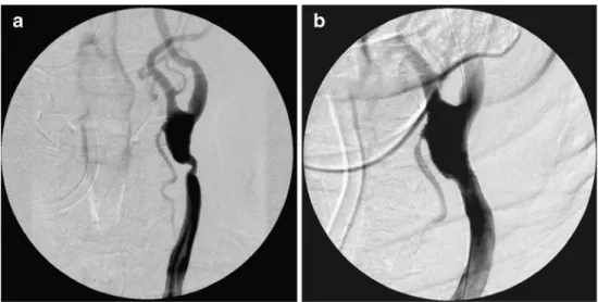
using a venous patch. At 6 months follow-up ultrasonog-raphy control showed significant recurrence of the stenosis. The patient was treated with carotid stenting using an Acculink stent (10×20 mm, Abbott Laboratories, Abbott Park, USA). Although the check angiogram after the intervention illustrated normal stent placement and structure (Fig. 1), a follow-up magnetic resonance angiography 6 months later depicted significant asymptomatic recur-rence of the stenosis (Fig. 2a). The stent structure showed deformity and fracture of the stent meshes at the distal end (Fig. 2b, c). A decision was made for re-stenting of the stenosis in combination with balloon angioplasty.
Under local anaesthesia, a routine cerebral angiogram using a biplane subtraction neuroangiography (Siemens Axiom Artis zee, Siemens, Erlangen, Germany) was performed showing a high-grade stenosis of the left common carotid artery. A Galeo Hydro 0.14 microwire (Biotronik Inc., Lake Oswego, USA) was carefully navi-gated through the stenosis.
A non-contrasted DynaCT was performed with a commercially available software (Dyna-CT; Siemens Med-ical Solution, Forchheim, Germany) using the following parameters: 20-s acquisition, 0.4° increment, 512 matrix in projections, 220° total angle, 20°/s, and 15 frames per second. Radiation dose applied for the DynaCT was 4,526.1 μGym2 and 222 mGy. Image post-processing, including axial and sagittal multiplanar reconstruction reconstructions as well as 3D reconstructions depicting the microwire passing within the stent, was performed on a commercially available workstation (Leonardo; Siemens Medical Solutions).
The DynaCT elegantly illustrated the correct placement of the mircrowire within the stent. Review of the sagittal and axial reconstructed images excluded that the microwire was navigated across the stent struts (Fig.3).
P. Mordasini
:
J. Gralla (*):
C. Brekenfeld:
G. SchrothInstitute of Diagnostic and Interventional Neuroradiology, University Hospital Inselspital,
Berne, Switzerland e-mail: [email protected] F. Al-Senani
Department of Neurology, King Fahad Medical City, Riyadh, Saudi Arabia
D.-D. Do
Department of Angiology, University Hospital Inselspital, Berne, Switzerland
Neuroradiology (2010) 52:629–632 DOI 10.1007/s00234-009-0556-1
A Cristallo ideale stent (6–9×30 mm, Invatec, Roncadelle, Italy) was navigated along the microwire, and the stent was deployed without complications. An Avion 4.5×20 mm balloon (Invatec, Roncadelle, Italy) was also navigated across the stenosis, and balloon angioplasty was performed without difficulty (Fig.4). The post-interventional duplex sonography control after 3 months showed no restenosis.
Discussion
The introduction of C-arm mounted flat-panel detector technology capable of high spatial resolution on detector-equipped angiographic devices has become a helpful tool for the neurointerventionalist for fast, “on the table” evaluation of treatment results or intraprocedural
compli-Fig. 1 a DSA depicting recur-rent high-grade stenosis after carotid endarterectomy using a venous patch.
b Post-interventional angiogram illustrating vessel lumen recon-struction and correct stent placement
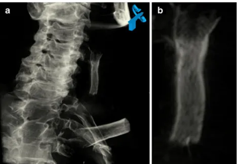
Fig. 2 a Pre-interventional follow-up magnetic resonance angiography showing an asymptomatic high-grade in-stent stenosis of the left CCA. b Right lateral 3D reconstruc-tion of the DynaCT data set showing the deformity of the carotid stent. c Right lateral fluoroscopic image depicting the
fractured stent meshes (arrows)
of the proximal stent
cations. Applications include assessment for the extent of subarachnoid haemorrhage and the width of the ventricles [1] and immediate evaluation of coil and stent placement in the treatment of intracranial aneuryms [2–5]. DynaCT has been shown to be capable to visualise the struts of a stent to illustrate stent placement during remodelling of intracranial aneurysms [3,4].
Restenosis rates of carotid arteries after stenting are up to 12.8% at 6 months and 15.9% at 1 year [6]. Most restenoses after carotid stenting are due to intimal hyper-plasia and smooth muscle hypercellularity, where naviga-tion through the stenosis is not a problem and which can be treated by balloon angioplasty and/or re-stenting. Stent fracture significantly increases the likelihood of in-stent stenosis [7]. Stent fractures of carotid stents leading to
high-grade in-stent restenosis have been described by several authors [8–12]. Ling et al. [13] reported a prevalence of stent fractures of up to 29.2%. Restenosis occurred in 21% of those fractured stents with an average follow-up of 15 months. These cases might necessitate a high-quality imaging technique to more precisely delineate the location of the microwire across the previously deployed stent in cases of re-stenting. Navigation through a previously deployed stent may be associated with a more complicated procedure, especially, if the stent is dislodged, deformed or even fractured. The advantage of DynaCT is the ability to immediately confirm the position of the microwire during intervention and the short processing times in acquiring high quality images. Its application allows a controlled and safe re-stenting in these selected cases.
Fig. 3 a, b Axial and sagittal multiplanar DynaCT reconstructions illustrating the correct position of the guidewire within the stent not entering through stent meshes (arrow)
Fig. 4 a Post-interventional 3D reconstruction showing stents in the internal carotid artery. b Magnification of the stent-in-stent reconstruction
Conclusion
The use of DynaCT to facilitate the navigation through a previously deployed and fractured stent has been shown to be feasible. The procedure allows a controlled and safe re-stenting procedure minimising the risk of inadvertent navigation across the stent struts.
Conflict of interest statement We declare that we have no conflict of interest.
References
1. Doelken M, Struffert T, Richter G et al (2008) Flat-panel detector volumetric CT for visualization of subarachnoid hemorrhage and ventricles: preliminary results compared to conventional CT.
Neuroradiology 50:517–523
2. White PM, Gilmour JN, Weir NW, Innes B, Sellar RJ (2008) AngioCT in the management of neurointerventional patients: a prospective, consecutive series with associated dosimetry and resolution data. Neuroradiology 50:321–330
3. Richter G, Engelhorn T, Struffert T et al (2007) Flat panel detector angiographic CT for stent-assisted coil embolization of broad-based cerebral aneurysms. AJNR Am J Neuroradiol 28:1902–1908
4. Benndorf G, Claus B, Strother CM, Chang L, Klucznik RP (2006) Increased cell opening and prolapse of struts of a neuroform stent in curved vasculature: value of angiographic computed tomogra-phy: technical case report. Neurosurgery 58:ONS-E380
5. Jou LD, Mohamed A, Lee DH, Mawad ME (2007) 3D rotational digital subtraction angiography may underestimate intracranial aneurysms: findings from two basilar aneurysms. AJNR Am J Neuroradiol 28:1690–1692
6. Bussiere M, Pelz DM, Kalapos P et al (2008) Results using a self-expanding stent alone in the treatment of severe symptomatic
carotid bifurcation stenosis. J Neurosurg 109:454–460
7. Scheinert D, Scheinert S, Sax J et al (2005) Prevalence and clinical impact of stent fractures after femoropopliteal stenting. J
Am Coll Cardiol 45:312–315
8. de Vries JP, Meijer RW, van den Berg JC, Meijer JM, van de Pavoordt ED (2005) Stent fracture after endoluminal repair of a
carotid artery pseudoaneurysm. J Endovasc Ther 12:612–615
9. Valibhoy AR, Mwipatayi BP, Sieunarine K (2007) Fracture of a
carotid stent: an unexpected complication. J Vasc Surg 45:603–606
10. Diehm N, Baum S, Katzen BT, Kovacs M, Benenati J (2008) Fracture
of a carotid stent: a word of caution. J Vasc Interv Radiol 19:622–623
11. Surdell D, Shaibani A, Bendok B, Eskandari MK (2007) Fracture of a nitinol carotid artery stent that caused restenosis. J Vasc Interv Radiol 18:1297–1299
12. Habib N, Heyden JV (2009) Acute fracture of ACCULINK carotid
stent during post dilation. Catheter Cardiovasc Interv. doi:10.1002/
ccd.22132
13. Ling AJ, Mwipatayi P, Gandhi T, Sieunarine K (2008) Stenting for carotid artery stenosis: fractures, proposed etiology and the need
for surveillance. J Vasc Surg 47:1220–1226