Design and Fabrication of Sharp Bend Photonic Crystal Slab
Waveguides
by
Solomon Assefa Submitted to the
Department of Electrical Engineering and Computer Science and
Department of Physics
in Partial Fulfillment of the Requirements of the
Degree of Bachelor of Science in Electrical Engineering and Computer Science, Degree of Master of Engineering in Electrical Engineering and Computer Science
and
Degree of Bachelor of Science in Physics at the Massachusetts Institute of Technology
May 11, 2001
Copyright 2001 Solomon Assefa. All rights reserved.
BARKER The author hereby grants to M.I.T. permission to reproduce and
distribute publicly paper and electronic copies of this the'Sg- TiTfl; UTE and to grant others the right to do so.
Jul1 1 2001
/x/ LIBRARIES
Author..-#~
Solomon Assefa Department of Electrical Engineering and Computer Science
Department of Physics May 11, 2001 Certified by... . . ... ...
Leslie A. Kolodziejski Professor of E M 1 Engineering and Computer Science -hesis Sunervisor
Design and Fabrication of Sharp Bend Photonic Crystal Slab
Waveguides
by
Solomon Assefa
Submitted to the Department of Electrical Engineering and
and
Department of Physics
Computer Science
May 11, 2001
In Partial Fulfillment of the Requirements of the
Degree of Bachelor of Science in Electrical Engineering and Computer Science, Degree of Master of Engineering in Electrical Engineering and Computer Science
and
Degree of Bachelor of Science in Physics
Abstract
A periodic arrangement of atoms in a crystal presents a periodic potential for
elec-trons. The bragg diffraction of electrons from the atoms may lead to a band gap, which represents a range of frequencies the electrons cannot have in the crystal. An analogous effect takes place in photonic crystals, which are periodic arrangement of layers of dielec-tric media. Scattering at the interface of the different layers produces a band gap for pho-tons.
The current effort to make integrated optical chips requires guiding light around sharp corners with a radius of curvature on the order of a wavelength. Photonic crystals serve this purpose by providing efficient sharp bend waveguides. An array of defects in a photonic crystal localizes modes in the band gap. This effect is used to build linear and
Acknowledgement
I would like to thank my advisor Professor Leslie Kolodziejski for the help and
guidance she has given me. I have been working with Leslie since I joined her group as a U.R.O.P. student in my junior year. The collaborative effort and the good spirit she
pro-motes within her group and with other groups has contributed towards my education.
I would like to thank Dr. Gale Petrich for sharing with me his expertise in all
aspects of my research. He is an invaluable resource of information in the theory and fab-rication of III-V material systems.
I would like to thank Alexei Erchak for familiarizing me with all the labs at M.I.T.
and for helping me understand the different fabrication processes. I have learned a lot from the discussions that we have everyday. I would also like to thank group members Aleksandra Markina and Reginald Briant. Additional thanks are extended to Mark
Mon-dol, Jim Daley, and Prof. H. Smith.
Finally, my deepest gratitude goes to my parents, brothers, sisters and my friends who have encouraged me throughout my MIT experience. You have made my life very enjoyable.
Table of Contents
Chapter I
I. Introdution... 9
II. Maxwell's Equations in Dielectric Media...11
III. Methods of Computation...13
IV. Lattice Structure of Photonic Crystals... 16
V. Bloch States in Photonic Crystals... 20
VI. Symmetry of Modes in Photonic Crystals...23
VII. Photonic Band Gaps... 24
VIII. Defects in Photonic Crystals... 26
IX. Photonic Crystal Slab Waveguides... 28
Chapter II
D evice D esign... 32Chapter III
Fabrication I. Epitaxial Growth and E-Beam Evaporation... 38II. Direct Write Electron Beam Lithography...41
III. Photolithography...45
IV . E tching... 50
Chapter IV
Conclusion and Future Work I. Fabrication conclusion
a. Electron Beam Lithography and Photolithography...61
b. Deep Etch... 64 c. Process Flow... 65 II. Future Work
a. Lapping and Cleaving... 66 b. Testing PBG Devices... 66
List of Figures
Figure 1. Dielectric layers with discrete translational symmetry. This periodic
system has a lattice constant a and a repeated unit cell (the wire box) in the x-direction...17
Figure 2. a) real space square lattice structure b) reciprocal square lattice structure ... 19
Figure 3. Construction of the brillouin zone (shaded region) ... 23
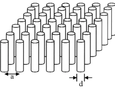
Figure 4. Two dimensional photonic crystal made of rods with lattice constant a and diameter d...24
Figure 5. Band diagram for the periodic structure given in Figure 1. I = 62 in (a); 6 1 = 12 and 62 = 13 in (b); 6 1 = 1 and 62 = 13 in (c)... 25
Figure 6. a) defect is created in a ID PBG by reducing the thickness of the middle stack b) defect is created in a 2D PBG by reducing the diameter of he middle dielectric cylinder...27
Figure 7. A straight waveguide made of a linear defect of rods whose radii is smaller than the radii of the surrounding array of PBG cylinders ... 30
Figure 8. A sharp 900 bend photonic crystal slab waveguide created using rods of smaller radii...31
Figure 9. Slab with a) uniform low-index dielectric background b) periodic low-index dielectric backgrou nd ... 32
Figure 10. Asymmetric slab with low index stilts and caps...34
Figure 11. Band diagram for the asymmetric structure design (shaded region indicates the light con e)...36
Figure 12. PBG test structures with varying number of columns a) 9x3 b) 9x5 c) 9x7 d) 9x9...37
Figure 13. Initial material system of PBG structure...39
Figure 14. SEM of material system showing the grown structure of AlGaAs, GaAs, and the evaporated Si0 2 which is etched to make a cross shaped alignment mark... 41
Figure 15. a) PMMA before exposure b) PMMA after exposure c,d) SEM of patterned PMMA with average hole size of 300 nm and input waveguide width of 1 pm...44
Figure 16. Sample a) after nickel evaporation b) after nickel liftoff... 45
Figure 17. SEM of a sample after UV exposure. Figure shows some of the mask patterns that have been transferred to the photoresist... 47
Figure 18. Sample a) after spinning photoresist b) after exposure and development...49
Figure 21. SEM of oxide mask after nickel wet etch (the nickel has not been completely lifted off the sid ew all)...5 2
Figure 22. a) sample after GaAs/AlGaAs etch b) cross section of (a)...53
Figure 23. GaAs/AlGaAs etch data in NSL... 55
Figure 24. GaAs/AlGaAs etch in EML... 58
Figure 25. Final profile of PBG structure... 60
Figure 26. Misaligned cross shaped alignment marks (left); the background cross is written by E-beam and covered with nickel while the cross on the foreground is unexposed photoresist. Alignment of 1 pm lines (right). The left section is exposed photoresist lines while the right section is E-beam written and nickel coated lines...61
Figure 27. Stitching error between two field written with two different beam currents (left) and stitch-ing error between two fields written with the same beam current (right)...63
Figure 28 a) 15 minutes straight etch of GaAs/AlGaAs layers (no interval)... 64
Figure 28 b) Interval Etch of GaAs/AlGaAs layers ... 65
Figure 29. Linear PBG waveguide...67
Figure 30. a) a total 1800 PBG bend with straight input and output couplers b) a 900 PBG bend with a curved output coupler...68
List of Tables
Table I. NSL Etch Results... 56
Chapter I
I. Introduction
A periodic arrangement of dielectric materials with different indices of refraction,
called a photonic crystal, creates a photonic band gap (PBG), that represents a frequency range where light is forbidden from propagating. Photonic crystals have led to new ways of controlling light. To this end, many groups are trying to fabricate three dimensional photonic crystals that have a complete band gap in all directions of propagation. For example, three dimensionally periodic structures can be made easily by using block copolymers', but these structures exhibit very small index contrast which results in a nar-row band gap. The necessary index contrast can be achieved by planar gnar-rowth or deposi-tion of a layered structure. However, the planar nature of the growth makes the
fabrication steps difficult because of the complex connectivity and alignment require-ments.2
Alternatively, photonic crystal slabs can be used to approximate the properties of three dimensional photonic crystals. Photonic crystal slabs are two-dimensionally (2D) periodic structures that have a band gap for propagation in a plane and also use index guiding to confine light in the third dimension. Photonic crystal slabs are different from
2D photonic crystals due to the presence of a light cone, which is the projection of
which no guided modes exist. However, there is still some radiation at those frequencies due to the light cone, which results from the higher effective index in the slab than in the background regions.
Photonic crystals have many applications in modem optoelectronics technology. Small scale optical signal processing requires waveguiding of an optical signal around sharp bends with a radius of curvature on the order of an optical wavelength. Conven-tional waveguiding is the result of total internal reflection at the interface between the high refractive index guiding layer and its low-index surroundings. However, waveguiding bends in the conventional index contrast waveguides may cause large optical losses depending on the radius of curvature of the bend.
Efficient waveguides with small radius of curvature are being created by introduc-ing a row of smaller diameter dielectric cylinders, called a line defect, into a photonic crystal composed of an array of cylindrical rods. The 2D array of dielectric rods, which surrounds the line defect, creates a PBG. Thus, an optical signal with a frequency in the PBG has its energy confined inside the line defect. The radius of the cylinders in the line
defect remains large enough to provide index guiding in the direction normal to the plane of periodicity of the crystal. Localization of the defect mode inside the line defect allows guiding around sharp corners including a 900 bend with low loss. This thesis concentrates on the design and fabrication of waveguides using photonic crystals.
II. Maxwell's Equations in Dielectric Media
The propagation of waves in dielectric media is governed by Maxwell's equations. These equations can be expressed as follows:
VeD(, t) = p (EQ 1) VOB(r, t) = 0
a-vxj ( , t)
= -(i?, t)
VxH(i, t) = aD(, t) + J(, t) (EQ 2) (EQ 3) (EQ 4)where P(, t) is the electric field strength, ft(i, t) is the magnetic field strength, D(Q, t)
is the electric displacement, and B(i, t) is the magnetic flux density.
Photonic crystal slabs are created using material that can be modeled as isotropic
and low loss, which implies D(i, t) = s(r)[E(i, t)], where s(r) is approximated as real and constant.3 We consider source free regions and let the permeability to be unity so that
B(-, t) = H(Q, t). Using these observations, Maxwell's equations can be rewritten as:
VxE(), t) t) (EQ 7)
at
VxH(I, t) = 6(r)-E(P, t) (EQ 8)
at
These equations are best solved by time-harmonic fields. These monochromatic waves or continuous waves (CW) are useful because (i) they can be easily used to elimi-nate the time dependence in the equations, (ii) they cover the whole spectrum of the elec-tromagnetic waves, (iii) Fourier analysis can be used to understand the time-domain phenomena, and (iv) any solution can be built by combining the time-harmonic solutions through Fourier analysis.4
The time harmonic fields with angular frequency o can be represented as
EQ, t) = Re{EQ)}e (EQ 9)
HQ, t) = Re{()}e (EQ 10)
Substituting these fields into EQ 7 and EQ 8 gives time independent equations
VxH(r, t) = -iwos(r)E(*) (EQ 11)
VxE(r, t) = ioH(?) (EQ 12)
By decoupling the two equations, the final expression in terms of H(Q) becomes
III. Methods of Computation
Light propagation and confinement in photonic crystals can be analyzed using a frequency-domain approach or a time-domain approach. In the frequency-domain
approach, Maxwell's equations are decoupled using time-harmonic modes. This approach is important in determining the band structure of photonic crystals by writing the fields as a linear combination of eigenmodes.
The detailed treatment of this approach, summarized below, is given in a paper by Whittaker.5 The approach uses the fact that s(r) is periodic, which lets us expand the transverse magnetic field into transverse plane waves summed over reciprocal lattice vec-tors G:
H(r, z) = jHk(G, z)ei(k + G) 14)
G
We also define a Fourier space vector h(z) = [Hk(G1, z), Hk(G2, z), ... ] . From the
Fourier expansion of the dielectric function, we define & . = i(G - G'), which is the
dielectric matrix. Moreover, we define the inverse of the dielectric function as
ri(r) = , I(r), which has a Fourier expansion fi~(G) and matrix representation rj
H(r, z) = ( G
+ ,(G) - (k,+ GY) J ei(k + G) -r + iqz
By defining vectors X = [,(G 1), *JG2), ... ]T and y = [4Y(Gj), OY(G2), ...] , we
can translate the above equation into a momentum representation as
h(z) = eiz4X+ Y (kq + ky4y)
(EQ 15)
(EQ 16)
where k, k are diagonal matrices with (kx)GG = (k, + Gx) and (ky)GG = (ky + G,).
The elements of the matrices are kx + Gx and ky+ GY, which are propagation vectors in
the plane of periodicity. By substituting the expression for the magnetic field into EQ 12, the expression for the electric field becomes
(EQ 17)
e(z) = leiz 2 + kykx)oy].
q
- [kxk,4, + (q +kxx)o,]y
+ q[ky 4X -kxo ] }
Substituting EQ 18 and EQ 19 into EQ 11 yields
2 ^ ^ 2 ^ ^
24, = [kylk,+ i(q + kxkx)]Ox+ [ikxky -kyijk] I
(2 ^ ^^ ^ 2 2^ ^ ^ ^ ^ ^^ [ kx,
= [k k,+(q x +kyky) ] O,+ [ ikykx - kxq ky IO
(EQ 18)
(EQ 19)
G) _ -(k, +G )A]
k+
xkx kx y + kky -kyi ._2x (EQ 20)
-L qkJ k yk_ _ -kxi ky kx
JkJLy
byThese eigenstates are used as basis states to construct the fields (EQ 18) in each layer of the structure. The field is expressed in a matrix as a combination of forward and backward propagating waves while the boundary conditions are set at the interfaces of the structure. Then, the matrix, call an S-matrix, is used to relate and determine the vectors and the amplitudes of forward and backward scattering waves in the different layers.5 Fast and optimized numerical simulations are used to solve Maxwell's equations and get band structures of photonic crystals in the frequency domain.6
Time-domain approach is useful in determining the transient properties and the quality factor of localized modes. A time domain finite difference method can be employed to study the evolution of fields in photonic crystals.7 For TM waves with the
electric field pointing in the z direction, the wave equation is written as
aE(x, y) + a E(x, y) = ,(x, y) E(x, y) (EQ 21)
8x 2y 2t
This can be changed into difference equations by discretization of space into units of As and time into units of At .8 Taking n to be time index, ij to be space indices, the field
E(iAs,jAs, nAt) is written as E ni,, and the discrete dielectric constant s(iAs,jAs, nAt)
n -2n+E n 7 n n El. + 1, 2E - + E + I - 2E + E (EQ 22) (As) 2 (As) E= , -2E + Ei (At)2
This equation is solved for E and used to update the previous time values. Since com-putation is done in finite-sized unit cells, information outside the cell is not available and the fields at the edges must be updated using boundary conditions, such as Mur's second order absorbing boundary condition in order to minimize back reflection into the cell.9 A detailed treatment of boundary conditions and the potential problems of the FDTD
approach is given by Winn et al.10
IV. Lattice Structure of Photonic Crystals
Photonic crystals have discrete translational symmetry. For example, a distributed Bragg reflector (DBR) which is constructed from alternating layers of two dielectric
mate-rials with different refractive indices and a layer thickness of as shown in Figure 1 is a
4
simple 1 dimensional (ID) photonic crystal. In two dimensional (2D) photonic crystals, the periodicity is along two axes and can consist of an array of high index columns sur-rounded by a low index material or an array of air holes in a high dielectric material.
(EQ 23)
where iii are the primitive lattice vectors.
7J
Unit cell
4-z
Figure 1. Dielectric layers with discrete translational symmetry. This periodic system has a lattice constant a and a repeated unit cell (the wire box) in the x-direction
The lattice vectors are defined as
a1 = ar,. (EQ 24)
The basis is the underlying pattern that is repeated with lattice constant a. The primitive cell is the smallest unit cell containing the basis that can be repeated. The one dimensional photonic crystal given in Figure 1 has a dielectric constant s,(?) such that
c(-) = ( + R), (EQ 25)
whereR = na.
R n I - + n2a2 +n3a3
y
Taking the Fourier transform of both sides of EQ 25 gives s(-) = _f (2 ) 3 + (=R)j. ( 2n 3 = ei R)f(i)ei~-k-f~ (2 7)3
From this, it follows that EQ 25 is true only if
e =1,
which is equivalent to saying,
Ij e Ri = 27c(l)
where 1 is an integer."
K. is called the reciprocal lattice vector, and is written in terms of primitive recip-rocal lattice vectors as
K = m1b1+ m2b2 + m3b3 (EQ 30)
with the condition that
ie bj = 27c8 i (EQ 31) (EQ 26) (EQ 27) (EQ 28) (EQ 29) s-+ Ri) =
a2x a3 By construction, b1 = 27c 2 3 , al * (a2 x a3) a3xa, b2 = 27c _ a] * (a 2 x a3)
a
xaE3
= 2 7 2 al *(a2 x a3)For orthogonal primitive lattice vectors, as in a 2D photonic crystal with a rectangular lat-tice of rods, the reciprocal latlat-tice vector becomes
- 27- 2r- 271 K = m-r + M2-r2 + M3-r3 (EQ 32) a , a2 a3 0 0 0 0 00@
@@0
0@0@
b, I*b
0 0 bFigure 2. a) real space square lattice structure b) reciprocal squre lattice structure
The reciprocal lattice can be constructed pictorially as well (Figure 2). Given the lattice of the structure, the basis can be identified. If the basis vectors are labelled as a j, then the
above equation tells us that the reciprocal lattice b is perpendicular to the lattice basis vectors where i wj.
V. Bloch States in Photonic Crystals
By making analogy to quantum mechanics, electromagnetism can be stated as an
eigenvalue problem in the frequency-domain approach. If the result of an operation on a function is just the function itself, multiplied by some constant, then the function is called an eigenfunction or eigenvector of that operator, and the multiplicative constant is called the eigenvalue.3 For the photonic crystals, the eigenfunction is the harmonic field H(r)
and eigenvalue would be o (EQ 13).
In quantum mechanics, a Hermitian operator Q acting on two field vectors A and B has the following property
(A, B) = (QA, B) (EQ 33)
where (A,B), the inner product of normalized vector fields A and B, is given as
(A, B) = JA* * B dr (EQ 34)
A Hermitian operator implies that the eigenfunctions are orthogonal and they have real
eigenvalues. From EQ 13, an electromagnetic operator which takes the curl of an eigen-function, divides the result by the dielectric constant, and takes the curl again is defined as
H = VX( V x4 (EQ 35)
s(r)
By making an analogy to the Hamiltonian operator in quantum mechanics, it can be shown that H is Hermitian as follows 3
(A, HB) = A* Vx (VxB dr (EQ 36)
=(VxA)* e 1(VxB) dr
= Vx(!VxA) B
= (HA, B)
Thus, since H is an eigenfunction of H , which is linear and Hermitian, H can be expressed as a linear combination of eigenmodes. For example, for two dimensional photonic crys-tals with k,, ky as the wavevectors in the direction of periodicity,
Hk, ky(r) = Ick xm(z)ckv,,(z) e i(kx +mbx)x i(ky+nby)y (EQ 37)
= e kyy kxxck c (z)e imb*x e inbyQ
m, n
= e ik~y ik uk (x,z)uk,(y, z)
where uk (x, z) = ZckM(z)eimbxx and uk (y, z) = L(z)einbyy
ck (z) and ck, (z) are calculated by using the frequency domain approach, which results in an expression like EQ 18. Using the concept of reciprocal lattice, it can be deduced that u(x + la, z) = u(x, z) and u(y + la, z) = u(y, z).
The above is an example of Bloch's theorem and u(x, z) is called a Bloch state.
The equation implies that the Bloch frequencies o (k,) and o (kx + are the same. It
follows from Bloch's theorem that E,(k) = k +
2m( . As a result, the extended
zone band diagram representation can be replaced with a reduced zone, having the range
-E < k < " , which is defined as the first brillouin zone (the edges are labelled by
2 2
F, M ,X. Bloch frequencies imply that at a given k in the first brillouin zone there are
bands of different energies E(k) that are labeled by the band index n. Because k repeats
itself outside the [- , ] range, in the reduced zone representation the energy bands fold 22
back into the Brillouin zone when they reach the zone edges. This is called band folding. The brillouin zone can be constructed pictorially from the reciprocal lattice. A basis is chosen as an origin and straight lines are drawn to the neighboring cells. Then,
perpendic-ular bisectors to these lines are drawn. As shown in Figure 3, the intersection of these per-pendicular lines forms the first brillouin zone.
Figure 3. Construction of the brillouin zone (shaded rectangular region)
VI. Symmetry of Modes in Photonic Crystals
Electromagnetic modes in photonic crystals can be often characterized by their mirror symmetry. Any mode y can be characterized by its mirror symmetry. For a mir-ror reflection M, the mode is defined as odd if My = -y and even if My = y .
For the two dimensional photonic crystal, the plane of periodicity is the x-y plane, and the axis of symmetry is the z-axis. The x-y mirror plane, called M,, transforms z into
-z. If k is parallel to the plane Mz, reflection through the plane gives back the wavevec-tor itself. This mirror is used to classify the modes as TE and TM.3 For TE modes,
k - - = 0, and for TM modes, RI - Z = 0.
V11. Photonic Band Gaps
A photonic band gap exists when there is a frequency gap between the minimum
point of the upper energy band and maximum point of the lower energy band; no propaga-tion modes exist in the gap regardless of the k values. The degeneracy at the symmetry points F, M, X is broken and the density of optical states in the gap is zero (p(o) = 0).
d
Figure 4. Two dimensional photonic crystal made of rods with lattice constant a and diameter d
For an infinite stack of dielectric layers with all of the layers having the same dielectric constant, the dispersion relationship gives two straight lines with slope
AO -
Il
in the extended zone representation. In the reduced zone, the folded bands Akmeet either at the zone center or the zone edges. As a result, there is no band gap with a range of forbidden frequency for photons.
The effect of index contrast on band gaps can be seen from the electromagnetic variation theorem. The functional energy Ef is given as3
E 1(H, HH)
2 (H,H) (EQ 38)
By considering the effect of a small perturbation, we finally find that
E$H)) - D,
2(H, O c (EQ 39)
where (H,H) is the inner product of the given fields. This is worked out in detail in refer-ence 3. The equation gives the dependrefer-ence of the energy bands on the index (n = cc taking permeability p to be unity). As E increases E/H) decreases, and as c decreases
E$H) increases. 0.25 0 0.20 0.Q -.05 0. -0.5 -0.25 0 0.25 0.5 (a) so 25 20 15 10 -0. - W L5 -to -d M5 0.25 0 0.25 0.5 (b) us 3.20 3.15 X.10 Ws0 -025 -0.20 -0.15 -0IO
Figure 5. Band diagram for the periodic structure given in Figure 1. 6 I = F2 in (a); 6 1 = 12 and
62 = 13 in (b); , I = 1 and 62 = 13 in (c) 0,30 0.25 020 0.15 0.10 0.5 I 4- w .AI -05 -025 0 Q25 (c)
tric layer. While a uniform dielectric layer does not have a band gap (Figure 5a), a dielec-tric composed of alternating layers with 6 = 12 and 6 = 13 results in the formation of a
small gap (Figure 5b). Finally, when the contrast is increased from s = 1 to c = 13, a
larger gap is formed between the bands as in Figure 5 c.
The existence of a gap means that photons with a frequency in the gap will not be able to propagate in the plane of periodicity. The modes that have gap frequency become evanescent, meaning that their wave vector becomes imaginary in the direction of period-icity. As a result, the transmitted Poynting vector ( &t ) becomes complex and the real part in the direction of propagation becomes zero, which implies the time-average power trans-mitted in that direction, (&,) , is zero.
VIII. Defects in Photonic Crystals
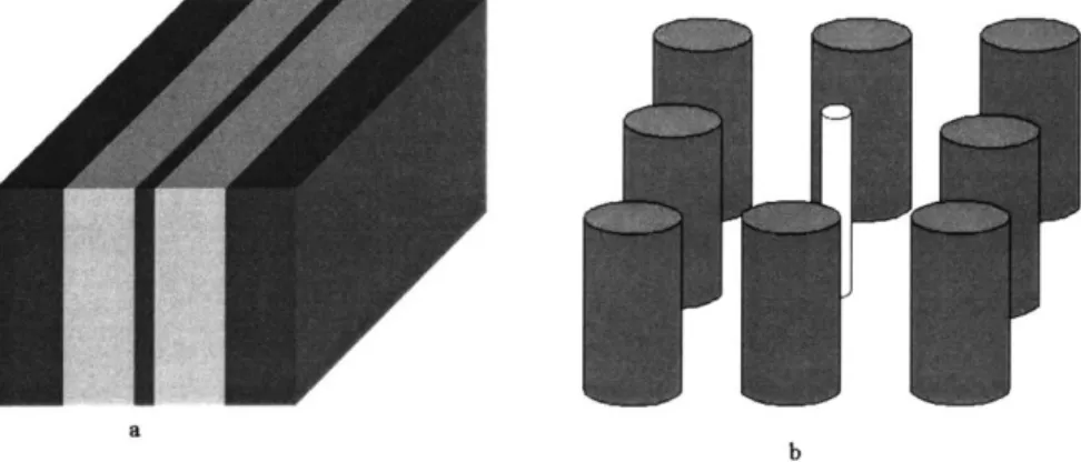
A defect is created in a one dimensional photonic crystal when the thickness of one
of the alternating stacks of dielectric layers is either increased or decreased, breaking the translational symmetry of the crystal as shown in Figure 6. The defect causes a mode, called a defect mode, to exist in the band gap.
a
b
Figure 6. a) defect is created in a ID PBG by reducing the thickness of the middle stack
b) defect is created in a 2D PBG by reducing the diameter of he middle dielectric cylinder
For a 2D array of cylinders, a single defect is produced by increasing or decreasing the radius of one of the rods. The 2D array of periodic dielectric rods, which surrounds the defect, creates a PBG in which light within that range of frequencies cannot propagate. A mode with a frequency in the PBG becomes localized at the defect and decays exponen-tially away from the defect. It cannot couple to the nearby rods because the defect mode has a frequency inside the PBG. The defect mode energy increases as the refractive index of the defect decreases; modes get pushed up into the gap from the lower band. The energy decreases as the index of the defect increases; modes get pushed down from the upper band. These modes are calculated by using the supercell approximation method, in which a supercell consisting of a crystal with a defect is repeated periodically in space.7
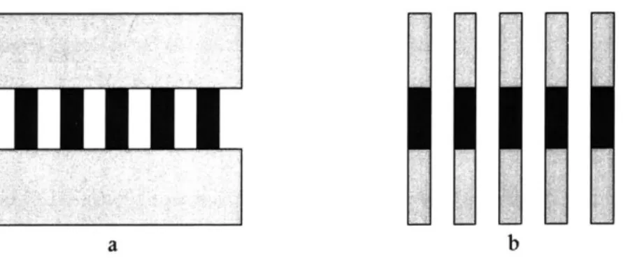
IX. Photonic Crystal Slab Waveguides
In two dimensions, a waveguide can be created from a linear array of defects. Increasing or decreasing the radius of one of the rows or columns of the periodic square lattice or rods produces a linear defect. The 2D array of periodic rods, which surrounds the line defect, creates a PBG. The mode that couples to the defects has a frequency that is
inside the PBG, which means the mode is forbidden from propagating anywhere else in the structure. As a result, the defect mode will not be able to couple to the surrounding rods. Instead, light will be forced to couple from one defect rod to the next defect rod which supports the same mode. Thus, the measured loss in this linear waveguide is prima-rily due to input and output coupling to the photonic crystal.
Photonic crystal slabs are 2D periodic structures that have a finite height. Guiding in the plane of propagation is due to the photonic crystal, while in the third dimension light is confined due to index contrast between the slab and the surrounding media. Hence, photonic crystal slabs are different from the ideal 2D photonic crystals discussed above, which are modeled with infinite height. The introduction of the light cone (a continuous region indicating all possible frequencies of the bulk background12) results from the index confinement in the z-direction; as a result, the presence of radiation modes prevents the band gap from being complete. Furthermore, the modes are classified as TE-like (even) and TM-like (odd) modes. The symmetry of the structure is broken due to the presence of the substrate (background region), which prevents the modes from being fully TE or TM. The thickness of the slab can be approximated as follows 12
h ~ I ,(EQ 40)
2
gap -bottom
(
where ogap - bottom is the frequency of the band at the bottom of the gap in units of c/a,
E is the effective dielectric constant, and h is the height in units of a. EQ 40 simply implies that using a wavelength as a lengthscale of variation in field amplitude, the opti-mal thickness will be on the order of gap-bottom wavelength.12 Photonic slab waveguides produce a practical way of creating three dimensional confinement, since the vertical index contrast can be achieved through planar growth and oxidation.
Photonic crystal slabs present a better alternative to conventional waveguiding, which is the result of total internal reflection at the interface between the high refractive index guiding layer and its low-index surrounding. These waveguides can be either sin-gle-mode or multi-mode. Waveguide bends in the conventional index-contrast
waveguides may cause large optical losses depending on the radius of curvature of the bend relative to the wavelength of the guided light. The loss that is incurred at bends due to coupling to radiation modes is dependent on the radius of curvature and the index con-trast of the material and the wavelength of the light being used.
In a photonic crystal slab, an efficient waveguide can be created by introducing a linear defect along one of the rows. The periodic structure creates a PBG for TM-like modes which prohibits the defect mode from escaping into the plane of the crystal. In the third dimension, the mode is index-confined and the size of the defects can be chosen to place the guided modes below the light line to prevent the coupling to radiation modes.
heavily localized in the defects. Furthermore, the TM-like modes are localized around the rod in air, which implies guiding in a low-index material.
Figure 7. A straight waveguide made of a linear defect of rods whose radii is smaller than the radii of the surrounding array of PBG cylinders
Sharp 900 bends in the waveguides can be constructed in photonic crystal slabs as shown in Figure 8. There will be high transmission through the bend, because the defect mode does not couple to any of the other guided modes and is also confined vertically. As a result, photonic crystal slabs provide efficient waveguides. Again, loss comes from input and output coupling to the photonic crystal region. The transmission can be quan-tized by normalizing the measured transmission of the bend waveguide to the measured transmission of the linear waveguide.
A good model for sharp bends in photonic crystals is given by Mekis.13 A sharp
con-for the (10) and (01) directions and as k2(w) in the (11) direction. The transmission
then can be modeled as a one dimensional scattering process. By using continuity of the electromagnetic field and an analogy of the Schr6 dinger equation, the derived reflection coefficient becomes
I 2k,(w)k2() 2--1
[k,2 (w)-k22(w)]sin[k2 (w)L
For a 900 bend, the effective length of the corner is extrapolated to be L = 0.33 2a; for
guided modes with frequency below o = 0.403 x 2c , the simulated transmission is
a
greater than 95%. For conventional dielectric guides that have index contrast of 3.5 to 1 with the surrounding, large radiation losses at the corner of a similar sharp 900 bend results in a power transmission of at most 30%.13
Chapter II
Device Design
In order to design the PBG waveguides, various structures were considered and simulated. The determining factors in choosing the final design were solid performance and amenability to fabrication. First, the design needs to have a band gap centered around a frequency of 0.323 (c/a) where a is 500 nm so that the device can operate at a wave-length of 1.55 pm; it also requires good localization of guided modes and a minimum mix-ing between the even and odd modes. Second, the design has to be feasible under the constraints of material systems and the limitation of current fabrication technology.
The first design considered was a slab of periodic rods sandwiched between uni-form dielectric substrate as shown in Figure 9 a. The substrate which occupies the back-ground regions above and below the array of rods has an index of refraction which is lower than the effective index of the slab.
1~
a V
If the substrate is several wavelengths thick, it can be considered to have infinite
thickness because the intensity of the guided modes dies off exponentially outside of the substrate. This index contrast between the slab and the substrate provides confinement in the vertical direction. Even though the modes can be classified as even or odd, they are somewhat less localized due to the smaller index contrast between the substrate and the slab.2 The major disadvantage of this design is the difficulty of fabrication. If all the lay-ers are grown first, then it will be difficult to make the rods by processing only the middle layer. Also, one might consider growing the lower substrate and the guiding layer, then doing the processing, and then growing or bonding the top substrate. Due to fabrication problems, the size scale of the rods and the size of the 2D array that is used here does not make the rods capable of supporting a thick substrate.
The second design considered was similar to the first, but with the background substrates patterned as shown in Figure 9b. The design leaves each rod with a dielectric layer above and below, which makes the structure more symmetric. The patterning of the background gives an effective index which is smaller than the substrate index in the first design, and leads to more confinement of the guided modes. Nevertheless, this is difficult to fabricate because of the nature of the material system being used despite the less com-plicated etching step. AlGasAs can be used as a low-index material below and above the guiding layer of GaAs. However, the top layer of AlGaAs which can be grown epitaxially will eventually oxidize when exposed to air during processing.
on top of them, which roughly restores mirror symmetry back to the system. The mirror symmetry determines which modes (even or odd) form the PBG so that light can be polar-ized as TE or TM during testing. In order to classify the guided modes as even or odd, it is only necessary to preserve mirror symmetry where the guided modes have non-negligible amplitude.2
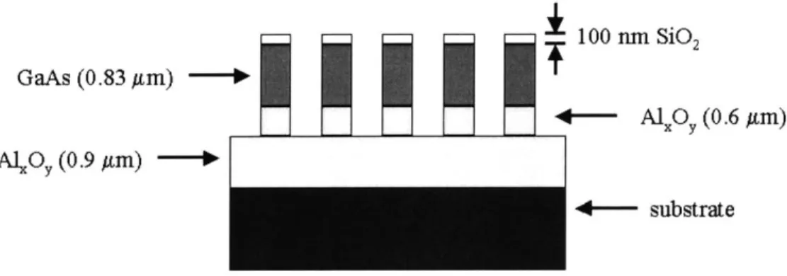
100 n Si02
GaAs (0.83 Am) f
4-- A(0m (0.6 Am)
-
4-- substrateFigure 10. Asymmetric slab with low index stilts and caps
The thickness of the slab was also a factor in the design of the structure. If the slab is too thick, then higher-order modes will be created close to the lowest-order mode which will cause the gap to disappear. If the slab is thicker than a wavelength, higher order modes are created by forming horizontal nodal planes. On the other hand, making the slab very thin will cause the modes to be weakly guided and very close to the light cone. If the slab thickness less than half a wavelength (size of the lowest order mode), the confinement of the mode is very small. It is postulated that the optimal thickness is half the two-dimen-sional gap-bottom wavelength, because the wavelength is the lengthscale for variations in field amplitude at the given frequency (EQ 40).2
For this design, simulations were done using lattice constant (a) values that range from 450 nm to 650 nm; the simulated heights varied from 1.36a to 1.66a while the diam-eter d varied from 0.4a to 0.6a. Both symmetric (GaAs rods with SiO2 caps and stilts, or
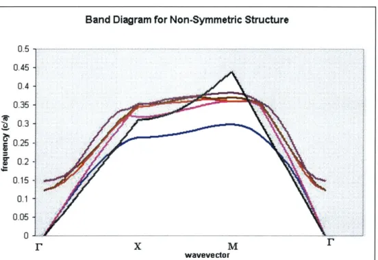
AlXOY caps and stilts) and asymmetric (GaAs rods with SiO2 caps and AlxOY stilts) were simulated. From simulations of slab thickness and gap size, the optimal thickness for the structure was chosen to be 0.83 pim. Given that light with wavelength between 1390 nm and 1680 nm can be used in the testing setup and given that the thickness will be 0.83 pm, the best structures were determined to have a lattice constant of 500 nm with rod diameter of 300 nm. Using these values, another simulation was performed on the following struc-ture: 100 nm thick SiO2 cap, 0.83 ptm thick GaAs guiding layer, 0.6 tm thick AlxOY stilts,
0.9 pm thick AlxOy layer, on a GaAs substrate. Figure 11 shows the calculated band
structure. Even though the structure is asymmetric, the index of refraction of the cap and the stilts are very close (1.5 for SiO2 and 1.6 for AlxOy). As a result, the extent of mirror symmetry increases and the guided modes can be classified as even and odd. The gap to midgap ratio for the odd modes for the above structure is found to be approximately 10%.
Figure 11. Band diagram for the asymmetric structure design (shaded region indicates the light cone)
Due to the small size, it is quite difficult to fabricate rods with an exact diameter of
300 nm. In order to be able to account for these errors and investigate the band edges, the
rods were designed with diameter within the range of 0.7d and 1.3d, where d is 300 nm. Furthermore, the design varied the number of columns from one structure to another, so that it would be possible to see the increasing width of the photonic bandgap as the num-ber of columns increases. As a method for normalization of data, straight dielectric waveguides were also included in the designed structures (Figure 12).
Band Diagram for Non-Symmetric Structure
0.45 0.4
I
0.3 0.25 0.2 0. X M wavevectorGoo 000 00* seeo 000000 600060 0000000 0000000 6666006 0000000 0000000M 0000000 0000000 Geee Goose .... Gees 00000 *neeeeeee
Chapter III
I. Epitaxial Growth and E-Beam Evaporation
The first part of the process involves the use of Gas Source Molecular Bean Epit-axy (GSMBE). This growth technique, which can deposit layers with atomic resolution", is done in an ultra high vacuum (UHV) environment (10-10 torr base vacuum). The basic requirements of a GSMBE system are in situ sample heating and cleaning, and
indepen-dently controlled sources for all constituent materials and dopants. High vacuum is achieved by baking and pumping the system at temperature ranging from 150 to 2500C,
which will increase the vapor pressure of gases and water adsorbed on the inside walls of the chamber. Most Solid Source MBEs use ion pumps or adsorption cryopumps, while GSMBEs use turbomolecular pumps.
Sample growth is performed at the CBE laboratory using the Riber 32P system. First, the sample is loaded into the Intro chamber, which is the load lock for the system and has a base pressure of 10-8 torr. Then, the sample is moved into the Transfer chamber, which has a base pressure of 9x 10- 10 torr, and degassed at 21 00C for one hour to remove
the absorbed moisture on the sample and the sample holder. The sample is then moved into a Buffer chamber that lies between the Transfer chamber and growth reactor. The base pressure of the Buffer chamber is at 5x10-O torr. Finally, the sample is moved into the Riber 32P growth chamber, which has a base pressure of 2x10⁻¹⁰ torr. The chamber contains elemental Ga, In, Al, Si and Be effusion cells and a AsH3and PH3cracker.
Dur-ing growth, the pressure increases to 2x 10⁻⁵ due to the presence of cracked or thermally decomposed (at 9000C) AsH3 and PH3. For a GaAs substrate, the sample is heated at
6000C, Ga source at
1000C,
In source at 8000C, and Al source at 11200C. Also, theRiber system is equipped with a Reflection High Energy Electron Diffraction (RHEED) which is used to characterize deposition rates and sample surface morphology, and an in situ spectroscopic ellipsometer.15
After the 300 pm thick p-type <100> GaAs substrate is baked and transferred into the growth chamber, a 1.5 pm thick AlO.9GaO.jAs layer is epitaxially grown. GaAs and AlO.9GaO. As are lattice matched, which means that they have the same lattice constant of
0.565 nm. As a result, stress/strain is not a problem in the growth. The composition can be varied by changing the AlGaAs cell temperature, which changes the flux ratio of the two types of atoms that arrive at the surface. By varying composition of Al and Ga, the index, electronic band gap, and some chemical properties can be varied. Next, a 0.85 pm thick GaAs layer is grown epitaxially on the AlO.9GaO. IAs.
Si02 (1 00n)
GaAs (0.86um)
GaAs
When the growth is finished, the sample is transferred to the Intro chamber which is then vented. A piece of the sample is cleaved and the thickness of the epitaxial growth is verified using x-ray diffraction. Then, a 100 nm thick layer of SiO2 is evaporated on the sample using an ebeam evaporator. The advantage of using an ebeam evaporator over be inductive or resistive evaporators is that it avoids contamination from the heating up of the crucible and the outgassing of coupling wires.'4 In an E-beam evaporator, a tungsten
fila-ment is heated, which makes electrons boil off with high flux. The filafila-ment is put far way from the sample to avoid contamination. The high flux electron beam passes through deflection plates and a bending magnet , which bends it 2700 and makes it incident onto the surface of the charge. As a result, the charge in the crucible melts and becomes evap-orated on the sample. To deposit oxide on the grown sample, an SiO2 target is put into the crucible of the evaporator, which is then put under vacuum.
The thickness of the evaporated oxide and its refractive index was checked using ellipsometry, in which a circularly polarized beam of light is reflected off the oxide sur-face at an angle and the resulting change in polarization is measured. Because different thicknesses can produce same intensity change, the measurements are done at different incidence angles and the incident and reflected intensities are compared. Ellipsometry on the evaporated sample piece gave a thickness of 96.7 nm.
100 m S1 2
GaAs
AIGnAs
GaAs Substrte
Figure 14. SEM of material system showing the grown structure of AlGaAs, GaAs, and the evaporated SiO2 which is etched to make a cross shaped alignment mark
II. Direct Write Electron Beam Lithography
Direct Write Electron Beam Lithography is used to define the square lattice of dielectric rods. In electron beam lithography systems, electrons are emitted from the cath-ode of an electron gun through thermionic emission, aided emission, thermal field-aided emission, or photoemission. The electrons are focussed into a narrow beam through the use of lens, deflection plates, and aperture arrangements. The direct write lithography can be done using a vector scan or raster scan system. In a vector scan system, the digital location of each area to be exposed is fed to x and y digital to analog converters causing
the beam is directed to those pixels that must be exposed. In raster scan systems, every pixel is scanned serially and a pattern is exposed by opening and closing a shutter."
Limitations on the smallest feature written by an E-beam lithography system arises from the size of the focussed beam as well as proximity effects. When a pattern is being written, forward and backscattered electrons can expose nearby areas that are not intended
for exposure. The beam becomes broadened because of forward scattering, which occurs over a small range of angles with respect to the incident velocity. Backscattering results in a large area fog of exposure.16 A disadvantage of E-beam lithography systems is their
small throughput due to the significantly longer write-time compared to contact or prox-imity lithography.
In this project, E-beam lithography is used to make these structures because optical lithography will not be able to properly produce features that are smaller than the wave-length of light. E-beam lithography was done by using an IBM VS2A Scanning Electron Beam Lithography (SEBL) system, which has 50 kV external potential applied to acceler-ate electrons from the source. This vector scan system is capable of having a beam spot size as small as 60 nm. To perform a write, the system breaks the pattern into fields as small as 100 ptm, which are exposed individually and stitched together. In order to write patterns within a field, electric and magnetic fields deflect the beam to the pixels that need to be exposed. To move from one field to another, the stage is physically moved under a laser interferometer control.
which is the incremental step of the electron gun. A field size of 250 pm is used, which gives a pixel size of 15 nm (214 pixels in a field). The patterned area of the file correspond to the area where the direct E-beam write takes place. From simulation, the optimal diam-eter of each rod is ddiam-etermined to be 300 nm. Nevertheless, E-beam write doesn't exactly simulate the pattern as designed with CAD due to proximity effects (forward and back-scattering of electrons) as well as the error introduced during the movement of the beam and the stage. To account for these effects, the patterns are programmed to be about 60 nm smaller than the desired size. Thus, in order to account for discrepancies as well as to cover a test frequency range, patterns with diameter ranging from 270 nm to 330 nm are designed.
Before the E-beam lithography exposure, each sample is coated with a 200 nm thick layer of polymethylmethacrylate (PMMA). The coating is done by spinning a drop-let of PMMA (solution of 3% by value in chlorobenzene) on the sample at 3600 rpm for one minute and baking it for one hour at 180C. Next, a thin 5 nm layer of chrome is evaporated on the sample. This conductive layer eliminates sample charging, which can deflect the beam, thus resulting in pattern elongation. The design parameters are made as accurate as possible through dose experiments, which include varying beam current, field size, and intensity values. A dose of 537 pC/cm2, current of 50 pA, and clock frequency of 0.04 MHz produced holes with the desired diameter.
Then, the chrome is etched off the written sample by submerging it in a Cr-7 pho-tomask remover solution for 15 seconds. The exposed PMMA is developed using 90
sec-PMMA layer and leave behind patterned holes; this is followed by a 30 second rinse in isopropanol (IPA). Finally, the sample is inspected using a Scanning Electron Microscope
(SEM) to determine the size of the holes (Figure 15).
/ PMMA (a) Exposed PMMA 77m77-77M 0111011I B11tm (b) (c) (d)
Figure 15. a) PMMA before exposure b) PMMA after exposure
III. Photolithography
After the PMMA is developed, a 30 nm thick nickel layer is evaporated on the samples. This process is necessary to provide a metal mask for the oxide etch that follow. The nickel evaporation leaves the sample coated with nickel, and a liftoff process strips off the PMMA with the nickel that is on top it (Figure 16 a). To perform the liftoff, 1-methyl-2-Pyrrolidinone (NMP) solution is heated up to 80*C in a beaker. The sample is then put into the beaker and touched carefully with smooth swaps to enhance the nickel removal. To ensure a successful liftoff process, the beaker was placed in an ultrasonic bath for 30 minutes. Finally, the sample is rinsed with acetone and methanol, and dried with a nitrogen gun. This process leaves behind a 2D array of 30 nm thick nickel
cylin-ders, and nickel waveguides that are 120 tm long, 1 ptm wide, and 30 nm thick (Figure
16b ).
Nickel Nick
a b
Figure 16. Sample a) after nickel evaporation b) after nickel liftoff
E-beam takes a very long time. Photolithography, which is a faster process with higher throughput, can be used to extend the waveguides longer. Photolithography consists of two steps. First, the patterns are created with the desired features using chrome on a quartz plate. The second step is a chemical process, in which a sample is coated with pho-toresist, exposed with ultraviolet (UV) light, and then developed with an alkaline solution (for positive resist).
Photoresist is a mixture of a photoactive compound (PAC), a matrix material, and a solvent. The matrix material, which is a thick resin of a long chain polymer, acts as the base material. The solvent helps control the mechanical properties such as viscosity. The
PAC, which usually is diazoquinone (DQ) in positive resist, acts as an inhibitor before
exposure. The PAC and the matrix material are chemically bonded at the surface of the resist. During exposure, the weakly bonded N2 in the PAC is liberated. Then, a Wolff
rearrangement takes place to stabilize the structure and forms ketene. When the solution reacts with water, it is converted into carboxylic acid. During the reaction with a base solution, the carboxylic acid breaks into water soluble amines." Thus, the exposed region is removed.
Figure 17. SEM of a sample after UV exposure. Figure shows some of the mask patterns that have been transferred to the photoresist.
Waveguide patterns with width ranging from 1 pm to 3 pm are designed on a
5-inch mask (Figure 17). Some of the device have one input and two outputs. Some of the waveguides are straight and some are curved to couple out light from the sharp bends. Due to the various designs and the small feature patterns, it was easier to make a dark field mask written by E-beam.
The samples with the nickel patterns are put in a hexamethyldisilazane (HMDS) oven. This process helps to promote adhesion of photoresist to the sample. Next, a drop of Shipley 1813 positive photoresist is spun on the sample at 4000 rpm for 30 seconds, which results in a layer that is approximately 1.3 pm thick (Figure 18 a). Then, the sample
is prebaked for 30 minutes to harden the resist.
The box shaped nickel alignment marks, which were written by E-beam with the PBG crystal, are visible through the resist layer. The alignment marks on the mask are crosses, whose size and position were designed in such a way that the box marks on the sample would fit around them. The cross-box patterns vary from 6 pm to 24 pm in size, which improves the focussing during the alignment process. By aligning the crosses with the boxes on the samples, the nickel coated waveguides are aligned with the waveguide patterns on the mask. This alignment process is difficult due to the resolution limit of the aligner. However, it is a crucial step because coupling loss, which would result from inac-curate alignment, has to be minimized.
Next, the resist is exposed to UV light through the clear part of the mask. Using vacuum contact, a 50 second exposure at 6 mW of power yields a patterns that are not over or under exposed. The samples are then developed in a Shipley MF-319 solution, which removes the exposed regions of the photoresist (Figure 18 b). One minute of development and a one minute rinse in deionized water results in a sample that is not over or under-developed.
After the samples are examined using an optical microscope, another nickel evap-oration and liftoff is performed to coat the newly defined input and output waveguides. The liftoff process leaves behind long waveguides and a 2D PBG crystal coated with nickel (Figure 19).
Photoresist
Figure 18. Sample a) after spinning photoresist b) after exposure and development
Nicke
(b)
(a)
(C)
IV. Etching
Material systems can be patterned and etched by using wet or dry etch mecha-nisms. Wet etch is a purely chemical process that usually consists of three process. First, the etchant species move to the surface of the sample. Second, a chemical reaction with the part of the sample that is not covered with a mask produces soluble byproducts. Third, these products move away from the surface of the sample. In order to facilitate etching, the solution is usually heated and stirred. Wet etch results in isotropic sidewalls.
Dry etches, which produce anisotropy and high selectivity, can be done in a reac-tive ion etcher (RIE) or a high density plasma etcher. RIEs are capable of generating ion density of 10 9 cm 3. In conventional parallel plate RIE systems, radio frequency power is
put into a thin sheath region around the electrode by capacitive coupling. A plasma starts as the radio frequency power accelerates electrons in the vertical direction. The electrons collide with gas molecules and cause electron-impact dissociation and excitation which
sustains the plasma. As a result, the sustained plasma will have many positive ions, few negative ions, radicals, and some neutral atoms. Because the electrons have lighter mass, they are able to move a larger distance compared to the ions. During the process, the elec-trons collide with the electrode and the sidewall, thus inducing a self-bias. This bias causes an electric field which forces many of the ions to accelerate and bombard the sam-ple. In RIEs, the ion density is coupled with the ion energy; to achieve a higher density of ions, higher RF power is required to ionize more molecules. However, the higher power causes a larger electric field and ion bombardment can damage the sample.
Advanced plasma etching techniques generate higher ion density by separating the power from the ion density. Electron cyclotron resonance (ECR) etchers use an external magnetic field and a resonance effect to confine the electrons. Resonance occurs in the systems when the applied microwave frequency is equivalent to the orbital frequency of electrons under an external magnetic field. The resonance accelerates the electrons and increases their mean free path before they collide with the electrode. As the electrons travel a longer distance, they are able to dissociate and excite more molecules, which pro-duces a higher plasma density (1012 cm-3). The ion energy is controlled separately by
power applied to the electrode. Another advanced technique is inductively coupled plasma (ICP) etching, in which a RF current circulating around the chamber in opposite directions causes an alternating magnetic field in the vertical direction. This induces a RF electric field, which accelerates electrons in a circular path and confines them in a circular motion. The circular motion of the electrons give them a longer mean free path and mini-mizes their collision with the electrode and the sidewall. The energy of the ions is con-trolled by applying another RF source to the electrode, which helps to reduce ion bombardment of the sample.
In the PBG fabrication process, the 100 nm thick SiO2 layer which has patterned nickel mask is etched first. Then, the nickel mask is removed using a wet etching process and the patterned oxide is used as a mask to etch the GaAs/AlGaAs layers.
A Fluorine containing gas is required to etch Si and SiO2. The etch of the SiO2 mask is done in the NanoStructures Lab (NSL) using a PlasmaTherm RIE, which has
Before the etch, the chamber is cleaned using 4 sccm of CF4 and 20 sccm of 02, striking the plasma at 25 mT pressure with 350 V DC bias. The etching is performed at 10 mT pressure, 300 V DC bias using 15 sccm of CHF3 (Figure 20 and 21). The etch rate was
measured to be 17 nm per minute.
Nickel Oidde Nickel Oxide (b) Oxide
z
Oxide (d) (c)Figure 20. a) sample after oxide etch b) cross section of (a) c) sample after nickel wet etch d) cross section of (c)
Figure 21. SEM of oxide mask after nickel wet etch (the nickel has not been completely lifted off the side-wall)
(b)
(a)
Figure 22. a) sample after GaAs/AlGaAs etch b) crossection of (a)
After the etch of the oxide mask, the nickel mask is removed using a wet etchant (Figure 20 and 21) which contains nitric acid. The next step in the process is to etch the GaAs and AlGaAs layers using the oxide mask (Figure 22) to a depth of 1.5 tm. The first etch experiment was done in the NanoStructures Lab (NSL) using a parallel plate RIE which has a chlorine (Cl2) line. The first etch was done at 50 W (resultant DC bias of 100
V), 20 mT pressure, and 40 sccm of C12 for 10 minutes. Deposition on the edges of the
mask was observed and the trench surface was not very smooth (Figure 23 a). In addition, the etch was not isotropic and underetching had taken place.
In the next trial, the pressure was reduced to 5 mT. This resulted in the destruction of the oxide mask (Figure 23 b). Electrons had a higher mean free path due to the pres-ence of fewer molecules in the chamber (thus less collision); they were able to directly couple to the electrode and cause a higher DC bias. As a result, the ionized molecules are accelerated with high energy and bombarded the sample.
The addition of He to reduce the chlorine partial pressure did not help the process (Figure 23 c). Even though it helped the mask to hold up, the etch of GaAs was uneven. This was because at a very low pressure, the etch limiting step is the adsorption and chem-ical reaction, which is the slowest step due to the small concentration of reactants. The ionized Cl atoms are transported away from the sample before there was enough time for them to arrive at and to react with the surface of the GaAs sample; since the Cl atoms are larger, they are easily pumped out when the system tries to maintain the low pressure. Instead, the smaller He atoms are accelerated to the sample. The mask was not damaged because the lighter (yet many) He atoms and the larger (yet fewer) Cl ions did not cause much physical etch.
In the next trial, the He was removed and the average pressure was increased to 13 mT (Figure 23 d). Even though the mask held up, polymer was deposited at the edge of the mask. Furthermore, the inside of the trench was very rough and full of "grass". Reducing the flow to 25 sccm and the pressure to 5 mT did not impact the etch (Figure 23e). In the trenches, chemical etching was insufficient while the high DC bias (1 15V) caused a stronger physical etch.
ab
C
e
f 2