HAL Id: hal-02796259
https://hal.inrae.fr/hal-02796259
Submitted on 5 Jun 2020HAL is a multi-disciplinary open access archive for the deposit and dissemination of sci-entific research documents, whether they are pub-lished or not. The documents may come from teaching and research institutions in France or abroad, or from public or private research centers.
L’archive ouverte pluridisciplinaire HAL, est destinée au dépôt et à la diffusion de documents scientifiques de niveau recherche, publiés ou non, émanant des établissements d’enseignement et de recherche français ou étrangers, des laboratoires publics ou privés.
probiotic properties
Muriel Thomas, Sylvie Miquel, Rebeca Martín, Luis Bermudez Humaran,
Philippe Langella
To cite this version:
Muriel Thomas, Sylvie Miquel, Rebeca Martín, Luis Bermudez Humaran, Philippe Langella. The indigenous microbiota and its potential to exhibit probiotic properties. Probiotics and Prebiotics : Current Research and Future Trends, Caister Academic Press, 508 p., 2015, 978-1-910190-09-8. �hal-02796259�
12
The Indigenous Microbiota and its Potential to
Exhibit Probiotic Properties
Sylvie Miquel, Rebeca Martin, Muriel Thomas, Luis G. Bermudez-Humaran and
Philippe Langella
Abstract
Humans harbour a different microbiota depending on the tissue considered. Most of the microorganisms are contained in the gastrointestinal tract (GIT) and this gut microbiota represents approximately 1014 cells that correspond to the highest
bacte-rial density for any ecosystem. Our microbiota represents a huge diversity in term of species and functions. A healthy gut microbiota is composed of a well-balanced community of three permanent residents termed symbionts (with beneficial effects), commensals (no effect), and pathobionts (potentially induce pathologies under certain situations), but no pathogens. The
term dysbiosis (microbial imbalance) has been related to many different kinds of pathologies although it is not clear whether the imbalance of such a microbiota is a cause or a consequence of the illness. Nowadays, the challenge of linking microbiota to human health and disease is being tackled by different research teams around the world with the aim to investigate the implica-tion of potential beneficial bacteria that could be decreased in the studied microbiota of patients. From this perspective, it could be interesting to use them as potential probiotics to try to resolve dysbioses.
Our indigenous microbiota: definition and composition
Few years ago, microorganisms of interest were essentially human pathogenic bacteria, but today, more and more studies aimed to characterize the interactions between commensal microbial and human physiology (Weinstock, 2012). The term microbiota refers to the microorganisms population present in a specific environment (soil, ocean, bowel, skin, vagina, etc.) including bacteria, viruses, archaea, protozoans and fungi. Humans could be considered as ‘meta-organisms’ constituted of 10-fold more microorganisms than human cells (Neish, 2009). Humans har-bour different microbiota depending on tissues considered; we can thus distinguish the cutaneous, the oral, the intestinal, the vaginal, and the airway microbiota. Nowadays, the area of metagenomics has given a new dimension to the analysis of microbiota based on classic microbiological techniques of the bacterial culture. In
fact, a complete characterization of the microbiota was difficult before the use of high-throughput sequencing and the emergence of ‘omics’ approaches (metagenomics, metatranscriptomics and metabolomics) (Neish, 2009). This approach has also allowed to define the term ‘microbiome’ referring to the genetic material of the catalogue of microbial taxa associated with humans (Ursell et al., 2012). Being frequently confused with the term microbiota,
the microbiome was first defined by Joshua Lederberg in 2001 (J. Lederberg and McCray, 2001) and is sometimes referred as our ‘second genome’ consisting of 150-fold more genes than the human genome itself (Bruls and Weissenbach, 2011; Qin et al.,
2010). Today, the availability of the reference gene catalogues obtained from MetaHIT project (Qin et al., 2010) and the
American Human Microbiome Project (Consortium, 2012) has allowed to evaluate richness of the human gut microbiome. The human microbiota (microbiome by extension), can be consid-ered as a additional human organ from a physiological standpoint with essential roles as an organ (Baquero and Nombela, 2012).
Depending on the considered tissues, the bacterial com-munities of a same individual have a very different composition (Weinstock, 2012). Most of the microorganisms are contained in the gastrointestinal tract (GIT) and represents approximately 1014 cells that correspond to the most important bacterial density
for an ecosystem (Backhed et al., 2005; Gill et al., 2006). These
microorganisms are for the greater part, around 70%, extremely oxygen sensitive (EOS) bacteria located in the colon (Ley et al.,
2006). Firmicutes (the Clostridum leptum and Clostridum coccoides
groups) and Bacteroidetes are the two dominant phyla, represent-ing ~ 90% of the microbiota, and the third most dominant group is Actinobacteria with only ~ 3% (Tap et al., 2009). In the vagina,
lactobacilli are the dominant bacteria (70% of the population) even if it could include some microorganisms also present in the GIT (Doderlein, 1982; Martin et al., 2008b). There is a huge
diversity in the bacterial species which constitute the intestinal microbiota: approximately 160 species of different bacteria are present within every individual (Qin et al., 2010). Besides, there
is variation in bacterial populations within a specific microbiota depending of the micro-localization. For instance, along the GIT, bacterial populations differ from the upper to the lower part of the
tract but also between intestinal mucosa and the luminal content (Eckburg et al., 2005; Swidsinski et al., 2008b). Furthermore, even
if at the level of phyla, the composition is relatively similar from one individual to another, an important interpersonal variability was observed at the level of species (Eckburg et al., 2005). In spite
of this relative heterogeneity, through faecal metagenomic analy-sis, it is possible to distinguish three main robust clusters named ‘Enterotypes’ in the gut microbiome, which are driven by species composition. Each of these three enterotypes are identifiable by the variation in the levels of one of three genera Bacteroides, Prevo-tella and Ruminococcus (Arumugam et al., 2011). Their abundance
and proportions vary between individuals and is associated with long-term dietary habits (Wu et al., 2011).
Our microbiota represents a huge diversity in term of species and functions. It will be interesting to understand how we could study their composition to better understand how they interact with the host. It fact, we will see that this ‘organ’ participate to the human health. Medicine has developed organ-based special-ties such as nephrology, hepatology, cardiology or pneumology. Perhaps, we can envisage ‘microbiomology’ as a future specialty of or a branch of clinical microbiology, devoted to the study of the physiology, pathology, diagnostics, therapy and prevention of alterations of the community structure of the microbiome (Baquero and Nombela, 2012).
How to study the microbiota
Nowadays, the most common microbiological approaches to study the human microbiota are divided depending on the need or not of the culture of the bacterium object of the study. The possibility to study bacteria without culturing them is a great suc-cess in the Microbiology as it has been one of the most important inflexion points in this science.
Classical microbiological approaches
The first analyses of the human microbiota have been developed thanks to culture dependent methodologies (Moore and Holde-man, 1974). For instance, Finegold and co-workers in the early 1970s have already studied the effect of diet on the human gut microbiota using this culture method (Finegold et al., 1974). The
protocols of identification were based on microscopic observa-tions and pure cultures (Lagier et al., 2012) joint to phenotypic
identification methods. The Gram staining (Biswas et al., 1970)
allowed describing a wide number of human ecosystems, such as the faeces population and the vaginal microbiota, and the use of biochemical systems, such as the Analytical Profile Index (API) galleries, is still a method of reference nowadays. These first data were very useful, but as it is known that only 1% of the bacteria can be easily grown in vitro (Vartoukian et al., 2010), new approaches
have been developed to avoid methodological bias due to culture restrictions.
The main problem of culture dependent methods is that they are focused on aerobic bacteria whereas a wide range of bacteria belonging to the human microbiota prefer micro-aerophilic con-ditions (Lagier et al., 2012). Furthermore, many microorganisms
need special growth conditions, such as the EOS bacteria, that
render their culture difficult and their detection even complicated (Miquel et al., 2013). Others have never been grown in culture and
may require specific, yet unknown, growth conditions impairing their identification by culture dependent methods (Martin et al.,
2014b). For example, the mucolytic bacteria Akkermansia mucin-iphila was isolated by dilution of faeces in anaerobic medium
containing gastric mucin as the sole carbon and nitrogen source (Derrien et al., 2004). Consequently, the major populations
iso-lated were those bacteria that grow quickly in aerobic conditions in classical rich nutrient media (Hugenholtz, 2002). In addition, the use of phenotypic identification methods is time consuming and expensive (Seng et al., 2009).
Can we consider that culture is an old method with a limited future? Our feeling is that the use of culture dependent meth-odologies is still required to achieve complete knowledge about bacterial physiology. We are sure that innovative methods have to be developed to optimize the growth of fastidious bacteria. ‘Omics’ approaches
Until the 1990s, knowledge of microbiota was limited because bacteriological culture was the only technique available to describe its composition. For example, only a small portion (esti-mated at < 30%) of the gut microbiota has been cultured to date (Fraher et al., 2012). This is because culture-based approaches,
even if being extremely helpful to understand the physiological potential of isolated microorganisms, do not essentially provide complete information on the composition of microbial com-munities (Orphan et al., 2000). In the 1990s, concomitantly
with the expansion of molecular tools, new culture independent methods significantly improved the knowledge about microbiota (Zoetendal et al., 2006). These techniques were mainly based
on divergences of the sequence of the small subunit ribosomal RNA (16S rRNA) giving quantitative and qualitative informa-tion about the microbiota through time and space (Fraher et al.,
2012). Examples of these techniques are denaturing gradient gel electrophoresis (DGGE), terminal restriction fragment length polymorphism (T-RFLP), fluorescence in situ hybridization
(FISH), DNA microarrays, and next-generation sequencing of the 16S rRNA gene or its amplicons (Fraher et al., 2012).
To study the human microbiota, it is imperative to understand the role of microbes in the host ecosystem and, for this, it is crucial to probe their genetic potential, expression, and ecological status (Lamendella et al., 2012). To achieve this new concept of
micro-biota as a collection of dynamic ecological communities, new techniques have been developed. These meta-omic or ‘systems biology’ approaches offer a complementary support to the clas-sical microbiology and are based on high-throughput methods (Del Chierico et al., 2012). The more relevant are those that study
the genome, metabolome, transcriptome and proteome of the human microbiota (metagenomics, metabolomics, transcriptom-ics and proteomtranscriptom-ics, respectively), although new fields of research show up frequently as the applications of these high-throughput methods are huge. The term metagenomic was originally coined for the shotgun characterization of total DNA and now is also used to name the study of marker genes such as the 16S rRNA gene (Ursell et al.). Its objective is to determine the microbiome
which is the catalogue of microbial taxa associated with humans and their genetic material (Ursell et al.). Metabolomics is the
study of the set of metabolites synthesized by a biological system (Fiehn, 2002). Transcriptomic refers originally to the identifica-tion of the critical messenger RNAs (Kiechle and Holland-Staley, 2003), nowadays the transcriptome is considered the set of all RNA molecules, including mRNA, rRNA, tRNA, and other non-coding RNA molecule produced in one or a population of cells, although most of the interest is in mRNA (Pinto et al., 2011).
Pro-teomics is defined as a large-scale study of proteins, in particular their functions and structures (Ghafourian et al., 2013) being the
entire set of proteins expressed by the genome.
All these powerful tools provide information on community diversity and structure (Gans et al., 2005) even if some bias linked
to sample extraction and preparation, data acquisition and data mining, may limit some interpretation. So for us, our next chal-lenge is to combine omics methods with culture approaches in order to better understand the functions and the physiological relevance of bacterial communities. The main purpose is to ben-efit of an integrated view of our physiology involving bacterial and eukaryotic cell communities.
The microbiota: our best companion
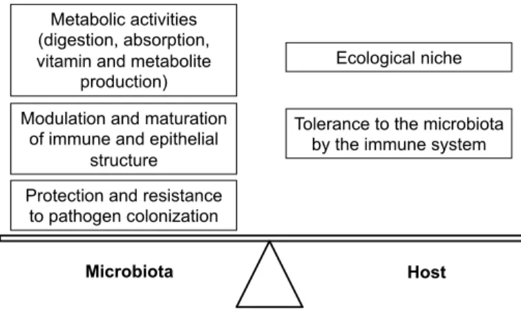
The microbial diversity of the GIT results from a co-evolution between the microbial communities and their hosts which represents its ecological niche. For a long time considered as com-mensal, from the Latin: cum (‘with’) and mesa (‘table’), which means the sharing of the meal, the term mutualism is preferred today to speak about the host-microbiota relation. Indeed, com-mensalism is a relation between individuals of two species in which one species obtains food or other benefits from the other without either harming or benefiting the latter. Commensalism is then a non-parasitic utilization of a live species by another. However, in the case of the host-intestinal microbiota relation, we speak about a symbiotic mutualism relation because both partners developed an intimate and long-term association with mutual profit (Fig. 12.1). In fact, there is a co-evolution between the human and its microbiota with an acquisition by the microbiota of functionalities
absent in human genome. In fact, the intestinal microbiota, by its enzymatic activities, is a complementary metabolic repertoire of the human digestive system. The intestinal bacteria present ben-eficial properties for the host in terms of synthesis of molecules, degradation of substrates and detoxification. For example, we could quote the acquisition of genes for degradation of the plant polysaccharides and other complex sugars, non-digestible by human enzymes (Ley et al., 2008). Their degradation by
bacte-rial fermentation in short-chain fatty acids (SCFA) constitutes an important source of energy for colonocytes (Macfarlane and Macfarlane, 2011). Moreover, the intestinal bacteria synthesize vitamins, amino acids and are involved in the metabolism of bile salts (Vyas and Ranganathan, 2012). The intestinal microbiota also participates in absorptive capacities of nutriments by the GIT by a direct action on its physiology. For example, the SCFA are not only involved in the energy metabolism but also present properties of regulation of the physiology of the host (differen-tiation, cellular growth, etc.) (Neish, 2009). A recent publication using gnotobiotic models showed that the colon evolved in paral-lel with the microbiota composition inducing a homeostasis of the two maturated partners (Tomas et al., 2013). In the same way,
during the post natal period, bacteria associated with the mucosa could regulate angiogenesis increasing the absorptive capacity of the intestine (Stappenbeck et al., 2002). Thus, the microbiota
supports human nutrition by two main mechanisms: increase of the availability in energy substrates for the host and regulating intestinal absorption capacity.
Besides being a nutritive supply, the microbiota also partici-pates in the protection of the host. Indeed, the organism defences at the level of the GIT have the delicate task to distinguish com-mensal microbiota from pathogenic agents. The mechanisms of the intestinal immune system allowing its tolerance to the microbiota remains poorly known (Littman and Pamer, 2011). To get a glimpse of an answer, it is necessary to better under-stand the recognition systems of microorganisms developed by the intestinal epithelial cells. Particularly, specific receptors to Microbial-associated molecular patterns (MAMPS) exist, the pattern recognition receptors (PRRs) (Wells et al., 2011).
Briefly, the activation of these receptors by commensal bacteria leads to the activation of the innate immunity system and is essential to maintain intestinal homeostasis and the protection of the epithelium against pathogenic bacteria (Lundin et al.,
2008; Rakoff-Nahoum and Medzhitov, 2008; Rakoff-Nahoum
et al., 2004). So, the microbiota limits the inflammation of the
intestinal epithelium and plays a major role in the development of the immune system (Macdonald and Monteleone, 2005). To illustrate the hypothesis, the best example is the use of germ free mice that present an immature immune system at the GIT level and the conventionalisation of these mice (reconstitution of a normal microbiota) restored the immunity of this mucosa (Umesaki et al., 1995). Furthermore, at birth, the immune system
is still immature and it was shown that contact with bifidobacteria influences its postnatal development via the production of IgA and an insufficient exposure to these microorganisms could lead to inappropriate immune responses later in the childhood and could induce asthma or allergies (Sjogren et al., 2009).
Protection and resistance to pathogen colonization Modulation and maturation
of immune and epithelial structure Metabolic activities (digestion, absorption, vitamin and metabolite
production)
Ecological niche
Microbiota Host
Tolerance to the microbiota by the immune system
Figure 12.1 Symbiotic mutualism relation forged during the
evolution between the host and its microbiota. (Adapted from Claire Cherbuy; http://mediatheque.inra.fr/media/detail/246161/private).
Thus, the microbiota participates to the protection of the host by supporting the development of the immune system, but commensal bacteria act also directly as a ‘microbiological barrier’ against pathogens. Indeed, it was shown that germ free mice are more sensitive to intestinal pathogens (Vollaard and Clasener, 1994). This protective capacity of the microbiota could be explained by different mechanisms: a competition with patho-genic bacteria for the access to nutriments and/or for a contact with the epithelium, the production of bactericidal or bacte-riostatic molecules and, a modification of the physiology of the epithelium through production of mucus or modulation of epi-thelial cell tight junctions (Martin et al., 2014b; Yu et al., 2012).
For instance, two major commensal bacteria B. thetaiotaomicron
and F. prausnitzii could directly modulate the intestinal mucus
barrier by their complementary metabolism by modifying goblet cells and mucin glycosylation (Wrzosek et al., 2013). Moreover,
commensal bacteria were able to induce maturation of intestinal barrier function through (i) modulation of the expression of tight junctions (claudin 3) that regulate paracellular permeability to maintain separation between tissue compartments by sealing the intercellular space, and (ii) intracellular permeability regulated by toll-like receptor signalling (Jakaitis and Denning, 2014).
It is now clear that the normal intestinal microbiota could influence numerous physiological aspects in the healthy host through the establishment of cross-species homeostatic regula-tion well detailed in the review of Sommer and Bäckhed (2013). Thus, the microbiota composition and its relation with the gut have resulted from the dynamics of selection and competition (Angelakis et al., 2012). For instance, the bacterial microbiome
of the inhabitants of Burkina-Faso was more adapted to the deg-radation of cellulose suggesting an adaptation to Burkina-Faso’s high-fibre diet (De Filippo et al., 2010); One could suggest that
our microbiota shelters unknown bacteria adapted to specific functions and represents a real richness for future research con-cerning human health. Our microbiota takes an important part in human health and an imbalance of its composition could partici-pate to the development of many diseases.
Diseases of microbiota: a break in the homeostasis
Since Robert Koch and Ilya Mechnikov were awarded two Nobel Prizes in physiology and medicine in the 1900s for their discover-ies linking microbes and human health, several determinants of host–microbe interactions, including spatial (e.g. skin, vagina, and gut), temporal (e.g. birth and senescence), and environ-mental (e.g. diet, antibiotics treatments), have been partially unravelled (Cani and Delzenne, 2011). For instance, a mature vaginal microbiota is established only in early adolescence after the hormonal changes typical of this period (Yamamoto et al.,
2009). Concerning intestinal microbiota, it varies all along the life, with the particularity to increase its population number and diversity during the first year and decline upon ageing (Claesson
et al., 2012; Sekirov et al., 2010). In adults the importance of the
microbiome has been highlighted by the microbial ‘abnormalities’ found in pathological conditions and particularly a disruption of
profile or biodiversity. In fact, many diseases are characterized by a dysbiosis, in other words, a microbial imbalance (Round and Mazmanian, 2009). A healthy gut microbiota is composed of a well-balanced community of three permanent residents termed symbionts (with beneficial effects), commensals (no effect), and pathobionts (potentially induce pathologies under certain situations) but no pathogens (Koboziev et al., 2013). The term
dysbiosis has been related to many different kinds of pathologies although it is not clear whether the imbalance of microbiota is a cause or a consequence of the illness (Martin et al., 2013). In
this way, ‘The hygiene hypothesis ‘, based on the observation of a positive correlation between chronic diseases and hygiene and socioeconomic status (Strachan, 1989), could be explained by the creation of dysbioses limiting the regulation of the immunity by the microbiota (von Mutius, 2007).This hypothesis proposes that a lack of early microbial stimulation in industrialized countries results in an aberrant immune response to innocuous antigens later in life (Wills-Karp et al., 2001). In parallel, the ‘microbiota
hypothesis’ claims that the gut microbiota dysbiosis, due to anti-biotic use and dietary changes, can disrupt the mechanisms of mucosal immunological tolerance (Noverr and Huffnagle, 2005). Recent epidemiological and clinical data (Droste et al.,
2000; Hoskin-Parr et al.), as well as experimental data obtained
in mouse models (Noverr et al., 2004; Olszak et al.), support this
latter theory. Nowadays, the challenge of linking microbiome to human health and disease is being tackled by different research teams around the world. They are currently studying different disease states to identify potential correlations and ecological models of community structure and function in order to under-stand the dynamics of all ecosystems that comprise the human microbiome (Martin et al., 2014b).
Impact of intestinal dysbiosis
The GIT is the major site of interaction between environmental microorganisms and host tissues. A high biodiversity of the gut microbiota is associated with a healthy status, while low biodiversity is linked to pathological conditions (Manichanh et al., 2006). More than 30 diseases are associated with intestinal
microbiota (dysbiosis) and the list is continuously increased. The pathogenesis of several autoimmune, atherosclerosis or rheumatoid arthritis, and chronic inflammatory diseases could be mentioned (Koboziev et al., 2013). Among these diseases we
could distinguish three important classes: metabolic syndromes, intestinal diseases and more prudently psychological syndromes (Fig. 12.2). The common point between all of these patients is that they are suffering of intestinal disorders with different severi-ties. The link between psychological syndrome and gut dysbiosis is a very recent hypothesis present in the literature. It is clear that gut microbiome plays a crucial role in the bidirectional gut–brain axis that integrates the gut and central nervous system activities (Wang and Kasper, 2013). Indeed, it has been shown that the niche-specific microbiome, prominently the gut microbiome, has the capacity to affect both local and distal sites within the host. In fact, intestinal dysbiosis is involved in metabolic syndromes such as diabetes, obesity, liver disease…etc. Interestingly, colonization of germ-free mice with microbiota obtained from obese but not
lean mice results in the generation of mice with a significantly greater amount of total body fat (Turnbaugh et al., 2006).
Simi-lar results were obtained in non-alcoholic fatty liver disease, the hepatic manifestation of metabolic syndrome (Le Roy et al.,
2012), suggesting that these kinds of diseases are transmissible via microbiota. Moreover, a disorder of the crosstalk between innate immune system and intestinal microbiota may promote the development of the metabolic syndrome (Cani and Delzenne, 2011). For instance, a recent study shows that mice genetically deficient in Toll-like receptor 5 (TLR5), a component of the innate immune system that is expressed in the gut mucosa and recognizes pathogen-associated molecular patterns (PAMPs) that are expressed on infectious microbes, develop hallmark features of metabolic syndrome (hyperlipidaemia, hypertension, insulin resistance, and increased adiposity) (Vijay-Kumar et al., 2010).
The best examples of the implication of microbiota in dis-eases and the importance of the homeostasis between these two partners are the development of intestinal diseases and particularly inflammatory bowel disease (IBD). IBD, including Crohn’s disease (CD) and ulcerative colitis (UC), are character-ized by a chronic activation of the immune system, of genetically predisposed patients in response to environmental factors and dysbiosis (De Cruz et al., 2012; Lepage et al., 2011) For instance,
the proportion of Firmicutes, in particular the symbiotic Faecali-bacterium prausnitzii, was found to be low in intestinal microbiota
of CD patients (Sokol et al., 2008) whereas there was an increased
in opportunistic pathogen bacteria, in particular Adherent inva-sive Escherichia coli (AIEC) (Darfeuille-Michaud, 2002; Miquel et al., 2010). A study showed that colitis (IBD like) and colonic
hypersensitivity (IBS-like syndrome) can be provoked in healthy rodents by the transfer of microbiota coming from mouse suf-fering from colitis (Crouzet et al., 2013; Garrett et al., 2007).
The excessive inflammation of the GIT would be particularly induced by an inappropriate stimulation of the immune system in the presence of an unbalanced commensal microbiota and its metabolites (Sartor, 2008). Dysbiosis has also been reported for numerous other intestinal patho-physiological contexts such as IBS (Rajilic-Stojanovic et al., 2011), colorectal cancer (Sobhani
et al., 2011), short bowel syndrome (SBS) (Joly et al., 2010) and
coeliac disease (De Palma et al., 2010).
Vaginal dysbiosis
Although dysbiosis has been considered at length in the gastroin-testinal tract, it can also occur on any exposed surface or mucous membrane such as skin (Grice et al., 2008; Grice and Segre,
2011), oral cavity, airway (Charlson et al., 2011; Gollwitzer and
Marsland, 2014), and the vagina.
The vaginal microbiota that is mainly formed by lactobacilli, has an important role in human development, physiology, and immunity (Martin et al., 2014b). Abnormal vaginal microbiota can
occur because of sexually transmitted pathogens or overgrowth of resident organisms (Lamont et al., 2011). The most common
pathologies are bacterial vaginosis (BV), the proliferation of
Candida sp. (mainly C. albicans) (candidiasis) and Trichomonas vaginalis (trichomoniasis) (Martin et al., 2008a). The community
of mutualistic vaginal bacteria, constitutes the first line of protec-tion for the host by excluding non-indigenous microbes that may cause illness (Martin et al., 2008a).
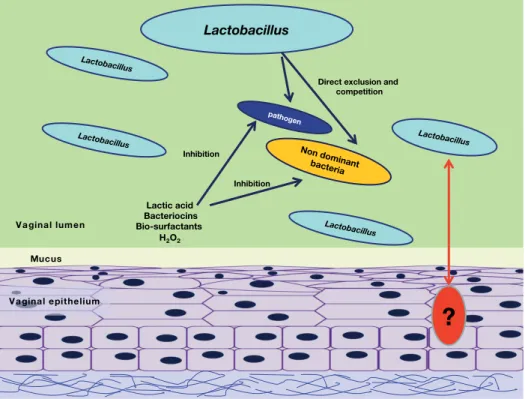
The lactobacilli exerts their beneficial role by two main mecha-nisms: (i) exclusion and (ii) inhibition of growth (Boris and Barbes, 2000) (Fig. 12.3). The exclusion is driven by the com-petition for epithelial cell receptors with urogenital pathogens, of which the most important are: group B Streptococcus species, Staphylococcus aureus, Gardnerella vaginalis, Neisseria gonorrhoeae, Pseudomonas aeruginosa, Klebsiella pneumoniae, Candida albicans
and Actinomyces neuii (Boris et al., 1998; Martin et al.; Osset et al.,
2001a,b; Vielfort et al., 2008; Zarate and Nader-Macias, 2006).
Furthermore, the ability of some lactobacilli to co-aggregate with pathogens like E. coli, C. albicans, and G. vaginalis has been found
(Boris et al., 1998; Osset et al., 2001a). Although the molecular
mechanism involved are not completely known and may depend on the Lactobacillus strain involved and the pathogen inhibited,
shaving and seeding treatments with proteases, lipases or periodic acid point out the proteinic, lipidic or polysaccharide nature of the adhesins and cellular receptors involved (Munoz-Provencio
et al., 2010; Sanchez et al., 2008; Tjalsma et al., 2008).
Inhibition of growth is due to the generation of antimicrobial compounds. Lactobacilli are able to produce mainly organic acids from the fermentation of sugars that contribute to the low pH of the vagina, the major factor in the inhibition of pathogen growth (Boskey et al., 2001). Besides, some vaginal lactobacilli are able
also to produce other antimicrobial compounds such as bacteri-ocins, biosurfactants and hydrogen peroxide (H2O2) (Velraeds et
al., 1998, 2000). The production of H2O2 seems to be the second major antimicrobial mechanisms after organic acid production. In fact, the prevalence of H2O2-producing strains has been cor-related with lower incidence of bacterial vaginosis (BV) and vaginal infections (Eschenbach et al., 1989; Hawes et al., 1996).
H2O2 exerts its bactericidal effect through generation of oxidizing metabolites such as the radical OH– that introduces breaks in the
DNA of the cell (Klebanoff and Belding, 1974). Furthermore, hydrogen peroxide is able to regulate the lactobacilli population, due to the ability of some lactobacilli to degrade and produce this molecule (Martin et al.) and the lysis of lysogenic lactobacilli
Intestinal disease
Metabolic
syndromes Psychologicalsyndrome
Intestinal dysbiosis IBD IBS Colorectal cancer Coeliac disease Allergy Obesity Diabetes Athero- sclerosis Autism Depression … Liver disease … SBS
Figure 12.2 Disease associated with intestinal dysbiosis. IBD,
inflammatory bowel disease; SBS, short bowel syndrome; IBS, irritable bowel syndrome.
by a H2O2-mediated prophage induction mechanism (Martin et
al., 2009; Pavlova et al., 1997). Although, the presence of
bac-teriocins is thoroughly described in numerous bacterial species (Cotter et al., 2013), only four bacteriocins have been found, to
our knowledge, up to day in vaginal Lactobacillus strains: Lactocin
160, Salivaricin CRL 1328, L23 and recently a bacteriocin-like substance produced by Lactobacillus fermentum CS57 isolated
from human vaginal secretions (Aroutcheva et al., 2001; Martin et al.; Ocana et al., 1999; Pascual et al., 2010; Sabia et al., 2014;
Simoes et al., 2001).
Despite all the defence mechanisms asserted by lactobacilli, sometimes the proportion of Lactobacillus drops under a
criti-cal level and this circumstance is used by other microorganisms that can act as opportunistic pathogens (Garcia-Rodriguez and Munoz, 1991). In general these microorganisms can be found in the normal vaginal microbiota, but in a lower abundance due to the antagonist effect of the lactobacilli. Among them, the most common aetiological agents are Gardnerella vaginalis, Mycoplasma hominis, Prevotella and Peptostreptococcus that can induce from an
asymptomatic infection up to vaginosis (Sobel, 2000; Thorsen
et al., 1998). Bacterial vaginosis patients are those who fulfil the
Amsel criteria: homogeneous vaginal discharge, amine (fishy) odour when potassium hydroxide solution is added to vaginal secretions (commonly called the ‘whiff test’), vaginal pH greater than 4.7 and presence of clue cells (greater than 20%) upon microscopy combined with a significant diminution of Gram-positive lactobacilli replaced by Gram-negative bacteria (Amsel
et al., 1983). BV is the most recurrent vaginal imbalance and has
been connected with a high diversity microbiota (Pavlova et al.,
2002) and the presence of unfamiliar bacteria such as Mobiluncus
sp., Atopobium sp., Megasphaera sp. and Ureaplasma urealyticum
(Doyle et al., 1995; Fredricks et al., 2005; Haggerty et al., 2009;
Hyman et al., 2005; Kazor et al., 2003). Thanks to metagenomic
approaches some uncultivable microorganisms have also been associated with BV (Fredricks et al., 2005; Lamont et al., 2011;
Oakley et al., 2008; Pavlova et al., 2002). Epidemiologically,
vagi-nal dysbiosis such as BV has been associated with preterm birth, development of pelvic inflammatory disease and acquisition of sexually transmitted infections produced by Neisseria gonorrhoeae, Chlamydia spp. and HIV (Brotman, 2011).
It is interesting to study the characterization of dysbiosis in more detail to investigate the implication of potential beneficial bacteria that could be decreased in the studied microbiota of patients. From this perspective, it could be interesting to use them as potential probiotics to try to resolve dysbioses.
The commensal beneficial bacteria
Commensal microorganisms offer a wide range of benefits to the host. The cross-talk between the host and its microbiota is fundamental for the maintenance of the host homeostasis (Leser and Molbak, 2009). The microbiota is capable to regulate the intestinal epithelium, the presence of pathogenic bacteria and the immune responses as well as to confer nutritional benefits as discussed before (Martin et al., 2013). Both host and indigenous
microorganisms, have then adapted to each other to maintain the advantages that this mutualism offers (Sekirov et al., 2010).
Figure 12.3 Beneficial effects of lactobacilli on the vaginal ecosystem. Lactobacilli protect the host epithelium thanks to two main
mechanisms: (i) exclusion, driven by the formation of a biofilm that mask the epithelial cell receptors and (ii) inhibition of growth, due to generation of antimicrobial compounds. The direct interactions between the lactobacilli and the host are not well known. Modified from (Martin et al., 2014b).
Lactobacillus
pathogen
Direct exclusion and competition Lactic acid Bacteriocins Bio-surfactants H2O2 Inhibition Lactobacillus Lactobacillus Non dominant bacteria Lactobacillus Inhibition
?
Vaginal epithelium MucusNumerous bacterial examples have been analysed in more or less detail. Bacteroides thetaiotaomicron and Faecalibacterium prausnitzii, both prominent members of the intestinal microbiota
of mice and humans, have been widely studied (Miquel et al.,
2013; Zocco et al., 2007). B. thetaiotaomicron exerts several
ben-eficial effects, such as the modulation of the expression of a large quantity of genes implicated in different aspects of host physiol-ogy (Zocco et al., 2007), the modulation of the colon epithelium
(Wrzosek et al., 2013) and the defence of the intestinal barrier
(Resta-Lenert and Barrett, 2006). Indeed, the acetate produced by B. thetaiotamicron seems to be key in the cell differentiation
(Wrzosek et al., 2013) and this bacterium prevents cytokine
induced increase in permeability (Resta-Lenert and Barrett, 2006). Thanks to the analysis of its genome and proteome, the huge ability to adapt and regulate gene expression in response to a changing ecosystem has been pointed out, highlighting the high level of adaptation of this bacterium to its niche (Comstock and Coyne, 2003).
F. prausnitzii, is a major EOS component of the intestinal
microbiota which represents around 5% of the bacterial popula-tion of the microbiota and 8% of Firmicutes (Eckburg et al., 2005;
Miquel et al., 2014). Its prevalence is low in many health disorders,
such as IBS, colorectal cancer, coeliac diseases, obesity, and par-ticularly in IBD patients, suggesting its potential as an indicator of intestinal health (Miquel et al., 2013; Sokol et al., 2008; Swidsinski et al., 2008a). Moreover, F. prausnitzii is a butyrate producer and
has demonstrated anti-inflammatory effects in vitro and in vivo
using mice colitis models making it a key member of the micro-biota that may contribute to intestinal homeostasis (Martin et al.,
2014a; Sokol et al., 2008). This bacterium also impacts physical
and chemical epithelial barrier functions though modulation of tight junction and mucus layer (Carlsson et al., 2013; Wrzosek et al., 2013). Thus, F. prausnitzii is a beneficial commensal bacterium
that could be a good candidate of our indigenous microbiota to elaborate new prophylactic or therapeutic applications in human health (Miquel et al., 2013).
Another Bacteroides species, B. fragilis, has been also widely
studied. This species is an important anaerobic gut commensal of humans that prevents and cures intestinal inflammation in mice (Mazmanian et al., 2008). B. fragilis NCTC 9343 presents
a polysaccharide (PSA) promoting lymphoid organogenesis that increases CD4+ T-cells in germ-free rodents (Mazmanian et al.,
2005) and corrects the Th1/Th2 balance as has been shown in
cellular and animal models (Mazmanian et al., 2005; Mazmanian et al., 2008). This strain corrects gut permeability, alters
micro-bial composition and ameliorates defects in communicative, stereotypic, anxiety-like and sensorimotor behaviours by being capable to modulate some metabolite levels (Hsiao et al., 2013). Bacteroides fragilis ATCC23745 plays a role in maintaining
physi-ological expression of heat-shock protein (Hsp)25 and Hsp72 which are cytoprotective in colonocytes (Kojima et al., 2003).
Escherichia coli Nissle 1917 (ECN) was isolated by Professor
Alfred Nissle in 1917 during the First World War. This strain has been used as a probiotic for several years. Its capacities are huge. It is capable to induce the expression of the antimicrobial pep-tide human beta-defensin-d2 (hBD-2) in the human intestinal
epithelial cell line Caco-2 in vitro. ECN strengthens the
intes-tinal barrier function by inducing pro-inflammatory pathways, mainly NF-kb and AP-1 as well as MAPKs (Schlee et al., 2007;
Wehkamp et al., 2004). This effect has been also demonstrated in vivo, where faecal hBD-2 peptide was increased by 78% after 3
weeks of E. coli Nissle 1917 administration (Mondel et al., 2009).
Furthermore, it is able to inhibit the invasion of various gut epi-thelial cells lines by adherent-invasive Escherichia coli (AIEC), S. typhimurium, Y. enterocolitica, S. flexneri, Legionella pneumophila
and L. monocytogenes (Altenhoefer et al., 2004; Boudeau et al.,
2003; Deriu et al., 2013) and to restore the barrier dysfunction
induced by E. coli (EPEC) and Salmonella dublin in vitro (Otte
and Podolsky, 2004). Some cellular components of ECN have been proposed in cell signalling. For instance, the host cell internalization of peptidoglycan fragment of ECN leads to inter-action with Noll-like receptor (NLR) 1 (Bron et al., 2012) and
its flagellins are recognized to interact with the TLR-5 (Hayashi
et al., 2001; Ogushi et al., 2004; Schlee et al., 2007). Another
mechanism of action could be a competition for iron uptake especially to reduce Salmonella typhimurium intestinal
coloniza-tion (Deriu et al., 2013).
Akkermansia muciniphila is an abundant resident of the human intestinal tract (Derrien et al., 2008) and mucus
degrad-ing specialist which correlates with health and disease (Joyce and Gahan). This bacterium modulates pathways involved in establishing homeostasis for basal metabolism and immune tolerance towards commensal microbiota (Derrien et al.) and
has been correlated with inflammation, obesity and diabetic parameters (Collado et al., 2007; Ellekilde et al.; Everard et al.;
Kang et al.).
Although the lactic acid bacteria (LAB) are not the major members of the human microbiota, due to their easy isolation and growth they have been thoroughly studied and most of the probiotic products available nowadays are based on bacteria belonging to this group. One of the main strains belonging to this group is Lactobacillus rhamnosus GG (LGG). LGG (ATCC
53103) was isolated in 1983 from the intestinal tract of a healthy human being by Sherwood Gorbach and Barry Goldin. LGG has shown positive effects in human patients with pouchitis, ulcerative colitis and Crohn’s disease (Hormannsperger and Haller, 2010) and have been found to increase faecal IgA levels (Bakker-Zierikzee et al., 2006; He et al., 2005). A clinical study
demonstrated that perinatal administration of this probiotic strain reduced the development of atopic eczema in children (Nermes et al., 2011). This effect may be due to the
anti-inflam-matory properties of this probiotic bacterium. Consumption of LGG by children with atopic dermatitis has been reported to enhance the production of the anti-inflammatory cytokine IL-10 by the host (Pessi et al., 2000). It has been demonstrated
that two soluble factors, proteins p75 and p40, present in the culture supernatant of LGG induce the expression of Hsp in
a p38- and JNK/MAPK-dependent way (Tao et al., 2006).
Additionally, LGG also prevents cytokine-induced apoptosis activating anti-apoptotic Akt in a phosphatidylinositol-3-kinase dependent manner and inhibiting pro-apoptotic p38/MAPK activation (Yan and Polk, 2002)(Yan et al., 2007). These factors
are also able to modulate hydrogen peroxide induced damage in Caco-2 cells (Seth et al., 2008). Other molecules produced
by lactobacilli have been found to have important characteris-tics. For example, the analysis of the currently known genomic sequences of Lactobacillus strains predicts a broad group of
bacteriocins active against Gram-positive bacteria such as Lac-tococcus, StrepLac-tococcus, Staphylococcus, Listeria and Mycobacteria
(Altermann et al., 2005; Chaillou et al., 2005; Makarova et al.,
2006; Pridmore et al., 2004). For instance, it is well known that L. salivarius UCC118 has the ability to protect mice against
infection with Listeria monocytogenes thanks to the production
of a Class II bacteriocin (Corr et al., 2007). Several other
lac-tobacilli strains have been tested in different in vivo and in vitro
test with positive results. In this regard, L. salivarius Ls33 and
its peptidoglycan were anti-inflammatory in a murine colitis model (Macho Fernandez et al., 2011a; Macho Fernandez et al.,
2011b). L. farciminis CIP 103136 prevents stress-induced
hyper-sensitivity, increase in colonic paracellular permeability, and colonocyte myosin light chain phosphorylation (Ait-Belgnaoui
et al., 2006). This antinociceptive effect occurs via inhibition of
contraction of the cytoskeleton of colonic epithelial cells and the subsequent tight junction opening, and may also involve direct or indirect effects of nitric oxide produced by this strain (Ait-Belgnaoui et al., 2006) or a decrease of the stress-induced
activation/sensitization of sensory neurons at the spinal and supraspinal level (Ait-Belgnaoui et al., 2009). Furthermore, L. farciminis CIP 103136 also improves TNBS-induced colitis
(Lamine et al., 2004a), mainly due to the nitric oxide released,
the normalization of colonic microbiota, the prevention of bac-terial translocation, the enhancement of barrier integrity and a decrease in the mucosal levels of IL-1β (Lamine et al., 2004a,b).
Contrary to the gut environment, lactobacilli are considered as the dominant organisms of the vaginal cavity (Doderlein, 1982) being more than 70% of all microorganisms isolated (Eschenbach et al., 1989; Redondo-Lopez et al., 1990). In this
ecosystem, the success obtained so far with the use of a cocktail containing the spermicide resistant L. rhamnosus GR-1 and the
H2O2 producing L. reuteri RC-14 strains by themselves or
asso-ciated with antimicrobial chemotherapy is well known (Anukam
et al., 2006; Reid et al., 2003). These indigenous lactobacilli
strains have beneficial effects on the host. GR-1 blocked the in vitro attachment of uropathogenic bacteria to human
uroepithe-lial cells and prevented onset of urinary tract infection in murine models (Reid et al., 1985) while RC-14 inhibited Staphylococcus aureus infection of surgical implants in rats (Gan et al., 2002).
Both strains, employed together, were able to interfere with the opportunistic pathogen Candida albicans (Kohler et al., 2012;
Martinez et al., 2009) and to prevent or treat BV (Homayouni et al., 2014; Hummelen et al., 2010).
All of presented bacteria are examples of commensal bacteria with beneficial effects in human health and some of them are already used to treat patients. Our microbiota is a real richness for future trend in ‘microbiomology’ research and could be a source of new probiotics that could impact on the gastrointesti-nal, nervous, and immune systems.
Future trends: could probiotics be indigenous?
Probiotics are defined as ‘live micro-organisms which, when administered in adequate amounts, confers a health benefit on the host’ (group, 2001). The use of them began to show clinical evidence of their impact on human health (Hungin et al., 2013).
The advent of probiotic treatments appears to be a promising ‘pharmaco-nutritional’ approach to reverse diseases linked to microbiota dysbiosis. Today, most micro-organisms marketed as probiotics in the food industry are lactic acid bacteria, belonging to the genera Lactobacillus and Bifidobacterium (Foligne et al., 2013).
Surprisingly, most of them, used for intestinal disorders manage-ment, are not currently present in our microbiota. However, the literature underscores the need for further understanding of the role of probiotics in health and disease (Klein et al., 2010). This
observation, associated with the rapid evolution of the knowledge of this complex ecosystem, suggests a large panel of potential new candidates that could be isolated directly from our indigenous microbiota. A good argument for this hypothesis is the recently gained renewed interest in microbiota transplantation. Proposed as a treatment for Clostridium difficile colitis, in a randomized
controlled trial, faecal microbiota transplantation was shown to be very efficient in more than 80% of the patients leading to increased bacterial diversity similar to healthy subjects (van Nood et al., 2013). Instead of using stools from healthy donors,
a cultured strain mix of sufficiently characterized beneficial com-mensal bacteria is proposed as an alternative (Petrof et al., 2013).
We speculate that newly discovered intestinal bacteria may be used for development of new micro-organisms containing products that could be novel probiotics with health claim. In fact, host physiology, gut maturation, innate and acquired immune responses and metabolism are largely influenced by the metabolic properties of the (gut) microbiota (Gaboriau-Routhiau et al.,
2009; Tomas et al., 2013). Moreover, the activity and the
com-position of microbiota are modulated by external factors making microbiota a highly ‘handleable’ tissue in humans (De Filippo et al.). Interestingly, some bacteria, depleted in many intestinal
dis-orders that displayed beneficial effects on the host, could be used to counterbalance the dysbiosis linked to certain diseases. It has been recently proposed that the anti-inflammatory F. prausnitzii
bacterium, could have prophylactic or therapeutic applications in human health (Miquel et al., 2013; Sokol et al., 2008). Today,
we could suggest other species as good candidates: B. thetaio-taomicron, B. fragilis, L. farciminis, etc. However, the description of
several microbial communities of our environment (particularly thus of our gut microbiota) allowed us to identify new bacterial species that were unknown so far for their health benefit. The recent description of the intestinal metagenome (all genomes of the bacterial populations of an environment), confirmed that the richness concerning bacterial species of the human gut microbiome correlates with metabolic markers (Le Chatelier et al., 2013). Therefore, besides being a reservoir of unexploited
bacteria for academic research, our microbiota also present potentially beneficial metabolic capacities for human health that could be exploited industrially. In parallel, it could be speculated
that intestinal probiotics prepared from commensal bacteria may be efficient at a lower threshold than an exogenous strain since they could occupy their own ecological niches more easily. References
Ait-Belgnaoui, A., Eutamene, H., Houdeau, E., Bueno, L., Fioramonti, J., and Theodorou, V. (2009). Lactobacillus farciminis treatment attenuates
stress-induced overexpression of Fos protein in spinal and supraspinal sites after colorectal distension in rats. Neurogastroenterol. Motil. 21,
567–573, e518–569.
Ait-Belgnaoui, A., Han, W., Lamine, F., Eutamene, H., Fioramonti, J., Bueno, L., and Theodorou, V. (2006). Lactobacillus farciminis treatment
suppresses stress induced visceral hypersensitivity: a possible action through interaction with epithelial cell cytoskeleton contraction. Gut 55,
1090–1094.
Altenhoefer, A., Oswald, S., Sonnenborn, U., Enders, C., Schulze, J., Hacker, J., and Oelschlaeger, T.A. (2004). The probiotic Escherichia coli strain
Nissle 1917 interferes with invasion of human intestinal epithelial cells by different enteroinvasive bacterial pathogens. FEMS immunology and medical microbiology 40, 223–229.
Altermann, E., Russell, W.M., Azcarate-Peril, M.A., Barrangou, R., Buck, B.L., McAuliffe, O., Souther, N., Dobson, A., Duong, T., Callanan, M.,
et al. (2005). Complete genome sequence of the probiotic lactic acid
bacterium Lactobacillus acidophilus NCFM. Proc. Natl. Acad. Sci. U.S.A. 102, 3906–3912.
Amsel, R., Totten, P.A., Spiegel, C.A., Chen, K.C., Eschenbach, D., and Holmes, K.K. (1983). Nonspecific vaginitis. Diagnostic criteria and microbial and epidemiologic associations. Am. J. Med. 74, 14–22.
Angelakis, E., Armougom, F., Million, M., and Raoult, D. (2012). The relationship between gut microbiota and weight gain in humans. Future Microbiol. 7, 91–109.
Anukam, K., Osazuwa, E., Ahonkhai, I., Ngwu, M., Osemene, G., Bruce, A.W., and Reid, G. (2006). Augmentation of antimicrobial metronidazole therapy of bacterial vaginosis with oral probiotic Lactobacillus rhamnosus
GR-1 and Lactobacillus reuteri RC-14: randomized, double-blind,
placebo controlled trial. Microbes Infect. 8, 1450–1454.
Aroutcheva, A.A., Simoes, J.A., and Faro, S. (2001). Antimicrobial protein produced by vaginal Lactobacillus acidophilus that inhibits Gardnerella
vaginalis. Infect. Dis. Obstet. Gynecol. 9, 33–39.
Arumugam, M., Raes, J., Pelletier, E., Le Paslier, D., Yamada, T., Mende, D.R., Fernandes, G.R., Tap, J., Bruls, T., Batto, J.M., et al. (2011). Enterotypes
of the human gut microbiome. Nature 473, 174–180.
Backhed, F., Ley, R.E., Sonnenburg, J.L., Peterson, D.A., and Gordon, J.I. (2005). Host-bacterial mutualism in the human intestine. Science 307,
1915–1920.
Bakker-Zierikzee, A.M., van Tol, E.A.F., Kroes, H., Alles, M.S., Kok, F.J., and Bindels, J.G. (2006). Faecal SIgA secretion in infants fed on pre- or probiotic infant formula. Pediatr. Allergy Immunol. 17, 134–140.
Baquero, F., and Nombela, C. (2012). The microbiome as a human organ. Clin. Microbiol. Infect. 18 Suppl. 4, 2–4.
Biswas, B.B., Basu, P.S., and Pal, M.K. (1970). Gram staining and its molcecular mechanism. International review of cytology 29, 1–27.
Boris, S., and Barbes, C. (2000). Role played by lactobacilli in controlling the population of vaginal pathogens. Microbes Infect. 2, 543–546.
Boris, S., Suarez, J.E., Vazquez, F., and Barbes, C. (1998). Adherence of human vaginal lactobacilli to vaginal epithelial cells and interaction with uropathogens. Infect. Immun. 66, 1985–1989.
Boskey, E.R., Cone, R.A., Whaley, K.J., and Moench, T.R. (2001). Origins of vaginal acidity: high D/L lactate ratio is consistent with bacteria being the primary source. Hum. Reprod. 16, 1809–1813.
Boudeau, J., Glasser, A.L., Julien, S., Colombel, J.F., and Darfeuille-Michaud, A. (2003). Inhibitory effect of probiotic Escherichia coli strain Nissle
1917 on adhesion to and invasion of intestinal epithelial cells by adherent-invasive E-coli strains isolated from patients with Crohn’s disease. Alimentary pharmacology and therapeutics 18, 45–56.
Bron, P.A., van Baarlen, P., and Kleerebezem, M. (2012). Emerging molecular insights into the interaction between probiotics and the host intestinal mucosa. Nat. Rev. Microbiol. 10, 66-U90.
Brotman, R.M. (2011). Vaginal microbiome and sexually transmitted infections: an epidemiologic perspective. J. Clin. Invest. 121, 4610–4617.
Bruls, T., and Weissenbach, J. (2011). The human metagenome: our other genome? Hum. Mol. Genet. 20, R142–148.
Cani, P.D., and Delzenne, N.M. (2011). The gut microbiome as therapeutic target. Pharmacology and therapeutics 130, 202–212.
Carlsson, A.H., Yakymenko, O., Olivier, I., Hakansson, F., Postma, E., Keita, A.V., and Soderholm, J.D. (2013). Faecalibacterium prausnitzii
supernatant improves intestinal barrier function in mice DSS colitis. Scand. J. Gastroenterol. 48, 1136–1144.
Chaillou, S., Champomier-Verges, M.C., Cornet, M., Crutz-Le Coq, A.M., Dudez, A.M., Martin, V., Beaufils, S., Darbon-Rongere, E., Bossy, R., Loux, V., et al. (2005). The complete genome sequence of the
meat-borne lactic acid bacterium Lactobacillus sakei 23K. Nat. Biotechnol. 23,
1527–1533.
Charlson, E.S., Bittinger, K., Haas, A.R., Fitzgerald, A.S., Frank, I., Yadav, A., Bushman, F.D., and Collman, R.G. (2011). Topographical continuity of bacterial populations in the healthy human respiratory tract. Am. J. Resp. Crit. Care Med. 184, 957–963.
Claesson, M.J., Jeffery, I.B., Conde, S., Power, S.E., O’Connor, E.M., Cusack, S., Harris, H.M., Coakley, M., Lakshminarayanan, B., O’Sullivan, O., et al.
(2012). Gut microbiota composition correlates with diet and health in the elderly. Nature 488, 178–184.
Collado, M.C., Derrien, M., Isolauri, E., de Vos, W.M., and Salminen, S. (2007). Intestinal integrity and Akkermansia muciniphila, a
mucin-degrading member of the intestinal microbiota present in infants, adults, and the elderly. Appl. Environ. Microbiol. 73, 7767–7770.
Comstock, L.E., and Coyne, M.J. (2003). Bacteroides thetaiotaomicron: a dynamic, niche-adapted human symbiont. BioEssays25, 926–929.
Consortium, T.H.M.P. (2012). A framework for human microbiome research. Nature 486, 215–221.
Corr, S.C., Li, Y., Riedel, C.U., O’Toole, P.W., Hill, C., and Gahan, C.G.M. (2007). Bacteriocin production as a mechanism for the antfinfective activity of Lactobacillus salivarius UCC118. Proc. Natl. Acad. Sci. U.S.A. 104, 7617–7621.
Cotter, P.D., Ross, R.P., and Hill, C. (2013). Bacteriocins – a viable alternative to antibiotics? Nat. Rev. 11, 95–105.
Crouzet, L., Gaultier, E., Del’Homme, C., Cartier, C., Delmas, E., Dapoigny, M., Fioramonti, J., and Bernalier-Donadille, A. (2013). The hypersensitivity to colonic distension of IBS patients can be transferred to rats through their fecal microbiota. Neurogastroenterol. Motil. 25,
e272–282.
Darfeuille-Michaud, A. (2002). Adherent-invasive Escherichia coli: a
putative new E. coli pathotype associated with Crohn’s disease. Int. J. Med. Microbiol. 292, 185–193.
De Cruz, P., Prideaux, L., Wagner, J., Ng, S.C., McSweeney, C., Kirkwood, C., Morrison, M., and Kamm, M.A. (2012). Characterization of the gastrointestinal microbiota in health and inflammatory bowel disease. Inflammatory bowel diseases 18, 372–390.
De Filippo, C., Cavalieri, D., Di Paola, M., Ramazzotti, M., Poullet, J.B., Massart, S., Collini, S., Pieraccini, G., and Lionetti, P. (2010). Impact of diet in shaping gut microbiota revealed by a comparative study in children from Europe and rural Africa. Proc. Natl. Acad. Sci. U.S.A. 107,
14691–14696.
De Palma, G., Nadal, I., Medina, M., Donat, E., Ribes-Koninckx, C., Calabuig, M., and Sanz, Y. (2010). Intestinal dysbiosis and reduced immunoglobulin-coated bacteria associated with coeliac disease in children. BMC Microbiol. 10, 63.
Del Chierico, F., Vernocchi, P., Bonizzi, L., Carsetti, R., Castellazzi, A.M., Dallapiccola, B., de Vos, W., Guerzoni, M.E., Manco, M., Marseglia, G.L., et al. (2012). Early-life gut microbiota under physiological and
pathological conditions: the central role of combined meta-omics-based approaches. Journal of proteomics 75, 4580–4587.
Deriu, E., Liu, J.Z., Pezeshki, M., Edwards, R.A., Ochoa, R.J., Contreras, H., Libby, S.J., Fang, F.C., and Raffatellu, M. (2013). Probiotic bacteria reduce salmonella typhimurium intestinal colonization by competing for iron. Cell Host Microbe 14, 26–37.
Derrien, M., Collado, M.C., Ben-Amor, K., Salminen, S., and de Vos, W.M. (2008). The mucin degrader Akkermansia muciniphila is an abundant
resident of the human intestinal tract. Appl. Environ. Microbiol. 74,
1646–1648.
Derrien, M., Van Baarlen, P., Hooiveld, G., Norin, E., Muller, M., and de Vos, W.M. Modulation of mucosal immune response, tolerance, and proliferation in mice colonized by the mucin-degrader Akkermansia muciniphila. Front. Microbiol. 2, 166.
Derrien, M., Vaughan, E.E., Plugge, C.M., and de Vos, W.M. (2004). Akkermansia muciniphila gen. nov., sp. nov., a human intestinal mucin-degrading bacterium. Int. J. Syst Evol. Microbiol. 54, 1469–1476.
Doderlein, A.S.G. (1982). Das Scheindensekret und seine bedeutung fur das puerperalfieber. Leipzig: O Durr.
Doyle, L.M., McInerney, J.O., Mooney, J., Powell, R., Haikara, A., and Moran, A.P. (1995). Sequence of the gene encoding the 16S rRNA of the beer spoilage organism Megasphaera cerevisiae. J. Ind Microbiol. 15, 67–70.
Droste, J.H., Wieringa, M.H., Weyler, J.J., Nelen, V.J., Vermeire, P.A., and Van Bever, H.P. (2000). Does the use of antibiotics in early childhood increase the risk of asthma and allergic disease? Clin. Exp. Allergy 30,
1547–1553.
Eckburg, P.B., Bik, E.M., Bernstein, C.N., Purdom, E., Dethlefsen, L., Sargent, M., Gill, S.R., Nelson, K.E., and Relman, D.A. (2005). Diversity of the human intestinal microbial flora. Science 308, 1635–1638.
Ellekilde, M., Krych, L., Hansen, C.H., Hufeldt, M.R., Dahl, K., Hansen, L.H., Sorensen, S.J., Vogensen, F.K., Nielsen, D.S., and Hansen, A.K. Characterization of the gut microbiota in leptin deficient obese mice – Correlation to inflammatory and diabetic parameters. Res. Vet. Sci. 96,
241–250.
Eschenbach, D.A., Davick, P.R., Williams, B.L., Klebanoff, S.J., Young-Smith, K., Critchlow, C.M., and Holmes, K.K. (1989). Prevalence of hydrogen peroxide-producing Lactobacillus species in normal women and women
with bacterial vaginosis. J. Clin. Microbiol. 27, 251–256.
Everard, A., Belzer, C., Geurts, L., Ouwerkerk, J.P., Druart, C., Bindels, L.B., Guiot, Y., Derrien, M., Muccioli, G.G., Delzenne, N.M., et al., Cross-talk
between Akkermansia muciniphila and intestinal epithelium controls diet-induced obesity. Proc. Natl. Acad. Sci. U.S.A. 110, 9066–9071.
Fiehn, O. (2002). Metabolomics – the link between genotypes and phenotypes. Plant Mol. Biol. 48, 155–171.
Finegold, S.M., Attebery, H.R., and Sutter, V.L. (1974). Effect of diet on human faecal flora: comparison of Japanese and American diets. Am. J. Clin. Nutr. 27, 1456–1469.
Foligne, B., Daniel, C., and Pot, B. (2013). Probiotics from research to market: the possibilities, risks and challenges. Curr. Opin. Microbiol. 16,
284–292.
Fraher, M.H., O’Toole, P.W., and Quigley, E.M. (2012). Techniques used to characterize the gut microbiota: a guide for the clinician. Nat. Rev. Gastroenterology and hepatology 9, 312–322.
Fredricks, D.N., Fiedler, T.L., and Marrazzo, J.M. (2005). Molecular identification of bacteria associated with bacterial vaginosis. N. Engl. J. Med. 353, 1899–1911.
Gaboriau-Routhiau, V., Rakotobe, S., Lecuyer, E., Mulder, I., Lan, A., Bridonneau, C., Rochet, V., Pisi, A., De Paepe, M., Brandi, G., et al. (2009).
The key role of segmented filamentous bacteria in the coordinated maturation of gut helper T-cell responses. Immunity 31, 677–689.
Gan, B.S., Kim, J., Reid, G., Cadieux, P., and Howard, J.C. (2002).
Lactobacillus fermentum RC-14 inhibits Staphylococcus aureus infection of
surgical implants in rats. J. Infect. Dis. 185, 1369–1372.
Gans, J., Wolinsky, M., and Dunbar, J. (2005). Computational improvements reveal great bacterial diversity and high metal toxicity in soil. Science
309, 1387–1390.
Garcia-Rodriguez, J.A., and Munoz, J.L. (1991). [Bacterial vaginosis: infectious disease or ecological change]. Enfermedades infecciosas y microbiologia clinica 9, 265–267.
Garrett, W.S., Lord, G.M., Punit, S., Lugo-Villarino, G., Mazmanian, S.K., Ito, S., Glickman, J.N., and Glimcher, L.H. (2007). Communicable ulcerative colitis induced by T-bet deficiency in the innate immune system. Cell
131, 33–45.
Ghafourian, S., Sekawi, Z., Raftari, M., and Ali, M.S. (2013). Application of proteomics in lab diagnosis. Clin. Lab. 59, 465–474.
Gill, S.R., Pop, M., Deboy, R.T., Eckburg, P.B., Turnbaugh, P.J., Samuel, B.S., Gordon, J.I., Relman, D.A., Fraser-Liggett, C.M., and Nelson, K.E. (2006). Metagenomic analysis of the human distal gut microbiome. Science 312, 1355–1359.
Gollwitzer, E.S., and Marsland, B.J. (2014). Microbiota abnormalities in inflammatory airway diseases – potential for therapy. Pharmacol. Ther.
141, 32–39.
Grice, E.A., Kong, H.H., Renaud, G., Young, A.C., Bouffard, G.G., Blakesley, R.W., Wolfsberg, T.G., Turner, M.L., and Segre, J.A. (2008). A diversity profile of the human skin microbiota. Genome Res. 18, 1043–1050.
Grice, E.A., and Segre, J.A. (2011). The skin microbiome. Nat. Rev. 9,
244–253.
group, J.F.W.w. (2001). Health and nutritional properties of probiotics in food including powder milk with live lactic acid bacteria.
Haggerty, C.L., Totten, P.A., Ferris, M., Martin, D.H., Hoferka, S., Astete, S.G., Ondondo, R., Norori, J., and Ness, R.B. (2009). Clinical characteristics of bacterial vaginosis among women testing positive for fastidious bacteria. Sex Transm Infect. 85, 242–248.
Hawes, S.E., Hillier, S.L., Benedetti, J., Stevens, C.E., Koutsky, L.A., Wolner-Hanssen, P., and Holmes, K.K. (1996). Hydrogen peroxide-producing lactobacilli and acquisition of vaginal infections. J. Infect. Dis. 174,
1058–1063.
Hayashi, F., Smith, K.D., Ozinsky, A., Hawn, T.R., Yi, E.C., Goodlett, D.R., Eng, J.K., Akira, S., Underhill, D.M., and Aderem, A. (2001). The innate immune response to bacterial flagellin is mediated by Toll-like receptor 5. Nature 410, 1099–1103.
He, F., Morita, H., Kubota, A., Ouwehand, A.C., Hosoda, M., Hiramatsu, M., and Kurisaki, J.I. (2005). Effect of orally administered non-viable
Lactobacillus cells on murine humoral immune responses. Microbiol.
Immunol. 49, 995–999.
Homayouni, A., Bastani, P., Ziyadi, S., Mohammad-Alizadeh-Charandabi, S., Ghalibaf, M., Mortazavian, A.M., and Mehrabany, E.V. (2014). Effects of probiotics on the recurrence of bacterial vaginosis: a review. Journal of lower genital tract disease 18, 79–86.
Hormannsperger, G., and Haller, D. (2010). Molecular crosstalk of probiotic bacteria with the intestinal immune system: clinical relevance in the context of inflammatory bowel disease. Int. J. Med. Microbiol. 300,
63–73.
Hoskin-Parr, L., Teyhan, A., Blocker, A., and Henderson, A.J. Antibiotic exposure in the first two years of life and development of asthma and other allergic diseases by 7.5 yr: a dose-dependent relationship. Pediatr. Allergy Immunol. 24, 762–771.
Hsiao, E.Y., McBride, S.W., Hsien, S., Sharon, G., Hyde, E.R., McCue, T., Codelli, J.A., Chow, J., Reisman, S.E., Petrosino, J.F., et al. (2013).
Microbiota modulate behavioral and physiological abnormalities associated with neurodevelopmental disorders. Cell 155, 1451–1463.
Hugenholtz, P. (2002). Exploring prokaryotic diversity in the genomic era. Genome biology 3, REVIEWS0003.
Hummelen, R., Changalucha, J., Butamanya, N.L., Cook, A., Habbema, J.D., and Reid, G. (2010). Lactobacillus rhamnosus GR-1 and L. reuteri
RC-14 to prevent or cure bacterial vaginosis among women with HIV. International journal of gynaecology and obstetrics: the official organ of the International Federation of Gynaecology and Obstetrics 111,
245–248.
Hungin, A.P., Mulligan, C., Pot, B., Whorwell, P., Agreus, L., Fracasso, P., Lionis, C., Mendive, J., Philippart de Foy, J.M., Rubin, G., et al. (2013).
Systematic review: probiotics in the management of lower gastrointestinal symptoms in clinical practice – an evidence-based international guide. Alimentary pharmacology and therapeutics 38, 864–886.
Hyman, R.W., Fukushima, M., Diamond, L., Kumm, J., Giudice, L.C., and Davis, R.W. (2005). Microbes on the human vaginal epithelium. Proc. Natl. Acad. Sci. U.S.A. 102, 7952–7957.
Jakaitis, B.M., and Denning, P.W. (2014). Commensal and probiotic bacteria may prevent NEC by maturing intestinal host defences. Pathophysiology. Joly, F., Mayeur, C., Bruneau, A., Noordine, M.L., Meylheuc, T., Langella, P., Messing, B., Duee, P.H., Cherbuy, C., and Thomas, M. (2010). Drastic changes in faecal and mucosa-associated microbiota in adult patients with short bowel syndrome. Biochimie 92, 753–761.
Joyce, S.A., and Gahan, C.G. The gut microbiota and the metabolic health of the host. Curr. Opin. Gastroenterol. 30, 120–127.
Kang, C.S., Ban, M., Choi, E.J., Moon, H.G., Jeon, J.S., Kim, D.K., Park, S.K., Jeon, S.G., Roh, T.Y., Myung, S.J., et al., Extracellular vesicles derived
from gut microbiota, especially Akkermansia muciniphila, protect the progression of dextran sulfate sodium-induced colitis. PloS One 8,
e76520.